IGFBP2 is one of six homologous proteins in the
IGFBP family. Based on its ability to bind IGFs with high affinity,
IGFBP2 can exert its functions by interacting with IGFs in the
circulation to control the function and activity of IGFs, and
prevent the interaction between IGFs and IGF receptors. Moreover,
it can also bind to IGFs and confer an IGF-dependent growth
inhibitory or stimulatory effect in many cell types (1,2). On
the other hand, IGFBP2 has intrinsic bioactivities as a result of
its protein structure, which includes an arginine-glycine-aspartic
acid (RGD) motif, heparin-binding domain (HBD) and nuclear
localization sequence (NLS) (1,3).
The role of IGFBP2 in the development of cancer is
complicated. IGFBP2 performs IGF-independent roles in cancer
development and progression through a variety of molecular
networks. For example, IGFBP2 can bind to integrins through its
C-terminal RGD motif, as well as bind to the extracellular matrix
components, such as glycosaminoglycans, through its heparin-binding
domain, and the cytoplasm-nuclear transporter importin-α through
its nuclear localization signal (4–6). It
also interacts with important cell signaling regulatory molecules,
which are involved in phosphatase and tensin homolog (PTEN)
regulation (7), EGFR/STAT3
modulation (8) and activation of
NF-kB (9). Aberrant overexpression
of IGFBP2 is associated with an aggressive phenotype of a broad
range of human cancers, including glioma (10–15),
ovarian (16–21), prostate (22–25),
pancreatic (9,26,27),
breast (28–31), lung (32,33),
colorectal (34–36), melanoma (37), liver cancer (38–40),
gastric (41,42), rhabdomyosarcoma (43) and leukemia (44,45).
Moreover, high circulating IGFBP2 levels may serve as a usable
diagnostic or prognostic tumor biomarker in many types of cancer
(20,26,35),
and is closely associated with relapse and a poorer outlook for
patients with cancer (20,33).
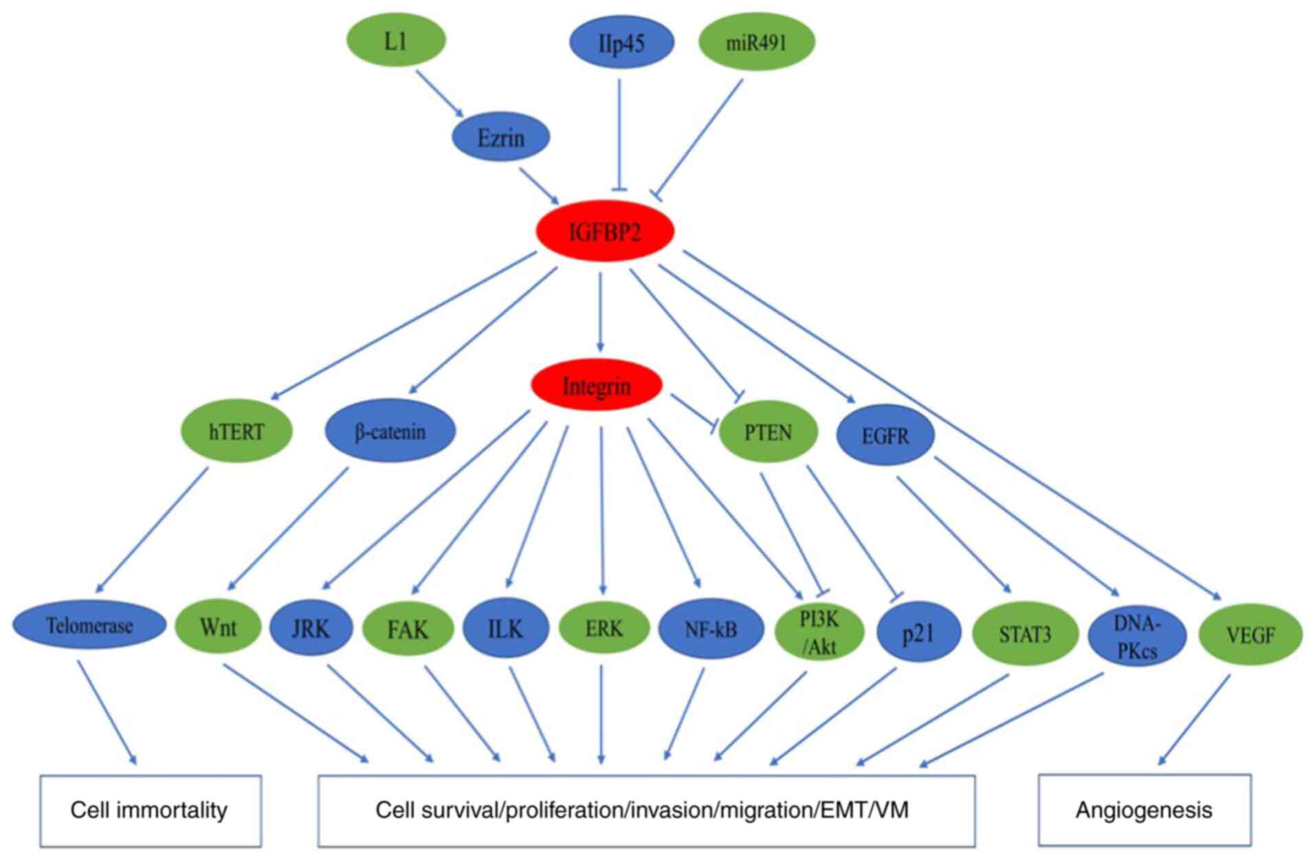
As a multifunctional oncogenic protein, IGFBP2
participates in oncogenic processes involving various signaling
pathways that are required for tumor initiation and progression
(Fig. 1). It is known that IGFBP2
promotes cancer metastasis by regulating invasion-associated
signaling networks (46); however,
the role of IGFBP2 on other oncogenic processes remains to be
elucidated. Therefore, we systematically examined the involvement
of IGFBP2 in several key oncogenic processes in the present review,
and summarized the potential regulatory network of IGFBP2 in
different cancer types. (Table
I).
Aberrant angiogenesis provides abundant blood
perfusion for the tumor, eventually promoting uncontrolled cell
proliferation and malignant progression. A large number of studies
demonstrated a crucial role for IGFBP2 in pathologic angiogenesis
in many cancers (63,68,69).
IGFBP2 is considered an inducer of angiogenesis in melanoma. IGFBP2
upregulates the expression of proangiogenic factor VEGF-A, and then
triggers angiogenesis by interacting with αVβ3 integrin and
activating the PI3K/AKT pathway (63). Similarly, IGFBP2 overexpression
upregulates the expression of VEGF in human neuroblastoma (68), and coexpression of IGFBP2 and VEGF
can be observed in the vicinity of tumor necrosis in glioblastoma
(70), suggesting that IGFBP2 is
involved in angiogenesis via the induction of VEGF expression.
Interestingly, nuclear IGFBP2, mediated by the NLS sequence within
the linker region, is responsible for the transcriptional
activation of VEGF (71). It is
known that endothelial cell (EC) mobility is crucial to
angiogenesis. In a breast cancer metastasis model, IGFBP2 secreted
by metastatic cells induces angiogenesis via recruiting endothelia
and endothelial cells in an IGF-I-dependent manner (69).
Vasculogenic mimicry (VM) is a novel manner of tumor
perfusion, and consists of vascular channel formation by aggressive
tumor cells rather than endothelial cells. In the case of
aggressive melanoma cells, a high expression of MMP-2, MT1-MMP and
laminin 5 γ2 chain is required for vasculogenic mimicry to
facilitate tumor perfusion independent of angiogenesis by
endothelial cells (72,73). Previous findings have shown that
IGFBP2 expression is positively associated with VM in patients with
glioma, and a higher expression of IGFBP2 promotes VM in
vitro and in vivo by enhancing CD144 and MMP2 expression
in an integrin/FAK/ERK pathway-dependent manner (48). As noted above, IGFBP2 has a pivotal
role in the pro-angiogenic process. Thus, further understanding of
the proangiogenic mechanism of IGFBP2 may offer novel insight for
vascular therapy.
It is being increasingly recognized that IGFBP2
plays a vital role in immune modulation, and numerous findings
indicate IGFBP2 induces antitumor immunity by not only serving as a
human tumor antigen, but also inducing an IGFBP2-specific T-cell
response in several cancers, including glioma, colorectal
carcinoma, and breast cancer (74–76).
Activated microglia/macrophages, which are important components of
brain tumor immune cells, may contribute to glioma development
(77). In addition, the
accumulation of IGFBP2 can be found in activated
microglia/macrophages (78).
Similarly, IGFBP2 immunoreactivity is observed in both glioma cells
and macrophages/microglia, with the IGFBP2-positive
macrophages/microglia and glioma cells observed to frequently
accumulate near the focal necrosis areas of glioma, suggesting an
important immune action for IGFBP2 in glioma (79). Recently, gene expression profiles
from 2447 glioma samples and bioinformatic analysis by Cai et
al demonstrated that IGFBP2 has immunosuppressive activities in
glioblastoma (GBM), and IGFBP2 is closely correlated with the
expression of immunosuppressive molecules, including CHI3L1,
TNFRSF1A, LGALS1, TIMP1, VEGFA, ANXA1 and LGALS3 (75). Furthermore, a recent study by Li and
coworkers also revealed a novel immune-associated tumor function of
IGFBP2 in malignant melanoma, where IGFBP2 upregulates the
expression of PD-L1 and contributes to the immune evasion of cancer
cells from host immunosurveillance (62). As mentioned above, there is evidence
that IGFBP2 plays a crucial role in tumor immune modulation. Thus,
it is important to thoroughly investigate immunoregulation by
IGFBP2 in human cancers, and search for potent anti-IGFBP2
therapy.
As an IGF-binding protein, IGFBP2 has been reported
as an inhibitory factor for IGF functions (80). In the human MCF-7β3 breast cancer
cell line, IGFBP2 inhibits IGF-I- and IGF-II-mediated migration,
but the mechanism underlying of this phenomenon is not clearly
understood (81). It has been
proposed that complex formation of IGFBP2 with αvβ3 integrin blocks
the amplification of IGF-I signaling, because αvβ3 integrin can
promote IGF-I-mediated proliferation and migration when binding to
vitronectin. IGFBP2 shows an inhibitory action for normal somatic
growth in vivo by directly interfering with the
growth-promoting effects of the GH/IGF-I axis (82). However, a paradoxical stimulatory
role of IGFBP2 in modulating IGF signaling has also been reported
in the majority of the literature. For example, IGFBP2 can bind to
IGF to form a binary complex in circulation, and this IGFBP2/IGF2
complex can stimulate osteoblast proliferation, although the
detailed mechanism is unclear (83). In addition, the IGFBP2/IGF2 complex
binds with increased affinity to 2- or 3-carbon O-sulfated
glycosaminoglycans (GAGs) via the IGFBP2 HBD, compared with free
intact IGFBP2, and could prevent IGF2 clearance by interfering with
IGF2R-mediated endocytosis (84).
It is known that the release of IGF-II from the IGF-II/IGFBP2
complex can occur by proteolysis by matrix metalloprotease-7
(MMP-7) and has a stimulatory effect on neoplastic transformation
of cells in colorectal cancer (85,86),
and contributes to the motility and growth in the case of LN229
astrocytoma cells (87). In this
context, IGFBP2 serves as a reservoir of IGFs in the pericellular
microenvironment. IGFBP2 binds to receptor protein tyrosine
phosphatase β (RPTPβ) via its HBD, leading to RPTPβ dimerization
and inactivation and then regulation of IGF-I signaling functions.
RPTPβ inhibits IGF-I-stimulated AKT activation via PTEN
dephosphorylation, whereas IGFBP2 functions via inhibition of this
signaling pathway, which consequently leads to the acceleration of
vascular smooth muscle cell (VSMC) proliferation (88). Similarly, IGFBP2 enhances
IGF-induced proliferation, migration and invasion of neuroblastoma
cells in a manner that requires an intact HBD domain, suggesting
that the growth-promoting effects of IGF are associated with
pericellular matrix proteins and/or proteoglycans or
glycosaminoglycans on the cell surface (89). Moreover, IGFBP2 can activate IGFRs
by increasing local IGF accumulation in the circulation or directly
interact with IGFs via its IGF-binding site. The secretion of
IGFBP2 from metastatic breast cancer cells can recruit endothelial
cells to the cancer site by regulating IGF1-mediated activation of
the IGFIR, which is a typical feature of metastatic breast cancer
(69).
The interaction between IGFBP2 and integrins is
mediated by the C-terminal RGD domain, which is a known
integrin-binding domain. The expression of integrin β and α can be
seen in various tumor cell types, and the crosstalk between IGFBP2
with integrin α5 and β1 has been significantly implicated in
tumorigenesis (47,48,52,54,90,91).
IGFBP2 enhances tumor cell proliferation and
mobility by directly binding with integrin and/or via
integrin-associated downstream signaling, which contributes to
tumor cell dissemination and tumor progression (91). For example, IGFBP2 enhances DU145
prostate cancer cell proliferation by binding to integrin β1
receptors, and the action may be blocked by a short RGD-containing
disintegrin peptide or by an integrin β1 receptor-blocking antibody
(54). Microarray studies indicate
that IGFBP2 activates the expression of integrin α5, but the
underlying regulatory mechanism is unclear. Furthermore, IGFBP2
promotes glioblastoma cell mobility in vitro via direct
binding to integrin α5, whereas decreasing expression of integrin
α5 by siRNA or RGD mutation may attenuate cell mobility (90). Moreover, exogenous IGFBP2 induces
glioma cell proliferation and invasion via integrin β1/ERK
signaling. However, blocking integrin β1 function by anti-integrin
β1-neutralizing antibody or integrin β1 knockdown inhibits
IGFBP2-induced ERK activation, and subsequent cell proliferation
and invasion (52). A recent study
in vitro also found that IGFBP2 promotes VM formation by
glioma cells via the binding to integrin α5 and β1 subunits through
its RGD domain (48). These
findings indicate that the IGFBP2/integrin pathway may provide a
strong driving force for tumor progression. Of note, the
interaction between IGFBP2 and integrins can regulate an array of
signaling pathways. An in vivo model of glioma progression
demonstrated that high IGFBP2 expression regulates downstream
invasion pathways, such as the NF-κB and integrin-linked kinase
(ILK) pathways, via activation of integrin β1. Most significantly,
the IGFBP2/integrin/ILK/NF-κB network is essential to glioma
progression and can be prevented by interfering at any point in the
pathway (47). A study by Mendes
et al showed that the activation of the JNK pathway is
closely related to the IGFBP2-integrin α5 signaling cascade, which
accelerates glioma cell migration (92).
PTEN is a tumor suppressor gene, and the loss of
PTEN function is often observed in many human cancers (50). The loss of PTEN function contributes
to the accumulation of the lipid
phosphatidylinositol-3,4,5-triphosphate (PIP3), which is the
product of PI3K, and then activates the Akt pathway to promote cell
survival and proliferation (93).
The interaction between IGFBP2 and PTEN has been linked to multiple
tumorigenic processes in breast, glioma, and prostate cancer
(29,50,54). A
study by Dean et al, using immunohistochemistry, revealed
that the loss of PTEN is tightly associated with IGFBP2
overexpression in triple-negative breast cancer, indicating that
IGFBP2 expression may be inversely associated with PTEN in breast
cancer (29). The evidence from
in vitro studies using glioma U251 cell lines also shows
that overexpression of PTEN can reduce IGFBP2 expression and
inhibit cell proliferation. Mechanically, it is proposed that the
lipid phosphatase activity of PTEN is responsible for restraining
IGFBP2 expression (94). Similarly,
in prostate cancer and GBM, the expression of serum IGFBP2
increases in PTEN-null tumors, but not PTEN-expressing tumors
(50). In addition,
microarray-based expression profiling identified IGFBP2 to be an
important marker for PTEN loss and activation of the PI3K/Akt
pathway in prostate cancer and GBM (50). In an acute myelocytic leukemia (AML)
transplantation mouse model, inhibition of IGFBP2 expression
impeded leukemia development and led to the upregulation of PTEN
expression and downregulation of AKT activation. However, this
condition was reversed by treatment with a PTEN inhibitor,
suggesting that the PTEN/Akt pathway is essential to IGFBP2-induced
leukemia development (61).
Notably, IGFBP2 has also been shown to suppress the activity of
PTEN. PTEN reduces tumor cell proliferation and increases
apoptosis, and these actions may be substantially attenuated in the
context of high IGFBP2 expression (94). In human breast cancer cells, PTEN
expression is stimulated by IGF-II forming a feedback loop where
IGFBP2 can block the feedback response for PTEN, which then leads
to an increase in p21-mediated pro-tumorigenic activity (56). In DU145 prostate cancer cells,
IGFBP2 increases PTEN phosphorylation in an integrin β1-dependent
manner, leading to the inactivation of PTEN, which eventually
potentiates cell proliferation (54). The same result is also observed in
MCF-7 breast cancer cells (56).
Therefore, PTEN may be a downstream mediator of
IGFBP2/integrin-mediated signaling. Moreover, IGFBP2 has been shown
to reduce the expression of PTEN protein levels, but not transcript
levels, suggesting that the mechanism by which IGFBP2 modulates
PTEN may be post-transcriptional (9).
Increasing evidence has shown that IGFBP2 possesses
a nuclear regulatory effect that is closely associated with EGFR
and STAT3. For example, co-overexpression of the EGFR and IGFBP2
genes has been reported to be strongly associated with poor
prognosis in astrocytoma (95). A
study by Chua et al also found a close relationship between
IGFBP2, STAT3 phosphorylation and nuclear co-localization of IGFBP2
and EGFR in glioma (8). Moreover,
IGFBP2 is considered to be involved in the regulation of an
EGFR-STAT3 pathway in glioma progression, since treatment with
IGFBP2-neutralizing antibody decreases the activation of EGFR and
STAT3, and reduces the expression of STAT3 downstream genes, such
as Bcl-xL and LSD1; however, the underlying mechanism by which
expression is regulated has not been well characterized (15). Moreover, high IGFBP2 expression has
been reported to increase the nuclear accumulation of EGFR and
eventually activate the nuclear EGFR signaling pathway, which
contributes to potentiating STAT3 transactivation (8). These findings provide a novel
oncogenic effect of IGFBP2 in glioma through activation of
EGFR/STAT3 signaling (8,15). Similarly, IGFBP2 upregulates PD-L1
expression by activating the nuclear EGFR-STAT3 signaling pathway
in malignant melanoma, which may contribute to tumor progression
(62).
Highly expressed in basal cell carcinoma (BCC),
IGFBP2 is considered as a mediator of the effects of Hedgehog (Hh)
signaling on epidermal progenitors, and is essential for promoting
BCC development (96). IGFBP2
induces colorectal cancer (CRC) cell motility and metastasis
mediated by an L1-ezrin-NF-κB pathway. It can be found that L1
induces IGFBP2 expression through activating ezrin phosphorylation
and NF-κB-mediated transactivation of the IGFBP2 promoter, which is
essential for increasing tumorigenesis and inducing liver
metastasis (59). Moreover, IGFBP2
promotes esophageal adenocarcinoma (EAC) cell survival via
stabilization and activation of an EGFR-DNA-PKcs signaling axis
(60). IGFBP2 promotion of tumor
development may also be linked to the β-catenin/Wnt signaling
pathway. In breast cancer cells, IGFBP2 regulates the expression of
β-catenin, an effector of the Wnt pathway, in an IGF1R- and
FAK-dependent manner, and combined overexpression of IGFBP2 and
β-catenin is associated with lymph node metastasis in breast tumors
(55). Similarly, IGFBP2 regulates
the β-catenin pathway involving the activation of Akt and
inactivation of GSK3b in glioma cells, and co-expression of high
levels of IGFBP-2 and β-catenin is correlated to worse prognosis
(51). In addition, IGFBP2 seems to
directly stimulate hypoxia-inducible factor 1α (HIF1α) expression.
HIF1α also promotes the expression of IGFBP2 in the absence of
oxygen, and the feedback between IGFBP2 and HIF1α may play an
important role in glioblastoma growth. Removing the feedback from
IGFBP2 to HIF1α results in the inhibition of cell growth (49). Foulstone et al found a
positive feedback loop between IGFBP2 and ER-α, which could
stimulate IGFBP2 expression. IGFBP2 also could increase the
expression of ER-α in breast cancer cells, and the crosstalk
between IGFBP2/ER-α may contribute to the growth and survival of
breast cancer cells (97). IGFBP2
significantly upregulates the expression of hTERT and stimulates
activity of telomerase in the absence of androgens, which plays a
role in prostate cancer cell immortality (53). Furthermore, the IGFBP2 function that
triggers pro-carcinogenic effects in prostate cancer appears to be
in part mediated by the MAP-kinase and PI3-kinase pathways
(53).
IGFBP2 is one of the most common and abundant
members of the IGFBP family in numerous human cancers. It became
evident that IGFBP2 exerts important roles in growth, metabolism
and cancer development (6).
Additionally, aberrant overexpression of IGFBP2 is intimately
associated with adverse cancer-associated clinical parameters
(6). Centered on several
malignancies, the expression of IGFBP2 in cancer is elaborated in
Table II, as is IGFBP2 expression
as a diagnostic/prognostic biomarker in different tumor types.
As a malignancy-associated secreted protein, high
IGFBP2 protein expression is detected both in tissues and blood of
glioma patients, and is positively correlated with tumor grade
(12,79). Moreover, higher plasma IGFBP2
protein expression is significantly associated with poorer
disease-free survival and recurrence after surgery in patients with
GBM (12). In addition, high
expression of tissue IGFBP2 protein may serve as an independent
poor prognosis biomarker in patients with GBM (11). In fact, except for the two
abovementioned studies, the prognostic value of IGFBP2 protein has
also been assessed in other several studies, as shown in Table II. From these studies, it can be
gleaned that a high expression of IGFBP2 protein is a usable
predictor for the progression and unfavorable outcome in patients
with glioma. In addition, tumor-specific immunity against IGFBP2
has been reported in glioma. Li et al (76) were, to the best of our knowledge,
the first to evaluate the diagnostic value of serum IGFBP2
antibodies in early detection of glioma. They ultimately found that
high IGFBP2 antibody expression distinguished patients with grade
II and III gliomas from normal controls, and the combination of
serum IGFBP2 and IGFBP2 antibodies improved the diagnostic power
for glioma.
IGFBP2 is consistently highly expressed in breast
cancer tissue compared with benign lesions in tissue microarray
analysis. In addition, increased IGFBP2 expression may be a useful
marker to predict lymph node metastasis in patients with T1
invasive breast carcinoma (31).
The expression of IGFBP2 is strongly correlated with the grade of
mammary neoplasms, as shown in a study by Busund et al,
where the expression of IGFBP2 increases in a step-wise manner from
hyperplasia, atypical hyperplasia, and carcinoma in situ to
invasive breast carcinoma (28).
Additionally, the prognostic value of IGFBP2 for breast cancer
patients has been shown in numerous reports (Table II). A prospective cohort study
showed that patients with higher pre-diagnostic serum levels of
IGFBP2 are more likely to have higher breast cancer-specific
mortality within 5 years of follow up (102). Moreover, IGFBP2 serves as a
predictive factor for recurrence-free survival (RFS) risk in
patients with residual triple receptor-negative breast cancer
(TNBC) after neoadjuvant chemotherapy (NCT) (103). Notably, IGFBP2 expression is an
independent predictor of shorter overall survival, and the
prognostic relevance of IGFBP2 partly is associated with hormonal
status and body weight in breast cancer (104). Similarly, a similar finding from
So et al showed that high IGFBP2 expression is related to
poorer disease-specific survival in patients with hormone
receptor-negative invasive breast cancer, but not in patients with
hormone receptor-positive tumors (101). Since breast cancer patients easily
become resistant to antiestrogen treatment, it may be a strong
clinical challenge to cure these hormone-resistant tumors. IGFBP2
is a potential marker for antiestrogen treatment response in breast
cancer, as both high IGFBP2 protein and mRNA levels can be examined
for resistance to antiestrogens such as tamoxifen, ICI 182,780 and
RU 58668 (114).
Increasing studies over the past decades has
investigated the expression of IGFBP2 with malignant prostate
disease. Elevated levels of serum IGFBP2 are detected in patients
with prostate cancer (24), and are
positively associated with high serum levels of prostate-specific
antigen (PSA) (23). In addition,
high serum IGFBP2 is an important risk factor for low-grade
prostate cancer (115). Both
IGFBP2 protein and mRNA expression are increased in high-grade
prostate intraepithelial neoplasia (PIN) tissues, and even further
elevated in malignant adenocarcinoma tissues, when compared to
normal epithelium (116).
Subsequently, Richardsen et al reported that overexpression
of IGFBP2 in prostate tissue is a useful marker for malignant
transformation of prostate epithelium, and may be a potential
auxiliary tool in the diagnosis of prostate cancer.
Immunohistochemical detection of IGFBP2 expression is negative or
very weakly positive in normal prostate epithelium or benign
prostatic hyperplasia (BPH) tissue, but increased extent and
intensity of IGFBP2 staining can be observed in prostatic
intraepithelial neoplasia (PIN) and carcinoma (105). IGFBP2 expression also appears to
be involved in the development of hormone-refractory prostate
cancer. Immunohistochemical analysis revealed that IGFBP2 protein
is significantly overexpressed in 100% of hormone-refractory
prostate cancers, and in 36% of primary tumors, but not in normal
specimens (117). Similarly,
increased expression of IGFBP2 plays a role in lymph node
metastasis of prostate cancer after hormone therapy, and is a
potential prognostic indicator in hormone-treated prostate cancer
patients (106).
IGFBP2 is overexpressed in ovarian cancer tissues,
serum and cyst fluids, and has a key role in the regulation of
human ovarian cancer progression (19,118,119). There are histological differences
of IGFBP2 expression in ovarian cancer. This was further
substantiated by Wang et al, using tissue microarray
analysis, who revealed that the expression of IGFBP2 was more often
higher in high-grade serous carcinoma, malignant-mixed Mullerian
tumors (MMMT), and undifferentiated carcinoma, but frequently
expressed at low levels or not at all in clear cell and mucinous
carcinoma (18). Moreover, IGFBP2
expression is closely correlated with tumor grade and
aggressiveness of ovarian carcinoma. IGFBP2 is also highly
expressed in invasive carcinomas compared to borderline tumors
(17,18).
Most importantly, IGFBP2 is a potential serum
biomarker in the detection and monitoring of epithelial ovarian
cancer (EOC) (107). Increased
levels of serum IGFBP2 are positively correlated with expression of
the ovarian tumor marker CA125 in patients with EOC (16), and the combination of IGFBP2 and
CA125 may improve the sensitivity of CA125 alone for the early
detection of ovarian cancer (21).
High serum IGFBP2 levels are closely correlated with increased risk
of mortality and poorer responses to chemotherapy, indicating that
IGFBP2 is an early predictor of EOC-related mortality and
chemotherapeutic response in EOC (108). Moreover, patients with higher
preoperative serum levels of IGFBP2 have a significantly higher
risk of relapse and worse overall survival (20).
High blood and tissue levels of IGFBP2 can be found
in patients with lung cancer (33,120).
Increased plasma IGFBP2 expression is positively associated with
tumor size and tumor stage, and patients with higher IGFBP2 levels
have shorter overall survival, suggesting that IGFBP2 may be a
useful prognostic biomarker in lung cancer (33). In addition, tissue IGFBP2
overexpression is correlated with lymph node metastasis and
dasatinib resistance in non-small cell lung cancer (NSCLC)
(58,120). A study by Migita et al,
using immunohistochemical analysis, found that high IGFBP2
expression is inversely correlated with procaspase-3 expression,
and may be an anti-apoptotic biomarker for lung adenocarcinoma
(LUAD) (32). Interestingly, it can
be found that high serum anti-IGFBP2 antibodies show diagnostic
relevance for early-stage lung cancer, and the combination of serum
IGFBP2 and anti-IGFBP2 antibodies can be more effective for lung
cancer diagnosis (109).
Although these findings show that a high IGFBP2
expression is associated with poor prognosis in lung cancer,
conflicting results have also been reported. Bioinformatics
analysis by Wang et al showed that increased IGFBP2 mRNA
levels are correlated with unfavorable OS in patients with NSCLC,
but with favorable OS in patients with lung squamous cell carcinoma
(LUSC) (121). Similarly, an
investigation in vitro revealed that the overexpression of
IGFBP2 may contribute to inhibitory effects on cell growth in small
cell lung cancer (SCLC) (122). As
mentioned above, these findings highlight the paradoxical role of
IGFBP2 in different subtypes of lung cancer. Therefore, further
research is required to explore the role of IGFBP2 and its exact
regulatory mechanism in different subtypes of lung cancer. As a
matter of fact, it has been reported that IGFBP2 functions as a
tumor suppressor in some tumors. Mechanistically, it has been
proposed that the suppressive/oncogenic role of IGFBP2 in
carcinogenesis may be associated with the cleaved status of IGFBP2.
Protease-resistant IGFBP2 is both able to suppress tumor growth
in vitro and in vivo when compared with wild-type
IGFBP2 (123).
Circulating IGFBP2 expression shows a close
relationship with the malignant development of patients with CRC.
For instance, serum IGFBP2 expression increases from Dukes B and C
to advanced cancer, and is highly associated with tumor load
(35,36). Plasma IGFBP2 levels can discriminate
between advanced colon polyps and CRC, and higher plasma IGFBP2
expression at diagnosis correlates with worse overall survival in
patients with CRC, suggesting that high plasma IGFBP2 expression
may serve as a valuable diagnostic and prognostic biomarker in CRC
(35). Also, Li et al
(76) reported that serum IGFBP2
antibody levels can discriminate patients with CRC I–II from normal
controls, and the combination of serum IGFBP2 and IGFBP2 antibodies
are more effective for early cancer detection of CRC.
The diagnosis and management of pancreatic cancer
remains a great challenge due to the lack of effective biomarkers.
Quantitative proteomics analysis by Chen et al identified
that IGFBP2 is aberrantly highly expressed in the pancreatic juice
and tumor tissue of pancreatic cancer patients compared to patients
with chronic pancreatitis (124).
Furthermore, IGFBP2 also appears to be elevated in riskier diseases
of pancreatic malignancy, such as intraductal papillary mucinous
neoplasms (IPMNs). Kendrick et al (26) reported that a high serum IGFBP2
expression is directly correlated with tumor burden, and may serve
as a potential diagnostic biomarker for PDAC. The expression of
IGFBP2 can distinguish early stage invasive ductal adenocarcinoma
of the pancreas (IDACP) patients from normal controls, and the use
of IGFBP2 with the CA19-9 biomarker may be more effective for the
diagnosis of IDACP (27).
Additionally, an in vivo model and clinical findings also
showed that IGFBP2 overexpression in primary tumors is more likely
to increase the risk of lymph node metastasis and is correlated
with shorter survival in patients with PDAC, suggesting that IGFBP2
may be a valuable biomarker for evaluating prognosis in PDAC
(9).
Previous findings have shown that the IGF-IGFBP
system may play an important role in gastric cancer development.
For example, IGFBP2 can be detected in all gastric cancer cell
lines, and the ubiquitous expression pattern of IGFBP2 suggests
that IGFBP2 may confer some growth advantage to the tumor cells
(125). High IGFBP2 expression can
be found in gastric carcinoma tissues, compared with normal gastric
mucosa, and participates in the malignant progression of gastric
carcinoma (42).
Immunohistochemical staining demonstrated that the expression of
IGFBP2 shows a positive correlation with Ki-67 expression,
suggesting that IGFBP2 may participate in carcinogenesis and
progression of gastric carcinoma by promoting cell proliferation.
Additionally, IGFBP2 expression is greatly elevated from early
gastric carcinoma to advanced gastric carcinoma, and is positively
correlated with lymph node metastasis and clinical stage of the
tumor (41,42). Patients with higher serum IGFBP2
levels are more likely to have a lower 5-year overall survival
rate, indicating that serum IGFBP2 may be a potential biomarker
predicting prognosis for patients with gastric cancer (110).
Recently, researchers have shown an increasing
interest in the role of IGFBP2 for esophageal cancer development.
Gene expression profiling has shown that the IGFBP2 gene is highly
expressed in early tumor stages (pT1-2) of esophageal squamous cell
carcinoma (ESCC) and esophageal adenocarcinoma compared to normal
tissues, and is a potential diagnostic candidate marker for
esophageal cancer (111), but more
clinical studies are needed to determine its diagnostic impact.
Interestingly, a high expression of IGFBP2 may be an important
predictive factor for recurrence and chemoresistance in patients
with EAC after chemotherapy following esophagectomy (112). In fact, few studies have
investigated the relationship between IGFBP2 with esophageal cancer
development. Therefore, more studies are needed to elaborate the
role of IGFBP2 in esophageal cancer.
The purpose of this review was to summarize recent
insights on the role of IGFBP2 and its underlying regulatory
mechanisms in the development and progression of different tumors.
To the best of our knowledge, there is compelling evidence
suggesting that IGFBP2 has pivotal tumorigenic functions in a great
majority of human cancers, but these findings are contradictory in
a few tumors, including LUSC and SCLC. According to these reports,
IGFBP2 can have both stimulatory and inhibitory effects on tumor
cell growth and survival, and these discrepancies may be ascribed
to the diversity of IGFBP2 functions in different cancers. A large
number of reports support the viewpoint that the oncogenic
properties of IGFBP2 are more likely to be IGF-independent, but the
exact mechanisms of how the IGFBP2 plays a role in different tumors
remains not fully elucidated. At present, IGFBP2 has received more
interest as a potential therapeutic target in multiple cancers.
However, there is no IGFBP2 inhibitor for clinical application. In
addition, the diagnostic or prognostic relevance of IGFBP2 has been
evaluated in a wide variety of human cancers. Nevertheless, it has
yet to provide diagnostic value for clinical use. Undoubtedly, more
studies are in great need to expound the underlying mechanisms of
IGFBP2 in detail, and improve the understanding of IGFBP2 as a
tumor biomarker and as a potent therapeutic target for better
clinical management of patients with malignant tumors.
Not applicable.
The present study was supported by funding from the
Natural Science Foundation of China (no. 81972801), the Guangdong
Basic and Applied Basic Research Foundation (nos. 2018A030307079
and 2019A1515011873), the Medical Project of Science and Technology
Planning of Shantou (no. 200605115266724), the 2020 Li Ka Shing
Foundation Cross-Disciplinary Research Grant (no. 2020LKSFG01B),
and the Science and Technology Special Fund of Guangdong Province
of China (nos. 190829105556145 and 180918114960704).
Not applicable.
Draft preparation, writing, and editing of the
review was carried out by LFW, XFW, XCH, HPG, YHP and YWX. HPG, YHP
and YWX drafted the review or revised it critically for important
intellectual content. All authors have read and agreed to the
published version of the manuscript.
Not applicable.
Not applicable.
The authors declare that they have no competing
interests.
|
1
|
Baxter RC: Insulin-like growth factor
(IGF)-binding proteins: Interactions with IGFs and intrinsic
bioactivities. Am J Physiol Endocrinol Metab. 278:E967–E976. 2000.
View Article : Google Scholar : PubMed/NCBI
|
|
2
|
Rajaram S, Baylink DJ and Mohan S:
Insulin-like growth factor-binding proteins in serum and other
biological fluids: Regulation and functions. Endocr Rev.
18:801–831. 1997. View Article : Google Scholar : PubMed/NCBI
|
|
3
|
Bach LA: IGF-binding proteins. J Mol
Endocrinol. 61:T11–T28. 2018. View Article : Google Scholar : PubMed/NCBI
|
|
4
|
Baxter RC: IGF binding proteins in cancer:
Mechanistic and clinical insights. Nat Rev Cancer. 14:329–341.
2014. View Article : Google Scholar : PubMed/NCBI
|
|
5
|
Yu H and Rohan T: Role of the insulin-like
growth factor family in cancer development and progression. J Natl
Cancer Inst. 92:1472–1489. 2000. View Article : Google Scholar : PubMed/NCBI
|
|
6
|
Russo VC, Azar WJ, Yau SW, Sabin MA and
Werther GA: IGFBP-2: The dark horse in metabolism and cancer.
Cytokine Growth Factor Rev. 26:329–346. 2015. View Article : Google Scholar : PubMed/NCBI
|
|
7
|
Zeng L, Perks CM and Holly JM:
IGFBP-2/PTEN: A critical interaction for tumours and for general
physiology? Growth Horm IGF Res. 25:103–107. 2015. View Article : Google Scholar : PubMed/NCBI
|
|
8
|
Chua CY, Liu Y, Granberg KJ, Hu L,
Haapasalo H, Annala MJ, Cogdell DE, Verploegen M, Moore LM, Fuller
GN, et al: IGFBP2 potentiates nuclear EGFR-STAT3 signaling.
Oncogene. 35:738–747. 2016. View Article : Google Scholar : PubMed/NCBI
|
|
9
|
Gao S, Sun Y, Zhang X, Hu L, Liu Y, Chua
CY, Phillips LM, Ren H, Fleming JB, Wang H, et al: IGFBP2 activates
the NF-κB pathway to Drive epithelial-mesenchymal transition and
invasive character in pancreatic ductal adenocarcinoma. Cancer Res.
76:6543–6554. 2016. View Article : Google Scholar : PubMed/NCBI
|
|
10
|
Fuller GN, Rhee CH, Hess KR, Caskey LS,
Wang R, Bruner JM, Yung WK and Zhang W: Reactivation of
insulin-like growth factor binding protein 2 expression in
glioblastoma multiforme: A revelation by parallel gene expression
profiling. Cancer Res. 59:4228–4232. 1999.PubMed/NCBI
|
|
11
|
McDonald KL, O'Sullivan MG, Parkinson JF,
Shaw JM, Payne CA, Brewer JM, Young L, Reader DJ, Wheeler HT, Cook
RJ, et al: IQGAP1 and IGFBP2: Valuable biomarkers for determining
prognosis in glioma patients. J Neuropathol Exp Neurol. 66:405–417.
2007. View Article : Google Scholar : PubMed/NCBI
|
|
12
|
Lin Y, Jiang T, Zhou K, Xu L, Chen B, Li
G, Qiu X, Jiang T, Zhang W and Song SW: Plasma IGFBP-2 levels
predict clinical outcomes of patients with high-grade gliomas.
Neuro Oncol. 11:468–476. 2009. View Article : Google Scholar : PubMed/NCBI
|
|
13
|
Dunlap SM, Celestino J, Wang H, Jiang R,
Holland EC, Fuller GN and Zhang W: Insulin-like growth factor
binding protein 2 promotes glioma development and progression. Proc
Natl Acad Sci USA. 104:11736–11741. 2007. View Article : Google Scholar : PubMed/NCBI
|
|
14
|
Wang H, Wang H, Shen W, Huang H, Hu L,
Ramdas L, Zhou YH, Liao WS, Fuller GN and Zhang W: Insulin-like
growth factor binding protein 2 enhances glioblastoma invasion by
activating invasion-enhancing genes. Cancer Res. 63:4315–4321.
2003.PubMed/NCBI
|
|
15
|
Phillips LM, Zhou X, Cogdell DE, Chua CY,
Huisinga A, R Hess K, Fuller GN and Zhang W: Glioma progression is
mediated by an addiction to aberrant IGFBP2 expression and can be
blocked using anti-IGFBP2 strategies. J Pathol. 239:355–364. 2016.
View Article : Google Scholar : PubMed/NCBI
|
|
16
|
Flyvbjerg A, Mogensen O, Mogensen B and
Nielsen OS: Elevated serum insulin-like growth factor-binding
protein 2 (IGFBP-2) and decreased IGFBP-3 in epithelial ovarian
cancer: Correlation with cancer antigen 125 and tumor-associated
trypsin inhibitor. J Clin Endocrinol Metab. 82:2308–2313. 1997.
View Article : Google Scholar : PubMed/NCBI
|
|
17
|
Lee EJ, Mircean C, Shmulevich I, Wang H,
Liu J, Niemistö A, Kavanagh JJ, Lee JH and Zhang W: Insulin-like
growth factor binding protein 2 promotes ovarian cancer cell
invasion. Mol Cancer. 4:72005. View Article : Google Scholar : PubMed/NCBI
|
|
18
|
Wang H, Rosen DG, Wang H, Fuller GN, Zhang
W and Liu J: Insulin-like growth factor-binding protein 2 and 5 are
differentially regulated in ovarian cancer of different histologic
types. Mod Pathol. 19:1149–1156. 2006. View Article : Google Scholar : PubMed/NCBI
|
|
19
|
Kanety H, Kattan M, Goldberg I, Kopolovic
J, Ravia J, Menczer J and Karasik A: Increased insulin-like growth
factor binding protein-2 (IGFBP-2) gene expression and protein
production lead to high IGFBP-2 content in malignant ovarian cyst
fluid. Br J Cancer. 73:1069–1073. 1996. View Article : Google Scholar : PubMed/NCBI
|
|
20
|
Baron-Hay S, Boyle F, Ferrier A and Scott
C: Elevated serum insulin-like growth factor binding protein-2 as a
prognostic marker in patients with ovarian cancer. Clin Cancer Res.
10:1796–1806. 2004. View Article : Google Scholar : PubMed/NCBI
|
|
21
|
Russell MR, Graham C, D'Amato A,
Gentry-Maharaj A, Ryan A, Kalsi JK, Ainley C, Whetton AD, Menon U,
Jacobs I and Graham RLJ: A combined biomarker panel shows improved
sensitivity for the early detection of ovarian cancer allowing the
identification of the most aggressive type II tumours. Br J Cancer.
117:666–674. 2017. View Article : Google Scholar : PubMed/NCBI
|
|
22
|
Biernacka KM, Uzoh CC, Zeng L, Persad RA,
Bahl A, Gillatt D, Perks CM and Holly JM: Hyperglycaemia-induced
chemoresistance of prostate cancer cells due to IGFBP2. Endocr
Relat Cancer. 20:741–751. 2013. View Article : Google Scholar : PubMed/NCBI
|
|
23
|
Kanety H, Madjar Y, Dagan Y, Levi J, Papa
MZ, Pariente C, Goldwasser B and Karasik A: Serum insulin-like
growth factor-binding protein-2 (IGFBP-2) is increased and IGFBP-3
is decreased in patients with prostate cancer: Correlation with
serum prostate-specific antigen. J Clin Endocrinol Metab.
77:229–233. 1993. View Article : Google Scholar : PubMed/NCBI
|
|
24
|
Cohen P, Peehl DM, Stamey TA, Wilson KF,
Clemmons DR and Rosenfeld RG: Elevated levels of insulin-like
growth factor-binding protein-2 in the serum of prostate cancer
patients. J Clin Endocrinol Metab. 76:1031–1035. 1993. View Article : Google Scholar : PubMed/NCBI
|
|
25
|
Shariat SF, Lamb DJ, Kattan MW, Nguyen C,
Kim J, Beck J, Wheeler TM and Slawin KM: Association of
preoperative plasma levels of insulin-like growth factor I and
insulin-like growth factor binding proteins-2 and −3 with prostate
cancer invasion, progression, and metastasis. J Clin Oncol.
20:833–841. 2002. View Article : Google Scholar : PubMed/NCBI
|
|
26
|
Kendrick ZW, Firpo MA, Repko RC, Scaife
CL, Adler DG, Boucher KM and Mulvihill SJ: Serum IGFBP2 and MSLN as
diagnostic and prognostic biomarkers for pancreatic cancer. HPB
(Oxford). 16:670–676. 2014. View Article : Google Scholar : PubMed/NCBI
|
|
27
|
Yoneyama T, Ohtsuki S, Honda K, Kobayashi
M, Iwasaki M, Uchida Y, Okusaka T, Nakamori S, Shimahara M, Ueno T,
et al: Identification of IGFBP2 and IGFBP3 as compensatory
biomarkers for CA19-9 in early-stage pancreatic cancer using a
combination of antibody-based and LC-MS/MS-based proteomics. PLoS
One. 11:e01610092016. View Article : Google Scholar : PubMed/NCBI
|
|
28
|
Busund LT, Richardsen E, Busund R, Ukkonen
T, Bjørnsen T, Busch C and Stalsberg H: Significant expression of
IGFBP2 in breast cancer compared with benign lesions. J Clin
Pathol. 58:361–366. 2005. View Article : Google Scholar : PubMed/NCBI
|
|
29
|
Dean SJ, Perks CM, Holly JM, Bhoo-Pathy N,
Looi LM, Mohammed NA, Mun KS, Teo SH, Koobotse MO, Yip CH and
Rhodes A: Loss of PTEN expression is associated with IGFBP2
expression, younger age, and late stage in triple-negative breast
cancer. Am J Clin Pathol. 141:323–333. 2014. View Article : Google Scholar : PubMed/NCBI
|
|
30
|
Garner CP, Ding YC, John EM, Ingles SA,
Olopade OI, Huo D, Adebamowo C, Ogundiran T and Neuhausen SL:
Genetic variation in IGFBP2 and IGFBP5 is associated with breast
cancer in populations of African descent. Hum Genet. 123:247–255.
2008. View Article : Google Scholar : PubMed/NCBI
|
|
31
|
Wang H, Arun BK, Wang H, Fuller GN, Zhang
W, Middleton LP and Sahin AA: IGFBP2 and IGFBP5 overexpression
correlates with the lymph node metastasis in T1 breast carcinomas.
Breast J. 14:261–267. 2008. View Article : Google Scholar : PubMed/NCBI
|
|
32
|
Migita T, Narita T, Asaka R, Miyagi E,
Nagano H, Nomura K, Matsuura M, Satoh Y, Okumura S, Nakagawa K, et
al: Role of insulin-like growth factor binding protein 2 in lung
adenocarcinoma: IGF-independent antiapoptotic effect via caspase-3.
Am J Pathol. 176:1756–1766. 2010. View Article : Google Scholar : PubMed/NCBI
|
|
33
|
Guo C, Lu H, Gao W, Wang L, Lu K, Wu S,
Pataer A, Huang M, El-Zein R, Lin T, et al: Insulin-like growth
factor binding protein-2 level is increased in blood of lung cancer
patients and associated with poor survival. PLoS One. 8:e749732013.
View Article : Google Scholar : PubMed/NCBI
|
|
34
|
Ladd JJ, Busald T, Johnson MM, Zhang Q,
Pitteri SJ, Wang H, Brenner DE, Lampe PD, Kucherlapati R, Feng Z,
et al: Increased plasma levels of the APC-interacting protein
MAPRE1, LRG1, and IGFBP2 preceding a diagnosis of colorectal cancer
in women. Cancer Prev Res (Phila). 5:655–664. 2012. View Article : Google Scholar : PubMed/NCBI
|
|
35
|
Liou JM, Shun CT, Liang JT, Chiu HM, Chen
MJ, Chen CC, Wang HP, Wu MS and Lin JT: Plasma insulin-like growth
factor-binding protein-2 levels as diagnostic and prognostic
biomarker of colorectal cancer. J Clin Endocrinol Metab.
95:1717–1725. 2010. View Article : Google Scholar : PubMed/NCBI
|
|
36
|
Renehan AG, Jones J, Potten CS, Shalet SM
and O'Dwyer ST: Elevated serum insulin-like growth factor (IGF)-II
and IGF binding protein-2 in patients with colorectal cancer. Br J
Cancer. 83:1344–1350. 2000. View Article : Google Scholar : PubMed/NCBI
|
|
37
|
Wang H, Shen SS, Wang H, Diwan AH, Zhang
W, Fuller GN and Prieto VG: Expression of insulin-like growth
factor-binding protein 2 in melanocytic lesions. J Cutan Pathol.
30:599–605. 2003. View Article : Google Scholar : PubMed/NCBI
|
|
38
|
Ranke MB, Maier KP, Schweizer R, Stadler
B, Schleicher S, Elmlinger MW and Flehmig B: Pilot study of
elevated levels of insulin-like growth factor-binding protein-2 as
indicators of hepatocellular carcinoma. Horm Res. 60:174–180.
2003.PubMed/NCBI
|
|
39
|
Zhou Q, Mao YQ, Jiang WD, Chen YR, Huang
RY, Zhou XB, Wang YF, Shi Z, Wang ZS and Huang RP: Development of
IGF signaling antibody arrays for the identification of
hepatocellular carcinoma biomarkers. PLoS One. 7:e468512012.
View Article : Google Scholar : PubMed/NCBI
|
|
40
|
Guo Q, Yu DY, Yang ZF, Liu DY, Cao HQ and
Liao XW: IGFBP2 upregulates ZEB1 expression and promotes
hepatocellular carcinoma progression through NF-κB signaling
pathway. Dig Liver Dis. 52:573–581. 2020. View Article : Google Scholar : PubMed/NCBI
|
|
41
|
Shi LH, Zhu XQ, Zhao GH, Xia YB and Zhang
YS: Expression of insulin-like growth factor binding protein-2 in
gastric carcinoma and its relationship with cell proliferation.
World J Gastroenterol. 12:6285–6289. 2006. View Article : Google Scholar : PubMed/NCBI
|
|
42
|
Zhang L, Huang W, Chen J, Zhou X, Lu Z and
Zhou H: Expression of IGFBP2 in gastric carcinoma and relationship
with clinicopathologic parameters and cell proliferation. Dig Dis
Sci. 52:248–253. 2007. View Article : Google Scholar : PubMed/NCBI
|
|
43
|
Tombolan L, Orso F, Guzzardo V, Casara S,
Zin A, Bonora M, Romualdi C, Giorgi C, Bisogno G, Alaggio R, et al:
High IGFBP2 expression correlates with tumor severity in pediatric
rhabdomyosarcoma. Am J Pathol. 179:2611–2624. 2011. View Article : Google Scholar : PubMed/NCBI
|
|
44
|
Vorwerk P, Mohnike K, Wex H, Röhl FW,
Zimmermann M, Blum WF and Mittler U: Insulin-like growth factor
binding protein-2 at diagnosis of childhood acute lymphoblastic
leukemia and the prediction of relapse risk. J Clin Endocrinol
Metab. 90:3022–3027. 2005. View Article : Google Scholar : PubMed/NCBI
|
|
45
|
Dawczynski K, Kauf E, Schlenvoigt D, Gruhn
B, Fuchs D and Zintl F: Elevated serum insulin-like growth factor
binding protein-2 is associated with a high relapse risk after
hematopoietic stem cell transplantation in childhood AML. Bone
Marrow Transplant. 37:589–594. 2006. View Article : Google Scholar : PubMed/NCBI
|
|
46
|
Yao X, Sun S, Zhou X, Guo W and Zhang L:
IGF-binding protein 2 is a candidate target of therapeutic
potential in cancer. Tumour Biol. 37:1451–1459. 2016. View Article : Google Scholar : PubMed/NCBI
|
|
47
|
Holmes KM, Annala M, Chua CY, Dunlap SM,
Liu Y, Hugen N, Moore LM, Cogdell D, Hu L, Nykter M, et al:
Insulin-like growth factor-binding protein 2-driven glioma
progression is prevented by blocking a clinically significant
integrin, integrin-linked kinase, and NF-κB network. Proc Natl Acad
Sci USA. 109:3475–3480. 2012. View Article : Google Scholar : PubMed/NCBI
|
|
48
|
Liu Y, Li F, Yang YT, Xu XD, Chen JS, Chen
TL, Chen HJ, Zhu YB, Lin JY, Li Y, et al: IGFBP2 promotes
vasculogenic mimicry formation via regulating CD144 and MMP2
expression in glioma. Oncogene. 38:1815–1831. 2019. View Article : Google Scholar : PubMed/NCBI
|
|
49
|
Lin KW, Liao A and Qutub AA: Simulation
predicts IGFBP2-HIF1α interaction drives glioblastoma growth. PLoS
Comput Biol. 11:e10041692015. View Article : Google Scholar : PubMed/NCBI
|
|
50
|
Mehrian-Shai R, Chen CD, Shi T, Horvath S,
Nelson SF, Reichardt JK and Sawyers CL: Insulin growth
factor-binding protein 2 is a candidate biomarker for PTEN status
and PI3K/Akt pathway activation in glioblastoma and prostate
cancer. Proc Natl Acad Sci USA. 104:5563–5568. 2007. View Article : Google Scholar : PubMed/NCBI
|
|
51
|
Patil SS, Gokulnath P, Bashir M, Shwetha
SD, Jaiswal J, Shastry AH, Arimappamagan A, Santosh V and Kondaiah
P: Insulin-like growth factor binding protein-2 regulates β-catenin
signaling pathway in glioma cells and contributes to poor patient
prognosis. Neuro Oncol. 18:1487–1497. 2016.PubMed/NCBI
|
|
52
|
Han S, Li Z, Master LM, Master ZW and Wu
A: Exogenous IGFBP-2 promotes proliferation, invasion, and
chemoresistance to temozolomide in glioma cells via the integrin
β1-ERK pathway. Br J Cancer. 111:1400–1409. 2014. View Article : Google Scholar : PubMed/NCBI
|
|
53
|
Moore MG, Wetterau LA, Francis MJ, Peehl
DM and Cohen P: Novel stimulatory role for insulin-like growth
factor binding protein-2 in prostate cancer cells. Int J Cancer.
105:14–19. 2003. View Article : Google Scholar : PubMed/NCBI
|
|
54
|
Uzoh CC, Holly JM, Biernacka KM, Persad
RA, Bahl A, Gillatt D and Perks CM: Insulin-like growth
factor-binding protein-2 promotes prostate cancer cell growth via
IGF-dependent or -independent mechanisms and reduces the efficacy
of docetaxel. Br J Cancer. 104:1587–1593. 2011. View Article : Google Scholar : PubMed/NCBI
|
|
55
|
Sehgal P, Kumar N, Praveen Kumar VR, Patil
S, Bhattacharya A, Vijaya Kumar M, Mukherjee G and Kondaiah P:
Regulation of protumorigenic pathways by insulin like growth factor
binding protein2 and its association along with β-catenin in breast
cancer lymph node metastasis. Mol Cancer. 12:632013. View Article : Google Scholar : PubMed/NCBI
|
|
56
|
Perks CM, Vernon EG, Rosendahl AH, Tonge D
and Holly JM: IGF-II and IGFBP-2 differentially regulate PTEN in
human breast cancer cells. Oncogene. 26:5966–5972. 2007. View Article : Google Scholar : PubMed/NCBI
|
|
57
|
Chakrabarty S and Kondratick L:
Insulin-like growth factor binding protein-2 stimulates
proliferation and activates multiple cascades of the
mitogen-activated protein kinase pathways in NIH-OVCAR3 human
epithelial ovarian cancer cells. Cancer Biol Ther. 5:189–197. 2006.
View Article : Google Scholar : PubMed/NCBI
|
|
58
|
Lu H, Wang L, Gao W, Meng J, Dai B, Wu S,
Minna J, Roth JA, Hofstetter WL, Swisher SG and Fang B: IGFBP2/FAK
pathway is causally associated with dasatinib resistance in
non-small cell lung cancer cells. Mol Cancer Ther. 12:2864–2873.
2013. View Article : Google Scholar : PubMed/NCBI
|
|
59
|
Ben-Shmuel A, Shvab A, Gavert N, Brabletz
T and Ben-Ze'ev A: Global analysis of L1-transcriptomes identified
IGFBP-2 as a target of ezrin and NF-κB signaling that promotes
colon cancer progression. Oncogene. 32:3220–3230. 2013. View Article : Google Scholar : PubMed/NCBI
|
|
60
|
Zhou Z, Lu H, Zhu S, Gomaa A, Chen Z, Yan
J, Washington K, El-Rifai W, Dang C and Peng D: Activation of
EGFR-DNA-PKcs pathway by IGFBP2 protects esophageal adenocarcinoma
cells from acidic bile salts-induced DNA damage. J Exp Clin Cancer
Res. 38:132019. View Article : Google Scholar : PubMed/NCBI
|
|
61
|
Chen X, Zheng J, Zou Y, Song C, Hu X and
Zhang CC: IGF binding protein 2 is a cell-autonomous factor
supporting survival and migration of acute leukemia cells. J
Hematol Oncol. 6:722013. View Article : Google Scholar : PubMed/NCBI
|
|
62
|
Li T, Zhang C, Zhao G, Zhang X, Hao M,
Hassan S, Zhang M, Zheng H, Yang D, Liu L, et al: IGFBP2 regulates
PD-L1 expression by activating the EGFR-STAT3 signaling pathway in
malignant melanoma. Cancer Lett. 477:19–30. 2020. View Article : Google Scholar : PubMed/NCBI
|
|
63
|
Das SK, Bhutia SK, Azab B, Kegelman TP,
Peachy L, Santhekadur PK, Dasgupta S, Dash R, Dent P, Grant S, et
al: MDA-9/syntenin and IGFBP-2 promote angiogenesis in human
melanoma. Cancer Res. 73:844–854. 2013. View Article : Google Scholar : PubMed/NCBI
|
|
64
|
Yao X, Wang Y, Duan Y, Zhang Q, Li P, Jin
R, Tao Y, Zhang W, Wang X, Jing C and Zhou X: IGFBP2 promotes
salivary adenoid cystic carcinoma metastasis by activating the
NF-κB/ZEB1 signaling pathway. Cancer Lett. 432:38–46. 2018.
View Article : Google Scholar : PubMed/NCBI
|
|
65
|
De Craene B and Berx G: Regulatory
networks defining EMT during cancer initiation and progression. Nat
Rev Cancer. 13:97–110. 2013. View Article : Google Scholar : PubMed/NCBI
|
|
66
|
Puisieux A, Brabletz T and Caramel J:
Oncogenic roles of EMT-inducing transcription factors. Nat Cell
Biol. 16:488–494. 2014. View Article : Google Scholar : PubMed/NCBI
|
|
67
|
Maier HJ, Schmidt-Strassburger U, Huber
MA, Wiedemann EM, Beug H and Wirth T: NF-kappaB promotes
epithelial-mesenchymal transition, migration and invasion of
pancreatic carcinoma cells. Cancer Lett. 295:214–228. 2010.
View Article : Google Scholar : PubMed/NCBI
|
|
68
|
Azar WJ, Azar SH, Higgins S, Hu JF,
Hoffman AR, Newgreen DF, Werther GA and Russo VC: IGFBP-2 enhances
VEGF gene promoter activity and consequent promotion of
angiogenesis by neuroblastoma cells. Endocrinology. 152:3332–3342.
2011. View Article : Google Scholar : PubMed/NCBI
|
|
69
|
Png KJ, Halberg N, Yoshida M and Tavazoie
SF: A microRNA regulon that mediates endothelial recruitment and
metastasis by cancer cells. Nature. 481:190–194. 2011. View Article : Google Scholar : PubMed/NCBI
|
|
70
|
Godard S, Getz G, Delorenzi M, Farmer P,
Kobayashi H, Desbaillets I, Nozaki M, Diserens AC, Hamou MF,
Dietrich PY, et al: Classification of human astrocytic gliomas on
the basis of gene expression: A correlated group of genes with
angiogenic activity emerges as a strong predictor of subtypes.
Cancer Res. 63:6613–6625. 2003.PubMed/NCBI
|
|
71
|
Azar WJ, Zivkovic S, Werther GA and Russo
VC: IGFBP-2 nuclear translocation is mediated by a functional NLS
sequence and is essential for its pro-tumorigenic actions in cancer
cells. Oncogene. 33:578–588. 2014. View Article : Google Scholar : PubMed/NCBI
|
|
72
|
Maniotis AJ, Folberg R, Hess A, Seftor EA,
Gardner LM, Pe'er J, Trent JM, Meltzer PS and Hendrix MJ: Vascular
channel formation by human melanoma cells in vivo and in vitro:
Vasculogenic mimicry. Am J Pathol. 155:739–752. 1999. View Article : Google Scholar : PubMed/NCBI
|
|
73
|
Seftor RE, Seftor EA, Koshikawa N, Meltzer
PS, Gardner LM, Bilban M, Stetler-Stevenson WG, Quaranta V and
Hendrix MJ: Cooperative interactions of laminin 5 gamma2 chain,
matrix metalloproteinase-2, and membrane
type-1-matrix/metalloproteinase are required for mimicry of
embryonic vasculogenesis by aggressive melanoma. Cancer Res.
61:6322–6327. 2001.PubMed/NCBI
|
|
74
|
Park KH, Gad E, Goodell V, Dang Y, Wild T,
Higgins D, Fintak P, Childs J, Dela Rosa C and Disis ML:
Insulin-like growth factor-binding protein-2 is a target for the
immunomodulation of breast cancer. Cancer Res. 68:8400–8409. 2008.
View Article : Google Scholar : PubMed/NCBI
|
|
75
|
Cai J, Chen Q, Cui Y, Dong J, Chen M, Wu P
and Jiang C: Immune heterogeneity and clinicopathologic
characterization of IGFBP2 in 2447 glioma samples. OncoImmunology.
7:e14265162018. View Article : Google Scholar : PubMed/NCBI
|
|
76
|
Li Y, Jiang T, Zhang J, Zhang B, Yang W,
You G, Xu K, Wu J, Luo C and Song SW: Elevated serum antibodies
against insulin-like growth factor-binding protein-2 allow
detecting early-stage cancers: Evidences from glioma and colorectal
carcinoma studies. Ann Oncol. 23:2415–2422. 2012. View Article : Google Scholar : PubMed/NCBI
|
|
77
|
Zhai H, Heppner FL and Tsirka SE:
Microglia/macrophages promote glioma progression. Glia. 59:472–485.
2011. View Article : Google Scholar : PubMed/NCBI
|
|
78
|
Chesik D, De Keyser J and Wilczak N:
Involvement of insulin-like growth factor binding protein-2 in
activated microglia as assessed in post mortem human brain.
Neurosci Lett. 362:14–16. 2004. View Article : Google Scholar : PubMed/NCBI
|
|
79
|
Elmlinger MW, Deininger MH, Schuett BS,
Meyermann R, Duffner F, Grote EH and Ranke MB: In vivo expression
of insulin-like growth factor-binding protein-2 in human gliomas
increases with the tumor grade. Endocrinology. 142:1652–1658. 2001.
View Article : Google Scholar : PubMed/NCBI
|
|
80
|
Firth SM and Baxter RC: Cellular actions
of the insulin-like growth factor binding proteins. Endocr Rev.
23:824–854. 2002. View Article : Google Scholar : PubMed/NCBI
|
|
81
|
Pereira JJ, Meyer T, Docherty SE, Reid HH,
Marshall J, Thompson EW, Rossjohn J and Price JT: Bimolecular
interaction of insulin-like growth factor (IGF) binding protein-2
with alphavbeta3 negatively modulates IGF-I-mediated migration and
tumor growth. Cancer Res. 64:977–984. 2004. View Article : Google Scholar : PubMed/NCBI
|
|
82
|
Hoeflich A, Nedbal S, Blum WF, Erhard M,
Lahm H, Brem G, Kolb HJ, Wanke R and Wolf E: Growth inhibition in
giant growth hormone transgenic mice by overexpression of
insulin-like growth factor-binding protein-2. Endocrinology.
142:1889–1898. 2001. View Article : Google Scholar : PubMed/NCBI
|
|
83
|
Conover CA: Insulin-like growth
factor-binding proteins and bone metabolism. Am J Physiol
Endocrinol Metab. 294:E10–E14. 2008. View Article : Google Scholar : PubMed/NCBI
|
|
84
|
Lund J, Søndergaard MT, Conover CA and
Overgaard MT: Heparin-binding mechanism of the IGF2/IGF-binding
protein 2 complex. J Mol Endocrinol. 52:345–355. 2014. View Article : Google Scholar : PubMed/NCBI
|
|
85
|
Šunderić M, Đukanović B, Malenković V and
Nedić O: Molecular forms of the insulin-like growth factor-binding
protein-2 in patients with colorectal cancer. Exp Mol Pathol.
96:48–53. 2014. View Article : Google Scholar : PubMed/NCBI
|
|
86
|
Miyamoto S, Nakamura M, Yano K, Ishii G,
Hasebe T, Endoh Y, Sangai T, Maeda H, Shi-Chuang Z, Chiba T and
Ochiai A: Matrix metalloproteinase-7 triggers the matricrine action
of insulin-like growth factor-II via proteinase activity on
insulin-like growth factor binding protein 2 in the extracellular
matrix. Cancer Sci. 98:685–691. 2007. View Article : Google Scholar : PubMed/NCBI
|
|
87
|
Rorive S, Berton A, D'Haene N, Takacs CN,
Debeir O, Decaestecker C and Salmon I: Matrix metalloproteinase-9
interplays with the IGFBP2-IGFII complex to promote cell growth and
motility in astrocytomas. Glia. 56:1679–1690. 2008. View Article : Google Scholar : PubMed/NCBI
|
|
88
|
Shen X, Xi G, Maile LA, Wai C, Rosen CJ
and Clemmons DR: Insulin-like growth factor (IGF) binding protein 2
functions coordinately with receptor protein tyrosine phosphatase β
and the IGF-I receptor to regulate IGF-I-stimulated signaling. Mol
Cell Biol. 32:4116–4130. 2012. View Article : Google Scholar : PubMed/NCBI
|
|
89
|
Russo VC, Schütt BS, Andaloro E, Ymer SI,
Hoeflich A, Ranke MB, Bach LA and Werther GA: Insulin-like growth
factor binding protein-2 binding to extracellular matrix plays a
critical role in neuroblastoma cell proliferation, migration, and
invasion. Endocrinology. 146:4445–4455. 2005. View Article : Google Scholar : PubMed/NCBI
|
|
90
|
Wang GK, Hu L, Fuller GN and Zhang W: An
interaction between insulin-like growth factor-binding protein 2
(IGFBP2) and integrin alpha5 is essential for IGFBP2-induced cell
mobility. J Biol Chem. 281:14085–14091. 2006. View Article : Google Scholar : PubMed/NCBI
|
|
91
|
Schütt BS, Langkamp M, Rauschnabel U,
Ranke MB and Elmlinger MW: Integrin-mediated action of insulin-like
growth factor binding protein-2 in tumor cells. J Mol Endocrinol.
32:859–868. 2004. View Article : Google Scholar : PubMed/NCBI
|
|
92
|
Mendes KN, Wang GK, Fuller GN and Zhang W:
JNK mediates insulin-like growth factor binding protein 2/integrin
alpha5-dependent glioma cell migration. Int J Oncol. 37:143–153.
2010.PubMed/NCBI
|
|
93
|
Carnero A and Paramio JM: The
PTEN/PI3K/AKT pathway in vivo, cancer mouse models. Front Oncol.
4:2522014. View Article : Google Scholar : PubMed/NCBI
|
|
94
|
Levitt RJ, Georgescu MM and Pollak M:
PTEN-induction in U251 glioma cells decreases the expression of
insulin-like growth factor binding protein-2. Biochem Biophys Res
Commun. 336:1056–1061. 2005. View Article : Google Scholar : PubMed/NCBI
|
|
95
|
Scrideli CA, Carlotti CG Jr, Mata JF,
Neder L, Machado HR, Oba-Sinjo SM, Rosemberg S, Marie SK and Tone
LG: Prognostic significance of co-overexpression of the
EGFR/IGFBP-2/HIF-2A genes in astrocytomas. J Neurooncol.
83:233–239. 2007. View Article : Google Scholar : PubMed/NCBI
|
|
96
|
Villani RM, Adolphe C, Palmer J, Waters MJ
and Wainwright BJ: Patched1 inhibits epidermal progenitor cell
expansion and basal cell carcinoma formation by limiting Igfbp2
activity. Cancer Prev Res (Phila). 3:1222–1234. 2010. View Article : Google Scholar : PubMed/NCBI
|
|
97
|
Foulstone EJ, Zeng L, Perks CM and Holly
JM: Insulin-like growth factor binding protein 2 (IGFBP-2) promotes
growth and survival of breast epithelial cells: Novel regulation of
the estrogen receptor. Endocrinology. 154:1780–1793. 2013.
View Article : Google Scholar : PubMed/NCBI
|
|
98
|
Yuan Q, Cai HQ, Zhong Y, Zhang MJ, Cheng
ZJ, Hao JJ, Wang MR and Wan JH: Overexpression of IGFBP2 mRNA
predicts poor survival in patients with glioblastoma. Biosci Rep.
39:BSR201900452019. View Article : Google Scholar : PubMed/NCBI
|
|
99
|
Han S, Meng L, Han S, Wang Y and Wu A:
Plasma IGFBP-2 levels after postoperative combined radiotherapy and
chemotherapy predict prognosis in elderly glioblastoma patients.
PLoS One. 9:e937912014. View Article : Google Scholar : PubMed/NCBI
|
|
100
|
Gállego Pérez-Larraya J, Paris S, Idbaih
A, Dehais C, Laigle-Donadey F, Navarro S, Capelle L, Mokhtari K,
Marie Y, Sanson M, et al: Diagnostic and prognostic value of
preoperative combined GFAP, IGFBP-2, and YKL-40 plasma levels in
patients with glioblastoma. Cancer. 120:3972–3980. 2014. View Article : Google Scholar : PubMed/NCBI
|
|
101
|
So AI, Levitt RJ, Eigl B, Fazli L,
Muramaki M, Leung S, Cheang MC, Nielsen TO, Gleave M and Pollak M:
Insulin-like growth factor binding protein-2 is a novel therapeutic
target associated with breast cancer. Clin Cancer Res.
14:6944–6954. 2008. View Article : Google Scholar : PubMed/NCBI
|
|
102
|
Kalledsøe L, Dragsted LO, Hansen L, Kyrø
C, Grønbæk H, Tjønneland A and Olsen A: The insulin-like growth
factor family and breast cancer prognosis: A prospective cohort
study among postmenopausal women in Denmark. Growth Horm IGF Res.
44:33–42. 2019. View Article : Google Scholar : PubMed/NCBI
|
|
103
|
Sohn J, Do KA, Liu S, Chen H, Mills GB,
Hortobagyi GN, Meric-Bernstam F and Gonzalez-Angulo AM: Functional
proteomics characterization of residual triple-negative breast
cancer after standard neoadjuvant chemotherapy. Ann Oncol.
24:2522–2526. 2013. View Article : Google Scholar : PubMed/NCBI
|
|
104
|
Probst-Hensch NM, Steiner JH, Schraml P,
Varga Z, Zürrer-Härdi U, Storz M, Korol D, Fehr MK, Fink D,
Pestalozzi BC, et al: IGFBP2 and IGFBP3 protein expressions in
human breast cancer: Association with hormonal factors and obesity.
Clin Cancer Res. 16:1025–1032. 2010. View Article : Google Scholar : PubMed/NCBI
|
|
105
|
Richardsen E, Ukkonen T, Bjørnsen T,
Mortensen E, Egevad L and Busch C: Overexpression of IGBFB2 is a
marker for malignant transformation in prostate epithelium.
Virchows Arch. 442:329–335. 2003. View Article : Google Scholar : PubMed/NCBI
|
|
106
|
Mita K, Nakahara M and Usui T: Expression
of the insulin-like growth factor system and cancer progression in
hormone-treated prostate cancer patients. Int J Urol. 7:321–329.
2000. View Article : Google Scholar : PubMed/NCBI
|
|
107
|
Lancaster JM, Sayer RA, Blanchette C,
Calingaert B, Konidari I, Gray J, Schildkraut J, Schomberg DW,
Marks JR and Berchuck A: High expression of insulin-like growth
factor binding protein-2 messenger RNA in epithelial ovarian
cancers produces elevated preoperative serum levels. Int J Gynecol
Cancer. 16:1529–1535. 2006. View Article : Google Scholar : PubMed/NCBI
|
|
108
|
Huang YF, Cheng WF, Wu YP, Cheng YM, Hsu
KF and Chou CY: Circulating IGF system and treatment outcome in
epithelial ovarian cancer. Endocr Relat Cancer. 21:217–229. 2014.
View Article : Google Scholar : PubMed/NCBI
|
|
109
|
Zhang Y, Ying X, Han S, Wang J, Zhou X,
Bai E, Zhang J and Zhu Q: Autoantibodies against insulin-like
growth factorbinding protein-2 as a serological biomarker in the
diagnosis of lung cancer. Int J Oncol. 42:93–100. 2013. View Article : Google Scholar : PubMed/NCBI
|
|
110
|
Hur H, Yu EJ, Ham IH, Jin HJ and Lee D:
Preoperative serum levels of insulin-like growth factor-binding
protein 2 predict prognosis of gastric cancer patients. Oncotarget.
8:10994–11003. 2017. View Article : Google Scholar : PubMed/NCBI
|
|
111
|
Warnecke-Eberz U, Metzger R, Holscher AH,
Drebber U and Bollschweiler E: Diagnostic marker signature for
esophageal cancer from transcriptome analysis. Tumour Biol.
37:6349–6358. 2016. View Article : Google Scholar : PubMed/NCBI
|
|
112
|
Myers AL, Lin L, Nancarrow DJ, Wang Z,
Ferrer-Torres D, Thomas DG, Orringer MB, Lin J, Reddy RM, Beer DG
and Chang AC: IGFBP2 modulates the chemoresistant phenotype in
esophageal adenocarcinoma. Oncotarget. 6:25897–25916. 2015.
View Article : Google Scholar : PubMed/NCBI
|
|
113
|
Zhang W, Wang H, Song SW and Fuller GN:
Insulin-like growth factor binding protein 2: Gene expression
microarrays and the hypothesis-generation paradigm. Brain Pathol.
12:87–94. 2002. View Article : Google Scholar : PubMed/NCBI
|
|
114
|
Juncker-Jensen A, Lykkesfeldt AE, Worm J,
Ralfkiaer U, Espelund U and Jepsen JS: Insulin-like growth factor
binding protein 2 is a marker for antiestrogen resistant human
breast cancer cell lines but is not a major growth regulator.
Growth Horm IGF Res. 16:224–239. 2006. View Article : Google Scholar : PubMed/NCBI
|
|
115
|
Neuhouser ML, Platz EA, Till C, Tangen CM,
Goodman PJ, Kristal A, Parnes HL, Tao Y, Figg WD, Lucia MS, et al:
Insulin-like growth factors and insulin-like growth factor-binding
proteins and prostate cancer risk: Results from the prostate cancer
prevention trial. Cancer Prev Res (Phila). 6:91–99. 2013.
View Article : Google Scholar : PubMed/NCBI
|
|
116
|
Tennant MK, Thrasher JB, Twomey PA,
Birnbaum RS and Plymate SR: Insulin-like growth factor-binding
protein-2 and −3 expression in benign human prostate epithelium,
prostate intraepithelial neoplasia, and adenocarcinoma of the
prostate. J Clin Endocrinol Metab. 81:411–420. 1996. View Article : Google Scholar : PubMed/NCBI
|
|
117
|
Bubendorf L, Kolmer M, Kononen J, Koivisto
P, Mousses S, Chen Y, Mahlamäki E, Schraml P, Moch H, Willi N, et
al: Hormone therapy failure in human prostate cancer: Analysis by
complementary DNA and tissue microarrays. J Natl Cancer Inst.
91:1758–1764. 1999. View Article : Google Scholar : PubMed/NCBI
|
|
118
|
Karasik A, Menczer J, Pariente C and
Kanety H: Insulin-like growth factor-I (IGF-I) and IGF-binding
protein-2 are increased in cyst fluids of epithelial ovarian
cancer. J Clin Endocrinol Metab. 78:271–276. 1994. View Article : Google Scholar : PubMed/NCBI
|
|
119
|
Yee D, Morales FR, Hamilton TC and Von
Hoff DD: Expression of insulin-like growth factor I, its binding
proteins, and its receptor in ovarian cancer. Cancer Res.
51:5107–5112. 1991.PubMed/NCBI
|
|
120
|
Hu Q, Huang L, Kuang X, Zhang H, Ling G,
Chen X, Li K, Deng Z and Zhou J: Is insulin-like growth factor
binding protein 2 associated with metastasis in lung cancer? Clin
Exp Metastasis. 31:535–541. 2014. View Article : Google Scholar : PubMed/NCBI
|
|
121
|
Wang J, Hu ZG, Li D, Xu JX and Zeng ZG:
Gene expression and prognosis of insulin-like growth factor-binding
protein family members in non-small cell lung cancer. Oncol Rep.
42:1981–1995. 2019.PubMed/NCBI
|
|
122
|
Yazawa T, Sato H, Shimoyamada H, Okudela
K, Woo T, Tajiri M, Ogura T, Ogawa N, Suzuki T, Mitsui H, et al:
Neuroendocrine cancer-specific up-regulating mechanism of
insulin-like growth factor binding protein-2 in small cell lung
cancer. Am J Pathol. 175:976–987. 2009. View Article : Google Scholar : PubMed/NCBI
|
|
123
|
Pickard A and McCance DJ: IGF-Binding
Protein 2 - Oncogene or tumor suppressor? Front Endocrinol
(Lausanne). 6:252015. View Article : Google Scholar : PubMed/NCBI
|
|
124
|
Chen R, Brentnall TA, Pan S, Cooke K,
Moyes KW, Lane Z, Crispin DA, Goodlett DR, Aebersold R and Bronner
MP: Quantitative proteomics analysis reveals that proteins
differentially expressed in chronic pancreatitis are also
frequently involved in pancreatic cancer. Mol Cell Proteomics.
6:1331–1342. 2007. View Article : Google Scholar : PubMed/NCBI
|
|
125
|
Yi HK, Hwang PH, Yang DH, Kang CW and Lee
DY: Expression of the insulin-like growth factors (IGFs) and the
IGF-binding proteins (IGFBPs) in human gastric cancer cells. Eur J
Cancer. 37:2257–2263. 2001. View Article : Google Scholar : PubMed/NCBI
|