Oncol Rep 41: [Related article:] 2389-2395, 2019; DOI:
10.3892/or.2019.7034
Following the publication of the above paper, the
authors realized that, in their follow-up experiments, the STAT3
and p65 antibodies they used had already expired prior to the
results being published. This affected the confidence that the
authors could place in the results published in Figs. 1 and 3E.
In addition, the findings were also inconsistent with Fig. 4A and B, potentially causing confusion
for the readers.
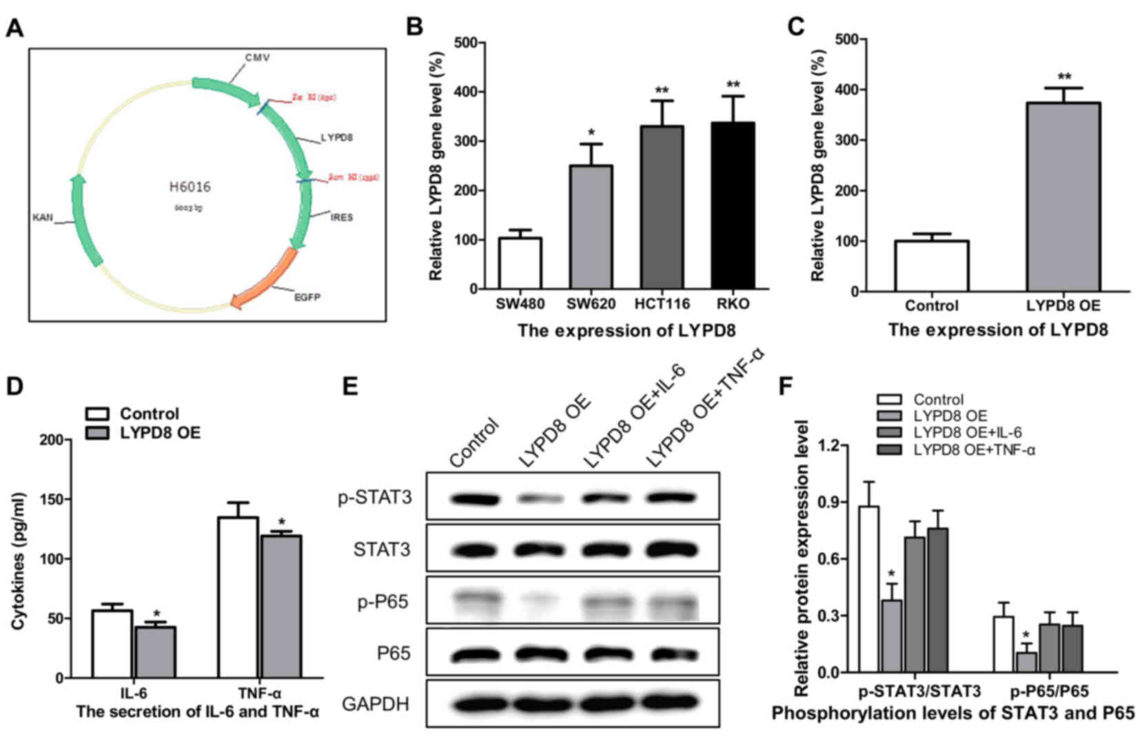 | Figure 3.Effects of the overexpression of LYPD8
on IL-6/TNF-α secretion and STAT3/P65 dephosphorylation in CRC
cells. (A) cDNA coding for the LYPD8 gene was cloned into the
eukaryotic expression vector. (B) Expression levels of LYPD8 in
different CRC cells were determined by RT-qPCR analysis, using
β-actin as a control. (C) Overexpression of LYPD8 via transient
transfection into SW480 cells was confirmed by RT-qPCR analysis.
(D) IL-6/TNF-α secretion was analyzed by ELISA in LYPD8 OE groups
of SW480 cells. (E) Western blotting revealed expression levels of
p-P65, P65, p-STAT3 and STAT3 in the control, LYPD8 OE, LYPD8 OE +
TNF-α and LYPD8 OE + IL-6 groups of SW480 cells. GAPDH was used as
a control. (F) Band intensities of western blotting for p-P65/P65
and p-STAT3/STAT3 in the control, LYPD8 OE, LYPD8 OE + TNF-α and
LYPD8 OE + IL-6 groups were analyzed. The data are reported as the
mean ± standard deviation of experiments (n=4). *P<0.05,
phosphorylation levels of P65 and STAT3 in LYPD8 OE groups vs.
control, LYPD8 OE + TNF-α and LYPD8 OE + IL-6 groups; **P<0.01,
expression of LYPD8 in control group vs. LYPD8 OE group. Control,
empty pIRES2; LYPD8, Ly6/Plaur domain-containing 8; OE,
overexpression; IL-6, interleukin-6; TNF-α, tumor necrosis
factor-α; STAT3, signal transducer and activator of transcription
3; p-, phosphorylated; RT-qPCR, reverse transcription-quantitative
polymerase chain reaction. |
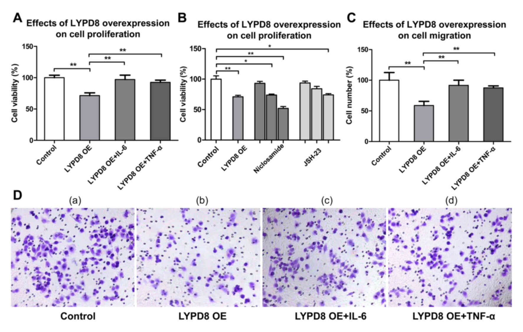 | Figure 4.Effects of the overexpression of LYPD8
on SW480 cell proliferation and migration. (A) Cell viability of
the Control, LYPD, LYPD8 OE + IL-6 and LYPD8 OE + TNF-α
groups of SW480 cells. (B) SW480 cells were treated with different
concentrations (0.5, 1 and 2 µM) of niclosamide and different
concentrations (5, 15 and 30 µM) of JSH-23, respectively. (C)
Numbers of migratory SW480 cells from the Control, LYPD8, LYPD8
OE + IL-6 and LYPD8 OE + TNF-α groups. (D) Transwell assay of
SW480 cells from the (a) Control, (b) LYPD8 OE, (c) LYPD8 OE +
IL-6 and (d) LYPD8 OE + TNF-α groups (magnification, ×200). The
data are reported as the mean ± standard deviation of experiments
(n=4). *P<0.05, **P<0.01. Control, empty pIRES2 group; LYPD8,
Ly6/Plaur domain-containing 8; OE, overexpression; IL-6,
interleukin-6; TNF-α, tumor necrosis factor-α. |
The authors therefore repeated some of these
experiments with newly purchased antibodies; they also changed the
RT-qPCR results. In the published manuscript, the authors
investigated the expression of LYPD8 mRNA expression in tissues
from stages I, II, and III whereas in the revised manuscript they
have investigated stages II and IV. In addition, the authors have
supplemented the manuscript with new transwell assays. The revised
figures are presented on the next two pages (Figs. 1–4).
Repeating these particular experiments has resulted in the
following changes being necessary to the text of the published
paper (changes are highlighted in bold):
i) The fourth sentence in the Abstract, on p. 2389,
should read as follows: “The results revealed that the expression
of LYPD8 was significantly reduced in the CRC tissue compared with
that in precancerous tissue and normal tissue, particularly in
stage IV tissue.” (‘III’ has been changed to ‘IV’).
ii) In the Materials and methods section,
“Histological analysis” subsection on p. 2390, the last
three sentences should be replaced with the following text: “The
histological sections were then stained with the DAB Kit (cat. no.
PV-9000; ZSGB-BIO, Beijing, China). All sections were observed
under a bright-field microscope (Nikon Corporation, Tokyo,
Japan).”
iii) In the “Cell culture” subsection in the
right-hand column, the first four sentences should be revised to
the following: “Four CRC cell lines (SW480, SW620, HCT116
and RKO) were used. SW480 (ATCC® CCL-228™,
organism, human; tissue, colon; disease, colorectal
adenocarcinoma), SW620 (ATCC® CCL-227™, organism, human;
tissue, colon; derived from metastatic site, lymph node; disease,
colorectal adenocarcinoma), HCT116 (ATCC® CCL-247™,
organism, human; tissue, colon; disease, colorectal carcinoma) and
RKO (ATCC® CRL-2577™, organism, human; tissue, colon;
disease, carcinoma) cells were purchased from the American Type
Culture Collection (Manassas, VA, USA). The four cell lines
within passages 10 were used in all experiments, and the cell
lines were maintained at 37°C in a humidified incubator
containing 5% CO2”. Also, in line 8 of p. 2391, the cell
lines here should be changed to “SW480, SW620 and HCT116
cells”, and on line 11, “HT29 cells” should be changed to “RKO
cells”.
iv) In the Results section, the following changes to
the text are necessary: In the “Correlation of the expression of
LYPD8 with STAT3/P65 phosphorylation and IL-6/TNF-α secretion in
patients with CRC” subsection, in the second sentence,
“immunofluorescence” should have been written as
“immunohistochemistry”, and the fourth sentence should have
read as follows: “The results of the western blotting showed that
the levels of p-P65/P65 and p-STAT3/STAT3 gradually increased
between stage II and IV (Fig. 1B
and C).” Then, the three sentences starting on line 7 on p.
2391 should now read as follows: “Following this, the gene
expression levels of LYWPD8 in stage II and IV CRC tissue,
precancerous tissue, and normal tissue were assessed using RT-qPCR
analysis (Fig. 2C). Compared with the
precancerous tissue and normal tissue, the gene expression of LYPD8
was significantly reduced in stage II and IV tissues.
Furthermore, the expression of LYPD8 was reduced in stage IV
tissue compared with that in stage II tissue.”
v) In the subsequent subsection, “Construction
and overexpression of LYPD8 in CRC cells”, the first sentence
should have read as follows: “The plasmid DNA for overexpressing
LYPD8 was constructed using the eukaryotic expression vector
(pIRES2), as shown in Fig. 3A and B,
and the relative expression levels of LYPD8 in the RTO,
SW480 HCT116 and SW620 cells were examined by RT-qPCR
analysis.
vi) In the “Overexpression of LYPD8 inhibits CRC
cell proliferation and migration” subsection, the penultimate
sentence as it appears towards the foot of p. 2392 should now read
as follows: “As shown in Fig. 4C and
D, a more marked inhibitory effect on cell migration was
observed in the LYPD8 OE group compared with that in the
control, LYPD8 OE + IL-6 and LYPD8 OE + TNF-α groups.”
vi) In the Discussion, the sentence starting on p.
2394, right-hand column, line 10 should read as follows: “By
contrast, the expression of LYPD8 was significantly reduced in
stage II and IV CRC tissues.”
vii) Finally, some revisions were necessary to the
descriptions in the figure legends for Figs. 1, 2 and
4, as follows (only the affected text
is included, and the changes are indicated in bold):
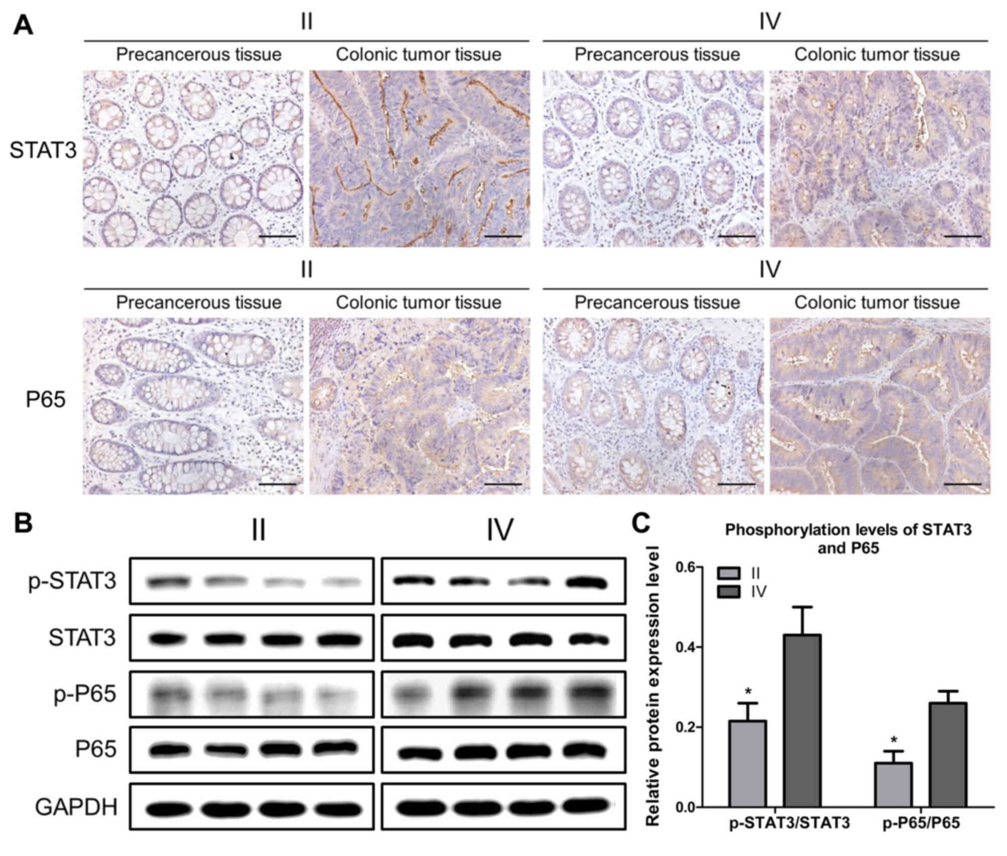
Figure 1. STAT3 and
P65 are activated in colonic tumor tissues from patients. (A)
Representative immunohistochemistry images revealing
activated STAT3 and P65 in colonic cancer tissue and precancerous
tissue. Scale bar, 100 µm. (B) Representative western blotting
revealing the expression of p-P65, P65, p-STAT3 and STAT3 in stages
II and IV colonic tumor tissues. GAPDH was used as a
control. (C) Band intensities of western blotting for p-P65/P65 and
p-STAT3/STAT3 in stage II and IV tissues were analyzed. The
data are reported as the mean ± standard deviation of experiments
(n=4). **P<0.05, phosphorylation levels of STAT3 in
stage II tissues vs. in stage IV tissues.
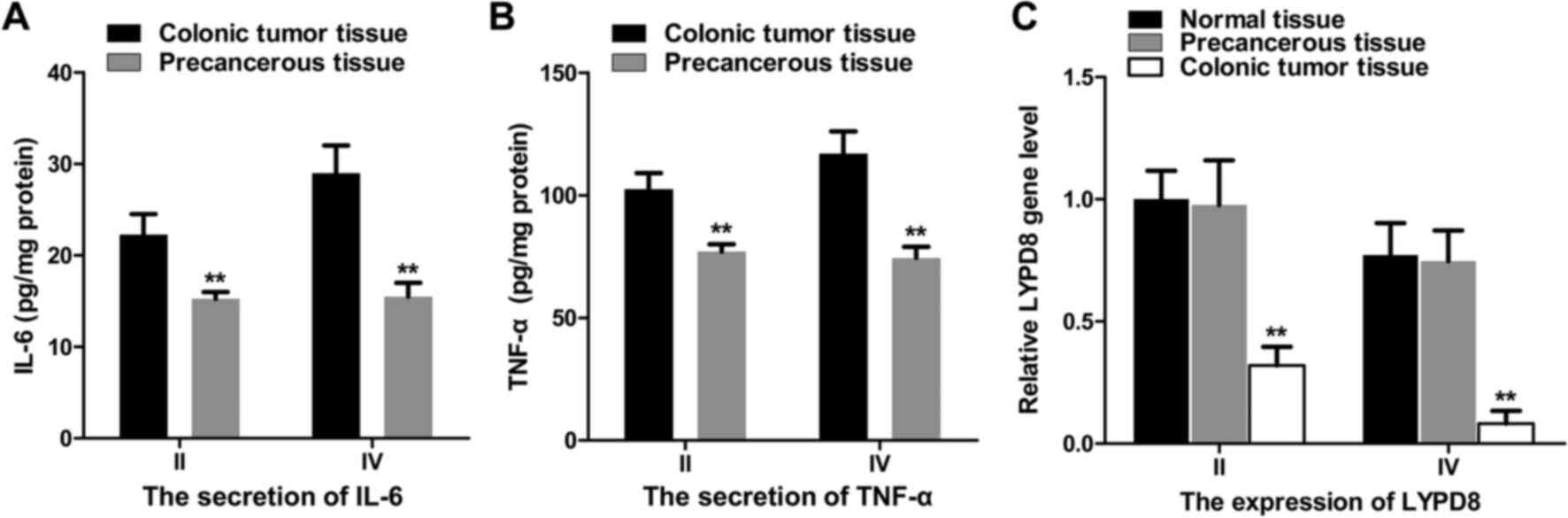
Figure 2. Association
between IL-6/TNF-α and the expression of LYPD8 in colonic tumor
tissue, precancerous tissue and normal tissue at different stages.
(A) IL-6 and (B) TNF-α secretion were analyzed by ELISA in stage II
and IV colonic tumor tissue and precancerous tissue. (C) Gene
expression of LYPD8 in stage II and IV colonic tumor tissue
and precancerous tissue. β-actin was used as a control. The data
are reported as the mean ± standard deviation of experiments (n=6).
**P<0.01, LYPD8 mRNA expression of normal tissue, precancerous
tissue vs. colonic tumor tissue in stage II and IV
tissues.
Figure 4. Effects of
the overexpression of LYPD8 on SW480 cell proliferation and
migration. (A) Cell viability of the Control, LYPD, LYPD8 OE +
IL-6 and LYPD8 OE + TNF-α groups of SW480 cells. (B) SW480
cells were treated with different concentrations (0.5, 1 and 2 µM)
of niclosamide and different concentrations (5, 15 and 30 µM) of
JSH-23, respectively. (C) Numbers of migratory SW480 cells from the
Control, LYPD8, LYPD8 OE + IL-6 and LYPD8 OE + TNF-α groups.
(D) Transwell assay of SW480 cells from the (a) Control, (b)
LYPD8 OE, (c) LYPD8 OE + IL-6 and (d) LYPD8 OE + TNF-α groups
(magnification, ×200).
Note that the replacement of the original figures
and these revisions made to the text do not drastically alter the
overall conclusions reported in the study. The authors are very
grateful to the Editor of Oncology Reports for allowing them
the opportunity to publish this Corrigendum; furthermore, they
apologize for any inconvenience caused to the readership of the
Journal.