Introduction
Toxicity is one of the major concerns in using drugs
in prevention settings since the recipients of the chemopreventive
drugs are normal subjects with high risk for developing cancer.
Similarly, most chemotherapy drugs possess severe toxicity and
numerous cases require dose reduction or treatment discontinuation.
Therefore, those agents with non-toxic or minimal side effects
would be ideal candidates as chemopreventive and chemotherapeutic
agents. Due to their proven high safety margin through centuries of
human consumption as food or as traditional medicines, natural
compounds present in fruits, vegetables and spices have drawn
special attention for chemoprevention and treatments (1–3). In the
last few decades, hundreds of different food-based natural
compounds have been investigated for their antitumor potentials.
However, only few of the promising compounds have been advanced to
clinical trials (4). Resveratrol and
epigallocatechin gallate (EGCG) are among the natural compounds
that have been tested in multiple clinical trials and their safety
profiles have been established through these trials (1).
Resveratrol is a phytoalexin present in grapes,
raspberries, blueberries, mulberries and peanuts. Red grapes,
particularly the skin, contain higher amounts of resveratrol. Some
wines also contain higher amounts of resveratrol (5,6). Several
in vitro and in vivo studies suggest that resveratrol
may demonstrate promising efficacy against head and neck cancer
(7–10). Resveratrol induced apoptosis of human
nasopharyngeal cancer cells (7) and
squamous cell carcinoma of the head and neck (SCCHN) cells
(8), prevented DMBA-induced oral
carcinogenesis in a hamster cheek pouch model (9) and selectively induced DNA damage in
SCCHN (10). Human studies reported
that orally administered resveratrol is reasonably well-tolerated,
and demonstrates only mild to moderate side effects at doses of up
to 5 g/day (11–17). However, resveratrol undergoes
extensive first-pass metabolism in the intestine and liver, and is
also a substrate for multi-drug resistant 1 (MDR1) efflux pumps
which further limit its intestinal absorption, resulting in a low
oral bioavailability (<1%) (18).
Green tea prepared from Camellia sinensis is
a widely consumed beverage in Southeast Asia and worldwide, and a
rich source of antioxidants (19).
The anti-tumor and chemopreventive properties of the constituents
of green tea have been demonstrated by multiple epidemiological,
cell culture and animal model studies (1,20). EGCG,
the most abundant catechin (10–50% of the total catechin) in green
tea, has been extensively studied for its chemopreventive,
chemotherapeutic and anticarcinogenic effects (20–22). The
safety of green tea preparations have been established by several
clinical trials (23–26). In a phase II trial in patients with
asymptomatic, Rai stage 0 to II chronic lymphocytic leukemia, 31%
patients experienced a reduction of ≥20 in the absolute lymphocyte
count at a dose of 2,000 mg polyphenon E (PPE), b.i.d (24). In another randomized,
placebo-controlled phase II trial in patients with high-risk oral
premalignant lesions, higher dose levels (750 and 1,000 mg, t.i.d)
showed clinical response and histological improvements (25). In a prostate cancer (PCa)
chemoprevention trial, green tea PPE did not reduce the likelihood
of PCa in men with baseline high grade prostatic intraepithelial
neoplasia and/or atypical small acinar proliferation, although the
number of incidents of PCa was less in the treatment arm (27). These trials clearly demonstrate that
green tea alone may not be sufficiently active for effective
chemoprevention/therapy and the combination of green tea with other
natural or synthetic agents has been suggested (24,25,28).
Although the use of drug combinations is a common
practice in the chemotherapy field, this approach has not been
widely explored using natural agents. Since ideal treatment or
chemopreventive strategies should involve non-toxic agents, a
combination of natural compounds is a logical approach. However,
the challenge is to find ideal combinations of natural compounds
which exhibit synergistic antitumor effects or synergistically
block carcinogenesis. In the present study, the antitumor effects
of the combination of resveratrol and EGCG against SCCHN cell lines
were investigated via apoptosis assay, western blotting, IHC and
using an in vivo xenograft model in nude mice.
Materials and methods
Cell lines
Cell lines used in the present study have been
previously described (29,30). Tu212, a cell line of hypopharyngeal
origin, was kindly provided by Dr Gary L. Clayman (University of
Texas M.D. Anderson Cancer Center, Houston, TX, USA). MDA686TU
(Tu686) and MSK-Leuk1 (MSK) were procured from Dr Peter G. Sacks
(New York University College of Dentistry, New York, NY, USA) in
2014 and 2012, respectively. Tu686 was established from primary
tongue cancer. The head and neck premalignant cell line MSK-Leuk1
(MSK) was established from a dysplastic leukoplakia lesion adjacent
to a squamous cell carcinoma of the tongue and was maintained in
keratinocyte basal media. SqCCY1 was established from a squamous
carcinoma of the buccal mucosa in Dr Sartorelli's laboratory (Yale
University, New Haven, CT, USA) (31)
and was obtained from Dr Shi-Yong Sun at Emory University (Atlanta,
GA, USA) in 2012. All cell lines were authenticated through
genotyping [short tandem repeats (STR) profiling]. SCCHN cell lines
were maintained in DMEM/F12 (Corning, Inc.) medium supplemented
with 10% heat-inactivated fetal bovine serum (Gibco; Thermo Fisher
Scientific, Inc.) in a 37°C and 5% CO2 humidified
incubator. STR profiles of these cell lines matched with those
published in Clinical Cancer Research 2011 (32), suggesting that Tu212 is identical with
some other SCCHN cell lines but other cell lines are unique.
Annexin V-phycoerythrin staining for
apoptosis
MDA686TU, Tu212 and SqCCy1 cells (1.5×10⁵
cells/6-cm plate) were treated with various concentrations of EGCG
(30–200 µM; Sigma-Aldrich; Merck KGaA), resveratrol (10–70 µM;
Sigma-Aldrich; Merck KGaA) and their combination (EGCG: 30–80 µM;
resveratrol: 10–20 µM) for 72 h, then trypsinized and washed in
cold 1X PBS (Corning, Inc.). The cells were then resuspended in 1X
Annexin V binding buffer (BD Biosciences), and stained with Annexin
V-phycoerythrin (Annexin V-PE; BD Biosciences) and 7-AAD (BD
Biosciences) for 15 min at room temperature. The stained samples
were analyzed using a fluorescence-activated cell sorting caliber
bench-top flow cytometer (BD Biosciences). FlowJo_v10.6.0_CL
software (Tree Star, Inc.) was used for apoptosis analysis. Total
apoptosis was considered the sum of early and late stage apoptosis.
Combination Index (CI) values were calculated by using CalcuSyn
2.11 software (Biosoft).
Western blot analysis
Whole cell lysates were extracted from Tu212,
MDA686TU and SqCCy1 cells using lysis buffer (for 200 ml: 10 ml 1 M
Tris HCl, 6 ml 5 M NaCl, 200 µl 20% sodium azide, 1 g sodium
deoxycholate, 4 ml Igepal, 2 ml 20% SDS) and the protein
concentration of each sample was determined by Quick Start Bradford
protein assay kit (Bio-Rad Laboratories, Inc.). Equal amounts of
protein (15–20 µg) from each sample were separated on 10 or 12%
sodium dodecyl sulfate-polyacrylamide gel electrophoresis
(SDS-PAGE). The proteins were transferred to polyvinylidene
difluoride (PVDF) membranes (EMD Millipore), blocked in 5% non-fat
skimmed milk for 2 h at room temperature and incubated with
appropriately diluted specific primary antibodies overnight at 4°C,
followed by incubation with a secondary antibody for 1 h at room
temperature. Mouse anti-β-actin (Sigma-Aldrich; Merck KGaA)
antibody was used as a sample loading control. Immunostained
protein bands were detected with an enhanced chemiluminescence kit
(Pierce; Thermo Fisher Scientific, Inc.). Phosphorylated (p)-AKT
(product no. 4060), AKT (product no. 4685), PARP (product no.
9542), cleaved caspase-3 (product no. 9661), p-mTOR (product no.
5536), p-S6 (product no. 4858), S6 (product no. 2317), p-4EBP-1
(product no. 2855), 4E-BP1 (product no. 9644) primary antibodies
were purchased from Cell Signaling Technology, Inc. and Mcl-1 (cat.
no. sc-377487) and survivin (cat. no. sc-374616) from Santa Cruz
Biotechnology, Inc. Anti-mouse (product no. W402B) and anti-rabbit
IgG (product no. W401B) were purchased from Promega Corporation.
All primary antibodies were diluted to 1:1,000 and secondary
antibodies to 1:10,000 in 5% non-fat skimmed milk.
In vivo xenograft model
The animal experiments were approved by the
Institutional Animal Care and Use Committee of Emory University
(DAR-2002630-050517BN). A total of 20 female nude mice (athymic
nu/nu; Taconic Biosciences, Inc.), aged 4–6 weeks (weight,
~20 g), were used in the present study. Mice were maintained on
Alpha-Dry bedding in temperature (22±2°C) and humidity (30–50%)
controlled rooms with a 12-h light/dark cycle. Rodent Chow No. 5010
(LabDiet) and autoclaved water were provided ad libitum.
After adaptation for a few days in the new environment, the mice
were subcutaneously injected with 2.5×10⁶ Tu212 cells
into the right flank. After approximately a week when visible
tumors had formed, the mice were randomly divided into four groups
(n=5 in each group). Each mouse was orally gavaged with vehicle
control (0.5% carboxymethyl cellulose), EGCG (125 mg/kg in water),
resveratrol (30 mg/kg in 0.5% carboxymethyl cellulose) or a
combination of EGCG (125 mg/kg) and resveratrol (30 mg/kg) 5 days a
week. The tumor size (larger diameter and smaller diameter) was
measured 2 times a week using a digital caliper. The tumor volume
was calculated using the formula: V=π/6 × larger diameter ×
(smaller diameter)2. Growth curves were plotted using
the average tumor volume within each experimental group at the set
time-points (0, 4, 6, 8, 11, 14, 20, 25 and 28 days). At the end of
the study, mice were euthanized with CO2 from a gas tank
(flow rate: 30% chamber vol/min); the mice were placed in a
euthanasia chamber to allow visualization during the procedure.
Each mouse was observed for lack of respiration and faded eye
color. CO2 flow was maintained for a minimum of 1 min
after respiration ceased and sacrifice was confirmed through fixed
and dilated pupils.
Immunohistochemistry and TUNEL
assay
Formalin-fixed (Z-Fix; 10% aqueous buffered zinc
formalin; Anatech Ltd.; overnight at room temperature)
paraffin-embedded (FFPE) xenograft tumor slides (3 microns) were
deparaffinized with xylene and dehydrated with a series of alcohol
treatments. The slides were incubated with 3% hydrogen peroxide in
water to block endogenous peroxidase activity and subjected to
antigen retrieval using 1X citrate buffer (pH 6.0) in a microwave
followed by cooling at room temperature. After washing, the slides
were incubated with a blocking buffer provided in a kit (Vectastain
Kit cat. no. PK7800; Vector Laboratories, Inc.) followed by
incubation with primary antibodies Ki67 (1:400 dilution; product
code ab15580; Abcam), p-AKT (1:50 dilution, product no. 4060), p-S6
(1:50 dilution; product no. 4858) and p-4EBP1 (1:100 dilution;
product no. 2855) (all from Cell Signaling Technology, Inc.)
overnight in a refrigerator. The slides were washed and incubated
with a ready to use secondary antibody provided with the kit for 30
min at room temperature followed by chromogen visualization using
3,3-diaminobenzideine (DAB) provided with the kit. The slides were
counterstained with hematoxylin for 1 min at room temperature for
nuclei visualization and five random areas were selected for
analysis. The TUNEL assay was performed by immunofluorescence using
the same specimens as aforementioned, following the procedure
provided by the manufacturer (cat. no. C10617; Life Technologies;
Thermo Fisher Scientific, Inc.). To analyze the assay results, the
total number of cells and the positive number of cells in the same
area were counted for five random areas; the result was presented
as an average ratio of the positive number of cells out of the
total number of cells. The magnification was ×200 for all IHC
images.
Statistical analysis
The effect of the combination of EGCG and
resveratrol on the inhibition of tumor growth was assessed through
in vivo and in vitro experiments. For in vivo
data analyses, the expression of TUNEL and Ki67 among the treatment
groups EGCG, resveratrol, combination of EGCG and resveratrol, and
control was compared by using two-way ANOVA after adjusting the
area from where the cells were obtained. Bonferroni adjustment was
performed for multiple comparisons between EGCG, resveratrol,
combination, and control. For in vivo tumor growth analyses,
a mixed effect model with random subject effect was used by
accounting the repeated outcomes over time. The data were collected
repeatedly 9 times from the same mouse and the tumor volume was
measured. In the model, compound symmetry covariance structure
based on minimum AIC criteria was used. The tumor volume was
compared between treatment groups EGCG, resveratrol, and
combination vs. control. The comparisons between the control and
treatment groups were adjusted using Bonferroni correction. All
statistical analyses were performed using SAS version 9.4 (SAS
Institute). Results were also validated by cross-checking with
other software, R (version 3.6.2; The R Foundation for Statistical
Computing) (33). The results were
considered statistically significant when the P-values were
<0.05.
Results
Synergistic apoptosis induced by the
combination of EGCG and resveratrol
It has been previously reported by our research
group that the combination of two natural compounds, EGCG and
luteolin, induced enhanced antitumor effects against both lung and
head and neck cancers both in vitro and in vivo
(29). In an effort to identify new
combinations of natural compounds with synergistic apoptosis, the
combination of EGCG and resveratrol, a combination never explored
in vivo and in in head and neck cancer were studied. First,
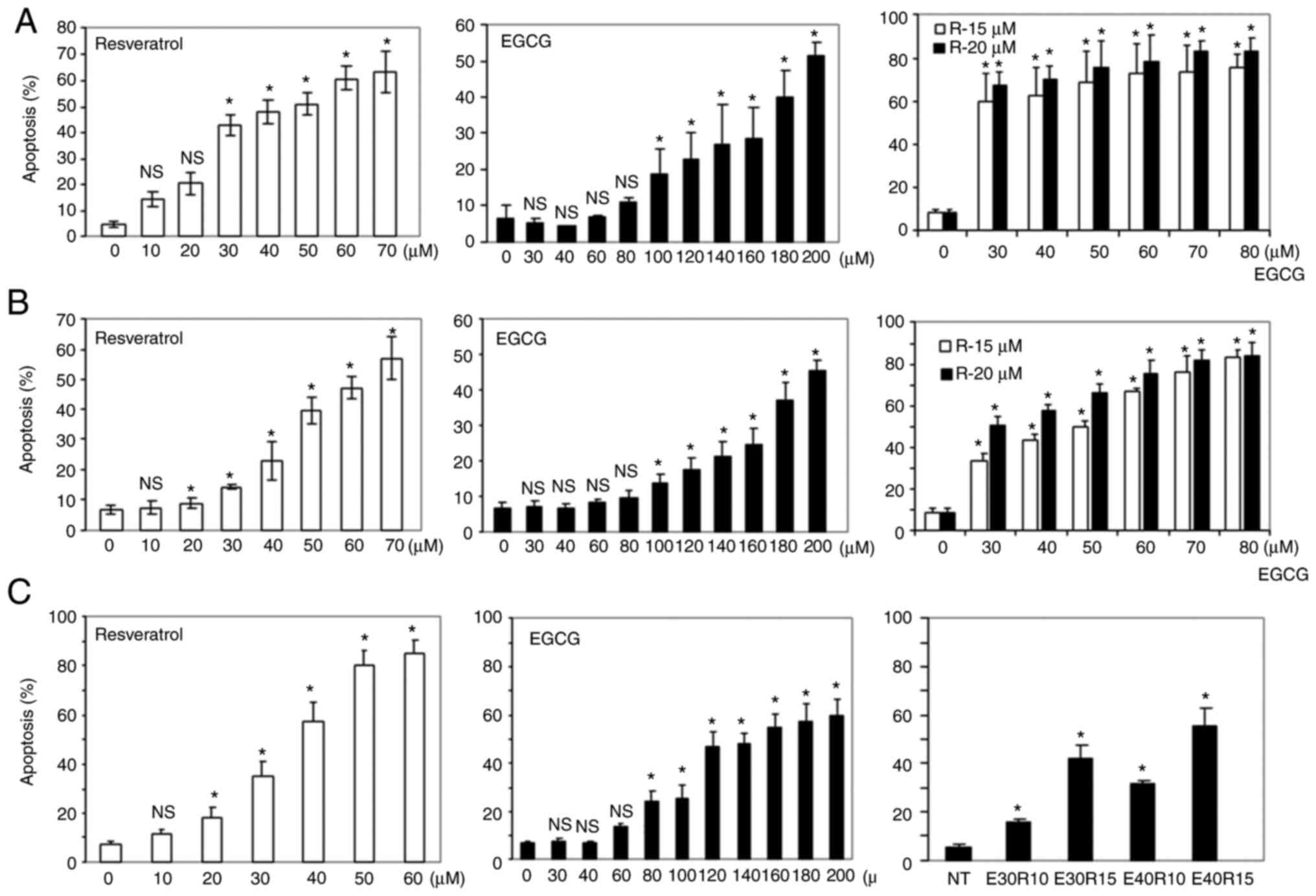
the sensitivity of three SCCHN cell lines to different doses of
EGCG and resveratrol were examined, and a dose-dependent apoptosis
induced by the compounds as single agents was revealed (Fig. 1A-C). Next, induction of apoptosis
using low doses of EGCG in combination with two doses of
resveratrol was examined. As revealed in Fig. 1A-C right panels, the combination
treatment significantly increased apoptosis although single agents
induced markedly little apoptosis at these doses. In order to
confirm synergistic apoptotic effects, the apoptosis data was
analyzed using CalcuSyn software and it was revealed that
combination of the two agents induced highly synergistic apoptosis
as indicated by combination index values <1 (Table I). Synergistic apoptosis was also
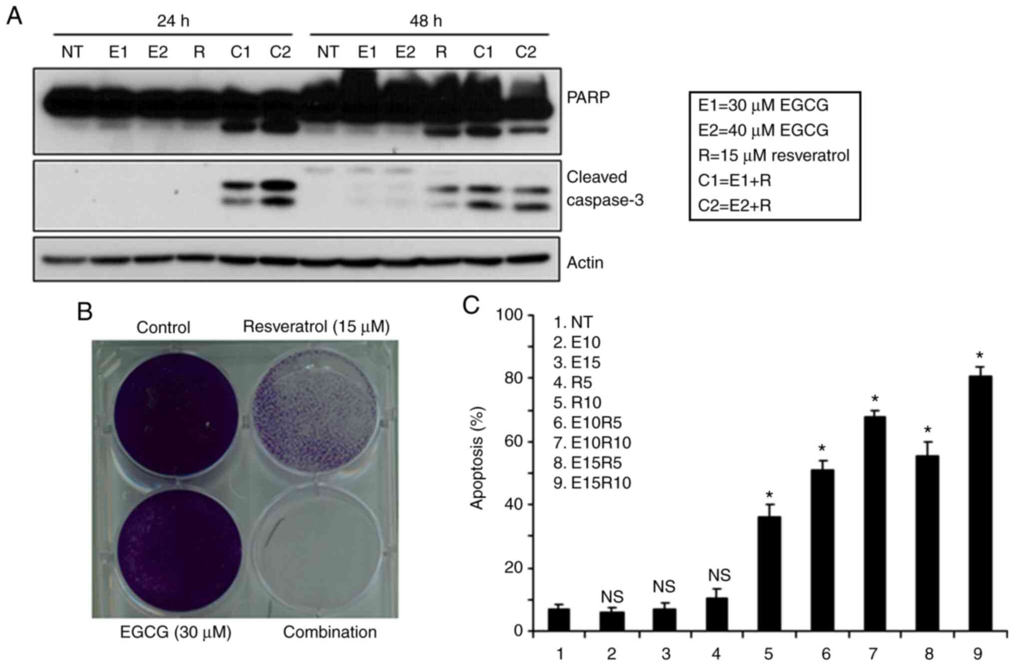
supported by cleavage of PARP and caspase-3 (Fig. 2A). Combination of the two agents at
lower doses also completely eradicated tumors cells (Fig. 2B). The efficacy of the combination of
EGCG and resveratrol against a premalignant cell line, MSK-LEUK1,
was next tested. As revealed in Fig.
2C, the premalignant cell line was sensitive to the combination
of considerably lower doses of each agent, suggesting that the
combination of EGCG and resveratrol is also suitable for
chemoprevention.
 | Table I.Combination index at different
combinations. |
Table I.
Combination index at different
combinations.
|
|
| Combination
Index |
|---|
|
|
|
|
|---|
| Cell line | EGCG (µM) | Resveratrol (15
µl) | Resveratrol (20
µl) |
|---|
| Tu212 | 30 | 0.93 | 0.73 |
|
| 35 | 0.68 | 0.63 |
|
| 40 | 0.66 | 0.62 |
|
| 45 | 0.62 | 0.68 |
|
| 50 | 0.63 | 0.74 |
|
| 55 | 0.61 | 0.43 |
|
| 60 | 0.59 | 0.47 |
| SqCCy1 | 30 | 0.41 | 0.42 |
|
| 40 | 0.43 | 0.44 |
|
| 50 | 0.43 | 0.43 |
|
| 60 | 0.47 | 0.44 |
|
| 70 | 0.49 | 0.43 |
|
| 80 | 0.49 | 0.44 |
| MDA686TU | 30 | 0.49 | 0.46 |
|
| 40 | 0.47 | 0.46 |
|
| 50 | 0.48 | 0.46 |
|
| 60 | 0.43 | 0.44 |
|
| 70 | 0.41 | 0.43 |
|
| 80 | 0.41 | 0.45 |
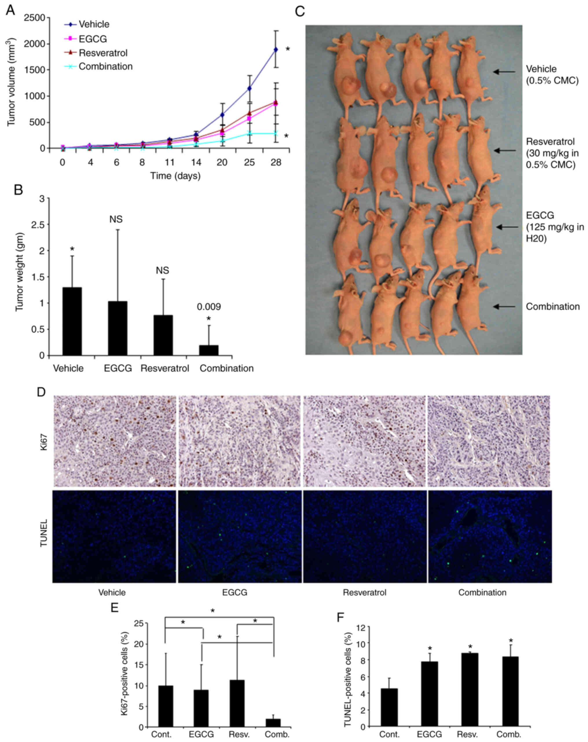
Inhibition of growth of xenograft
tumors in nude mice by the combination of EGCG and resveratrol
The in vivo antitumor efficacy of the
combined treatment with EGCG and resveratrol was investigated in
xenografted mice bearing Tu212 cells, an established SCCHN cell
line known to generate tumor in nude mice (34–36). Each
group consisted of five mice. Although EGCG and resveratrol as
single agents had minimal effect on tumor growth, their combination
inhibited tumor growth to a statistically significant level
(P=0.026) (Fig. 3A) compared to
single-agent resveratrol (P=0.182) and EGCG (P=0.130). The tumor
weight at the end of the study was also assessed. Combination of
the two agents also significantly inhibited tumor weight (Fig. 3B). Images of tumors obtained after
sacrifice are presented in Fig. 3C.
To confirm the aforementioned results in tissue levels, tumor
tissues were also stained for the expression of Ki67 (proliferation
marker) and TUNEL (apoptosis marker). Ki67- and TUNEL-positive
cells were quantified and it was revealed that the combination of
EGCG and resveratrol significantly inhibited Ki67-positive cells
(Fig. 3D and E). The number of
TUNEL-positive cells was increased similarly by EGCG, resveratrol
and their combination (Fig. 3D and
F). These in vivo data thus confirmed the in
vitro results.
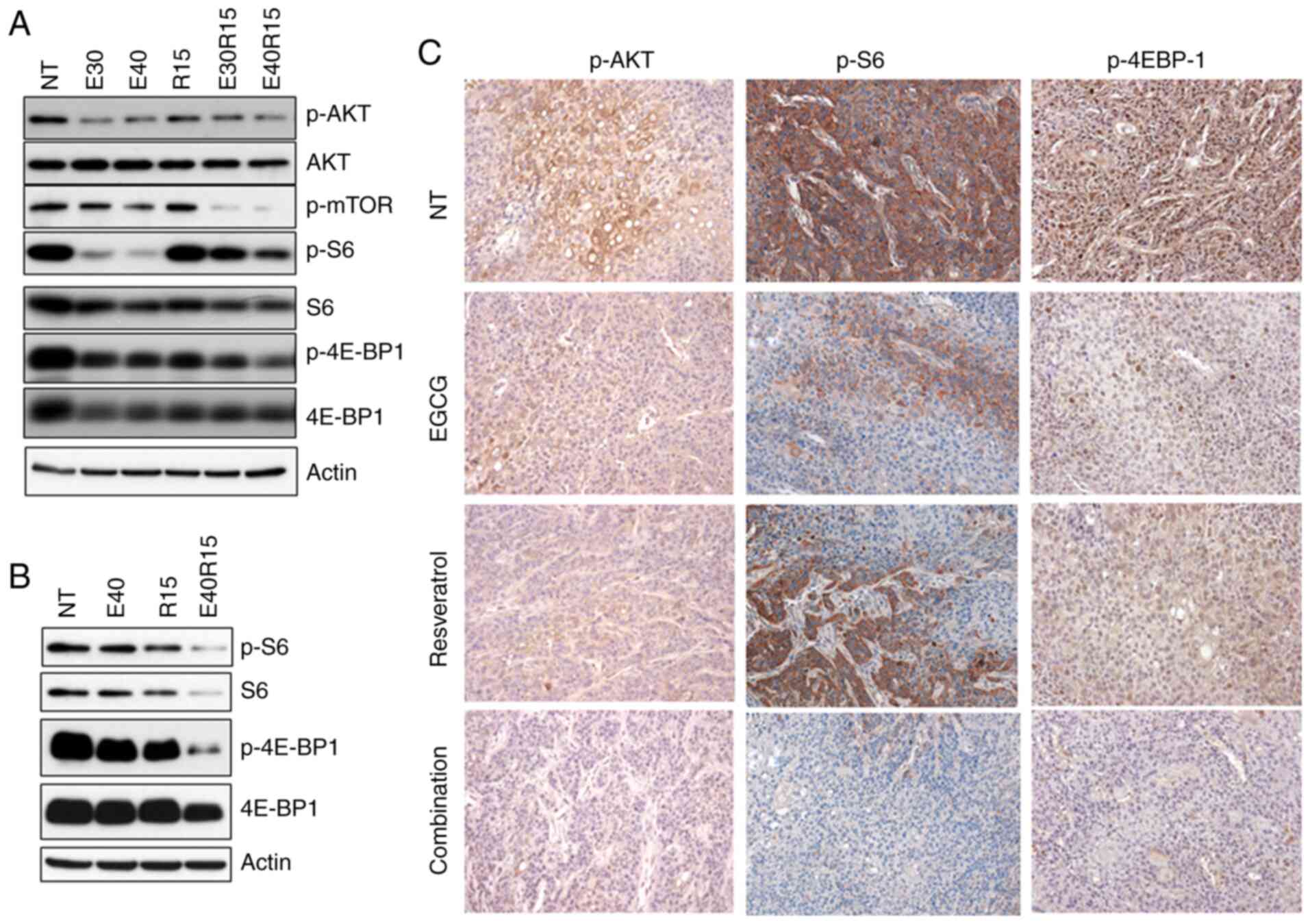
Inhibition of AKT-mTOR signaling by
the combination of EGCG and resveratrol
Activation of AKT and its target mTOR, has been
observed in >80% of SCCHN lesions (37,38). The
effect of EGCG, resveratrol and their combination on AKT-mTOR
pathway markers was next examined. As revealed in Fig. 4A and B, treatment of SCCHN cells with
EGCG, resveratrol and their combination markedly inhibited p-AKT,
p-mTOR and downstream p-S6 and p-4E-BP1 in vitro. In order
to associate in vitro data with in vivo biomarker
modulation, xenografted tumor tissues were stained for the
expression of p-AKT, p-S6 and p-4E-BP1 (Fig. 4C). As revealed in Fig. 4C, treatment with EGCG, resveratrol and
their combination inhibited the expression of p-AKT. On the other
hand, only the combination of EGCG and resveratrol strongly
inhibited the expression of p-S6. In the case of p-4E-BP1,
treatment with EGCG, resveratrol and their combination inhibited
the expression of p-4E-BP1. Combination of the two agents also
significantly inhibited the expression of p-4E-BP1 as compared with
single agent treatments. To confirm the role of inhibition of the
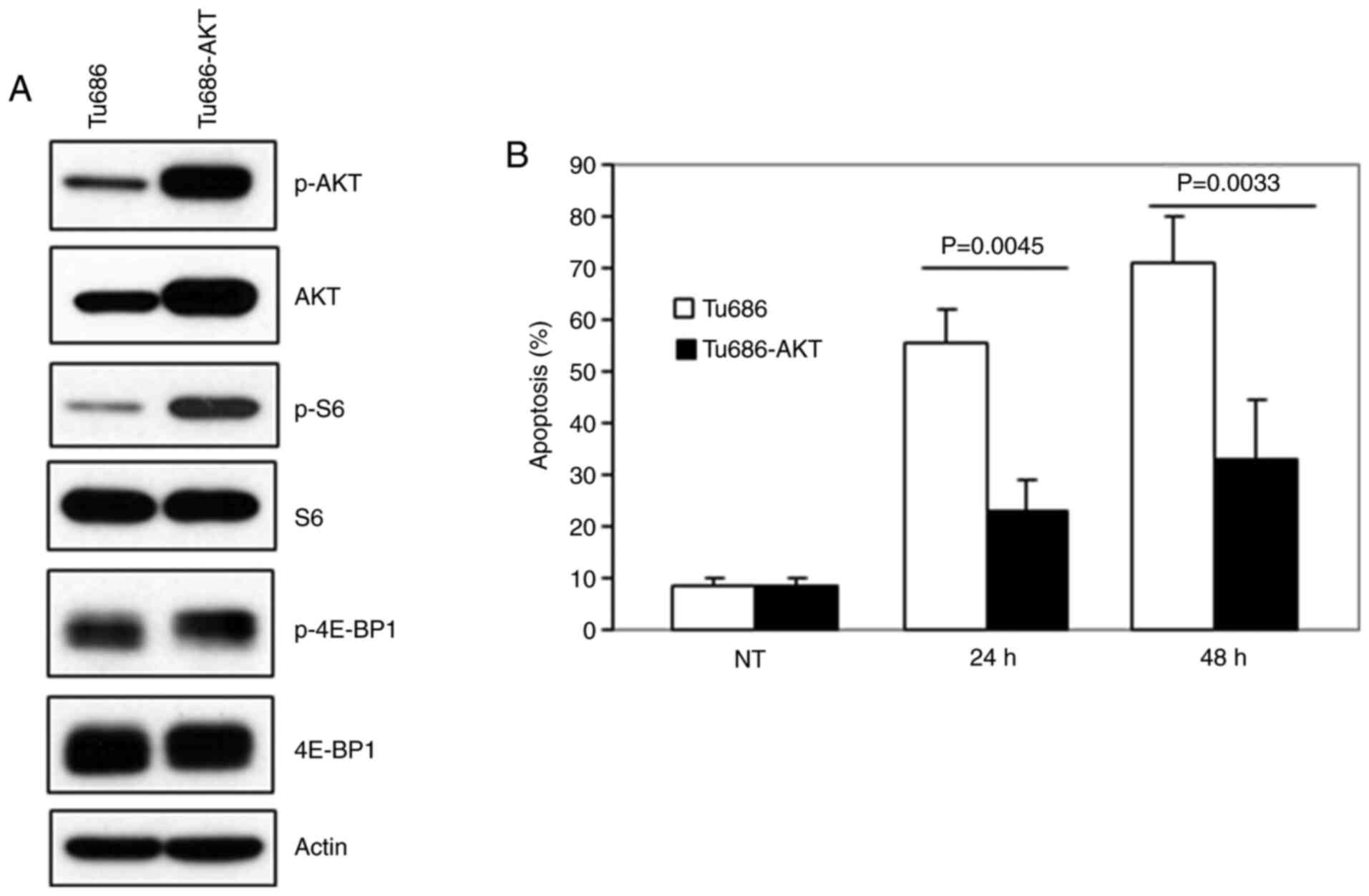
AKT pathway in apoptosis, MDA686TU cells overexpressing
constitutively active (CA-AKT) were used (39). As revealed in Fig. 5A, overexpression of CA-AKT increased
the basal level of S6 phosphorylation more strongly than 4E-BP1
phosphorylation. Overexpression of CA-AKT also significantly
protected cells from apoptosis induced by the combination of EGCG
and resveratrol, suggesting that inhibition of AKT-mTOR signaling
is required for the induction of apoptosis by the combination of
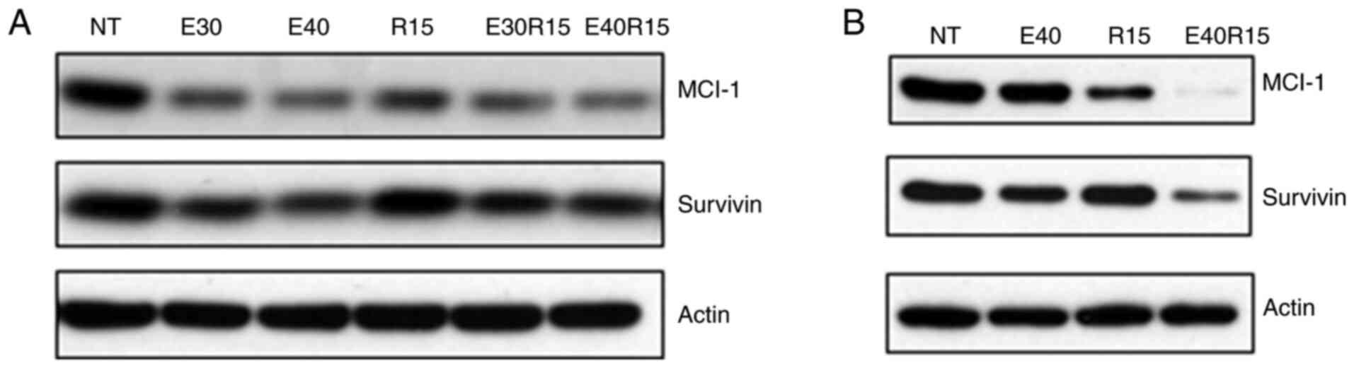
the two agents. It was also revealed that the combination of the
two agents more strongly inhibited the expression of the survival
proteins Mcl-1 and survivin as compared to single-agent treatment
(Fig. 6).
Discussion
To date, the most successful drugs for the cure of
cancer and eradication of tumor cells from the body are cytotoxic
chemotherapy drugs, molecularly targeted agents and immunotherapy
drugs (40,41). These agents are capable of inducing
apoptosis, although most have significant toxicities. In contrast,
cytostatic drugs induce growth arrest without eliminating cancer
cells from the body, thus the acquisition of resistance is a common
phenomenon (42,43). While relatively non-toxic, low
bioavailability limits the success of food-derived natural
compounds in chemoprevention and treatment of cancers (1,3),
combinatorial approaches with synergistic effects may be a solution
to improve the efficacy of food-derived natural agents. In the
present study, apoptosis using the combination of two natural
compounds, EGCG and resveratrol was investigated. The present data
clearly demonstrated that high concentrations are required for the
induction of significant apoptosis when these agents are used
alone. Due to the low bioavailability of these compounds, it may
not be possible to achieve such high concentrations in vivo
that will be effective for tumor regression. In contrast,
combination of the two agents induced synergistic apoptosis at
relatively low doses. The in vitro data is supported by
in vivo xenograft data revealing that only the combination
of two agents inhibited tumor growth (reduction of tumor volume and
tumor weight, and expression of Ki67) with a statistically
significant P-value. The in vitro data also suggest that
premalignant cells (MSK-LEUK1) are markedly more sensitive to the
combination of the two agents (i.e, a lower dose was required for
similar effects), and thus this combination may also be suitable in
chemoprevention settings. To the best of our knowledge, this is the
first such study reporting in vitro and in vivo
antitumor effects as well as synergistic apoptosis of the
combination of two natural compounds EGCG and resveratrol against
head and neck cancer.
The mechanism of synergistic apoptosis induced by
the combination was also explored. Anti-apoptotic Bcl-2 proteins
including Bcl-2, Bcl-xL and Mcl-1 serve as mitochondrial
gatekeepers and maintain mitochondrial membrane integrity (44). Inhibition of the expression of these
anti-apoptotic Bcl-2 proteins favors apoptosis by inhibiting
mitochondrial membrane potential, allowing release of cytochrome
c in the cytoplasm to activate the apoptotic cascade.
Survivin, a member of the inhibitors of apoptosis (IAP) family,
also protects cells from apoptosis. Multiple cell survival pathways
are associated with the pathogenesis of SCCHN. In the present
study, it was revealed that treatment of cells with EGCG,
resveratrol and their combination inhibited the expression of Mcl-1
and survivin.
Genome-wide sequence analyses of SCCHN have
identified the most frequent genetic alterations in these diseases
(37,38). Several mutations were identified in
the phosphoinositide 3-kinase (PI3K)/mammalian target of rapamycin
(mTOR) pathway, including PIK3CA (8–10%), TSC1/2
(5–8%), and PTEN (5–10%), all of which result in
PI3K/AKT/mTOR pathway activation (37,38,45–48).
Activation of AKT and its target mTOR has been observed in >80%
of SCCHN lesions, implicating the AKT/mTOR pathway as an excellent
target in SCCHN. The present data demonstrated that treatment with
EGCG, resveratrol or their combination inhibited p-AKT, p-mTOR and
downstream p-S6 and p-4E-BP1 both in vitro and in
vivo. Notably, overexpression of constitutively active AKT
(CA-AKT) which rescues p-AKT and downstream mTOR pathways,
significantly protected cells suggesting the critical role of p-AKT
inhibition in apoptosis induced by the combination of EGCG and
resveratrol. A number of previous studies suggest that inhibition
of mTOR pathways successfully prevents the development of SCCHN in
4NQO-induced and genetic mouse models (49–51). It
was also previously reported by our research group that inhibition
of pS6, a downstream target of mTOR signaling was correlated with
clinical response in our celecoxib/erlotinib trial of SCCHN
chemoprevention (52). Short
noncoding RNAs including microRNAs (also known as miRNAs or miRs)
modulate gene expression at the post transcriptional level via
sequence-specific interactions with the cognate mRNA targets
(53). miRNAs regulate gene silencing
through both degradations of the mRNA and inhibition of translation
and play crucial roles in carcinogenesis as well as in regulating
cell growth and survival (54).
Accumulated evidence suggests that both EGCG and resveratrol
modulate the expression of miRs. For example, EGCG suppressed
growth and tumorigenicity in breast cancer cells by downregulation
of miR-25 (55). Another study
demonstrated that EGCG modulated the expression of miR-203,
miR-125b, miR-210 and miR-29a in cervical carcinoma cell lines
(56). Similarly, triacetyl
resveratrol upregulated miR-200 in pancreatic cancer cell lines
(57). It is possible that the
combination of EGCG and resveratrol inhibits tumor cell growth by
modulating the expression of miRs, which needs to be confirmed
through future studies.
Unique identitity of cell lines used for evaluting
anticancer drugs is critical for efficacy studies as well as
elucidating their mechanism of action. Misidentification and cross
contamination is unfortunately common and can generate misleading
results. One of the limitations of the present study is the use of
the Tu212 cell line. The unique identity of this cell line is under
question since some other SCCHN cell lines used by the head and
neck community have a similar STR profile. Multiple cell lines were
used to confirm the present results. All other cell lines were
confirmed by STR profiling to have a unique identity. In
conclusion, the present study has identified that the combination
of EGCG and resveratrol induced synergistic apoptosis and inhibited
SCCHN xenograft growth in vivo, and explored the potential
mechanisms. The present results have high translational
significance and will pave the way for further clinical development
of the combination of EGCG and resveratrol, particularly for the
chemoprevention of head and neck cancers and other malignacies.
Acknowledgements
The authors thank Dr Anthea Hammond (Scientific
Writer, Department of Hematology and Medical Oncology, Winship
Cancer Institute of Emory University) for editing the
manuscript.
Funding
The present study was supported by the start-up
funding from Winship Cancer Institute of Emory University (ARA),
the Faculty Research Support grant from Marshall University School
of Pharmacy (ARA), the WV–INBRE grant (no. P20GM103434) (ARA) and
the Head and Neck Cancer SPORE (grant no. P50CA128613) (DMS and
GC).
Availability of data and materials
The datasets used during the present study are
available from the corresponding author upon reasonable
request.
Authors' contributions
All the authors contributed to the conception and
design of the study. ARMRA, DW and SN acquired the data. ARMRA, SN,
RL, ZGC and DMS analyzed and interpreted the data. ARMRA, SN, RL
drafted the manuscript. ARMRA, DW, ZGC and DMS critically revised
the manuscript for important intellectual content. All authors read
and approved the final version of the manuscript.
Ethics approval and consent to
participate
The animal experiments were approved by the
Institutional Animal Care and Use Committee of Emory University
(DAR-2002630-050517BN).
Patient consent for publication
Not applicable.
Competing interests
The authors declare that they have no competing
interests.
References
|
1
|
Amin AR, Kucuk O, Khuri FR and Shin DM:
Perspectives for cancer prevention with natural compounds. J Clin
Oncol. 27:2712–2725. 2009. View Article : Google Scholar : PubMed/NCBI
|
|
2
|
Rahman MA, Amin AR and Shin DM:
Chemopreventive potential of natural compounds in head and neck
cancer. Nutr Cancer. 62:973–987. 2010. View Article : Google Scholar : PubMed/NCBI
|
|
3
|
Gullett NP, Ruhul AR, Amin AR, Bayraktar
S, Pezzuto JM, Shin DM, Khuri FR, Aggarwal BB, Surh YJ and Kucuk O:
Cancer prevention with natural compounds. Semin Oncol. 37:258–281.
2010. View Article : Google Scholar : PubMed/NCBI
|
|
4
|
Park W, Amin AR, Chen ZG and Shin DM: New
perspectives of curcumin in cancer prevention. Cancer Prev Res
(Phila). 6:387–400. 2013. View Article : Google Scholar : PubMed/NCBI
|
|
5
|
Burns J, Yokota T, Ashihara H, Lean MEJ
and Crozier A: Plant foods and herbal sources of resveratrol. J
Agric Food Chem. 50:3337–3340. 2002. View Article : Google Scholar : PubMed/NCBI
|
|
6
|
Cione E, Torre CL, Cannataro R, Caroleo
MC, Plastina P and Gallelli L: Quercetin, epigallocatechin gallate,
curcumin, and resveratrol: From dietary sources to human MicroRNA
modulation. Molecules. 25:632020. View Article : Google Scholar
|
|
7
|
Huang TT, Lin HC, Chen CC, Lu CC, Wei CF,
Wu TS, Liu FG and Lai HC: Resveratrol induces apoptosis of human
nasopharyngeal carcinoma cells via activation of multiple apoptotic
pathways. J Cell Physiol. 226:720–728. 2011. View Article : Google Scholar : PubMed/NCBI
|
|
8
|
Lin HY, Sun M, Tang HY, Simone TM, Wu YH,
Grandis JR, Cao HJ, Davis PJ and Davis FB: Resveratrol causes
COX-2- and p53-dependent apoptosis in head and neck squamous cell
cancer cells. J Cell Biochem. 104:2131–2142. 2008. View Article : Google Scholar : PubMed/NCBI
|
|
9
|
Berta GN, Salamone P, Sprio AE, Di Scipio
F, Marinos LM, Sapino S, Carlotti ME, Cavalli R and Di Carlo F:
Chemoprevention of 7,12-dimethylbenz[a]anthracene (DMBA)-induced
oral carcinogenesis in hamster cheek pouch by topical application
of resveratrol complexed with 2-hydroxypropyl-beta-cyclodextrin.
Oral Oncol. 46:42–48. 2010. View Article : Google Scholar : PubMed/NCBI
|
|
10
|
Tyagi A, Gu M, Takahata T, Frederick B,
Agarwal C, Siriwardana S, Agarwal R and Sclafani RA: Resveratrol
selectively induces DNA Damage, independent of Smad4 expression, in
its efficacy against human head and neck squamous cell carcinoma.
Clin Cancer Res. 17:5402–5411. 2011. View Article : Google Scholar : PubMed/NCBI
|
|
11
|
Patel KR, Scott E, Brown VA, Gescher AJ,
Steward WP and Brown K: Clinical trials of resveratrol. Ann N Y
Acad Sci. 1215:161–169. 2011. View Article : Google Scholar : PubMed/NCBI
|
|
12
|
Brown VA, Patel KR, Viskaduraki M, Crowell
JA, Perloff M, Booth TD, Vasilinin G, Sen A, Schinas AM, Piccirilli
G, et al: Repeat dose study of the cancer chemopreventive agent
resveratrol in healthy volunteers: Safety, pharmacokinetics, and
effect on the insulin-like growth factor axis. Cancer Res.
70:9003–9011. 2010. View Article : Google Scholar : PubMed/NCBI
|
|
13
|
Patel KR, Brown VA, Jones DJ, Britton RG,
Hemingway D, Miller AS, West KP, Booth TD, Perloff M, Crowell JA,
et al: Clinical pharmacology of resveratrol and its metabolites in
colorectal cancer patients. Cancer Res. 70:7392–7399. 2010.
View Article : Google Scholar : PubMed/NCBI
|
|
14
|
Chow HH, Garland LL, Hsu CH, Vining DR,
Chew WM, Miller JA, Perloff M, Crowell JA and Alberts DS:
Resveratrol modulates drug- and carcinogen-metabolizing enzymes in
a healthy volunteer study. Cancer Prev Res (Phila). 3:1168–1175.
2010. View Article : Google Scholar : PubMed/NCBI
|
|
15
|
la Porte C, Voduc N, Zhang G, Seguin I,
Tardiff D, Singhal N and Cameron DW: Steady-State pharmacokinetics
and tolerability of trans-resveratrol 2000 mg twice daily with
food, quercetin and alcohol (ethanol) in healthy human subjects.
Clin Pharmacokinet. 49:449–454. 2010. View Article : Google Scholar : PubMed/NCBI
|
|
16
|
Almeida L, Vaz-da-Silva M, Falcao A,
Soares E, Costa R, Loureiro AI, Fernandes-Lopes C, Rocha JF, Nunes
T, Wright L and Soares-da-Silva P: Pharmacokinetic and safety
profile of trans-resveratrol in a rising multiple-dose study in
healthy volunteers. Mol Nutr Food Res. 53 (Suppl 1):S7–S15. 2009.
View Article : Google Scholar : PubMed/NCBI
|
|
17
|
Boocock DJ, Faust GE, Patel KR, Schinas
AM, Brown VA, Ducharme MP, Booth TD, Crowell JA, Perloff M, Gescher
AJ, et al: Phase I dose escalation pharmacokinetic study in healthy
volunteers of resveratrol, a potential cancer chemopreventive
agent. Cancer Epidemiol Biomarkers Prev. 16:1246–1252. 2007.
View Article : Google Scholar : PubMed/NCBI
|
|
18
|
Juan ME, Gonzalez-Pons E and Planas JM:
Multidrug resistance proteins restrain the intestinal absorption of
trans-resveratrol in rats. J Nutr. 140:489–495. 2010. View Article : Google Scholar : PubMed/NCBI
|
|
19
|
Shirakami Y, Shimizu M and Moriwaki H:
Cancer chemoprevention with green tea catechins: From bench to bed.
Curr Drug Targets. 13:1842–1857. 2012. View Article : Google Scholar : PubMed/NCBI
|
|
20
|
Kim JW, Amin AR and Shin DM:
Chemoprevention of head and neck cancer with green tea polyphenols.
Cancer Prev Res (Phila). 3:900–909. 2010. View Article : Google Scholar : PubMed/NCBI
|
|
21
|
Chakrawarti L, Agrawal R, Dang S, Gupta S
and Gabrani R: Therapeutic effects of EGCG: A patent review. Expert
Opin Ther Pat. 26:907–916. 2016. View Article : Google Scholar : PubMed/NCBI
|
|
22
|
Gan RY, Li HB, Sui ZQ and Corke H:
Absorption, metabolism, anti-cancer effect and molecular targets of
epigallocatechin gallate (EGCG): An updated review. Crit Rev Food
Sci Nutr. 58:924–941. 2018. View Article : Google Scholar : PubMed/NCBI
|
|
23
|
Pisters KM, Newman RA, Coldman B, Shin DM,
Khuri FR, Hong WK, Glisson BS and Lee JS: Phase I trial of oral
green tea extract in adult patients with solid tumors. J Clin
Oncol. 19:1830–1838. 2001. View Article : Google Scholar : PubMed/NCBI
|
|
24
|
Shanafelt TD, Call TG, Zent CS, LaPlant B,
Bowen DA, Roos M, Secreto CR, Ghosh AK, Kabat BF, Lee MJ, et al:
Phase I trial of daily oral Polyphenon E in patients with
asymptomatic Rai stage 0 to II chronic lymphocytic leukemia. J Clin
Oncol. 27:3808–3814. 2009. View Article : Google Scholar : PubMed/NCBI
|
|
25
|
Tsao AS, Liu D, Martin J, Tang XM, Lee JJ,
El-Naggar AK, Wistuba I, Culotta KS, Mao L, Gillenwater A, et al:
Papadimitrakopoulou V, Phase II randomized, placebo-controlled
trial of green tea extract in patients with high-risk oral
premalignant lesions. Cancer Prev Res (Phila). 2:931–941. 2009.
View Article : Google Scholar : PubMed/NCBI
|
|
26
|
Basu A, Du M, Sanchez K, Leyva MJ, Betts
NM, Blevins S, Wu M, Aston CE and Lyons TJ: Green tea minimally
affects biomarkers of inflammation in obese subjects with metabolic
syndrome. Nutrition. 27:206–213. 2011. View Article : Google Scholar : PubMed/NCBI
|
|
27
|
Kumar NB, Pow-Sang J, Egan KM, Spiess PE,
Dickinson S, Salup R, Helal M, McLarty J, Williams CR, Schreiber F,
et al: Randomized, placebo-controlled trial of green tea catechins
for prostate cancer prevention. Cancer Prev Res (Phila). 8:879–887.
2015. View Article : Google Scholar : PubMed/NCBI
|
|
28
|
Shin DM: Oral cancer prevention advances
with a translational trial of green tea. Cancer Prev Res (Phila).
2:919–921. 2009. View Article : Google Scholar : PubMed/NCBI
|
|
29
|
Amin AR, Wang D, Zhang H, Peng S, Shin HJ,
Brandes JC, Tighiouart M, Khuri FR, Chen ZG and Shin DM: Enhanced
anti-tumor activity by the combination of the natural compounds
(−)-epigallocatechin-3-gallate and luteolin: Potential role of p53.
J Biol Chem. 285:34557–34565. 2010. View Article : Google Scholar : PubMed/NCBI
|
|
30
|
Amin AR, Haque A, Rahman MA, Chen ZG,
Khuri FR and Shin DM: Curcumin induces apoptosis of upper
aerodigestive tract cancer cells by targeting multiple pathways.
PLoS One. 10:e01242182015. View Article : Google Scholar : PubMed/NCBI
|
|
31
|
Reiss M, Pitman SW and Sartorelli AC:
Modulation of the terminal differentiation of human squamous
carcinoma cells in vitro by all-trans-retinoic acid. J Natl Cancer
Inst. 74:1015–1023. 1985.PubMed/NCBI
|
|
32
|
Zhao M, Sano D, Pickering CR, Jasser SA,
Henderson YC, Clayman GL, Sturgis EM, Ow TJ, Lotan R, Carey TE, et
al: Assembly and initial characterization of a panel of 85
genomically validated cell lines from diverse head and neck tumor
sites. Clin Cancer Res. 17:7248–7264. 2011. View Article : Google Scholar : PubMed/NCBI
|
|
33
|
R Core Team (2013), . R: A language and
environment for statistical computing. R Foundation for Statistical
Computing; Vienna, Austria: http://www.R-project.org/2020
|
|
34
|
Shen LF, Zhao X, Zhou SH, Lu ZJ, Zhao K,
Fan J and Zhou ML: In vivo evaluation of the effects of
simultaneous inhibition of GLUT-1 and HIF-1α by antisense
oligodeoxynucleotides on the radiosensitivity of laryngeal
carcinoma using micro 18F-FDG PET/CT. Oncotarget.
8:34709–34726. 2017. View Article : Google Scholar : PubMed/NCBI
|
|
35
|
Zhu S, Moore TW, Lin X, Morii N, Mancini
A, Howard RB, Culver D, Arrendale RF, Reddy P, Evers TJ, et al:
Synthetic curcumin analog EF31 inhibits the growth of head and neck
squamous cell carcinoma xenografts. Integr Biol (Camb). 4:633–640.
2012. View Article : Google Scholar : PubMed/NCBI
|
|
36
|
Zhang X, Chen ZG, Choe MS, Lin Y, Sun SY,
Wieand HS, Shin HJ, Chen A, Khuri FR and Shin DM: Tumor growth
inhibition by simultaneously blocking epidermal growth factor
receptor and cyclooxygenase-2 in a xenograft model. Clin Cancer
Res. 11:6261–6269. 2005. View Article : Google Scholar : PubMed/NCBI
|
|
37
|
Cancer Genome Atlas Network, .
Comprehensive genomic characterization of head and neck squamous
cell carcinomas. Nature. 517:576–582. 2015. View Article : Google Scholar : PubMed/NCBI
|
|
38
|
Leemans CR, Snijders PJF and Brakenhoff
RH: The molecular landscape of head and neck cancer. Nat Rev
Cancer. 18:269–282. 2018. View Article : Google Scholar : PubMed/NCBI
|
|
39
|
Anisuzzaman AS, Haque A, Rahman MA, Wang
D, Fuchs JR, Hurwitz S, Liu Y, Sica G, Khuri FR, Chen ZG, et al:
Preclinical in vitro, in vivo, and pharmacokinetic evaluations of
FLLL12 for the prevention and treatment of head and neck cancers.
Cancer Prev Res (Phila). 9:63–73. 2016. View Article : Google Scholar : PubMed/NCBI
|
|
40
|
Gotwals P, Cameron S, Cipolletta D,
Cremasco V, Crystal A, Hewes B, Mueller B, Quaratino S,
Sabatos-Peyton C, Petruzzelli L, et al: Prospects for combining
targeted and conventional cancer therapy with immunotherapy. Nat
Rev Cancer. 17:286–301. 2017. View Article : Google Scholar : PubMed/NCBI
|
|
41
|
Carneiro BA and El-Deiry WS: Targeting
apoptosis in cancer therapy. Nat Rev Clin Oncol. 17:395–417. 2020.
View Article : Google Scholar : PubMed/NCBI
|
|
42
|
Faber AC, Ebi H, Costa C and Engelman JA:
Apoptosis in targeted therapy response: The role of BIM. Adv
Pharmacol. 65:519–542. 2012. View Article : Google Scholar : PubMed/NCBI
|
|
43
|
Camidge DR, Pao W and Sequist LV: Acquired
resistance to TKIs in solid tumors: Learning from lung cancer. Nat
Rev Clin Oncol. 11:473–481. 2014. View Article : Google Scholar : PubMed/NCBI
|
|
44
|
Hata AN, Engelman JA and Faber AC: The
BCL2 family: Key mediators of the apoptotic response to targeted
anticancer therapeutics. Cancer Discov. 5:475–487. 2015. View Article : Google Scholar : PubMed/NCBI
|
|
45
|
Agrawal N, Frederick MJ, Pickering CR,
Bettegowda C, Chang K, Li RJ, Fakhry C, Xie TX, Zhang J, Wang J, et
al: Exome sequencing of head and neck squamous cell carcinoma
reveals inactivating mutations in NOTCH1. Science. 333:1154–1157.
2011. View Article : Google Scholar : PubMed/NCBI
|
|
46
|
Stransky N, Egloff AM, Tward AD, Kostic
AD, Cibulskis K, Sivachenko A, Kryukov GV, Lawrence MS, Sougnez C,
McKenna A, et al: The mutational landscape of head and neck
squamous cell carcinoma. Science. 333:1157–1160. 2011. View Article : Google Scholar : PubMed/NCBI
|
|
47
|
Poetsch M, Lorenz G and Kleist B:
Detection of new PTEN/MMAC1 mutations in head and neck squamous
cell carcinomas with loss of chromosome 10. Cancer Genet Cytogenet.
132:20–24. 2002. View Article : Google Scholar : PubMed/NCBI
|
|
48
|
Shao X, Tandon R, Samara G, Kanki H, Yano
H, Close LG, Parsons R and Sato T: Mutational analysis of the PTEN
gene in head and neck squamous cell carcinoma. Int J Cancer.
77:684–688. 1998. View Article : Google Scholar : PubMed/NCBI
|
|
49
|
Madera D, Vitale-Cross L, Martin D,
Schneider A, Molinolo AA, Gangane N, Carey TE, McHugh JB, Komarck
CM, Walline HM, et al: Prevention of tumor growth driven by PIK3CA
and HPV oncogenes by targeting mTOR signaling with metformin in
oral squamous carcinomas expressing OCT3. Cancer Prev Res (Phila).
8:197–207. 2015. View Article : Google Scholar : PubMed/NCBI
|
|
50
|
Sun ZJ, Zhang L, Hall B, Bian Y, Gutkind
JS and Kulkarni AB: Chemopreventive and chemotherapeutic actions of
mTOR inhibitor in genetically defined head and neck squamous cell
carcinoma mouse model. Clin Cancer Res. 18:5304–5313. 2012.
View Article : Google Scholar : PubMed/NCBI
|
|
51
|
Vitale-Cross L, Molinolo AA, Martin D,
Younis RH, Maruyama T, Patel V, Chen W, Schneider A and Gutkind JS:
Metformin prevents the development of oral squamous cell carcinomas
from carcinogen-induced premalignant lesions. Cancer Prev Res
(Phila). 5:562–573. 2012. View Article : Google Scholar : PubMed/NCBI
|
|
52
|
Shin DM, Zhang H, Saba NF, Chen AY,
Nannapaneni S, Amin AR, Muller S, Lewis M, Sica G, Kono S, et al:
Chemoprevention of head and neck cancer by simultaneous blocking of
epidermal growth factor receptor and cyclooxygenase-2 signaling
pathways: Preclinical and clinical studies. Clin Cancer Res.
19:1244–1256. 2013. View Article : Google Scholar : PubMed/NCBI
|
|
53
|
Bartel DP: MicroRNAs: Target recognition
and regulatory functions. Cell. 136:215–233. 2009. View Article : Google Scholar : PubMed/NCBI
|
|
54
|
Hammond SM: An overview of microRNAs. Adv
Drug Deliv Rev. 87:3–14. 2015. View Article : Google Scholar : PubMed/NCBI
|
|
55
|
Zan L, Chen Q, Zhang L and Li X:
Epigallocatechin gallate (EGCG) suppresses growth and
tumorigenicity in breast cancer cells by downregulation of miR-25.
Bioengineered. 10:374–382. 2019. View Article : Google Scholar : PubMed/NCBI
|
|
56
|
Zhu Y, Huang Y, Liu M, Yan Q, Zhao W, Yang
P, Gao Q, Wei J, Zhao W and Ma L: Epigallocatechin gallate inhibits
cell growth and regulates miRNA expression in cervical carcinoma
cell lines infected with different high-risk human papillomavirus
subtypes. Exp Ther Med. 17:1742–1748. 2019.PubMed/NCBI
|
|
57
|
Fu J, Shrivastava A, Shrivastava SK,
Srivastava RK and Shankar S: Triacetyl resveratrol upregulates
miRNA200 and suppresses the Shh pathway in pancreatic cancer: A
potential therapeutic agent. Int J Oncol. 54:1306–1316.
2019.PubMed/NCBI
|