Introduction
Biliary cancer has a poor prognosis. The 5-year
survival rate ranges from 24.2 to 61.3% (39.8% for gallbladder
cancer, 24.2% for perihilar bile duct cancer, 39.1% for distal bile
duct cancer, and 61.3% for ampullary region cancer) (1). Biliary cancer is difficult to detect
and is often already advanced at the time of diagnosis. Therefore,
it is necessary to develop simple diagnostic tools for detection.
One such method is tumor marker measurement, which is possible with
a minimally invasive blood test. However, tumor markers are
primarily used for identifying advanced cancers, specifically for
monitoring therapeutic effects and possible recurrence. Yamada
et al (2) emphasized the
significance of serum carbohydrate antigen 19-9 (CA19-9) as a tumor
marker with high diagnostic accuracy for para-aortic lymph node and
regional lymph node metastases in intrahepatic cholangiocarcinoma.
However, CA19-9 levels may also be abnormally high in response to
inflammation (3). Thus,
identifying new tumor markers is important because CA19-9 alone has
a limited diagnostic ability for biliary cancer. Serum p53
antibodies are highly expressed in esophageal, colorectal and
uterine cancers (4). Zhang et
al (5) also reported that
serum p53 antibodies may be a tumor marker for various cancers,
including breast, colon, lung, and ovarian cancers. Previously,
these antibodies have been recognized as markers for extrahepatic
cholangiocarcinoma (6). Therefore,
it was sought to identify tumor markers for biliary cancer.
Serological identification of antigens by
recombinant cDNA expression cloning (SEREX), a screening method
that utilizes the antigen-antibody response, employs cDNA libraries
from tumor specimens expressed in Escherichia coli (E.
coli) to screen for clones reactive to patient serum IgG. This
platform has enabled the large-scale screening of tumor antigens
and has identified numerous novel antigens (7). SEREX is an effective screening method
to isolate tumor markers that elicit high-titer IgG response, which
can be measured in patient serum. SEREX has been used for various
malignancies, such as esophageal cancer (8,9),
gastric cancer (10,11), colon cancer (12,13),
pancreatic cancer (14,15) and hepatocellular carcinoma
(16,17). Thus far, SEREX has not yet been
employed to identify markers for biliary cancer.
In the present study, SEREX was performed to search
for novel tumor markers of biliary cancer. The levels of the
antibody against the tumor antigen obtained by SEREX were measured
in the serum using amplified luminescence proximity homogeneous
assay-linked immunosorbent assay (AlphaLISA).
Materials and methods
Human samples
Sera were obtained from 56 patients with biliary
cancer who underwent surgical intervention at the Department of
Gastroenterological Surgery II, Faculty of Medicine, Hokkaido
University between 2014 and 2016; biliary cancer was pathologically
diagnosed in these patients using their surgical specimens or
biopsy tissue. The inclusion criteria were histologically diagnosed
intrahepatic biliary cancer, extrahepatic biliary cancer,
gallbladder cancer, cystic ductal cancer, or cancer of the ampulla
of Vater. Patients with other synchronous cancers or autoimmune
diseases were excluded. The patients in the present study were
classified according to the tumor-node-metastasis classification of
the Union for International Cancer Control, 7th edition as follows:
stages 0 (n=3), I (n=2), II (n=22), III (n=18), IV (n=9), and X
(stage unknown; n=2). These patients underwent surgical methods
such as radical operation, palliative operation, and exploratory
laparotomy. Blood was collected in tubes without
ethylenediaminetetraacetic acid from the 56 patients who provided
written informed consent upon admission. The blood samples were
then centrifuged at 3,000 × g for 10 min at room temperature, and
the supernatants (sera) were stored at −80°C. Sera from 30 healthy
donors were obtained from Tokyo Future Style (Tsukuba, Japan).
Serum samples were also collected from 14 patients with biliary
cancer, 87 with esophageal cancer, 92 with gastric cancer, 93 with
colorectal cancer, 39 with pancreatic cancer, and 91 with breast
cancer and from 114 healthy donors from the Department of Frontier
Surgery, Graduate School of Medicine, Chiba University. The study
protocol was approved by the Ethics Committee of Hokkaido
University Hospital (approval no. 015-0098; Sapporo, Japan) and
Chiba University Graduate School of Medicine (approval no. 973;
Chiba, Japan).
Screening of cDNA libraries by
SEREX
The cDNA libraries were screened for clones as
previously reported (7–9,18,19)
using a commercially available λ ZAP II phage-based human testis
cDNA library (Stratagene; Agilent). XL1-Blue MRF' (Stratagene;
Agilent) E. coli cells infected with the λ ZAP II phage were
cultured at 37°C on NZY agar plates for 4–5 h until plaques
appeared. NitroBind nitrocellulose transfer membrane (cat. no.
1215471; GVS Japan; gvsjapan.co.jp) pretreated with isopropyl
ß-D-1-thiogalactopyranoside for 30 min was placed on the NZY agar
plates and incubated for 2 h to transfer expressed proteins from
the agar plates. The membranes were washed thrice with TBS-T (0.05%
Tween 20; 150 mM NaCl; 20 mM Tris-HCl, pH 7.5) and incubated for 1
h at room temperature with biliary cancer patient sera (1:2,000
dilution), which served as the source of the antibodies. After
three washes with TBS-T, the membranes were incubated with alkaline
phosphatase-conjugated AffiniPure goat anti-human IgG (1:5,000
dilution; RRID: AB_2337577; Jackson ImmunoResearch Laboratories,
Inc.). Positive reactions were visualized using a color development
solution (0.3 mg/ml nitroblue tetrazolium chloride; 0.15 mg/ml
5-bromo-4-chloro-3-indolyl phosphate; 100 mM NaCl; 5 mM
MgCl2; 100 mM Tris-HCl, pH 9.5). The positive plaques
were further screened twice, after which the clones were
monoclonalized.
Identification of antigen-encoding
genes by sequence analysis
Screened monoclonal phage cDNA clones were converted
to pBluescript phagemid constructs via in vivo excision
using the ExAssist Helper Phage (Stratagene; Agilent). Plasmid DNA
obtained from the E. coli SOLR (Stratagene; Agilent) strain
was sequenced using the BigDye Terminator v3.1 Cycle Sequencing Kit
(Applied Biosystems; Thermo Fisher Scientific, Inc.) and an ABI
PRISM 3130-Avant Genetic Analyzer (Applied Biosystems; Thermo
Fisher Scientific, Inc.). A search using the Basic Local Alignment
Search Tool (BLAST) from the National Center for Biotechnology
Information database (http://blast.ncbi.nlm.nih.gov/Blast.cgi) for the
resulting sequences was performed to identify the genes encoded by
candidate antigens.
Expression and purification of
glutathione S-transferase (GST)-fusion antigenic proteins
Recombinant GST-tagged proteins were constructed by
recombining the insertion sequence of pBluescript with that of the
pGEX-4T-1 vector plasmid (Cytiva). The pGEX-4T-1 vector was
digested using EcoRI and XhoI (Nippon Gene Co., Ltd.). Inserted
cDNAs in the pBluescript plasmids were amplified via polymerase
chain reaction using PrimeSTAR HS (premix; Takara Bio, Inc.). The
DNA fragments of the insert cDNAs and the vector pGEX-4T-1 were
isolated using QIAquick Gel Extraction kit (Qiagen GmbH) and
ligated using In-Fusion HD Cloning Kit (Takara Bio, Inc.) according
to the manufacturer's protocol. ECOS competent E. coli JM109
cells (Nippon Gene Co., Ltd.) were transformed with the ligation
mixtures and selected on Luria-Bertani agar plates containing 50
µg/ml ampicillin and incubated overnight at 37°C. Plasmid DNA was
purified, and successful recombinants were confirmed via DNA
sequencing. The recombinant plasmids were introduced into ECOS
competent E. coli BL21 (DE3; Nippon Gene Co., Ltd.) cells
for protein expression. The expression of GST-fusion proteins was
induced by incubating the cells with 0.5 mM isopropyl
ß-D-1-thiogalactopyranoside for 4 h at 25°C. GST-fusion proteins
were purified using the GSTrap FF column (Cytiva), and the buffer
was exchanged with phosphate-buffered saline using Amicon Ultra-15
Centrifugal Filters (MilliporeSigma).
Solubilization of insoluble
proteins
Insoluble GST-fusion proteins were dissolved using 8
M urea in TED buffer (50 mM Tris-HCl, pH 8.0; 1 mM
ethylenediaminetetraacetic acid; and 1 mM dithiothreitol), and the
samples were dialyzed stepwise against 4 M urea in TED buffer, 2 M
urea in TED buffer, and 50 mM NaCl in TED buffer.
Preparation of antigenic proteins and
peptides
Wingless-type MMTV integration site family, member 7
(WNT7B) was selected as the target antigen. One GST-tagged
recombinant protein (WNT7B245-353; GST + WNT7B residues
245–353) was purchased from Abnova (cat. no. NP_478679), and four
deletion mutants (WNT7B−92-2, −92-260,
2-260 and 184-260) were constructed using the
KOD-Plus-Mutagenesis kit (Toyobo Life Science) according to the
manufacturer's protocol. The other four mutants, apart from
WNT7B245-353, were used to transform ECOS competent
E. coli BL21 (DE3) cells to express the cDNA products.
N-terminal biotinylated peptides were purchased from Peptide 2.0,
Inc. The names and residue numbers of the GST-tagged recombinant
proteins and N-terminal biotinylated peptides are shown in Table SI.
Analysis of serum antibody levels by
AlphaLISA
The serum levels of antibodies against candidate
antigens in the patient and healthy donor groups were compared
using AlphaLISA immunoassay. Serum samples (2.5 µl, 1:100 dilution)
with AlphaLISA ImmunoAssay Buffer (PerkinElmer, Inc.) and 2.5 µl of
GST, GST-fusion protein (10 µg/ml), biotin, or N-terminal
biotinylated peptides (400 ng/ml) were placed in 384-well
microtiter plates (white opaque OptiPlate). The mixture was
incubated for at least 3 h at room temperature in the dark.
AlphaLISA anti-human IgG Acceptor Beads (2.5 µl of 40 µg/ml) and
Glutathione-Donor Beads (2.5 µl of 40 µg/ml) or Streptavidin-Donor
Beads were added, followed by 1 to 14 days of incubation. The Alpha
photon counts representing the antigen-antibody reaction were
measured using an EnSpire Alpha microplate reader (PerkinElmer,
Inc.). The serum levels of antibodies against the GST-tagged
proteins were determined by subtracting the Alpha photon counts for
GST from those for the GST-tagged proteins. The serum levels of
antibodies against the N-terminal biotinylated peptides were
determined by subtracting the Alpha counts for biotin from those
for N-terminal biotinylated peptides (20). Each measurement was performed in
triplicate.
Analysis of the tertiary structure of
WNT7B
The tertiary structures of WNT7B234-253
and WNT7B244-260 were predicted using computational
modeling (I-TADSSER: Iterative Threading ASSEmbly Refinement,
http://zhanggroup.org/I-TASSER/)
(21). A peptide consisting of 20
amino acid residues was applied to the prediction. Hence, residues
234–253 were assigned to the model for the WNT7B234-253
domain and residues 244–263 for WNT7B244-260.
Cell lines
Human biliary cancer cell lines, G-415, HuCCT1,
TFK-1, and YSCCC, were purchased from RIKEN. A normal human dermal
fibroblast (NHDF) cell line was purchased from Takara Bio, Inc.
Biliary cancer cell lines were cultured at 37°C in RPMI-1640
(Nacalai Tesque, Inc.), while NHDF was cultured in DMEM (Nacalai
Tesque, Inc.). All media were supplemented with 10% fetal bovine
serum (Sigma-Aldrich; Merck KGaA) and 1% penicillin/streptomycin
(Thermo Fisher Scientific, Inc.).
Reverse transcription-quantitative PCR
(RT-qPCR)
RNA was extracted from cells using RNeasy Plus Mini
Kit (Qiagen) according to the manufacturer's protocol and used for
cDNA synthesis (PrimeScript RT Master Mix; Takara Bio, Inc.). cDNA
was then used to amplify target genes using Fast SYBR Green Master
Mix (Thermo Fisher Scientific, Inc.) and gene-specific primers. PCR
reactions and data analysis were performed via the StepOne
Real-Time PCR System (Thermo Fisher Scientific, Inc.), using the
comparative 2−ΔΔCq method, normalized against the
housekeeping gene, glyceraldehyde-3-phosphate dehydrogenase
(GAPDH). The 2−ΔΔCq method was based on a previous study
(22). The thermocycling
conditions of qPCR were as follows: 95°C for 20 sec, followed by 40
cycles of denaturation, 3 sec each at 95°C, and annealing/extension
at 60°C for 30 sec. Primer specificity was confirmed by peaks in
the melting curve. All experiments were performed in triplicate for
each sample. The primers used in the present study were as follows:
GAPDH forward, 5′-GAAGGTGAAGGTCGGAGTC-3′ and reverse,
5′-GAAGATGGTGATGGGATTTC-3′; WNT7B forward, 5′-TGGCGTCCTGTACGTGAA-3′
and reverse, 5′-TCTTGTTGCAGATGATGTTGG-3′.
Western blotting of whole cell
lysates
Total cell lysates were prepared using
radioimmunoprecipitation buffer with phenylmethylsulfonyl fluoride.
Protein samples (10 µg/sample) were resolved using Mini-PROTEAN TGX
Precast Gels 4–20% (Bio-Rad Laboratories, Inc.) and transferred to
a polyvinylidene difluoride membrane (Merck Millipore). Membranes
were probed at 25°C with target-specific primary antibodies [WNT7B
(1:5,000; cat. no. ab155313; Abcam) for 1 h and β-actin (1:5,000;
cat. no. MAB1501; Merck Millipore) for 16 h] followed by incubation
at 25°C for 1 h with secondary antibodies [horseradish peroxidase
(HRP)-conjugated AffiniPure Goat Anti-Mouse IgG (H+L) (1:30,000;
cat. no. 115-035-003; Jackson ImmunoResearch Laboratories, Inc.)
against the β-actin and HRP-conjugated AffiniPure Goat Anti-Rabbit
IgG (H+L) (1:30,000; cat. no. 111-035-003; Jackson ImmunoResearch
Laboratories, Inc.) against the WNT7B antibody]. Immunoreactivity
was detected with an Enhanced Chemiluminescence Detection System
(GE Healthcare Life Sciences).
Statistical analyses
All statistical analyses were performed using JMP
Pro 14 (SAS Institute Inc.). Significant differences in serum
antibody levels between healthy donors and patients with cancer
were analyzed using Wilcoxon rank-sum test, which was also used to
analyze the correlation among sex, age, and serum antibody levels.
The serum antibody level cutoff value was fixed based on the
receiver operating characteristic curve analysis. The correlation
between the serum level of CA19-9 and that of the antibody against
WNT7B234-253 was evaluated using Spearman's rank
correlation coefficient. Overall survival was analyzed via the
Kaplan-Meier method and compared using the log-rank test. P<0.05
was considered to indicate a statistically significant
difference.
Results
Identification of biliary
cancer-associated antigens by SEREX
A total of 1×10⁶ clones in the testis
cDNA library were screened using sera from 10 biliary cancer
patients, and seven immunoreactive clones were isolated. DNA
sequence analysis and BLAST search identified six distinct
antigens: zinc finger and SCAN domain-containing 18 (ZSCAN18),
WNT7B, cilia- and flagella-associated protein 53 (CFAP53), GAPDH,
nascent polypeptide-associated complex alpha subunit (NACA), and
BRCA1-associated protein (BRAP) (Table
I). The following nucleotide regions were isolated from each
gene: 82-2596 in ZSCAN18, 164-3683 in WNT7B, 47-1828
in CFAP53, 77-1084 in GAPDH, 311-1038 in NACA,
and 799-2035 and 286-2100 in BRAP.
 | Table I.Genes identified by SEREX screening
of sera of patients with biliary cancer. |
Table I.
Genes identified by SEREX screening
of sera of patients with biliary cancer.
| Clone name | Gene identify | NCBI Accession
no. |
|---|
| BC1B1A2-3 | Zinc finger and
SCAN domain-containing 18 | XM_017027170.2 |
| BC2H1C2-3 | Wingless-type MMTV
integration site family, member 7B | XM_011530366.1 |
| BC3B1-3 | Cilia- and
flagella-associated protein 53 | NM_145020.4 |
| BC6D1-3 |
Glyceraldehyde-3-phosphate
dehydrogenase | NM_002046.6 |
| BC8A1-3 | Nascent
polypeptide-associated complex subunit alpha | NM_001320193.1 |
| BC9C1, BC9E1 | BRCA1-associated
protein | NM_006768.4 |
mRNA and protein expression of WNT7B
in human cell lines
WNT7B mRNA was not detected in the
non-biliary cancer cell line (NHDF) but was observed in all biliary
cancer cell lines (G-415, HuCCT1, TFK-1 and YSCCC) (Fig. S1A). WNT7B protein expression was
not observed in the non-biliary cancer cell line but was detected
in all biliary cancer cell lines (Fig. S1B).
Quantification of antibodies against
WNT7B antigens using AlphaLISA
Serum levels of antibodies against WNT7B of the
following three cohorts were analyzed depending on the step
(Table II): Cohort 1 consisted of
sera from 44 biliary cancer patients and 30 healthy donors, cohort
2 consisted of sera from 44 biliary cancer patients and 44 healthy
donors, and cohort 3 consisted of sera from 70 biliary cancer
patients, 114 healthy donors, and 402 patients with other kinds of
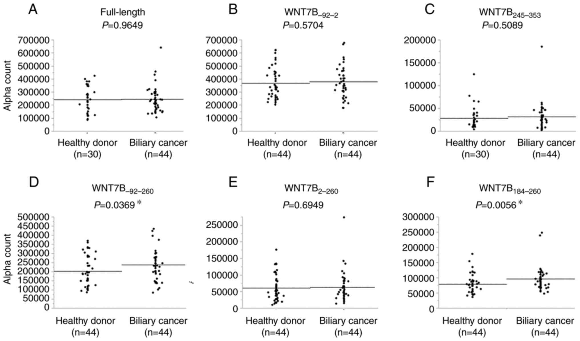
cancer. GST-fused full-length WNT7B protein was expressed in E.
coli and purified by affinity chromatography. AlphaLISA
immunoassay revealed that the serum levels of the antibody against
full-length WNT7B were not significantly different between the
healthy donors and patients with biliary cancer (P=0.9649; Fig. 1A). However, the identification of
WNT7B by SEREX screening indicated the presence of anti-WNT7B
antibodies in the sera of patients with biliary cancer. A total of
3 deletion mutants were then prepared (WNT7B−92-2,
WNT7B−92-260 and WNT7B245-353; Fig. 2) to evaluate the response to serum
antibodies. There was no significant difference in the serum levels
of antibodies against WNT7B−92-2 (P=0.5704, Fig. 1B) and WNT7B245-353
(P=0.5089, Fig. 1C) between
healthy donors and patients with biliary cancer, whereas a
significant difference was observed in the serum levels of
anti-WNT7B−92-260 antibodies (P=0.0369, Fig. 1D). To further narrow down the
epitope site, two deletion mutants, WNT7B2-260 and
WNT7B184-260, were prepared, and the serum levels of the
antibodies against these proteins were measured (Fig. 2). The serum levels of the antibody
against WNT7B2-260 (P=0.6949, Fig. 1E) did not differ significantly
between healthy donors and patients with biliary cancer, whereas
the serum levels of the antibodies against WNT7B184-260
did (P=0.0056, Fig. 1F). This
suggested that the serum antibodies recognize the epitope
comprising the amino acid residues 184–260 of WNT7B. The cDNA clone
isolated by SEREX contained the pre-splicing sequence of WNT7B X1
mRNA at the 5′-end of WNT7B X1 cDNA (Fig. 2). However, the serum levels of the
antibodies against WNT7B−92-2, which contained only this
pre-splicing sequence, did not differ significantly between healthy
donors and patients with biliary cancer (P=0.5704; Fig. 1B). Therefore, this pre-splicing
sequence site did not affect the serum levels of antibodies against
WNT7B.
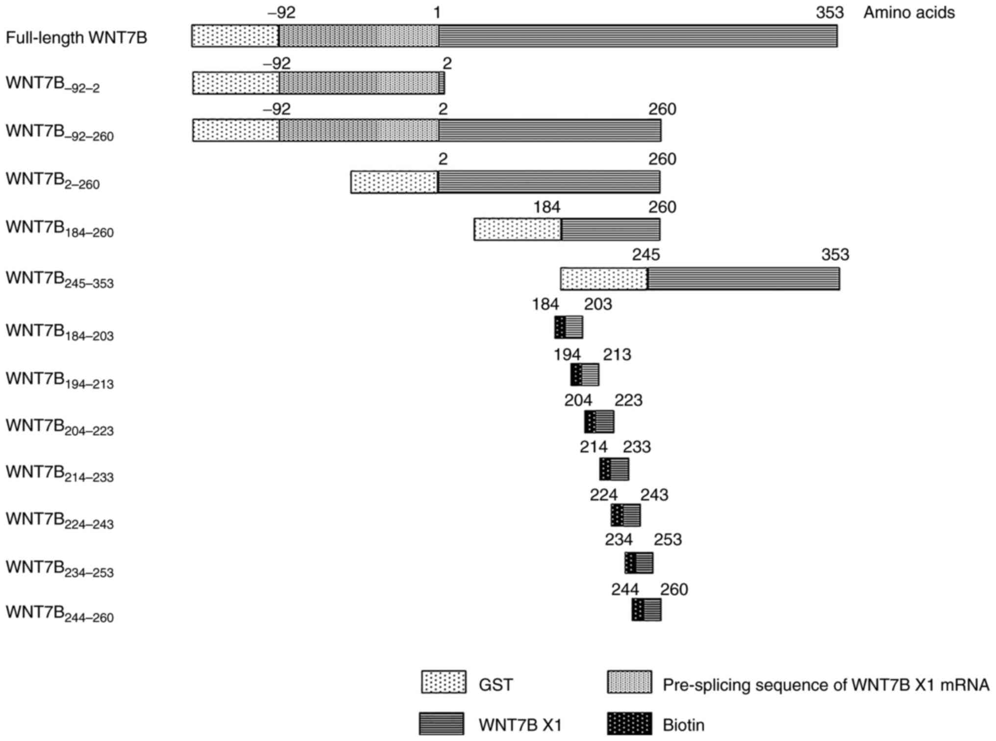 | Figure 2.Schemas of deletion mutants and
peptides. To investigate the antibody response to WNT7B,
full-length WNT7B and WNT7B deletion mutants covering all coding
sequences (WNT7B92-2, 92-260,
2-260, 184-260 and 245-353) were
prepared. The epitope site was inferred from the antibody reaction
and divided into peptides (WNT7B184-203,
194-213, 204-223, 214-233,
224-243, 234-253 and 244-260).
GST-tag was used for proteins and biotin tag was used for peptides.
The proteins identified in the present study also include the
pre-splicing sequences of Homo sapiens WNT7B X1, which are
indicated by ‘- (minus)’. WNT7B, wingless-type MMTV integration
site family, member 7; GST, glutathione S-transferase. |
| Table II.The background of healthy donors and
patients with cancer using AlphaLISA analysis. |
Table II.
The background of healthy donors and
patients with cancer using AlphaLISA analysis.
|
|
Backgroud
of HD and patients used AlphaLISA analysis |
|---|
|
|
|
|---|
|
| Cohort 1
(n=74) | Cohort 2
(n=88) | Cohort 3
(n=586) |
|---|
|
|
|
|
|
|---|
|
| HD (30) | BC (44) | HD (44) | BC (44) | HD (114) | BC (70) | EC (87) | GC (92) | CC (93) | PC (39) | BrC (91) |
|---|
| Sex |
|
|
|
|
|
|
|
|
|
|
|
|
Male | 20 (66.7) | 29 (65.9) | 25 (56.8) | 29 (65.9) | 64 (56.1) | 47 (67.1) | 76 (87.4) | 62 (67.4) | 55 (59.0) | 23 (59.0) | 0 (0) |
|
Female | 10 (33.3) | 15 (34.1) | 19 (43.2) | 15 (34.1) | 50 (43.9) | 23 (32.9) | 11 (12.6) | 30 (32.6) | 38 (40.9) | 16 (41.0) | 91 (100) |
| Age |
|
|
|
|
|
|
|
|
|
|
|
|
<65 | 7 (23.3) | 11 (25.0) | 22 (50.0) | 11 (25.0) | 95 (83.3) | 20 (28.6) | 25 (28.7) | 24 (26.1) | 24 (25.8) | 10 (25.6) | 62 (68.1) |
|
≥65 | 23 (76.7) | 33 (75.0) | 22 (50.0) | 33 (75.0) | 19 (16.7) | 50 (71.4) | 62 (71.3) | 68 (73.9) | 69 (74.2) | 29 (74.4) | 29 (31.9) |
| Stage |
| 3 (6.8) |
| 3 (6.8) |
| 3 (4.3) | 4 (4.6) |
| 3 (3.2) |
| 23 (25.3) |
| I |
| 1 (2.3) |
| 1 (2.3) |
| 3 (4.3) | 6 (6.9) | 47 (51.0) | 19 (20.4) | 2 (5.1) | 28 (30.8) |
| II |
| 19 (43.2) |
| 19 (43.2) |
| 22 (31.4) | 12 (13.8) | 15 (16.3) | 28 (30.1) | 0 (0) | 22 (24.2) |
|
III |
| 15 (34.1) |
| 15 (34.1) |
| 20 (28.6) | 25 (28.7) | 5 (5.4) | 25 (26.9) | 4 (10.3) | 2 (2.2) |
| IV |
| 5 (11.4) |
| 5 (11.4) |
| 20 (28.6) | 21 (24.1) | 17 (18.5) | 15 (16.1) | 28 (71.8) | 2 (2.2) |
| X |
| 1 (2.3) |
| 1 (2.3) |
| 2 (2.9) | 19 (21.8) | 8 (8.7) | 3 (3.2) | 5 (12.8) | 14 (5.4) |
| Location |
|
|
|
|
|
|
|
|
|
|
|
| G |
| 8 (18.2) |
| 8 (18.2) |
| 9 (12.9) |
|
|
|
|
|
| C |
| 0 (0) |
| 0 (0) |
| 1 (1.4) |
|
|
|
|
|
| Bh |
| 1 (2.3) |
| 1 (2.3) |
| 7 (10.0) |
|
|
|
|
|
| Bp |
| 24 (54.4) |
| 24 (54.4) |
| 36 (51.4) |
|
|
|
|
|
| Bd |
| 10 (22.7) |
| 10 (22.7) |
| 15 (21.4) |
|
|
|
|
|
| A |
| 1 (2.3) |
| 1 (2.3) |
| 2 (2.9) |
|
|
|
|
|
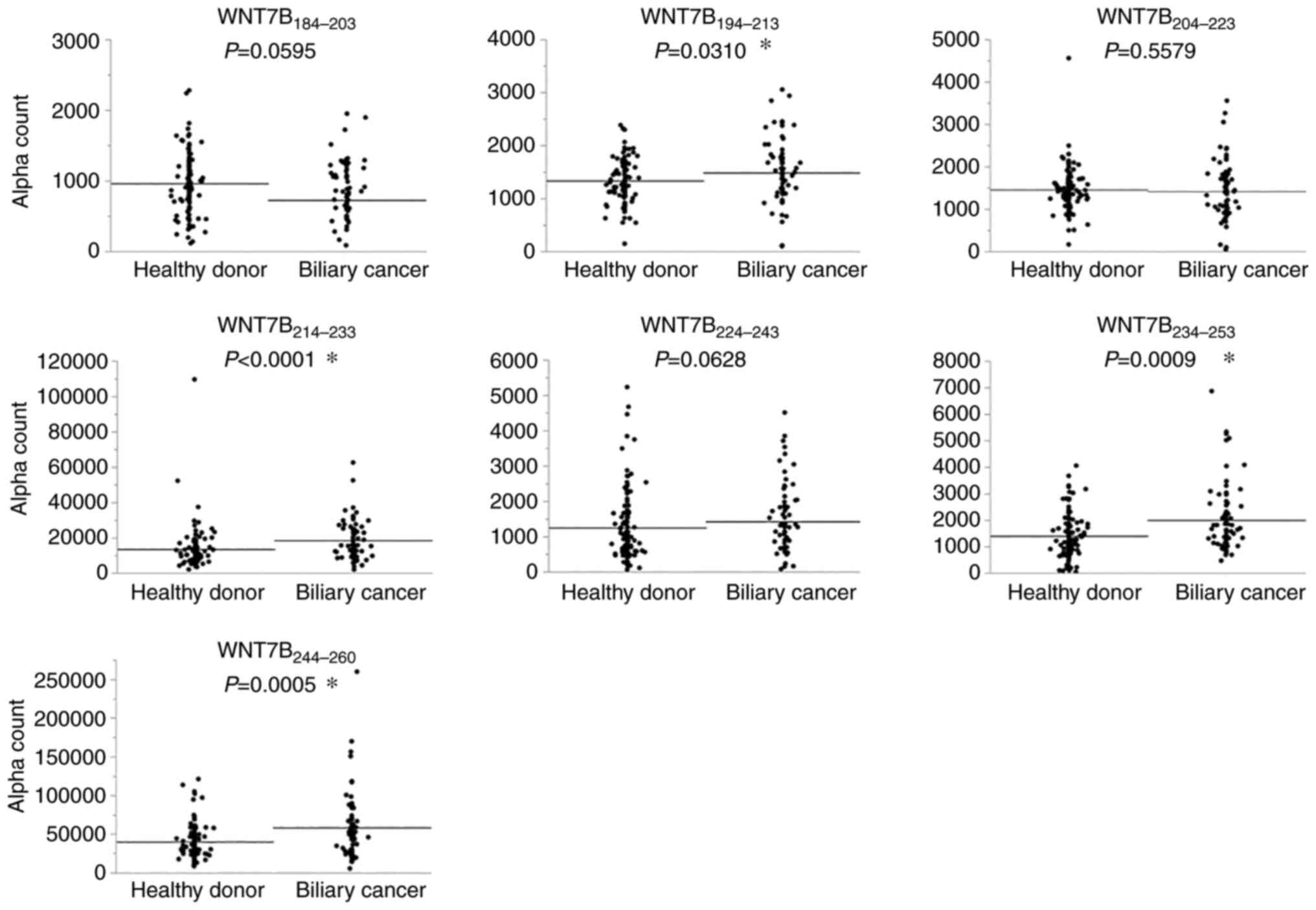
To further narrow down the epitope site, the region
was decomposed into seven peptides, and the serum levels of
antibodies against these were measured. The serum levels of
antibodies against WNT7B194-213,
WNT7B214-233, WNT7B234-253 and
WNT7B244-260 were significantly higher in patients with
biliary cancer than in healthy donors (WNT7B194-213,
P=0.0310; WNT7B214-233, P<0.0001;
WNT7B234-253, P=0.0009; WNT7B244-260,
P=0.0005) (Fig. 3). By contrast,
no significant difference was observed in the levels of antibodies
against other peptides (WNT7B184-203, P=0.0595;
WNT7B204-223, P=0.5579; WNT7B224-243,
P=0.0628) between healthy donors and patients with biliary cancer.
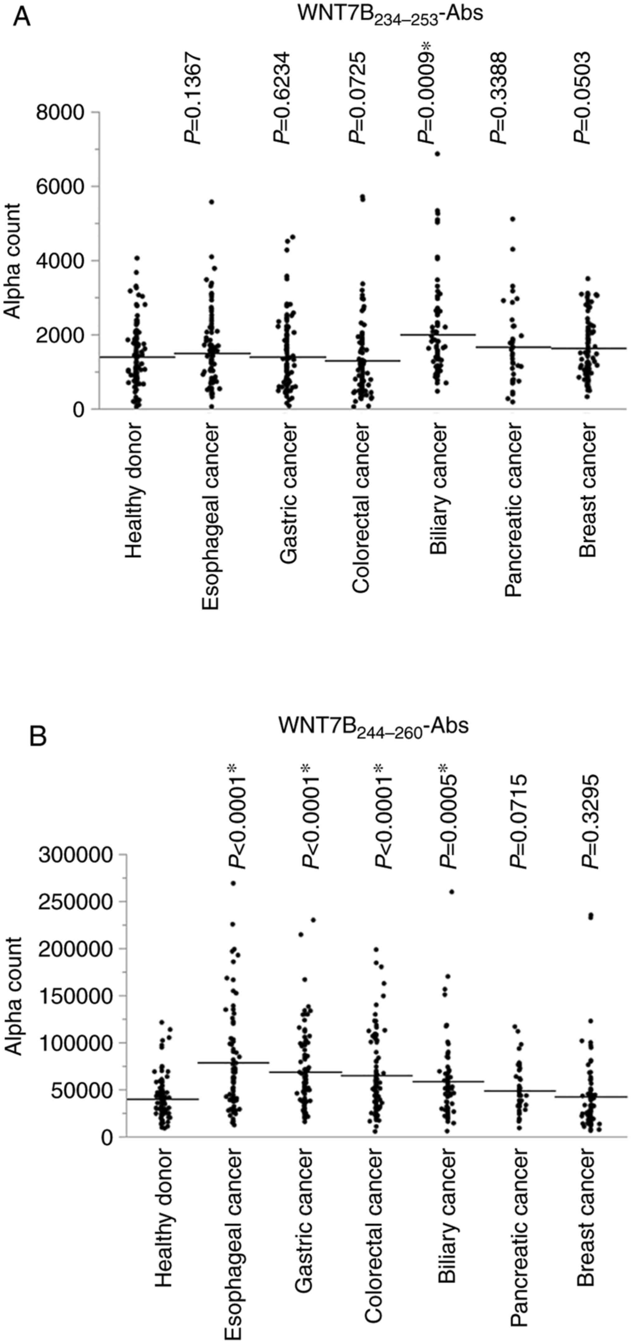
The serum levels of anti-WNT7B234-253 antibodies
(WNT7B234-253-Abs) and anti-WNT7B244-260
antibodies (WNT7B244-260-Abs) were then examined in
patients with other types of cancer, including esophageal, gastric,
colorectal, pancreatic and breast cancer.
WNT7B234-253-Ab levels were significantly increased only
in patients with biliary cancer compared with those in healthy
donors (Fig. 4A). Furthermore,
WNT7B244-260-Ab levels were significantly higher in
patients with esophageal, gastric, and colorectal cancer than in
healthy donors but not in patients with pancreatic or breast cancer
(Fig. 4B).
Correlation between
WNT7B234-253-Ab levels and characteristics of patients
with biliary cancer
The correlation between sex and age and
WNT7B234-253-Ab levels was analyzed in 70 patients with
biliary cancer (Fig. S2). There
was no significant difference in WNT7B234-253-Ab levels
between the sexes (P=0.0549). In addition, there was no significant
difference in WNT7B234-253-Ab levels between the two age
groups: ≥65 and <65 years (P=0.8585). The correlation between
WNT7B234-253-Ab and CA19-9 levels was analyzed in 55
patients with biliary cancer whose CA19-9 data could be confirmed,
and no correlation was found between WNT7B234-253-Ab and
CA19-9 levels (ρ=−0.0029; P=0.9831).
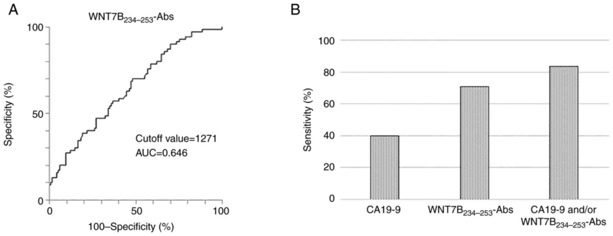
The cutoff value for
WNT7B234-253-Ab levels
The cutoff value for WNT7B234-253-Ab
levels fixed based on receiver operating characteristic curve
analysis was 1271 Alpha count (area under the curve=0.646; Fig. 5A). Based on this cutoff value,
WNT7B234-253-Ab levels ≥1271 Alpha count were defined as
positive, and those <1271 Alpha count were defined as negative.
Thus, the positivity rate of WNT7B234-253-Abs was 66.7,
33.3, 81.8, 70 and 70% for Stages 0, I, II, III, and IV of biliary
cancer, respectively.
Diagnostic accuracy of CA19-9 and
WNT7B234-253-Ab levels in detecting biliary cancer
The accuracy of CA19-9 and serum
WNT7B234-253-Ab levels to serve as a tumor marker for
biliary cancer was examined. Preoperative CA19-9 measurements were
evaluated based on a cutoff value of 37 U/ml, which is our facility
criteria. The sensitivity of WNT7B234-253-Abs alone was
analyzed in 70 patients with biliary cancer. The sensitivity of
CA19-9 alone and that of WNT7B234-253-Ab- and/or
CA19-9-positivity were analyzed in 55 patients with biliary cancer
for whom CA19-9 data was available. The specificity of
WNT7B234-253-Abs was analyzed using the sera of 144
healthy donors. The sensitivity of CA19-9 alone in diagnosing
biliary cancer was 40% (22 of 55 cases), and that of
WNT7B234-253-Abs alone was 70% (49 of 70 cases).
Conversely, the sensitivity of CA19-9- and/or
WNT7B234-253-Ab-positivity in detecting biliary cancer
was 83.6% (46 of 55 cases) (Fig.
5B). In addition, the sensitivity of
WNT7B234-253-Abs in diagnosing relatively early-stage
biliary cancer (Stage 0-II) was 75% (21 of 28 cases). The
specificity of WNT7B234-253-Abs was 51.8% (59 of 114
cases).
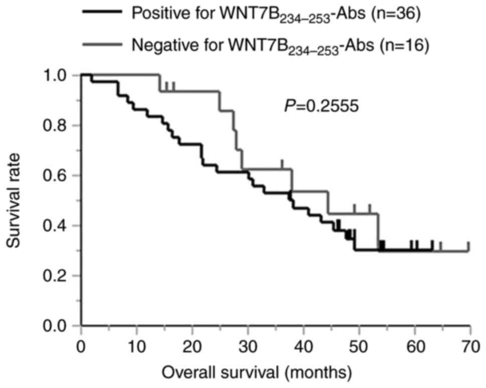
Relationship between
WNT7B234-253-Ab levels and biliary cancer prognosis
The association between WNT7B234-253-Ab
levels and the prognosis of biliary cancer was analyzed in 52
patients with a known prognosis. A total of 36 patients with
biliary cancer were WNT7B234-253-Ab-positive and 16 were
WNT7B234-253-Ab-negative. Overall survival analysis
demonstrated a lower survival rate in the
WNT7B234-253-Ab-positive group than in the
WNT7B234-253-Ab-negative group, but there was no
significant difference (P=0.2555, Fig.
6).
Discussion
The purpose of the present study was to discover
novel serum markers for biliary cancer by measuring serum antibody
levels. SEREX analyses of serum from patients with gastrointestinal
cancers have previously been used to identify antigens against
which the antibodies produced can serve as useful tumor markers
(9,18,23).
Hence, in the present study, target antigen proteins were
identified via SEREX using sera from patients with biliary cancer.
Among these candidate antigens, WNT7B were targeted in the present
study because RT-qPCR showed high mRNA levels of WNT7B in
biliary cancer cell lines, which was further verified via western
blotting (Fig. S1). It was
revealed that the serum levels of antibodies against
WNT7B−92-260 and WNT7B184-260 proteins were
significantly increased in patients with biliary cancer compared
with those in healthy donors. Further analysis using peptide
antigens showed that the serum levels of
WNT7B234-253-Abs were exclusively higher in biliary
cancer patients than in healthy donors. Therefore,
WNT7B234-253-Abs may have the potential to be a specific
biliary cancer marker.
The serum levels of antibodies (Alpha counts)
against WNT7B244-260 were higher than those against
WNT7B234-253. Such a difference in specificity and
reactivity between the two peptide antigens may be attributable to
the tertiary structure of peptides and may cause a difference in
disease specificity. The tertiary structure of
WNT7B234-253 and WNT7B244-260 was then
analyzed using a computational model. Although both have a helical
structure, WNT7B244-263 but not WNT7B234-253
has a bend at the amino acid sequence portion ‘RQP’ (Fig. S3). BLAST search found five
proteins that had the same sequence as the bend. Among them,
protein kinase C is carcinogenic and widely studied (24,25).
According to the Expression Atlas (https://www.ebi.ac.uk/gxa/home), a high expression of
protein kinase C epsilon-type was confirmed in numerous
gastrointestinal cancers. Antibodies developed against this
sequence of protein kinase C in numerous gastrointestinal cancers
may cross-react with WNT7B244-260.
Notably, the present study found that the levels of
the antibody against WNT7B in the sera of patients with biliary
cancer were high and that the specific epitope was present among
the amino acids 234–253. It was demonstrated that
WNT7B234-253-Abs are more sensitive than CA19-9 in
detecting biliary cancer, and the combination of
WNT7B234-253-Abs and CA19-9 yielded a high sensitivity
of 83.6%, rendering it useful as a novel tumor marker. In addition,
relatively early-stage biliary cancer can also be identified with
high sensitivity (75%), and improved prognosis can be expected
through early diagnosis and treatment. Nevertheless, the low
specificity of WNT7B234-253-Abs makes it difficult to
use them as tumor markers in clinical practice. If the number of
cases analyzed could be increased to set a more appropriate cutoff
value, WNT7B234-253-Abs could be expected to play a
major role in diagnosing biliary cancer in clinical practice.
Since the first discovery of WNT1 (initially named
int-1) in 1982 (26), 19
human WNT members have been identified, including WNT7B.
When Wnt binds to its receptors, the β-catenin, planar cell
polarity, and Ca2+ pathways become activated (27). The β-catenin pathway, which
controls cell proliferation and differentiation, has been linked to
carcinogenesis. In fact, mutations and abnormal accumulation of
β-catenin have been reported in various types of cancer, including
colorectal, liver, gastric, ovarian, prostate and pancreatic cancer
(28). Mila et al (29) reported that the nuclear expression
of β-catenin in gallbladder cancer correlates with tumor grade and
depth of invasion, thus suggesting the role of this gene in tumor
progression.
The anti-WNT7B antibody may also offer additional
benefits in terms of applicability aside from serving as a tumor
marker. In the β-catenin pathway, one of the intracellular signal
transduction pathways of WNT7B, stabilized β-catenin enters the
nucleus and stimulates the transcription of target genes, such as
the oncogene cyclin D1 and c-myc, and exerts an important function
in carcinogenesis (30,31). In biliary cancer, suppression of
β-catenin by siRNA has been shown to inhibit the expression of
cyclin D1, leading to decreased viability of biliary cancer cell
lines. Therefore, the WNT/β-catenin pathway is considered to
contribute to cell proliferation in biliary cancer (32). In the present study, significantly
elevated levels of anti-WNT7B antibodies were observed in the sera
of patients with biliary cancer compared with those in healthy
donors and patients diagnosed with other types of cancer.
A major limitation to the present study was that
most of the cases analyzed were stage II or higher, primarily
because few cases of biliary cancer are detected at an early stage.
Hence, in the future, further examination of
WNT7B234-253-Abs in patients at an early stage of
biliary cancer is warranted for a more precise and comprehensive
analysis. Another drawback is the low specificity of the
WNT7B234-253-Abs for clinical applications. Further
investigation of optimal cutoff values by increasing the number of
cases may increase their clinical significance.
In conclusion, it was found that
WNT7B234-253-Ab is a possible new tumor marker for
biliary cancer and the appropriate cutoff value for serum
anti-WNT7B234-253 levels to diagnose biliary cancer was
determined.
Supplementary Material
Supporting Data
Supporting Data
Acknowledgements
The authors would like to thank Professor Masaki
Takiguchi (Department of Biochemistry and Genetics, Graduate School
of Medicine, Chiba University) for experimental instruction and
equipment provision. The authors are also grateful to Dr Shotaro
Furukawa, Dr Saseem Paudel, Dr Shoki Sato, Dr Hirotake Abe and Dr
Shintaro Takeuchi (Department of Gastroenterological Surgery II,
Faculty of Medicine, Hokkaido University) for their cooperation in
the present study.
Funding
The present study was supported by JSPS KAKENHI (grant no.
JP17K10532).
Availability of data and materials
The datasets used and/or analyzed during the
current study are available from the corresponding author on
reasonable request.
Authors' contributions
MT made substantial contributions to the
conceptualization, design, acquisition, analysis and interpretation
of data for the work. TT, TNa, KM, HM, MO, HS, KT, YN, TA, TNo, KO,
TS and SH made substantial contributions to the conceptualization
and design of the work. THi, KH, YH and THo made substantial
contributions to the design, acquisition, analysis, and
interpretation of data for the work. TK, KI and HT made substantial
contributions to acquisition and analysis. All authors read and
approved the final version of the manuscript. MT and TT confirm the
authenticity of all the raw data.
Ethics approval and consent to
participate
The study protocol was approved by the Ethics
Committee of Hokkaido University Hospital (approval no. 015-0098;
Sapporo, Japan) and Chiba University Graduate School of Medicine
(approval no. 973; Chiba, Japan). Informed consent was obtained
from all participants of the present study. Written consent or
opt-out consent was provided by the 56 patients with biliary cancer
in our institute. For specimens from collaborating institutions,
consent was obtained in accordance with the standards of the
collaborating institutions. It was determined that there are no
ethical issues with the 30 healthy donor sera purchased from Tokyo
Future Style.
Patient consent for publication
Not applicable.
Competing interests
The authors declare that they have no competing
interests.
Glossary
Abbreviations
Abbreviations:
|
AlphaLISA
|
amplified luminescence proximity
homogeneous assay-linked immunosorbent assay
|
|
CA19-9
|
carbohydrate antigen 19-9
|
|
GST
|
glutathione S-transferase
|
|
SEREX
|
serological identification of
antigens by recombinant cDNA expression cloning
|
|
E. coli
|
Escherichia coli
|
|
WNT7B
|
wingless-type MMTV integration site
family, member 7
|
|
BLAST
|
Basic Local Alignment Search Tool
|
References
|
1
|
Ishihara S, Horiguchi A, Miyakawa S, Endo
I, Miyazaki M and Takada T: Biliary tract cancer registry in Japan
from 2008 to 2013. J Hepatobiliary Pancreat Sci. 23:149–157. 2016.
View Article : Google Scholar : PubMed/NCBI
|
|
2
|
Yamada T, Nakanishi Y, Okamura K,
Tsuchikawa T, Nakamura T, Noji T, Asano T, Tanaka K, Kurashima Y,
Ebihara Y, et al: Impact of serum carbohydrate antigen 19-9 level
on prognosis and prediction of lymph node metastasis in patients
with intrahepatic cholangiocarcinoma. J Gastroenterol Hepatol.
33:1626–1633. 2018. View Article : Google Scholar
|
|
3
|
Blechacz BG and Feldman GJ: Sleisenger and
Fordtran's gastrointestinal and liver disease. 9. Vol 1 Tumors of
the Bile Ducts, Gallbladder, and Ampulla. Saunders. 1171–1176.
2010.
|
|
4
|
Shimada H, Ochiai T and Nomura F; Japan
p53 Antibody Research Group, : Titration of serum p53 antibodies in
1,085 patients with various types of malignant tumors: A
multiinstitutional analysis by the Japan p53 antibody research
group. Cancer. 97:682–689. 2003. View Article : Google Scholar : PubMed/NCBI
|
|
5
|
Zhang J, Xu Z, Yu L, Chen M and Li K:
Assessment of the potential diagnostic value of serum p53 antibody
for cancer: A meta-analysis. PLoS One. 9:e992552014. View Article : Google Scholar : PubMed/NCBI
|
|
6
|
Okada R, Shimada H, Otsuka Y, Tsuchiya M,
Ishii J, Katagiri T, Maeda T, Kubota Y, Nemoto T and Kaneko H:
Serum p53 antibody as a potential tumor marker in extrahepatic
cholangiocarcinoma. Surg Today. 47:1492–1499. 2017. View Article : Google Scholar : PubMed/NCBI
|
|
7
|
Sahin U, Türeci O, Schmitt H, Cochlovius
B, Johannes T, Schmits R, Stenner F, Luo G, Schobert I and
Pfreundschuh M: Human neoplasms elicit multiple specific immune
responses in the autologous host. Proc Natl Acad Sci USA.
92:11810–11813. 1995. View Article : Google Scholar : PubMed/NCBI
|
|
8
|
Nakashima K, Shimada H, Ochiai T,
Kuboshima M, Kuroiwa N, Okazumi S, Matsubara H, Nomura F, Takiguchi
M and Hiwasa T: Serological identification of TROP2 by recombinant
cDNA expression cloning using sera of patients with esophageal
squamous cell carcinoma. Int J Cancer. 112:1029–1035. 2004.
View Article : Google Scholar : PubMed/NCBI
|
|
9
|
Shimada H, Shiratori T, Yasuraoka M,
Kagaya A, Kuboshima M, Nomura F, Takiguchi M, Ochiai T, Matsubara H
and Hiwasa T: Identification of Makorin 1 as a novel SEREX antigen
of esophageal squamous cell carcinoma. BMC Cancer. 9:2322009.
View Article : Google Scholar : PubMed/NCBI
|
|
10
|
Obata Y, Takahashi T, Sakamoto J, Tamaki
H, Tominaga S, Hamajima N, Chen YT and Old LJ: SEREX analysis of
gastric cancer antigens. Cancer Chemother Pharmacol. 46
(Suppl):S37–S42. 2000. View Article : Google Scholar : PubMed/NCBI
|
|
11
|
Linē A, Stengrēvics A, Slucka Z, Li G,
Jankevics E and Rees RC: Serological identification and expression
analysis of gastric cancer-associated genes. Br J Cancer.
86:1824–1830. 2002. View Article : Google Scholar : PubMed/NCBI
|
|
12
|
Song MH, Ha JC, Lee SM, Park YM and Lee
SY: Identification of BCP-20 (FBXO39) as a cancer/testis antigen
from colon cancer patients by SEREX. Biochem Biophys Res Commun.
408:195–201. 2011. View Article : Google Scholar : PubMed/NCBI
|
|
13
|
Garifulin OM, Kykot VO, Gridina NY,
Kiyamova RG, Gout IT and Filonenko VV: Application of
serex-analysis for identification of human colon cancer antigens.
Exp Oncol. 37:173–180. 2015. View Article : Google Scholar : PubMed/NCBI
|
|
14
|
Nakatsura T, Senju S, Yamada K, Jotsuka T,
Ogawa M and Nishimura Y: Gene cloning of immunogenic antigens
overexpressed in pancreatic cancer. Biochem Biophys Res Commun.
281:936–944. 2001. View Article : Google Scholar : PubMed/NCBI
|
|
15
|
Heller A, Zörnig I, Müller T, Giorgadze K,
Frei C, Giese T, Bergmann F, Schmidt J, Werner J, Buchler MW, et
al: Immunogenicity of SEREX-identified antigens and disease outcome
in pancreatic cancer. Cancer Immunol Immunother. 59:1389–1400.
2010. View Article : Google Scholar : PubMed/NCBI
|
|
16
|
Wang K, Xu X, Nie Y, Dai L, Wang P and
Zhang J: Identification of tumor-associated antigens by using SEREX
in hepatocellular carcinoma. Cancer Lett. 281:144–150. 2009.
View Article : Google Scholar : PubMed/NCBI
|
|
17
|
Song MH, Choi KU, Shin DH, Lee CH and Lee
SY: Identification of the cancer/testis antigens AKAP3 and CTp11 by
SEREX in hepatocellular carcinoma. Oncol Rep. 28:1792–1798. 2012.
View Article : Google Scholar : PubMed/NCBI
|
|
18
|
Kagaya A, Shimada H, Shiratori T,
Kuboshima M, Nakashima-Fujita K, Yasuraoka M, Nishimori T, Kurei S,
Hachiya T, Murakami A, et al: Identification of a novel SEREX
antigen family, ECSA, in esophageal squamous cell carcinoma.
Proteome Sci. 9:312011. View Article : Google Scholar : PubMed/NCBI
|
|
19
|
Machida TT, Kubota M, Kobayashi E, Iwadate
Y, Saeki N, Yamaura A, Nomura F, Takiguchi M and Hiwasa T:
Identification of stroke-associated-antigens via screening of
recombinant proteins from the human expression cDNA library
(SEREX). J Transl Med. 13:712015. View Article : Google Scholar : PubMed/NCBI
|
|
20
|
Hiwasa T, Wang H, Goto KI, Mine S, Machida
T, Kobayashi E, Yoshida Y, Adachi A, Matsutani T, Sata M, et al:
Serum anti-DIDO1, anti-CPSF2, and anti-FOXJ2 antibodies as
predictive risk markers for acute ischemic stroke. BMC Med.
19:1312021. View Article : Google Scholar : PubMed/NCBI
|
|
21
|
Yang J, Yan R, Roy A, Xu D, Poisson J and
Zhang Y: The I-TASSER Suite: Protein structure and function
prediction. Nat Methods. 12:7–8. 2015. View Article : Google Scholar : PubMed/NCBI
|
|
22
|
Livak KJ and Schmittgen TD: Analysis of
relative gene expression data using real-time quantitative PCR and
the 2(−Delta Delta C(T)) method. Methods. 25:402–408. 2001.
View Article : Google Scholar : PubMed/NCBI
|
|
23
|
Kobayashi S, Hiwasa T, Arasawa T, Kagaya
A, Ishii S, Shimada H, Ito M, Suzuki M, Kano M, Rahmutulla B, et
al: Identification of specific and common diagnostic antibody
markers for gastrointestinal cancers by SEREX screening using
testis cDNA phage library. Oncotarget. 9:18559–18569. 2018.
View Article : Google Scholar : PubMed/NCBI
|
|
24
|
Gorin MA and Pan Q: Protein kinase C
epsilon: An oncogene and emerging tumor biomarker. Mol Cancer.
8:92009. View Article : Google Scholar : PubMed/NCBI
|
|
25
|
Cacace AM, Guadagno SN, Krauss RS, Fabbro
D and Weinstein IB: The epsilon isoform of protein kinase C is an
oncogene when overexpressed in rat fibroblasts. Oncogene.
8:2095–2104. 1993.PubMed/NCBI
|
|
26
|
Nusse R and Varmus HE: Many tumors induced
by the mouse mammary tumor virus contain a provirus integrated in
the same region of the host genome. Cell. 31:99–109. 1982.
View Article : Google Scholar : PubMed/NCBI
|
|
27
|
Kikuchi A and Yamamoto H: Tumor formation
due to abnormalities in the beta-catenin-independent pathway of Wnt
signaling. Cancer Sci. 99:202–208. 2008. View Article : Google Scholar : PubMed/NCBI
|
|
28
|
Kikuchi A: Tumor formation by genetic
mutations in the components of the Wnt signaling pathway. Cancer
Sci. 94:225–229. 2003. View Article : Google Scholar : PubMed/NCBI
|
|
29
|
Ghosh M, Sakhuja P, Singh S and Agarwal
AK: p53 and beta-catenin expression in gallbladder tissues and
correlation with tumor progression in gallbladder cancer. Saudi J
Gastroenterol. 19:34–39. 2013. View Article : Google Scholar : PubMed/NCBI
|
|
30
|
Tetsu O and McCormick F: Beta-catenin
regulates expression of cyclin D1 in colon carcinoma cells. Nature.
398:422–426. 1999. View
Article : Google Scholar : PubMed/NCBI
|
|
31
|
Kikuchi A: Roles of Axin in the Wnt
signalling pathway. Cell Signal. 11:777–788. 1999. View Article : Google Scholar : PubMed/NCBI
|
|
32
|
Loilome W, Bungkanjana P, Techasen A,
Namwat N, Yongvanit P, Puapairoj A, Khuntikeo N and Riggins GJ:
Activated macrophages promote Wnt/β-catenin signaling in
cholangiocarcinoma cells. Tumour Biol. 35:5357–5367. 2014.
View Article : Google Scholar : PubMed/NCBI
|