|
1
|
Burris HA III, Moore MJ, Andersen J, Green
MR, Rothenberg ML, Modiano MR, Cripps MC, Portenoy RK, Storniolo
AM, Tarassoff P, et al: Improvements in survival and clinical
benefit with gemcitabine as first-line therapy for patients with
advanced pancreas cancer: A randomized trial. J Clin Oncol.
15:2403–2413. 1997. View Article : Google Scholar : PubMed/NCBI
|
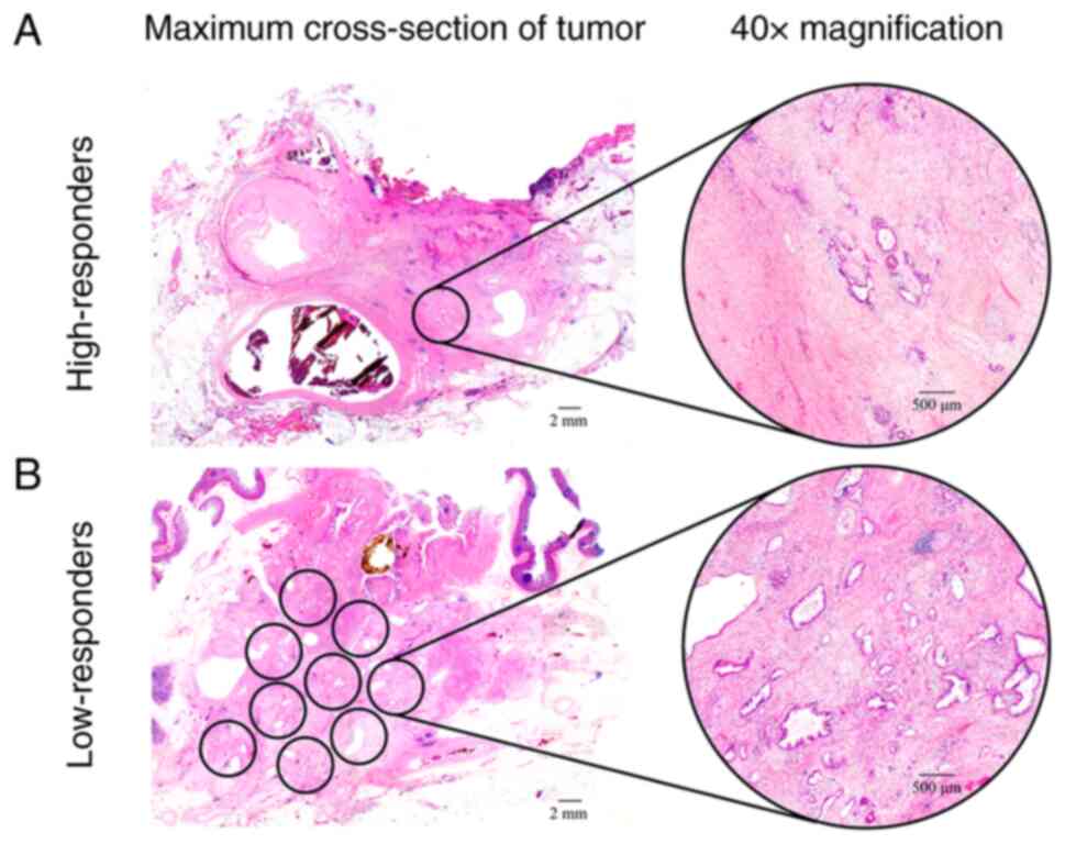
|
2
|
Whatcott CJ, Diep CH, Jiang P, Watanabe A,
LoBello J, Sima C, Hostetter G, Shepard HM, Von Hoff DD and Han H:
Desmoplasia in primary tumors and metastatic lesions of pancreatic
cancer. Clin Cancer Res. 21:3561–3568. 2015. View Article : Google Scholar : PubMed/NCBI
|
|
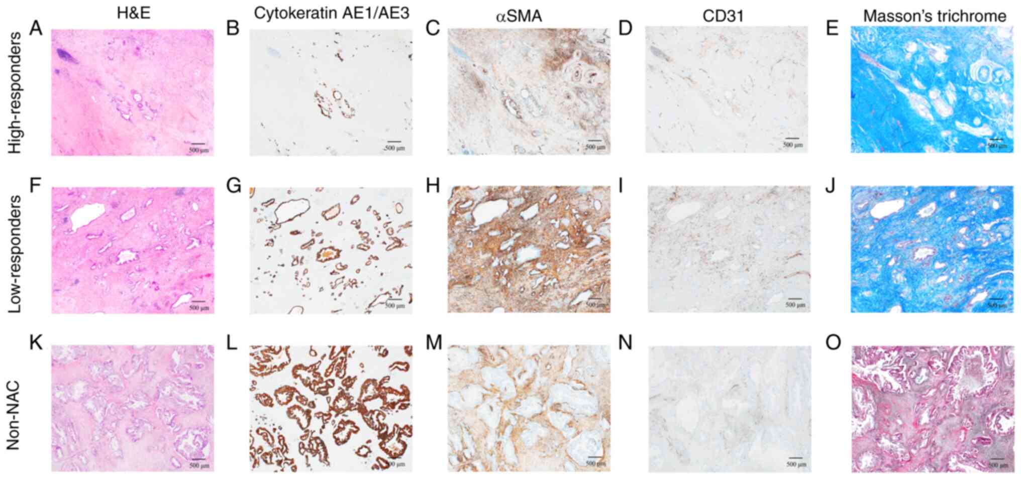
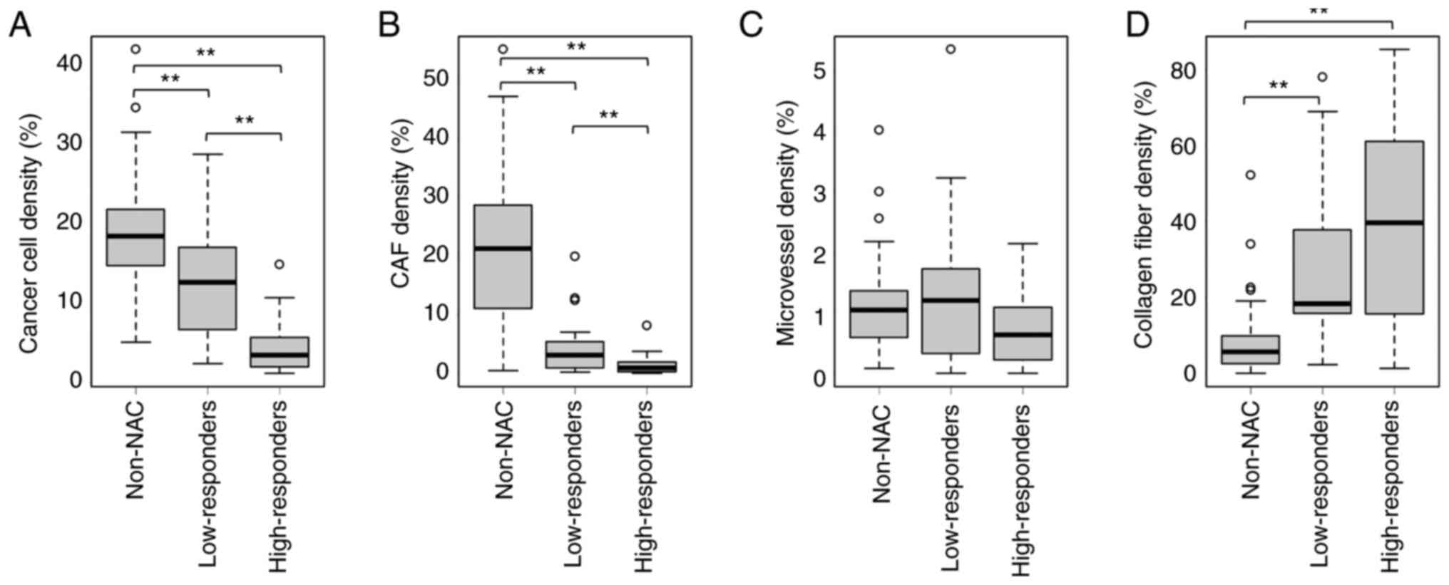
3
|
Neesse A, Bauer CA, Öhlund D, Lauth M,
Buchholz M, Michl P, Tuveson DA and Gress TM: Stromal biology and
therapy in pancreatic cancer: Ready for clinical translation? Gut.
68:159–171. 2019. View Article : Google Scholar : PubMed/NCBI
|
|
4
|
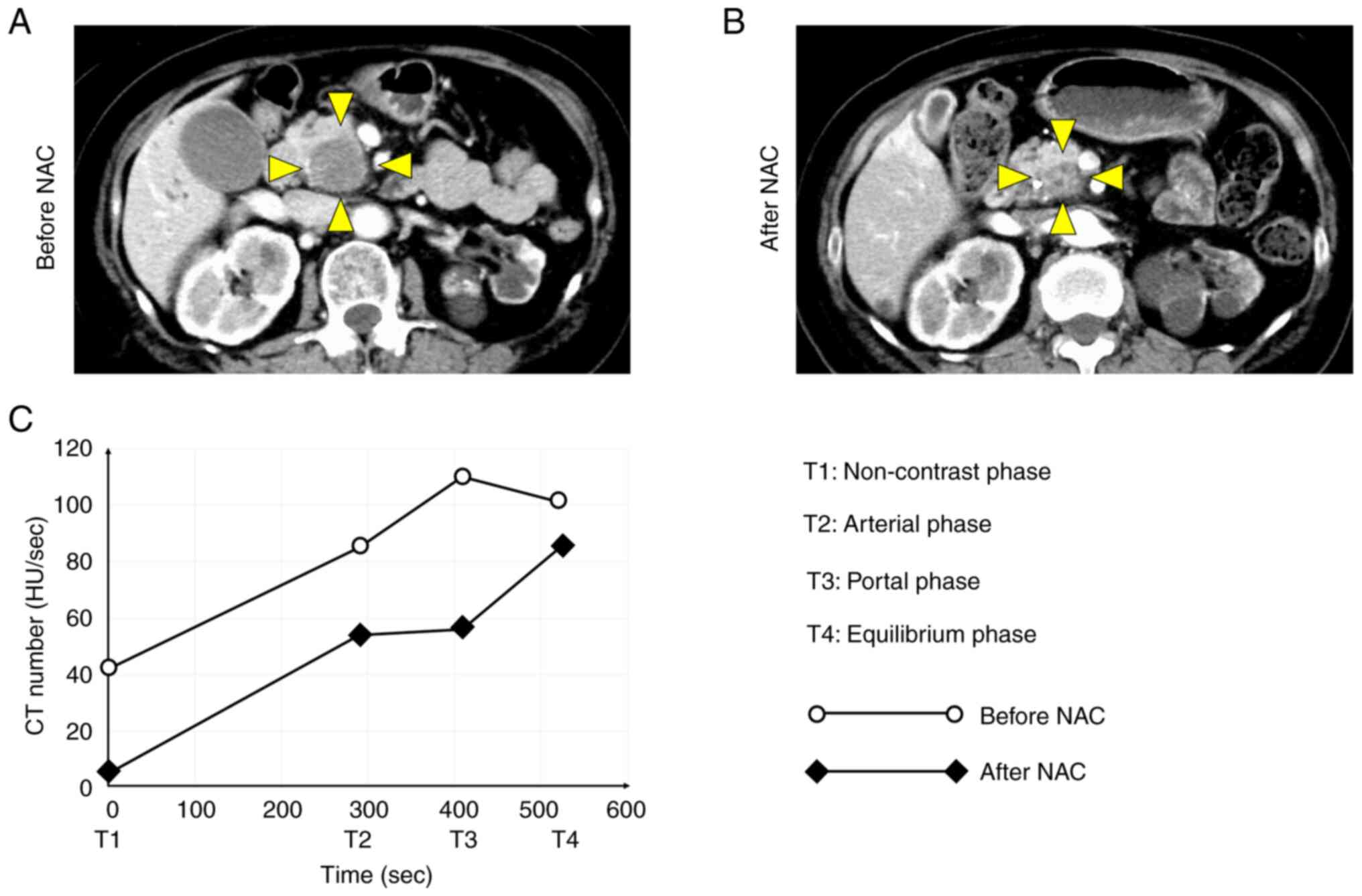
Oba A, Ho F, Bao QR, Al-Musawi MH,
Schulick RD and Del Chiaro M: Neoadjuvant treatment in pancreatic
cancer. Front Oncol. 10:2452020. View Article : Google Scholar : PubMed/NCBI
|
|
5
|
Motoi F and Unno M: Adjuvant and
neoadjuvant treatment for pancreatic adenocarcinoma. Jpn J Clin
Oncol. 50:483–489. 2020. View Article : Google Scholar : PubMed/NCBI
|
|
6
|
Versteijne E, van Dam JL, Suker M, Janssen
QP, Groothuis K, Akkermans-Vogelaar JM, Besselink MG, Bonsing BA,
Buijsen J, Busch OR, et al: Neoadjuvant chemoradiotherapy versus
upfront surgery for resectable and borderline resectable pancreatic
cancer: Long-term results of the Dutch randomized PREOPANC trial. J
Clin Oncol. 40:1220–1230. 2022. View Article : Google Scholar : PubMed/NCBI
|
|
7
|
Von Hoff DD, Ervin T, Arena FP, Chiorean
EG, Infante J, Moore M, Seay T, Tjulandin SA, Ma WW, Saleh MN, et
al: Increased survival in pancreatic cancer with nab-paclitaxel
plus gemcitabine. N Engl J Med. 369:1691–1703. 2013. View Article : Google Scholar : PubMed/NCBI
|
|
8
|
Goldstein D, El-Maraghi RH, Hammel P,
Heinemann V, Kunzmann V, Sastre J, Scheithauer W, Siena S,
Tabernero J, Teixeira L, et al: nab-Paclitaxel plus gemcitabine for
metastatic pancreatic cancer: Long-term survival from a phase III
trial. J Natl Cancer Inst. 107:dju4132015. View Article : Google Scholar : PubMed/NCBI
|
|
9
|
Welsh JL, Bodeker K, Fallon E, Bhatia SK,
Buatti JM and Cullen JJ: Comparison of response evaluation criteria
in solid tumors with volumetric measurements for estimation of
tumor burden in pancreatic adenocarcinoma and hepatocellular
carcinoma. Am J Surg. 204:580–585. 2012. View Article : Google Scholar : PubMed/NCBI
|
|
10
|
Chatterjee D, Katz MH, Rashid A, Wang H,
Iuga AC, Varadhachary GR, Wolff RA, Lee JE, Pisters PW, Crane CH,
et al: Perineural and intraneural invasion in posttherapy
pancreaticoduodenectomy specimens predicts poor prognosis in
patients with pancreatic ductal adenocarcinoma. Am J Surg Pathol.
36:409–417. 2012. View Article : Google Scholar : PubMed/NCBI
|
|
11
|
Lee SM, Katz MH, Liu L, Sundar M, Wang H,
Varadhachary GR, Wolff RA, Lee JE, Maitra A, Fleming JB, et al:
Validation of a proposed tumor regression grading scheme for
pancreatic ductal adenocarcinoma after neoadjuvant therapy as a
prognostic indicator for survival. Am J Surg Pathol. 40:1653–1660.
2016. View Article : Google Scholar : PubMed/NCBI
|
|
12
|
Kalimuthu SN, Serra S, Dhani N,
Hafezi-Bakhtiari S, Szentgyorgyi E, Vajpeyi R and Chetty R:
Regression grading in neoadjuvant treated pancreatic cancer: An
interobserver study. J Clin Pathol. 70:237–243. 2017. View Article : Google Scholar : PubMed/NCBI
|
|
13
|
Matsuda Y, Ohkubo S, Nakano-Narusawa Y,
Fukumura Y, Hirabayashi K, Yamaguchi H, Sahara Y, Kawanishi A,
Takahashi S, Arai T, et al: Objective assessment of tumor
regression in post-neoadjuvant therapy resections for pancreatic
ductal adenocarcinoma: Comparison of multiple tumor regression
grading systems. Sci Rep. 10:182782020. View Article : Google Scholar : PubMed/NCBI
|
|
14
|
Ferrone CR, Marchegiani G, Hong TS, Ryan
DP, Deshpande V, McDonnell EI, Sabbatino F, Santos DD, Allen JN,
Blaszkowsky LS, et al: Radiological and surgical implications of
neoadjuvant treatment with FOLFIRINOX for locally advanced and
borderline resectable pancreatic cancer. Ann Surg. 261:12–17. 2015.
View Article : Google Scholar : PubMed/NCBI
|
|
15
|
Wagner M, Antunes C, Pietrasz D,
Cassinotto C, Zappa M, Sa Cunha A, Lucidarme O and Bachet JB: CT
evaluation after neoadjuvant FOLFIRINOX chemotherapy for borderline
and locally advanced pancreatic adenocarcinoma. Eur Radiol.
27:3104–3116. 2017. View Article : Google Scholar : PubMed/NCBI
|
|
16
|
Goto S, Seino H, Yoshizawa T, Morohashi S,
Ishido K, Hakamada K and Kijima H: Time density curve of dynamic
contrast-enhanced computed tomography correlates with histological
characteristics of pancreatic cancer. Oncol Lett. 21:2762021.
View Article : Google Scholar : PubMed/NCBI
|
|
17
|
Brierley JD, Gospodarowicz MK and
Wittekind C: The TNM classification of malignant tumours. 8th
edition. Wiley-Blackwell; Oxford: pp. 93–95. 2017
|
|
18
|
WHO Classification of Tumours Editorial
Board, . WHO classification of tumours of the digestive system.
IARC Press; Lyon: pp. 322–332. 2019
|
|
19
|
Inoue C, Miki Y, Saito R, Hata S, Abe J,
Sato I, Okada Y and Sasano H: PD-L1 induction by cancer-associated
fibroblast-derived factors in lung adenocarcinoma cells. Cancers
(Basel). 11:12572019. View Article : Google Scholar : PubMed/NCBI
|
|
20
|
Itou RA, Uyama N, Hirota S, Kawada N, Wu
S, Miyashita S, Nakamura I, Suzumura K, Sueoka H, Okada T, et al:
Immunohistochemical characterization of cancer-associated
fibroblasts at the primary sites and in the metastatic lymph nodes
of human intrahepatic cholangiocarcinoma. Hum Pathol. 83:77–89.
2019. View Article : Google Scholar : PubMed/NCBI
|
|
21
|
Zhang J, Li S, Zhao Y, Ma P, Cao Y, Liu C,
Zhang X, Wang W, Chen L and Li Y: Cancer-associated fibroblasts
promote the migration and invasion of gastric cancer cells via
activating IL-17a/JAK2/STAT3 signaling. Ann Transl Med. 8:8772020.
View Article : Google Scholar : PubMed/NCBI
|
|
22
|
Matsuda K, Ohga N, Hida Y, Muraki C,
Tsuchiya K, Kurosu T, Akino T, Shih SC, Totsuka Y, Klagsbrun M, et
al: Isolated tumor endothelial cells maintain specific character
during long-term culture. Biochem Biophys Res Commun. 394:947–954.
2010. View Article : Google Scholar : PubMed/NCBI
|
|
23
|
Calvi EN, Nahas FX, Barbosa MV, Calil JA,
Ihara SS, Silva Mde S, Franco MF and Ferreira LM: An experimental
model for the study of collagen fibers in skeletal muscle. Acta Cir
Bras. 27:681–686. 2012. View Article : Google Scholar : PubMed/NCBI
|
|
24
|
Kanda Y: Investigation of the freely
available easy-to-use software ‘EZR’ for medical statistics. Bone
Marrow Transplant. 48:452–458. 2013. View Article : Google Scholar : PubMed/NCBI
|
|
25
|
Miyashita T, Tajima H, Makino I, Okazaki
M, Yamaguchi T, Ohbatake Y, Nakanuma S, Hayashi H, Takamura H,
Ninomiya I, et al: Neoadjuvant chemotherapy with gemcitabine plus
nab-paclitaxel reduces the number of cancer-associated fibroblasts
through depletion of pancreatic stroma. Anticancer Res. 38:337–343.
2018.PubMed/NCBI
|
|
26
|
Mancini ML and Sonis ST: Mechanisms of
cellular fibrosis associated with cancer regimen-related
toxicities. Front Pharmacol. 5:512014. View Article : Google Scholar : PubMed/NCBI
|
|
27
|
Arimoto A, Uehara K, Tsuzuki T, Aiba T,
Ebata T and Nagino M: Role of bevacizumab in neoadjuvant
chemotherapy and its influence on microvessel density in rectal
cancer. Int J Clin Oncol. 20:935–942. 2015. View Article : Google Scholar : PubMed/NCBI
|
|
28
|
Eefsen RL, Engelholm L, Willemoe GL, Van
den Eynden GG, Laerum OD, Christensen IJ, Rolff HC, Høyer-Hansen G,
Osterlind K, Vainer B and Illemann M: Microvessel density and
endothelial cell proliferation levels in colorectal liver
metastases from patients given neo-adjuvant cytotoxic chemotherapy
and bevacizumab. Int J Cancer. 138:1777–1784. 2016. View Article : Google Scholar : PubMed/NCBI
|
|
29
|
Awai K and Date S: Basic knowledge to
achieve optimal enhancement of CT. Nichidoku Iho. 56:13–32.
2011.
|
|
30
|
Harder FN, Jungmann F, Kaissis GA, Lohöfer
FK, Ziegelmayer S, Havel D, Quante M, Reichert M, Schmid RM, Demir
IE, et al: [18F]FDG PET/MRI enables early chemotherapy response
prediction in pancreatic ductal adenocarcinoma. EJNMMI Res.
11:702021. View Article : Google Scholar : PubMed/NCBI
|
|
31
|
Hamdy A, Ichikawa Y, Toyomasu Y, Nagata M,
Nagasawa N, Nomoto Y, Sami H and Sakuma H: Perfusion CT to assess
response to neoadjuvant chemotherapy and radiation therapy in
pancreatic ductal adenocarcinoma: Initial experience. Radiology.
292:628–635. 2019. View Article : Google Scholar : PubMed/NCBI
|
|
32
|
Abdelrahman AM, Goenka AH, Alva-Ruiz R,
Yonkus JA, Leiting JL, Graham RP, Merrell KW, Thiels CA, Hallemeier
CL, Warner SG, et al: FDG-PET predicts neoadjuvant therapy response
and survival in borderline resectable/locally advanced pancreatic
adenocarcinoma. J Natl Compr Canc Netw. 20:1023–1032.e3. 2022.
View Article : Google Scholar : PubMed/NCBI
|
|
33
|
Koay EJ, Truty MJ, Cristini V, Thomas RM,
Chen R, Chatterjee D, Kang Y, Bhosale PR, Tamm EP, Crane CH, et al:
Transport properties of pancreatic cancer describe gemcitabine
delivery and response. J Clin Invest. 124:1525–1536. 2014.
View Article : Google Scholar : PubMed/NCBI
|
|
34
|
Myllyharju J and Kivirikko KI: Collagens
and collagen-related diseases. Ann Med. 33:7–21. 2001. View Article : Google Scholar : PubMed/NCBI
|