Introduction
Breast cancer is a malignancy originating from the
epithelial cells of the breast and has become one of the most
commonly diagnosed cancers worldwide, ranking first among all
cancers in women. According to the 2025 Cancer Statistics report,
breast cancer is the most common cancer among women, accounting for
32% of all new cancer cases in females, and ranks as the second
leading cause of cancer-related mortality in women. The lifetime
risk of developing breast cancer for women is 13.1% (approximately
one in eight). Since 2012, the incidence rate has been increasing
slowly at an annual rate of ~1%, with the fastest rise observed
among younger women (1.4% per year) (1). Current clinical management of breast
cancer includes surgical resection, radiotherapy, chemotherapy,
endocrine therapy, immunotherapy, and targeted therapy. These
modalities are applied in combination based on molecular subtype,
clinical stage, and individual patient differences (2). Despite advances in modern medicine and
the continuous development of targeted and chemotherapeutic
regimens, treatment resistance remains a major obstacle, leading to
disease progression and reduced survival (3).
The complexity of drug resistance stems largely from
the high heterogeneity and adaptability of tumor cells, involving
multiple interrelated biological processes such as upregulation of
drug efflux pumps, influences of the tumor microenvironment (TME),
aberrant epigenetic regulation, enhanced DNA damage repair (DDR)
capacity, and cellular metabolic reprogramming. Unlike prior
reviews that typically examine resistance mechanisms in isolation,
the present review provides a systematically interconnected
framework integrating five core axes and directly links each to
corresponding therapeutic solutions. Critically, the emerging dual
role of the microbiome in resistance modulation is incorporated and
an artificial intelligence-driven adaptive therapy model for
real-time monitoring and intervention is proposed.
Therefore, a systematic understanding of the core
molecular mechanisms and regulatory networks underlying drug
resistance in breast cancer will facilitate the identification of
novel biomarkers and therapeutic targets. This will provide a
theoretical foundation for the design of effective treatment
strategies aimed at improving patient prognosis and quality of
life.
Molecular subtypes of breast cancer and
targeted therapies
Breast cancer can be classified into several major
molecular subtypes based on gene expression profiles, including
Luminal A, Luminal B, human epidermal growth factor receptor
2-positive (HER2)-positive, and triple-negative breast cancer
(TNBC) (2). The Luminal A subtype
is defined as hormone receptor (HR)-positive [estrogen receptor
(ER)-positive and/or progesterone receptor-positive],
HER2-negative, and accompanied by a low Ki-67 index. Luminal B
shares similarities with Luminal A but exhibits higher Ki-67
expression and may have HER2 overexpression, conferring a
relatively poorer prognosis (4).
HER2-positive breast cancer is defined by HER2 gene overexpression
or amplification, is typically HR-negative, and is associated with
high aggressiveness and an unfavorable prognosis (5). TNBC is characterized by the lack of
both HR and HER2 expression, often shows high Ki-67 expression, and
represents the most aggressive subtype with the worst prognosis
(6). As a result of substantial
differences in biological behavior, treatment response, and
clinical outcomes among subtypes, the development of personalized
treatment strategies tailored to specific molecular profiles is
required.
HR-positive breast cancer is commonly treated with
endocrine therapy, including selective ER modulators (SERMs),
selective ER degraders (SERDs), and aromatase inhibitors (AIs). The
U.S. Food and Drug Administration (FDA)-approved SERMs include
tamoxifen, raloxifene, and toremifene; SERDs include fulvestrant
and the oral agent elacestrant; and AIs include anastrozole,
exemestane, and letrozole (7).
Resistance to long-term endocrine therapy primarily stems from ER 1
(ESR1) mutations and aberrant activation of signaling pathways such
as phosphatidylinositol 3-kinase/protein kinase B/mammalian target
of rapamycin (PI3K/AKT/mTOR) and cyclin-dependent kinases 4 and 6
(CDK4/6), which reduces treatment efficacy (8). Elacestrant, the first oral SERD
approved by the FDA, is indicated for postmenopausal women with
ER-positive/HER2-negative advanced breast cancer harboring ESR1
mutations who have experienced disease progression on prior
endocrine therapy (9).
Additionally, targeted agents such as CDK4/6 inhibitors (including
palbociclib, ribociclib, and abemaciclib) and PI3K inhibitors
(including alpelisib and inavolisib) can precisely inhibit these
pathways, significantly prolonging survival in patients with
advanced HR-positive breast cancer (10).
HER2-positive breast cancer accounts for 15–30% of
all breast cancer cases. Therapeutic agents for this subtype are
primarily classified into the following categories: Monoclonal
antibodies, including trastuzumab, pertuzumab, and margetuximab,
which inhibit tumor cell proliferation by targeting the HER2
receptor; tyrosine kinase inhibitors, including lapatinib,
tucatinib, neratinib, and pyrotinib; and antibody-drug conjugates
(ADCs), which deliver cytotoxic drugs specifically to tumor sites
by coupling tumor antigen-specific antibodies with potent
chemotherapeutic agents. Three ADCs have been approved by the FDA:
Trastuzumab emtansine, trastuzumab deruxtecan, and sacituzumab
govitecan (11,12).
TNBC is frequently treated with cytotoxic
chemotherapeutic agents such as doxorubicin, cyclophosphamide,
paclitaxel, cisplatin, carboplatin, and capecitabine. However, the
efficacy of chemotherapy is limited by tumor heterogeneity and drug
resistance, and is accompanied by significant toxicity (13). Additionally, patients with breast
cancer susceptibility genes 1 and 2 (BRCA1/2; involved in DNA
repair) mutations may benefit from poly(ADP-ribose) polymerase
inhibitors (PARPi). Olaparib and talazoparib are FDA-approved for
breast cancer treatment, while rucaparib and niraparib are approved
for ovarian and prostate cancer (14). Immune checkpoint inhibitors (ICIs),
such as pembrolizumab, atezolizumab, durvalumab, and camrelizumab,
enhance anti-tumor immune responses by blocking the programmed cell
death protein 1 and programmed death ligand 1 (PD-1/PD-L1) pathway.
Bevacizumab inhibits tumor angiogenesis by targeting vascular
endothelial growth factor (VEGF). The combination of ICIs with
chemotherapy, PARPi, and ADCs has demonstrated considerable
clinical potential (15). In
conclusion, elucidating the molecular mechanisms of drug resistance
in breast cancer is of paramount scientific and clinical
significance, facilitating drug discovery, optimization of
therapeutic strategies, overcoming resistance, and improving
patient prognosis. The therapeutic drugs for each molecular subtype
are detailed in Table I.
 | Table I.Therapeutic agents for various
molecular subtypes of breast cancer. |
Table I.
Therapeutic agents for various
molecular subtypes of breast cancer.
| Molecular
subtype | Drug category | Representative
drugs |
|---|
| HR+
breast cancer | Selective estrogen
receptor modulators Selective estrogen receptor degraders Aromatase
inhibitors CDK4/6 inhibitors PI3K inhibitors | Tamoxifen,
raloxifene, and toremifene Fulvestrant and elacestrant Anastrozole,
exemestane and letrozole Palbociclib, ribociclib and abemaciclib
Alpelisib and inavolisib |
| HER2-positive
breast cancer | Tyrosine kinase
inhibitors Antibody-drug conjugates | Lapatinib,
tucatinib, neratinib and pyrotinib Trastuzumab emtansine,
trastuzumab deruxtecan and sacituzumab govitecan |
| Triple-negative
breast cancer | Cytotoxic
chemotherapeutic agents Poly(ADP-ribose) polymerase inhibitors
Immune checkpoint inhibitors | Doxorubicin,
cyclophosphamide, paclitaxel, cisplatin, carboplatin and
capecitabine |
|
|
| Olaparib and
talazoparib |
|
|
| Pembrolizumab,
atezolizumab, durvalumab and camrelizumab |
|
| Anti-angiogenic
drugs | Bevacizumab |
Aberrant expression of drug efflux
proteins
ATP-binding cassette (ABC) transporters are a family
of transmembrane proteins that mediate the transport of various
molecules, immune recognition, and drug efflux, with their aberrant
expression being closely associated with the development of tumor
multidrug resistance (MDR) (16).
In breast cancer, the overexpression of three major ABC
transporters, ABC subfamily C member 1 (ABCC1; also known as
multidrug resistance protein 1), P-glycoprotein (P-gp; also known
as ABC subfamily B member 1), and ABC subfamily G member 2 (ABCG2;
also known as breast cancer resistance protein), mediates
chemoresistance by actively pumping out chemotherapeutic drugs,
reducing intracellular drug concentrations (17). The drug efflux function of ABCC1
relies on the synergistic effect of glutathione (GSH). Modulators
targeting the ABCC1 and GSH axis that inhibit ABCC1 function and
facilitate GSH efflux have been suggested as potential therapeutic
interventions (18,19). Furthermore, the expression and
activity of ABC transporters are regulated by multiple signaling
pathways, including PI3K/AKT/mTOR, mitogen-activated protein
kinase/extracellular signal-regulated kinase (MAPK/ERK), nuclear
factor-κB (NF-κB), and hypoxia-inducible factor (HIF-1α).
Activation of these pathways promotes ABC transporter expression
and the development of drug-resistant phenotypes, making them
potential targets for reversing MDR (20,21).
Currently, ABC transporter inhibitors have advanced
to the third generation. Tariquidar (XR-9576), one of these
inhibitors, has entered clinical trials due to its high efficacy
and favorable pharmacokinetic properties (22). However, nonspecific targeting and
cytotoxicity remain major challenges for the clinical application
of ABC inhibitors. To further improve therapeutic specificity and
reduce toxicity, strategies have been explored that target upstream
signaling pathways regulating ABC transporter expression. For
example, the use of PI3K inhibitors (such as alpelisib) or mTOR
inhibitors (such as everolimus) can downregulate the expression of
P-gp and ABCG2, thereby restoring chemosensitivity in resistant
cancer cells (23). Furthermore,
novel nanomaterials such as liposomes, polymeric micelles, and
mesoporous silica nanoparticles exploit the enhanced permeability
and retention effect or targeting ligands to achieve tumor-specific
enrichment. These systems can enter cells via endocytosis to bypass
efflux pump recognition while co-delivering inhibitors, thereby
synergistically increasing the intracellular retention
concentration of chemotherapeutic drugs and effectively reversing
drug resistance (24,25). Therefore, by downregulating ABC
transporter expression using upstream signaling pathway inhibitors
and employing nano-based co-delivery systems to bypass efflux
pumps, a synergistic dual-level strategy can overcome the
off-target toxicity associated with traditional ABC transporter
inhibitors, offering a more promising combination treatment
approach for patients with advanced chemotherapy-resistant breast
cancer (Fig. 1).
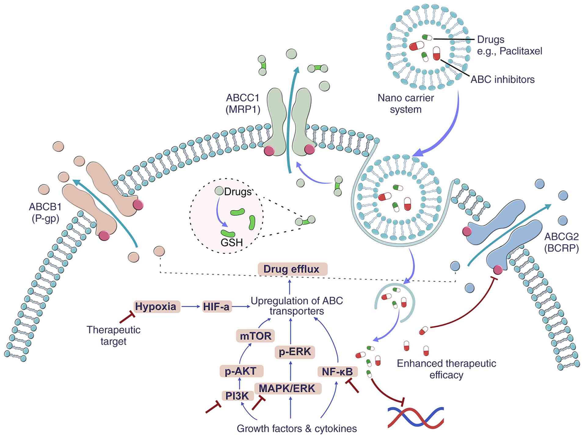 | Figure 1.ABC transporter-mediated multidrug
resistance in breast cancer. ABCB1 (P-gp), ABCC1 (MRP1), and ABCG2
(BCRP) actively efflux chemotherapeutic drugs, such as paclitaxel,
with ABCC1 function assisted by GSH. Hypoxia/HIF-α and growth
factor-activated PI3K/AKT/mTOR, MAPK/ERK, and NF-κB pathways
upregulate these transporters. Nanocarrier systems co-delivering
ABC inhibitors and drugs bypass efflux to enhance intracellular
retention and therapeutic efficacy. ABC, ATP binding cassette;
ABCB1, ABC subfamily B member 1; P-gp P-glycoprotein; ABCC1, ABC
subfamily C member 1; MRP1, multidrug resistance protein 1; ABCG2,
ABC subfamily G member 2; BCRP, breast cancer resistance protein;
GSH, glutathione; HIF-1α, hypoxia-inducible factor; PI3K,
phosphatidylinositol 3-kinase; AKT, protein kinase B; mTOR,
mammalian target of rapamycin; MAPK, mitogen-activated protein
kinase; ERK, extracellular signal-regulated kinase; NF-κB, nuclear
factor-κB; p-, phosphorylated. |
Tumor microenvironment reprogramming
Hypoxic microenvironment and abnormal
angiogenesis
The hypoxic nature of the TME is a critical
pathological feature that drives angiogenesis and the development
of drug resistance. As tumors proliferate rapidly, the local oxygen
partial pressure decreases significantly, leading to the
stabilization and activation of HIF-1α (26). As a master regulator of the hypoxic
response, HIF-1α promotes the formation of disorganized and
dysfunctional tumor vasculature by upregulating pro-angiogenic
factors such as VEGF (27). This
aberrant vascular network not only impedes effective drug delivery
but also supports the survival and stemness of cancer stem cells
(CSCs), thereby enhancing chemoresistance (28). Meanwhile, hypoxia facilitates the
infiltration of immunosuppressive cells, including myeloid-derived
suppressor cells (MDSCs) and regulatory T cells, while impairing
the function of cytotoxic T cells, which compromises the response
to immunotherapy (29). Moreover,
the hypoxia-HIF-α axis reprograms cellular metabolism and induces
epigenetic remodeling, further conferring tolerance to chemotherapy
and radiotherapy in breast cancer cells (30). It is noteworthy that the expression
and activity of HIF-1α are regulated by multiple signaling
pathways, including PI3K/AKT/mTOR and MAPK/ERK (31). Therefore, targeting these upstream
pathways or directly inhibiting HIF-1α, in combination with
anti-angiogenic and immunomodulatory strategies, may represent a
promising therapeutic approach to overcome hypoxia-associated
treatment resistance (Fig. 2).
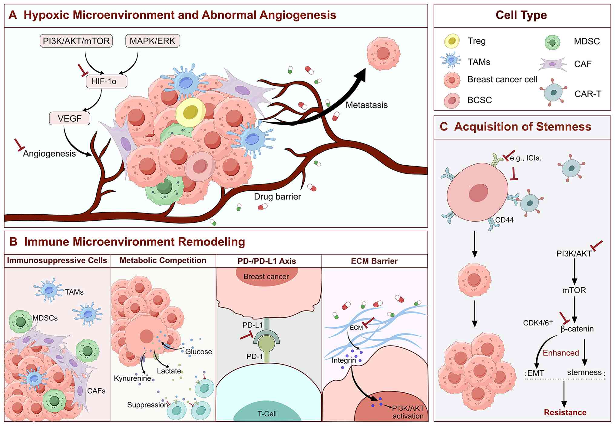 | Figure 2.Schematic of tumor
microenvironment-mediated drug resistance in breast cancer. (A)
Hypoxia-induced HIF-1α/VEGF signaling promotes abnormal
angiogenesis and drug barrier formation. (B) Immunosuppressive
cells (TAMs, MDSCs, Tregs and CAFs), metabolic competition,
PD-1/PD-L1 axis and ECM barrier suppress antitumor immunity. (C)
PI3K/AKT/mTOR and Wnt/β-catenin pathways drive BCSC stemness and
EMT, conferring therapeutic resistance. HIF-1α, hypoxia-inducible
factor; VEGF, vascular endothelial growth factor; TAMs,
tumor-associated macrophages; MDSCs, myeloid-derived suppressor
cells; Tregs, regulatory T cells; CAFs, cancer-associated
fibroblasts; PD-1, programmed cell death protein 1; PD-L1,
programmed death ligand 1; ECM, extracellular matrix; PI3K,
phosphatidylinositol 3-kinase; AKT, protein kinase B; mTOR,
mammalian target of rapamycin; BCSC, breast cancer stem cell; EMT,
epithelial-mesenchymal transition; MAPK, mitogen-activated protein
kinase; ERK, extracellular signal-regulated kinase; CAR-T, chimeric
antigen receptor T cell; ICIs, Immune checkpoint inhibitors;
CDK4/6, cyclin-dependent kinases 4 and 6. |
Immune microenvironment
remodeling
The TME in breast cancer constitutes a complex
ecosystem composed of tumor cells, immune cells, cancer-associated
fibroblasts (CAFs), and the extracellular matrix (ECM), among other
components. Through their interactions, these elements suppress
immune responses, thereby promoting tumor progression and
conferring drug resistance (32).
For instance, the infiltration of immunosuppressive cells, such as
CAFs, tumor-associated macrophages, and MDSCs, can secrete
inhibitory cytokines that impair the activity of cytotoxic T cells,
leading to resistance to immunotherapy (33,34).
Furthermore, metabolic competition and immunosuppression contribute
to therapeutic resistance. Due to metabolic reprogramming, breast
cancer cells compete with T cells for nutrients such as glucose,
amino acids, and lipids. This process results in the accumulation
of metabolites including lactic acid and kynurenine, which suppress
T-cell activity. The combined effects of metabolic stress and
metabolite-induced immunosuppression significantly enhance
immunotherapy resistance in breast cancer (35). Abnormal immune checkpoint activation
further promotes tumor evasion. The binding of PD-L1 on tumor cells
to PD-1 on T cells inhibits T-cell activation and facilitates
immune escape. Clinical studies show that blocking the PD-1/PD-L1
interaction can restore T-cell function (36). ECM-mediated physical barrier also
contributes to resistance. Excessive ECM deposition not only
impedes drug penetration but also promotes tumor cell survival by
activating the PI3K/AKT pathway via integrin signaling. Targeting
ECM remodeling can improve drug delivery and restore immune cell
infiltration (37). Therefore,
developing combination strategies targeting the immunosuppressive
microenvironment, such as ICIs combined with metabolic modulators
or ECM targeting agents, may provide a novel approach to overcome
drug resistance in breast cancer. A comprehensive analysis of the
dynamic interaction network among various components within the
tumor microenvironment will establish a theoretical foundation for
developing more effective therapeutic strategies (Fig. 2).
Acquisition of stemness
Breast CSCs (BCSCs), a subpopulation within tumors
endowed with self-renewal and multilineage differentiation
potential, are a major driver of chemotherapy and targeted therapy
resistance due to their enrichment and expansion (38). The maintenance of BCSC stemness is
mediated by aberrant activation of multiple signaling pathways. For
example, activation of the PI3K/AKT/mTOR axis enhances BCSC
stemness, thereby conferring therapy resistance (39). Activation of the Wnt/β-catenin
signaling pathway induces epithelial-mesenchymal transition (EMT)
to promote metastasis and confers resistance to endocrine therapy.
Notably, studies have shown that inhibiting this pathway, in
combination with CDK4/6 inhibitors, can partially reverse this
resistance (40,41). The stemness of BCSCs is also
regulated by epigenetic mechanisms. For instance, aberrant
expression of long non-coding RNAs (lncRNAs) and alterations in DNA
methylation patterns promote the stem cell-like phenotype, thereby
reducing treatment efficacy (42).
Moreover, inflammatory factors in the TME contribute to stemness
maintenance by inhibiting apoptosis, promoting drug efflux protein
expression, and enhancing inherent stem-like properties, thereby
reinforcing therapy resistance (43). Several therapeutic strategies
targeting BCSCs have been developed, including chimeric antigen
receptor T-cell (CAR-T) therapy, ICIs, and approaches using BCSCs
as drug delivery vehicles. However, these treatments still face
challenges such as insufficient targeting specificity, acquired
resistance, and safety concerns. Future efforts should focus on
in-depth characterization of BCSCs, optimization of treatment
strategies, and enhanced clinical validation to improve therapeutic
outcomes (44) (Fig. 2).
Dysregulation of epigenetics
Aberrant DNA methylation
DNA methylation plays a pivotal role in breast
cancer drug resistance. Aberrant DNA methylation patterns can lead
to the silencing of tumor suppressor genes or activation of drug
resistance-associated genes, thereby altering cellular response to
therapy (45). Research has
demonstrated a significant correlation between the mRNA expression
of drug resistance genes and their DNA methylation status, with
variations observed across different molecular subtypes (46). In ER-positive breast cancer,
aberrant DNA methylation was shown to be closely associated with
resistance to endocrine therapy (47). The DNA methyltransferase inhibitor
decitabine has been shown to reverse epigenetic alterations in
drug-resistant cells, restoring their sensitivity to
chemotherapeutic agents (48).
Protein arginine methyltransferases (PRMTs) are fundamental
epigenetic enzymes; pharmacological inhibition of PRMTs has been
shown to markedly sensitize tumors to diverse anticancer therapies,
supporting the potential of combination strategies with
conventional agents to circumvent treatment resistance (49). In addition, dysregulation of m6A RNA
methylation regulators may contribute to tumor progression.
Although their precise role in DNA repair remains incompletely
elucidated, preliminary evidence suggests a potential influence on
cancer drug resistance (50). DNA
methylation-based biomarkers have the potential to identify
patients who respond to platinum-based chemotherapy and may offer
novel therapeutic targets to combat tumor resistance. However,
research in this field continues to face several challenges. The
heterogeneity of DNA methylation patterns across distinct breast
cancer subtypes, the complexity of the DNA methylation regulatory
network, and the limited selectivity of current epigenetic drugs
pose significant challenges. The development of liquid biopsy
technology has provided new opportunities for the clinical
translation of DNA methylation biomarkers by enabling the
simultaneous assessment of tumor gene mutations, resistance genes,
and DNA methylation status through detection of circulating tumor
DNA in blood, thereby offering multidimensional evidence for
precision breast cancer therapy (51). Future studies should focus on
elucidating the crosstalk between DNA methylation and other
epigenetic mechanisms, such as histone modifications, to facilitate
the development of more effective combination treatment regimens
(Fig. 3).
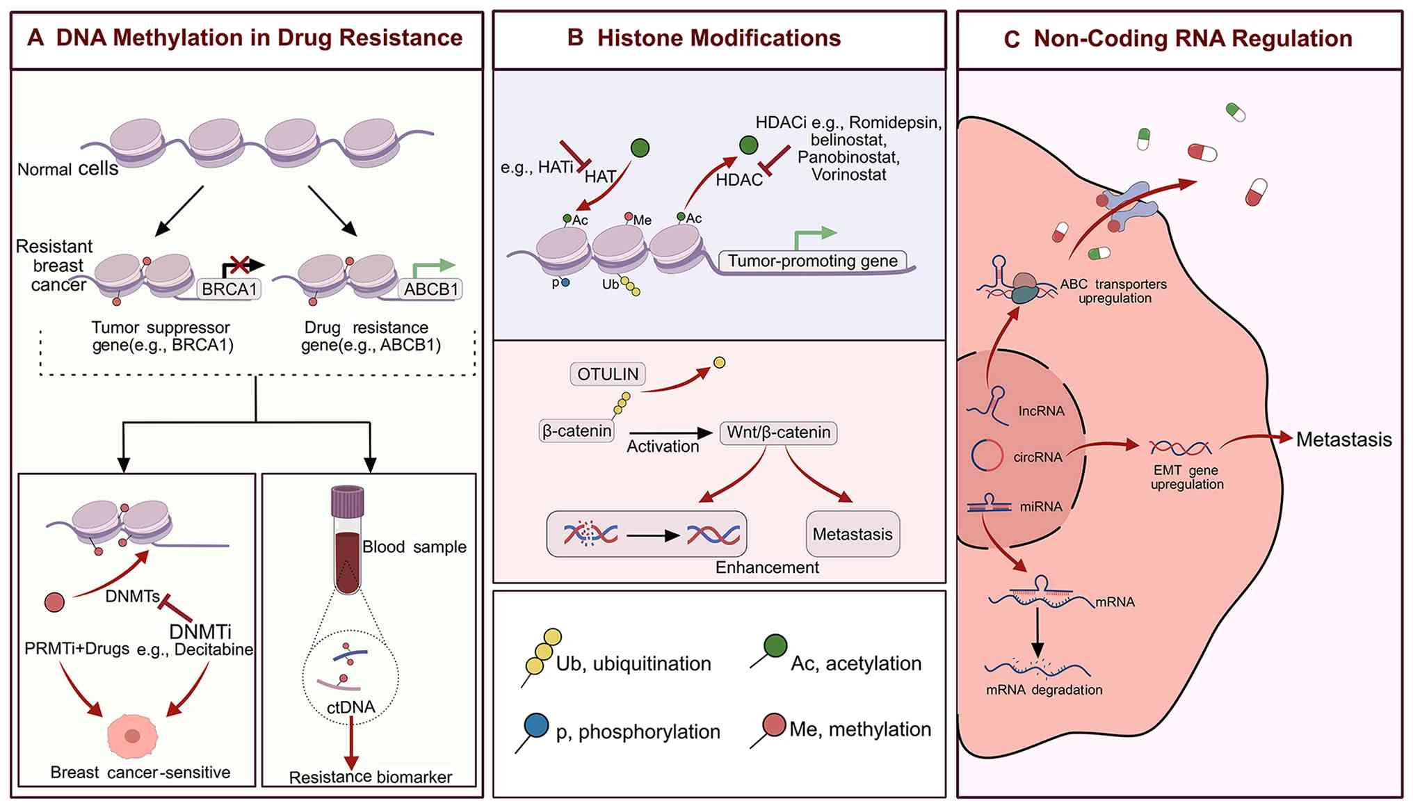 | Figure 3.Epigenetic regulation of MDR in
breast cancer. (A) DNA methylation by DNMTs silences tumor
suppressors, such as BRCA1, and activates resistance genes, such as
ABCB1, reversed by DNMTi/PRMTi. (B) Histone modifications
(acetylation, phosphorylation, ubiquitination) regulate oncogene
expression and Wnt/β-catenin-mediated DNA repair and metastasis.
(C) Non-coding RNAs (lncRNAs, circRNAs, miRNAs) modulate ABC
transporter expression, EMT and mRNA stability. MDR, multidrug
resistance; DNMTs, DNA methyltransferases; BRCA1, breast cancer
susceptibility gene 1; ABCB1, ABC subfamily B member 1; DNMTi, DNA
methyltransferase inhibitors; PRMTi, protein arginine
methyltransferase inhibitors; lncRNAs, long non-coding RNAs;
miRNAs, microRNAs; circRNAs, circular RNAs; ABC, ATP binding
cassette; EMT, epithelial-mesenchymal transition; HATi, histone
acetyltransferase inhibitors; HAT, histone acetyltransferase; HDAC,
histone deacetylase; HDACi, histone deacetylase inhibitors. |
Histone modifications
Histone modifications refer to various chemical
changes, such as methylation, acetylation, phosphorylation, and
ubiquitination, that occur on histone proteins. These modifications
regulate gene transcription by altering chromatin structure or
recruiting modifier proteins, and they play a critical role in
epigenetic regulation. Dysregulation of histone modifications is a
core epigenetic mechanism underlying acquired drug resistance in
breast cancer. In ER-positive breast cancer, aberrant histone H3
modifications, such as H3K27 acetylation, are associated with
resistance to endocrine therapy (52). The combination of histone
deacetylase inhibitors (HDACi; such as vorinostat) with
conventional chemotherapeutic drugs significantly enhances
treatment sensitivity in resistant cells (53). Furthermore, following chemotherapy
for breast cancer, OTULIN (a deubiquitinating enzyme) stabilizes
β-catenin via deubiquitination, activating the Wnt/β-catenin
pathway. This activation promotes DDR and metastasis, suggesting
that inhibition of this pathway may enhance chemosensitivity
(54). The FDA has approved several
HDACi, including romidepsin, belinostat, panobinostat, and
vorinostat, for clinical use. In addition, small-molecule
inhibitors targeting histone acetyltransferases are under clinical
investigation (45). In summary,
histone modifications drive drug resistance by regulating gene
expression, chromatin architecture, and signaling pathway activity.
Developing small-molecule inhibitors against specific
histone-modifying enzymes, either alone or in combination with
other epigenetic drugs, may provide novel therapeutic strategies to
overcome drug resistance in breast cancer (Fig. 3).
Non-coding RNA (ncRNA)-mediated
regulation
ncRNAs, as crucial players in epigenetic regulation,
play key roles in mediating treatment resistance in breast cancer.
Diverse ncRNAs, including microRNAs, lncRNAs, and circular RNAs
(circRNAs) contribute to resistance by regulating gene expression,
modulating the activation of signaling pathways, and remodeling the
TME (55,56). For example, in ER-positive breast
cancer, ~40% of patients relapse due to acquired tamoxifen
resistance, a process involving ncRN-mediated regulation of
resistance-related genes (57). HOX
transcript antisense intergenic RNA, a well-characterized oncogenic
lncRNA, promotes breast cancer metastasis and chemoresistance
through epigenetic modifications, regulation of target genes, and
activation of signaling pathways (58). Certain lncRNAs enhance
chemoresistance by upregulating ABC transporter expression, which
reduces intracellular drug accumulation and impairs the efficacy of
agents such as gemcitabine (59).
Moreover, aberrant expression of circRNAs has been shown to promote
tumor cell invasiveness and drug resistance by regulating
EMT-related genes (60). Thus,
ncRNA expression patterns show promise as biomarkers for predicting
the risk of resistance and as potential therapeutic targets.
Although antisense oligonucleotide-based interventions have
demonstrated efficacy in restoring chemosensitivity, challenges
such as a lack of assay standardization and off-target effects have
impeded their translation into routine clinical use (61). Future research should focus on
elucidating ncRNA-mediated crosstalk within the immune
microenvironment, developing highly specific ncRNA antagonists to
overcome endocrine resistance through personalized therapeutic
approaches, and integrating multi-omics data to accelerate
translational applications (62)
(Fig. 3).
Enhanced DNA damage repair mechanisms
Homologous recombination repair (HRR)
restoration
HRR deficiency is closely associated with drug
resistance mechanisms in breast cancer, primarily involving
restoration of homologous recombination function, alterations in
DNA replication fork stability, aberrant epigenetic regulation, and
compensatory activation of alternative DNA repair pathways. As core
components of the HRR pathway, loss-of-function mutations in
BRCA1/2 sensitize tumor cells to PARPi and platinum-based agents.
However, clinical observations indicate that restoration of HRR
function may lead to acquired resistance (63). Specific mechanisms of resistance
include epigenetic regulation-mediated restoration of HRR,
exemplified by demethylation of BRCA gene promoters, which restores
BRCA protein expression and reactivates the HRR pathway (64) and compensatory upregulation of key
DNA repair proteins. For instance, loss of BRCA1/2 induces
compensatory activation of RAD51 recombinase (RAD51), which
bypasses HRR defects to restore double-strand break (DSB) repair
capacity, thereby conferring chemotherapy resistance (65). Loss of 53BP1 partially restores HRR
in BRCA-deficient cells by modulating DNA end resection, thereby
enabling escape from PARPi-induced synthetic lethality (66,67).
Dysfunction of HRR-associated proteins such as partner and
localizer of BRCA2 (PALB2), which works together with BRCA1/2 and
RAD51 to mediate HRR, may also lead to HRR deficiency and sensitize
tumors to PARPi. For example, loss of PALB2 similarly disrupts HRR
and increases sensitivity to PARPi. However, certain PALB2
mutations may impair its binding to BRCA1/2, resulting in partially
retained HRR function and subsequent drug resistance. Targeting the
PALB2 recruitment machinery may thus represent a promising
therapeutic strategy for BRCA1-mutated tumors (68–70).
Clinical data have demonstrated that combining PARPi with
chemotherapeutic agents, such as paclitaxel and gemcitabine, or
immunotherapeutic agents, such as pembrolizumab, shows potential in
overcoming drug resistance (71).
Future precision therapies targeting HRR deficiency are expected to
expand the eligible patient population in breast cancer. Further
investigation of pathways that crosstalk with the HRR network may
reveal novel targets for overcoming PARPi resistance. In clinical
practice, molecular profiling of HRR gene alterations could enable
early identification of patients at high risk of treatment
resistance and guide personalized therapeutic strategies (Fig. 4).
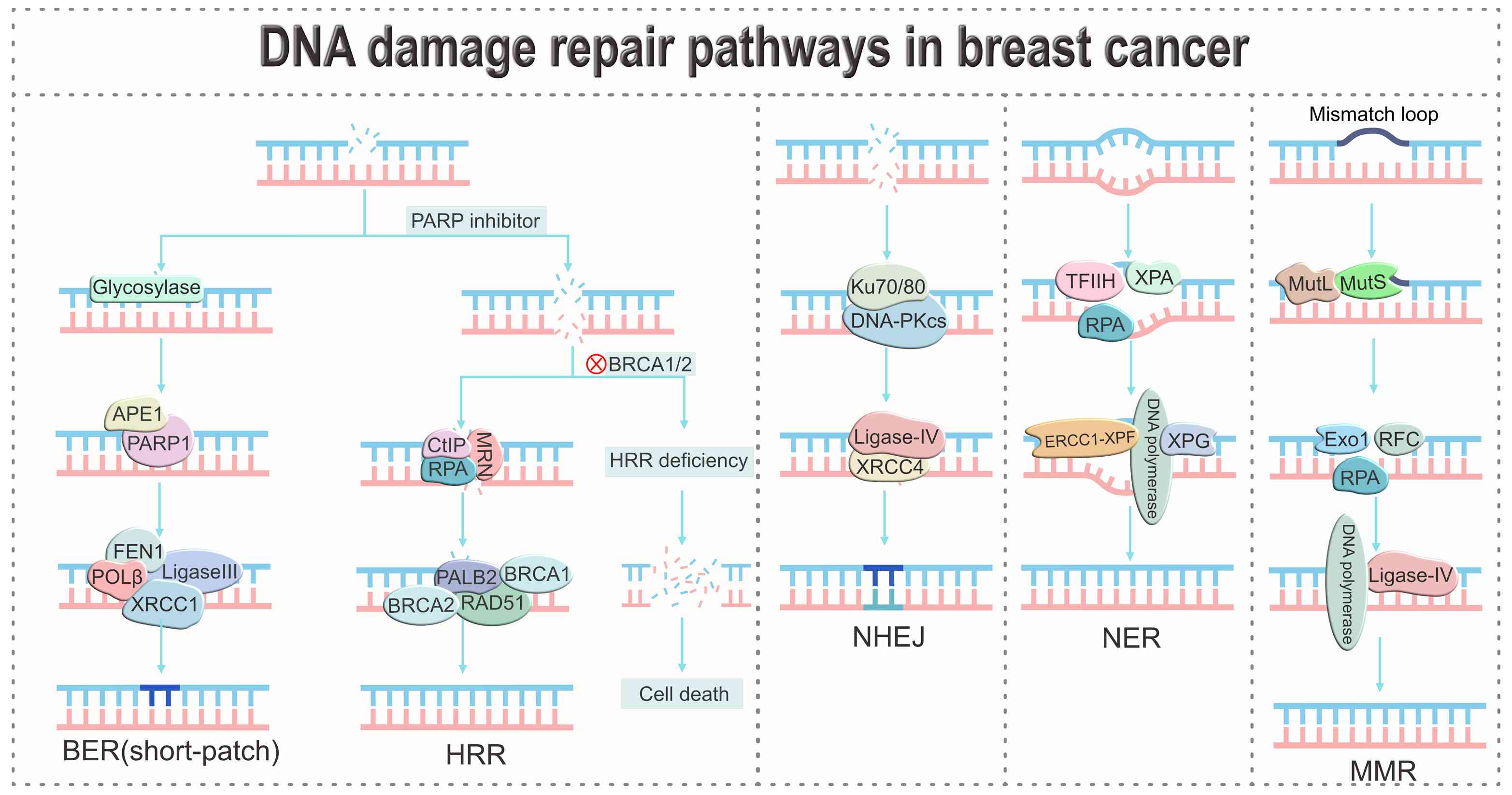 | Figure 4.DNA damage repair pathways in breast
cancer. The schematic depicts BER, HRR, NHEJ, NER, and MMR
mechanisms. PARPi selectively induce cell death via synthetic
lethality in BRCA1/2-deficient tumors. Compensatory upregulation of
these pathways mediates resistance to chemotherapy and PARPi. BER,
base excision repair; HRR, homologous recombination repair; NHEJ,
non-homologous end joining; NER, nucleotide excision repair; MMR,
mismatch repair; PARP, poly(ADP-ribose) polymerase inhibitors;
BRCA1/2, breast cancer susceptibility genes 1/2; APE1,
apurinic/apyrimidinic endonuclease 1; CtIP, CtBP-interacting
protein; RPA, replication protein A; MRN, MRE11-RAD50-NBS1 complex;
HR, homologous recombination; FEN1, flap endonuclease 1; POLβ, DNA
polymerase β; LigaseIII, DNA ligase III; XRCC1, X-ray repair
cross-complementing 1; PALB2, partner and localizer of BRCA2;
BRCA1, breast cancer susceptibility gene 1; BRCA2, breast cancer
susceptibility gene 2; RAD51, RAD51 recombinase; DNA-PKcs,
DNA-dependent protein kinase catalytic subunit; Ligase-IV, DNA
ligase IV; XRCC4, X-ray repair cross-complementing protein 4-like
factor; TFIIH, transcription factorIIH; XPA, xeroderma pigmentosum
complementation group A protein; ERCC1, excision repair
cross-complementation group 1; XPF, xeroderma pigmentosum group
F-complementing protein; XPG, xeroderma pigmentosum complementation
group G protein; MutL, mismatch repair protein complex; MutS,
mismatch recognition protein complex; Exo1, exonuclease 1; RFC,
replication factor C. |
Non-homologous end joining (NHEJ)
pathway activation
NHEJ is a primary pathway for repairing DSBs in
mammalian cells. Upon the occurrence of DSBs, the Ku70/80
heterodimer is rapidly recruited to the broken ends and binds to
them in a sequence-nonspecific manner. This recruitment is followed
by the assembly and activation of the DNA-dependent protein kinase
catalytic subunit, facilitating synapsis of the broken DNA ends.
Subsequently, end-processing factors, including the Artemis
nuclease, are engaged to prepare the termini for ligation.
Ultimately, the X-ray repair cross-complementing (XRCC) 4-like
factor complex, in conjunction with DNA ligase IV, catalyzes the
final ligation step to complete the repair process (72–74).
The repair process frequently introduces base deletion or insertion
mutations, leading to reduced sequence fidelity. In breast cancer,
core NHEJ components are frequently upregulated. This elevation is
regarded as a compensatory adaptation to genomic instability or
impairments in other DSB repair pathways, thereby conferring
resistance to radiotherapy and numerous chemotherapeutic agents
(75,76). Additionally, the alternative
non-homologous end joining (Alt-NHEJ) pathway is mediated by key
factors such as DNA ligase III, DNA polymerase θ, and PARP1. Their
upregulation is closely associated with tumor progression and drug
resistance. When canonical NHEJ is impaired, Alt-NHEJ is activated
as a backup pathway to maintain genome integrity, paradoxically
promoting drug resistance (77).
The aforementioned evidence indicates that NHEJ and its alternative
pathways contribute to drug resistance in breast cancer via a
multi-molecular interaction network. Targeting key factors in the
NHEJ pathway may represent a promising strategy to overcome
NHEJ-mediated resistance (Fig.
4).
Base excision repair (BER) pathway
activation
The BER pathway serves as a primary cellular defense
mechanism against DNA single-base lesions and single-strand breaks,
primarily repairing damage caused by both endogenous and exogenous
factors such as reactive oxygen species (ROS), alkylating agents,
and ionizing radiation (78–80).
First, chemotherapy-induced DDR is considered. The BER pathway
effectively reverses DNA lesions induced by chemotherapeutic agents
such as cisplatin, significantly reducing the cytotoxic efficacy of
these drugs against cancer cells (81). Flap endonuclease 1 (FEN1), a key
enzyme in BER, is often overexpressed in TNBC and confers
resistance to chemotherapeutic agents. Inhibition of FEN1 has been
shown to sensitize tumor cells to treatment (82). Second, the influence of genetic
variation, such as single-nucleotide polymorphisms in BER-related
genes, including XRCC1, may alter BER efficiency and consequently
affect treatment response and prognosis in patients with breast
cancer (83). Currently, no
specific therapeutic agents targeting the BER pathway are available
beyond PARPi. Therefore, systematic identification of key
biomarkers in the BER pathway, such as the expression level of DNA
polymerase β, XRCC1 mutation status, and FEN1 activity, will
contribute to the precise screening of patients likely to benefit,
and provide a theoretical basis for developing novel targeted
strategies (Fig. 4).
Hyperactivation of nucleotide excision
repair (NER)
NER is an essential DNA repair mechanism in
mammalian cells, dedicated to removing bulky DNA lesions that
distort the helical structure, such as UV-induced cyclobutane
pyrimidine dimers and DNA damage generated by chemotherapeutic
agents. In breast cancer, aberrantly elevated NER activity
efficiently removes cisplatin-induced DNA adducts and impedes
damage accumulation, thereby mediating both intrinsic and acquired
resistance to this chemotherapeutic agent (84,85).
Excision repair cross-complementation (ERCC) group 1, a
rate-limiting enzyme in the NER pathway, confers cellular
resistance to platinum-based drugs when highly expressed due to
enhanced DNA repair capacity (86).
Further studies revealed that methylation of the ERCC4 gene
promoter results in loss of its encoded product, xeroderma
pigmentosum group F-complementing protein (XPF), thereby
suppressing NER function and increasing cisplatin sensitivity.
Conversely, restoring XPF expression reactivates NER and reduces
cisplatin efficacy, suggesting that epigenetic modulation of ERCC4
to target NER activity holds therapeutic potential (87,88).
Moreover, under specific contexts, BRCA1 facilitates the clearance
of chemotherapy-induced DNA lesions and promotes genomic stability
through co-activation of NER (89).
However, the functional landscape of NER activity in breast cancer
remains incompletely defined. Precise identification of NER
deficiencies or hyperactivation is expected to guide the
development of NER-targeting agents, thereby increasing
chemosensitivity and overcoming drug resistance (Fig. 4).
Mismatch repair (MMR) deficiency
The MMR system maintains genomic stability by
recognizing and correcting base-base mismatches during DNA
replication. In breast cancer chemoresistance, it mediates
context-dependent resistance through bidirectional epigenetic
reprogramming. On one hand, loss of MMR function, such as that
induced by MutL Homolog 1 (MLH1) or MutS Homolog 2 (MSH2) gene
silencing, causes microsatellite instability-high and may initially
increase tumor sensitivity to DNA-damaging agents. However,
sustained MMR deficiency ultimately promotes genomic instability.
For example, in doxorubicin resistance, hypermethylation of the
MSH2 promoter compromises MMR function, facilitates the
accumulation of acquired mutations, and leads to secondary
resistance (90–92). On the other hand,
anthracycline-induced hypomethylation of MLH1 or MSH2 promoters can
enhance MMR gene expression and repair activity. This enables
cancer cells to excessively clear DNA damage, resulting in
pan-chemoresistance (93). The MutL
complex genes (MLH1, PMS1, PMS2, and MLH3) are core components of
the MMR pathway. Defects in these genes disrupt MMR function,
preventing ER-positive breast cancers from effectively suppressing
CDK4 activity during endocrine therapy, thereby conferring
treatment resistance. Consequently, MMR deficiency may serve as a
predictive biomarker for response to CDK4/6 inhibitors (94). Furthermore, it has been shown that
the HR and MMR pathways are functionally interconnected in DNA
repair-deficient tumors and share key protein components. Although
the precise mechanisms remain incompletely elucidated, this
crosstalk offers novel insights and potential therapeutic targets
for precision oncology (95). In
summary, co-targeting complementary DNA damage response pathways,
such as combining PARPi with MMR targeting strategies, represents a
promising therapeutic approach to overcome drug resistance in
breast cancer (Fig. 4).
Alterations in metabolic reprogramming
Enhanced glycolysis
Metabolic reprogramming in breast cancer cells is
characterized by markedly enhanced glycolysis (the Warburg effect),
which operates synergistically with the tricarboxylic acid cycle
and the hexosamine biosynthesis pathway to supply energy and
biosynthetic precursors that promote tumor cell survival, invasion,
and maintenance of stemness under chemotherapy-induced stress
(96). For instance, Pim-2
proto-oncogene, serine/threonine kinase was shown to bind to and
promote phosphorylation of 6-phosphofructo-2-kinase at Ser478,
thereby enhancing glycolytic activity and conferring paclitaxel
resistance in breast cancer cells (97). Moreover, pyruvate kinase M2 was
demonstrated to activate autophagy, supplying tumor cells with
energy and metabolic intermediates that bolster survival advantage
under drug pressure. Thus, the ‘glycolysis-autophagy axis’
represents a core mechanism underlying chemoresistance (98). Hexokinase 2 (HK2) also activates the
NF-κB pathway, leading to upregulation of PD-L1 expression, which
drives immune escape and influences tumor immune infiltration and
patient prognosis. These findings suggest that combining PD-L1
blockade with HK2-targeted therapy may offer a novel therapeutic
strategy for breast cancer (99).
The end product of glycolysis, lactate, contributes to tumor
progression by acidifying the tumor microenvironment, suppressing
immune responses, and modulating oncogene expression (100). Therefore, developing specific
inhibitors targeting key glycolytic enzymes and using them in
combination with existing anticancer drugs may overcome therapy
resistance by inhibiting tumor glycolytic metabolism and enhancing
immune responses, thereby providing a novel strategy for breast
cancer treatment (96) (Fig. 5).
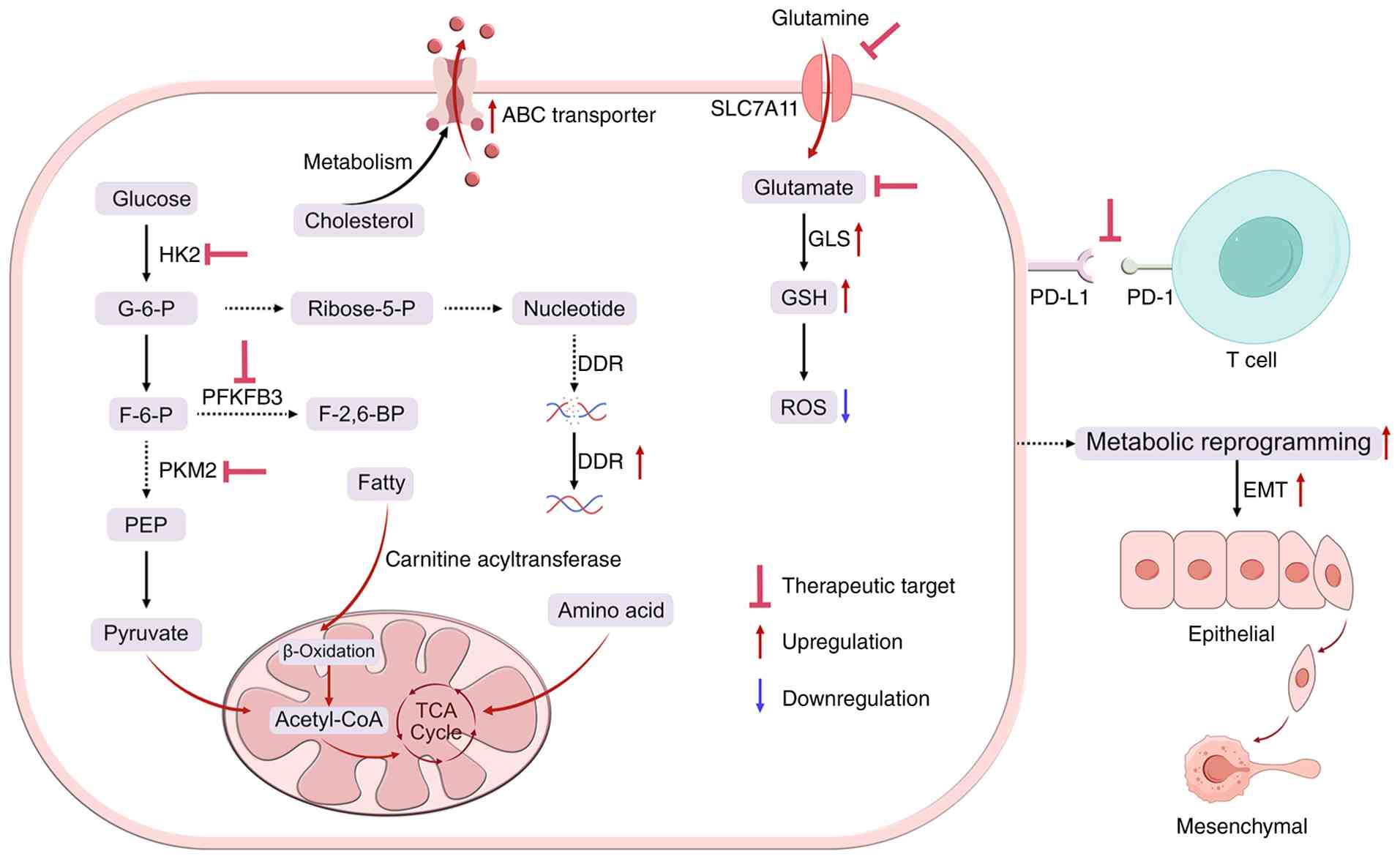 | Figure 5.Metabolic reprogramming in breast
cancer and its therapeutic targets. Upregulated glycolysis,
glutamine metabolism and nucleotide synthesis sustain tumor
bioenergetics, redox balance (GSH/ROS) and DDR. Lipid metabolism
and the TCA cycle provide additional energetic support. These
adaptations drive PD-L1-mediated immune evasion and EMT, promoting
drug resistance. GSH, glutathione; ROS, reactive oxygen species;
DDR, DNA damage repair; TCA, tricarboxylic acid; PD-L1, programmed
death ligand 1; EMT, epithelial-mesenchymal transition; SLC7A11,
solute carrier family 7 member 11; ABC, ATP binding cassette; HK2,
hexokinase 2; G-6-P, glucose-6-phosphate; F-6-P,
fructose-6-phosphate; PFKFB3,
6-phosphofructo-2-kinase/fructose-2,6-biphosphatase 3; F-2,6-BP,
fructose-2,6-bisphosphate; PKM2, pyruvate kinase M2; PEP,
phosphoenolpyruvate; Ribose-5-P, ribose-5-phosphate; GLS,
glutaminase; PD-1, programmed cell death protein 1. |
Dysregulation of lipid metabolism
Disorders in lipid metabolism contribute to
treatment evasion in breast cancer cells by regulating fatty acid
synthesis, cholesterol metabolism, and phospholipid remodeling,
thereby providing a sustained energy supply and maintaining TME
homeostasis (101,102). Key lipid metabolic enzymes are
tightly regulated by transcription factors that drive aberrant
synthesis and accumulation of fatty acids and cholesterol, thereby
enhancing resistance to chemotherapy, endocrine therapy, and
targeted therapy. Sterol regulatory element-binding protein 1
promotes fatty acid synthesis via Akt/mTOR activation, thereby
increasing cell membrane fluidity and reducing drug influx, leading
to doxorubicin resistance (103).
Dysregulated cholesterol metabolism enhances chemoresistance by
upregulating ABC transporter expression and facilitating lipid
raft-mediated signaling (104).
The ketone body-producing rate-limiting enzyme
3-hydroxy-3-methylglutaryl-CoA synthase 2 is highly expressed in
tamoxifen-resistant breast cancer, and developing inhibitors
against it represents a promising strategy for reversing endocrine
resistance (105). Notably, lipid
metabolic reprogramming also modulates EMT and stemness, remodels
the immune microenvironment, and alters chemotherapeutic
metabolism, collectively exacerbating drug resistance (106–108). This multi-layered and
multi-targeted metabolic network highlights targeting key nodes of
lipid metabolism as a potential therapeutic strategy to overcome
drug resistance in breast cancer (Fig.
5).
Aberrant amino acid metabolism
Amino acids serve as essential nutrients for
sustaining cellular life. Metabolic reprogramming of amino acids
has been demonstrated to significantly promote the proliferation,
metastasis, and therapy resistance of breast cancer cells, a
process closely associated with dysregulated expression of amino
acid transporters and altered activity of key enzymes. Research has
revealed that solute carrier family 7 member 5 (SLC7A5), an amino
acid transporter, is highly expressed across different breast
cancer subtypes, particularly in Luminal B breast cancer, where its
elevated expression is associated with poor patient prognosis,
suggesting its potential as a therapeutic target in this subtype
(109). In TNBC,
chemotherapy-induced reactive ROS stress prompts cancer cells to
enhance glutamine metabolism by upregulating glutaminase (GLS) and
glutamate transporters, leading to massive glutamate efflux. This
facilitates GSH synthesis to scavenge ROS and maintain redox
homeostasis, thereby driving chemoresistance. Targeting glutamate
depletion or inhibiting GLS and the cystine/glutamate transporter
SLC7A11 (xCT) disrupts this antioxidant system, resulting in ROS
accumulation and resensitization to treatment (110,111). Amino acid metabolism also
interfaces with the TME: Tumor cells compete with immune cells,
such as T cells, for key amino acids such as tryptophan and
arginine, supporting tumor survival and proliferation. Meanwhile,
accumulation of the tryptophan metabolite kynurenine suppresses
T-cell function and fosters an immunosuppressive TME, ultimately
compromising immunotherapy efficacy and indirectly contributing
drug resistance in breast cancer (112,113). In summary, targeting amino acid
transporters, such as with SLC7A5 inhibitors, in combination with
metabolic inhibitors and immunotherapy, along with metabolism-based
precision therapy, represents a promising strategy for overcoming
drug-resistant breast cancer (Fig.
5).
Alterations in nucleotide
metabolism
Nucleotides function as the fundamental building
blocks for DNA/RNA synthesis and participate in cellular energy
metabolism, signaling transduction, and proliferation regulation.
In breast cancer, upregulation of rate-limiting enzymes in the
de novo nucleotide synthesis pathway promotes excessive
purine and pyrimidine accumulation, which activates downstream
signaling cascades, enhances tumor stemness and metastatic
potential, and facilitates DDR, ultimately leading to
chemoresistance (114,115). For instance, elevated expression
of the purine metabolic enzyme phosphoribosylaminoimidazole
succinocarboxamide synthetase enhances ERα activity through the
cyclic adenosine monophosphate-protein kinase A-mTOR signaling
axis, resulting in tamoxifen resistance in breast cancer (116). Furthermore, nucleotide metabolic
reprogramming contributes to therapy resistance by modulating the
TME. Tumor cells consume nucleotide precursors and release
immunosuppressive metabolites, leading to T-cell dysfunction
through nutrient competition and subsequent immune escape (117,118). Therefore, targeting nucleotide
metabolism in combination with conventional therapies may offer a
novel multi-target strategy to overcome drug resistance in breast
cancer by synergizing metabolic intervention with immune modulation
(Fig. 5).
Conclusions and future perspectives
Breast cancer is one of the most common malignancies
in women. Standard treatment regimens include chemotherapy, as well
as endocrine and targeted therapies; however, acquired drug
resistance significantly shortens patient survival and remains a
major clinical challenge. Drug resistance in breast cancer arises
from a multifactorial, interconnected network encompassing ABC
transporter-mediated drug efflux, hypoxic and immunosuppressive TME
remodeling, aberrant epigenetic reprogramming via DNA methylation,
histone modifications and ncRNAs, compensatory activation of DDR
pathways including HRR and NHEJ, and adaptive metabolic rewiring
involving glycolysis, lipid, amino acid, and nucleotide metabolism.
These mechanisms are co-regulated by key signaling axes such as
PI3K/AKT/mTOR, Wnt/β-catenin, and HIF-1α. Correspondingly,
effective therapeutic solutions include combination regimens
integrating targeted inhibitors, such as PI3K/mTOR inhibitors and
HDACi, immune-based modalities such as CAR-T, CAR-natural killer
cells, bispecific T-cell engagers, oncolytic viruses, and ADCs,
alongside stimuli-responsive nanocarriers and proteolysis-targeting
chimeras that enhance delivery precision and reduce off-target
toxicity.
Additionally, it is worth noting that the microbiome
can regulate estrogen metabolism and host immune responses,
directly promoting breast cancer development and the emergence of
chemotherapy resistance. Conversely, certain probiotic
Lactobacillus species can enhance antitumor immunity,
thereby playing a bidirectional regulatory role in disease
progression and treatment (119).
This duality is well exemplified by previous studies.
Pro-tumorigenic effects were observed in the findings of Ma et
al (120), who reported that
enterotoxigenic Bacteroides fragilis activates
nucleotide-binding oligomerization domain-containing 1-Notch
receptor 1 signaling via Bacteroides fragilis toxin-1,
enriching BCSCs and driving chemoresistance; similarly, Fu et
al (121) showed that
intratumoral bacteria remodel the cytoskeleton of circulating tumor
cells, facilitating metastatic colonization (121). Conversely, anti-tumorigenic
effects were demonstrated by Wu et al (122), who found that flaxseed lignans,
converted by the gut microbiota into enterolactone, downregulate
CD38 and enrich Akkermansia, synergizing with PD-1/PD-L1 inhibitors
to suppress tumor growth (122).
Therefore, through the integration of prospective interventional
trials with multi-omics and artificial intelligence, the clinical
value of gut and breast microbiota as predictive biomarkers for
therapeutic response and as druggable targets for reversing drug
resistance in breast cancer can be systematically validated.
In summary, future efforts should focus on
developing switch-controlled immunotherapies and local prodrug
strategies to improve treatment safety, constructing
smart-responsive nanocarriers and exosome-based platforms to
overcome drug delivery challenges, and adopting staged combination
regimens along with microenvironmental synergistic modulation to
reduce drug toxicity. Artificial intelligence should assist in
designing highly potent, low-toxicity candidate molecules,
recommending personalized regimens based on dynamic changes in drug
resistance, and forming a closed-loop feedback with intelligent
delivery systems to establish a precision framework that
transitions from real-time monitoring to adaptive therapy.
Ultimately, this would transform breast cancer resistance into a
long-term controllable paradigm characterized by predictability,
real-time monitoring, and timely intervention, thereby delaying or
even reversing resistance.
Acknowledgements
Not applicable.
Funding
This work was supported by the National Natural Science
Foundation of China (grant no. 82503243), the Yunnan High-level
Personnel Training Support Program (grant no. YNWR-QNBJ-2020-243),
the Yunnan Province Science and Technology Program, China (grant
no. 202401AT070372), and the Kunming University of Science and
Technology and Lijiang People's Hospital Joint Special Project on
Medical Research (grant no. KUST-LJ2022001Y).
Availability of data and materials
Not applicable.
Authors' contributions
WX, MS and YZ conceived the review and wrote the
original draft. SK designed the scope and structure of the review.
SX and YW performed structured literature searches. MS and YZ
critically synthesized and interpreted the findings. MS and WX
revised major sections of the manuscript. All authors read and
approved the final manuscript. Data authentication is not
applicable.
Ethics approval and consent to
participate
Not applicable.
Patient consent for publication
Not applicable.
Competing interests
The authors decalre that they have no competing
interests.
Glossary
Abbreviations
Abbreviations:
|
TME
|
tumor microenvironment
|
|
BCSCs
|
breast cancer stem cells
|
|
DDR
|
DNA damage repair
|
|
HRR
|
homologous recombination repair
|
|
NHEJ
|
non-homologous end joining
|
|
BER
|
base excision repair
|
|
NER
|
nucleotide excision repair
|
|
MMR
|
mismatch repair
|
|
PARPi
|
poly(ADP-ribose) polymerase
inhibitors
|
|
HER2
|
human epidermal growth factor receptor
2
|
|
TNBC
|
triple-negative breast cancer
|
|
HR
|
hormone receptor
|
|
ER
|
estrogen receptor
|
|
SERMs
|
selective estrogen receptor
modulators
|
|
SERDs
|
selective estrogen receptor
degraders
|
|
AIs
|
aromatase inhibitors
|
|
FDA
|
U.S. Food and Drug Administration
|
|
ESR1
|
estrogen receptor 1
|
|
PI3K
|
phosphatidylinositol 3-kinase
|
|
AKT
|
protein
|
|
ICIs
|
immune checkpoint inhibitors
|
|
PD-1
|
programmed cell death protein 1
|
|
PD-L1
|
programmed death ligand 1
|
|
VEGF
|
vascular endothelial growth factor
|
|
MDR
|
multidrug resistance
|
|
ABCB1
|
ATP-binding cassette subfamily B
member 1
|
|
ABCC1
|
ATP-binding cassette subfamily C
member 1
|
|
P-gp
|
P-glycoprotein
|
|
ABCG2
|
ATP-binding cassette subfamily G
member 2
|
|
GSH
|
glutathione
|
|
MAPK
|
mitogen-activated protein kinase
|
|
ERK
|
extracellular signal-regulated
kinase
|
|
NF-κB
|
nuclear factor-κB
|
|
HIF-1α
|
hypoxia-inducible factor-1α
|
|
CSCs
|
cancer stem cells
|
|
MDSCs
|
myeloid-derived suppressor cells
|
|
CAFs
|
cancer-associated fibroblasts
|
|
ECM
|
extracellular matrix
|
|
EMT
|
epithelial-mesenchymal transition
|
|
lncRNAs
|
long non-coding RNAs
|
|
CAR-T
|
chimeric antigen receptor T cell
|
|
PRMTs
|
protein arginine
methyltransferases
|
|
HDAC
|
histone deacetylase
|
|
ncRNAs
|
non-coding RNAs
|
|
circRNAs
|
circular RNAs
|
|
DSB
|
double-strand break
|
|
BRCA1/2
|
breast cancer susceptibility genes
1/2
|
|
Alt-NHEJ
|
alternative non-homologous end
joining
|
|
ROS
|
reactive oxygen species
|
|
FEN1
|
flap endonuclease 1
|
|
XPF
|
xeroderma pigmentosum group
F-complementing protein
|
|
MLH1
|
MutL homolog 1
|
|
HK2
|
hexokinase 2
|
|
GLS
|
glutaminase
|
|
RAD51
|
RAD51 recombinase
|
|
PALB2
|
partner and localizer of BRCA2
|
References
|
1
|
Siegel RL, Kratzer TB, Giaquinto AN, Sung
H and Jemal A: Cancer statistics, 2025. CA Cancer J Clin. 75:10–45.
2025.PubMed/NCBI
|
|
2
|
Xiong X, Zheng LW, Ding Y, Chen YF, Cai
YW, Wang LP, Huang L, Liu CC, Shao ZM and Yu KD: Breast cancer:
Pathogenesis and treatments. Signal Transduct Target Ther.
10:492025. View Article : Google Scholar : PubMed/NCBI
|
|
3
|
Guiu S, Michiels S, André F, Cortes J,
Denkert C, Di Leo A, Hennessy BT, Sorlie T, Sotiriou C, Turner N,
et al: Molecular subclasses of breast cancer: How do we define
them? The IMPAKT 2012 working group statement. Ann Oncol.
23:2997–3006. 2012. View Article : Google Scholar : PubMed/NCBI
|
|
4
|
Dai X, Xiang L, Li T and Bai Z: Cancer
hallmarks, biomarkers and breast cancer molecular subtypes. J
Cancer. 7:1281–1294. 2016. View Article : Google Scholar : PubMed/NCBI
|
|
5
|
Fedele P, Sanna V, Santoro AN, Iaia ML and
Fancellu A: Tailoring antiHer2 treatment strategies in breast
cancer and beyond. Curr Probl Cancer. 46:1008922022. View Article : Google Scholar : PubMed/NCBI
|
|
6
|
Yin L, Duan JJ, Bian XW and Yu SC:
Triple-negative breast cancer molecular subtyping and treatment
progress. Breast Cancer Res. 22:612020. View Article : Google Scholar : PubMed/NCBI
|
|
7
|
Roskoski R Jr: Targeted and cytotoxic
inhibitors used in the treatment of breast cancer. Pharmacol Res.
210:1075342024. View Article : Google Scholar : PubMed/NCBI
|
|
8
|
Khan A, Sisodiya S, Aftab M, Tanwar P,
Hussain S and Gupta V: Mechanisms and therapeutic strategies for
endocrine resistance in breast cancer: A comprehensive review and
meta-analysis. Cancers (Basel). 17:16532025. View Article : Google Scholar : PubMed/NCBI
|
|
9
|
Hoy SM: Elacestrant: First approval.
Drugs. 83:555–561. 2023. View Article : Google Scholar : PubMed/NCBI
|
|
10
|
Ziyeh S, Wong L and Basho RK: Advances in
endocrine therapy for hormone receptor-positive advanced breast
cancer. Curr Oncol Rep. 25:689–698. 2023. View Article : Google Scholar : PubMed/NCBI
|
|
11
|
Stanowicka-Grada M and Senkus E: Anti-HER2
drugs for the treatment of advanced HER2 positive breast cancer.
Curr Treat Options Oncol. 24:1633–1650. 2023. View Article : Google Scholar : PubMed/NCBI
|
|
12
|
Randall M, Akers R and Rao R: A review of
current and future antibody drug conjugates in breast cancer. Curr
Treat Options Oncol. 25:1506–1516. 2024. View Article : Google Scholar : PubMed/NCBI
|
|
13
|
Bhise K, Gavande NS and Iyer AK:
Leveraging hypoxia in triple-negative breast cancer as a promising
treatment strategy. Drug Discov Today. 28:1037612023. View Article : Google Scholar : PubMed/NCBI
|
|
14
|
Li Y, Liu CF and Rao GW: A review on poly
(ADP-ribose) polymerase (PARP) inhibitors and synthetic
methodologies. Curr Med Chem. 28:1565–1584. 2021. View Article : Google Scholar : PubMed/NCBI
|
|
15
|
Wu S, Ge A, Deng X, Liu L and Wang Y:
Evolving immunotherapeutic solutions for triple-negative breast
carcinoma. Cancer Treat Rev. 130:1028172024. View Article : Google Scholar : PubMed/NCBI
|
|
16
|
Muriithi W, Macharia LW, Heming CP,
Echevarria JL, Nyachieo A, Filho PN and Neto VM: ABC transporters
and the hallmarks of cancer: Roles in cancer aggressiveness beyond
multidrug resistance. Cancer Biol Med. 17:253–269. 2020. View Article : Google Scholar : PubMed/NCBI
|
|
17
|
Stefan K, Schmitt SM and Wiese M:
9-Deazapurines as broad-spectrum inhibitors of the ABC transport
proteins P-glycoprotein, multidrug resistance-associated protein 1,
and breast cancer resistance protein. J Med Chem. 60:8758–8780.
2017. View Article : Google Scholar : PubMed/NCBI
|
|
18
|
Hanssen KM, Wheatley MS, Yu DMT, Conseil
G, Norris MD, Haber M, Cole SPC and Fletcher JI: GSH facilitates
the binding and inhibitory activity of novel multidrug resistance
protein 1 (MRP1) modulators. FEBS J. 289:3854–3875. 2022.
View Article : Google Scholar : PubMed/NCBI
|
|
19
|
Nasr R, Lorendeau D, Khonkarn R, Dury L,
Pérès B, Boumendjel A, Cortay JC, Falson P, Chaptal V and
Baubichon-Cortay H: Molecular analysis of the massive GSH transport
mechanism mediated by the human multidrug resistant protein
1/ABCC1. Sci Rep. 10:76162020. View Article : Google Scholar : PubMed/NCBI
|
|
20
|
Anish Ruban S, Raj FJ and Thangaraj P:
Phytochemical intervention in BCRP-driven cancer drug resistance: A
comprehensive review. Biochim Biophys Acta Rev Cancer.
1880:1893492025. View Article : Google Scholar : PubMed/NCBI
|
|
21
|
Dong J, Yuan L, Hu C, Cheng X and Qin JJ:
Strategies to overcome cancer multidrug resistance (MDR) through
targeting P-glycoprotein (ABCB1): An updated review. Pharmacol
Ther. 249:1084882023. View Article : Google Scholar : PubMed/NCBI
|
|
22
|
Mohammad IS, He W and Yin L: Understanding
of human ATP binding cassette superfamily and novel multidrug
resistance modulators to overcome MDR. Biomed Pharmacother.
100:335–348. 2018. View Article : Google Scholar : PubMed/NCBI
|
|
23
|
Alves CL and Ditzel HJ: Drugging the
PI3K/AKT/mTOR pathway in ER+ breast cancer. Int J Mol Sci.
24:45222023. View Article : Google Scholar : PubMed/NCBI
|
|
24
|
Shah D, Ajazuddi n and Bhattacharya S:
Role of natural P-gp inhibitor in the effective delivery for
chemotherapeutic agents. J Cancer Res Clin Oncol. 149:367–391.
2023. View Article : Google Scholar : PubMed/NCBI
|
|
25
|
Zhu S, Sun C, Cai Z, Li Y, Liu W, Luan Y
and Wang C: Effective therapy of advanced breast cancer through
synergistic anticancer by paclitaxel and P-glycoprotein inhibitor.
Mater Today Bio. 26:1010292024. View Article : Google Scholar : PubMed/NCBI
|
|
26
|
de Heer EC, Jalving M and Harris AL: HIFs,
angiogenesis, and metabolism: Elusive enemies in breast cancer. J
Clin Invest. 130:5074–5087. 2020. View Article : Google Scholar : PubMed/NCBI
|
|
27
|
Shi S, Ou X, Liu C, Wen H and Ke J:
Research progress of HIF-1a on immunotherapy outcomes in immune
vascular microenvironment. Front Immunol. 16:15492762025.
View Article : Google Scholar : PubMed/NCBI
|
|
28
|
De Francesco EM, Maggiolini M and Musti
AM: Crosstalk between Notch, HIF-1α and GPER in breast cancer EMT.
Int J Mol Sci. 19:20112018. View Article : Google Scholar : PubMed/NCBI
|
|
29
|
Zhang G, Tao X, Ji B and Gong J:
Hypoxia-driven M2-polarized macrophages facilitate cancer
aggressiveness and temozolomide resistance in glioblastoma. Oxid
Med Cell Longev. 2022:16143362022. View Article : Google Scholar : PubMed/NCBI
|
|
30
|
Capatina AL, Malcolm JR, Stenning J, Moore
RL, Bridge KS, Brackenbury WJ and Holding AN: Hypoxia-induced
epigenetic regulation of breast cancer progression and the tumour
microenvironment. Front Cell Dev Biol. 12:14216292024. View Article : Google Scholar : PubMed/NCBI
|
|
31
|
Rashid M, Zadeh LR, Baradaran B, Molavi O,
Ghesmati Z, Sabzichi M and Ramezani F: Up-down regulation of HIF-1α
in cancer progression. Gene. 798:1457962021. View Article : Google Scholar : PubMed/NCBI
|
|
32
|
Dominguez-Cejudo MA, Gil-Torralvo A,
Cejuela M, Molina-Pinelo S and Salvador Bofill J: Targeting the
tumor microenvironment in breast cancer: Prognostic and predictive
significance and therapeutic opportunities. Int J Mol Sci.
24:167712023. View Article : Google Scholar : PubMed/NCBI
|
|
33
|
Akinsipe T, Mohamedelhassan R, Akinpelu A,
Pondugula SR, Mistriotis P, Avila LA and Suryawanshi A: Cellular
interactions in tumor microenvironment during breast cancer
progression: New frontiers and implications for novel therapeutics.
Front Immunol. 15:13025872024. View Article : Google Scholar : PubMed/NCBI
|
|
34
|
Kundu M, Butti R, Panda VK, Malhotra D,
Das S, Mitra T, Kapse P, Gosavi SW and Kundu GC: Modulation of the
tumor microenvironment and mechanism of immunotherapy-based drug
resistance in breast cancer. Mol Cancer. 23:922024. View Article : Google Scholar : PubMed/NCBI
|
|
35
|
Zou J, Mai C, Lin Z, Zhou J and Lai G:
Targeting metabolism of breast cancer and its implications in T
cell immunotherapy. Front Immunol. 15:13819702024. View Article : Google Scholar : PubMed/NCBI
|
|
36
|
Kaufman B, Abu-Ahmad M, Radinsky O, Gharra
E, Manko T, Bhattacharya B, Gologan D, Erlichman N, Meshel T, Nuta
Y, et al: N-glycosylation of PD-L1 modulates the efficacy of immune
checkpoint blockades targeting PD-L1 and PD-1. Mol Cancer.
24:1402025. View Article : Google Scholar : PubMed/NCBI
|
|
37
|
Liang D, Liu L, Zhao Y, Luo Z, He Y, Li Y,
Tang S, Tang J and Chen N: Targeting extracellular matrix through
phytochemicals: A promising approach of multi-step actions on the
treatment and prevention of cancer. Front Pharmacol.
14:11867122023. View Article : Google Scholar : PubMed/NCBI
|
|
38
|
Bhavnagari H, Raval A and Shah F:
Deciphering potential role of Hippo signaling pathway in breast
cancer: A comprehensive review. Curr Pharm Des. 29:3505–3518. 2023.
View Article : Google Scholar : PubMed/NCBI
|
|
39
|
Zhang C, Xu S, Yin C, Hu S and Liu P: The
role of the mTOR pathway in breast cancer stem cells (BCSCs):
Mechanisms and therapeutic potentials. Stem Cell Res Ther.
16:1562025. View Article : Google Scholar : PubMed/NCBI
|
|
40
|
Sakunrangsit N and Ketchart W: Plumbagin
inhibits cancer stem-like cells, angiogenesis and suppresses cell
proliferation and invasion by targeting Wnt/β-catenin pathway in
endocrine resistant breast cancer. Pharmacol Res. 150:1045172019.
View Article : Google Scholar : PubMed/NCBI
|
|
41
|
Ham A, Cho MH, Won HS, Jo J and Lee KE:
β-catenin blockers enhance the effect of CDK4/6 inhibitors on
stemness and proliferation suppression in endocrine-resistant
breast cancer cells. Oncol Rep. 48:1302022. View Article : Google Scholar : PubMed/NCBI
|
|
42
|
Luo F, Zhang M, Sun B, Xu C, Yang Y, Zhang
Y, Li S, Chen G, Chen C, Li Y and Feng H: LINC00115 promotes
chemoresistant breast cancer stem-like cell stemness and metastasis
through SETDB1/PLK3/HIF1α signaling. Mol Cancer. 23:602024.
View Article : Google Scholar : PubMed/NCBI
|
|
43
|
Liu Y, Sun X, Wei C, Guo S, Song C, Zhang
J and Bai J: Targeted drug nanodelivery and immunotherapy for
combating tumor resistance. Comb Chem High Throughput Screen.
28:561–581. 2025. View Article : Google Scholar : PubMed/NCBI
|
|
44
|
Zhang Z, Li T, Li Y, Wang XI, Liu H, Shen
X, Xu A, Xia T and Xu BO: Cancer stem cell specificity as new
targets in breast tumor treatment. Oncol Res. 33:811–819. 2025.
View Article : Google Scholar : PubMed/NCBI
|
|
45
|
Wang N, Ma T and Yu B: Targeting
epigenetic regulators to overcome drug resistance in cancers.
Signal Transduct Target Ther. 8:692023. View Article : Google Scholar : PubMed/NCBI
|
|
46
|
Torabian P, Yousefi H, Fallah A, Moradi Z,
Naderi T, Delavar MR, Ertas YN, Zarrabi A and Aref AR: Cancer stem
cell-mediated drug resistance: A comprehensive gene expression
profile analysis in breast cancer. Pathol Res Pract.
246:1544822023. View Article : Google Scholar : PubMed/NCBI
|
|
47
|
Dimitrakopoulos FI, Kottorou A and Tzezou
A: Endocrine resistance and epigenetic reprogramming in estrogen
receptor positive breast cancer. Cancer Lett. 517:55–65. 2021.
View Article : Google Scholar : PubMed/NCBI
|
|
48
|
Buocikova V, Longhin EM, Pilalis E,
Mastrokalou C, Miklikova S, Cihova M, Poturnayova A, Mackova K,
Babelova A, Trnkova L, et al: Decitabine potentiates efficacy of
doxorubicin in a preclinical trastuzumab-resistant HER2-positive
breast cancer models. Biomed Pharmacother. 147:1126622022.
View Article : Google Scholar : PubMed/NCBI
|
|
49
|
Zhu Y, Xia T, Chen DQ, Xiong X, Shi L, Zuo
Y, Xiao H and Liu L: Promising role of protein arginine
methyltransferases in overcoming anti-cancer drug resistance. Drug
Resist Updat. 72:1010162024. View Article : Google Scholar : PubMed/NCBI
|
|
50
|
Qu L, Liu SJ, Zhang L, Liu JF, Zhou YJ,
Zeng PH, Jing QC and Yin WJ: The role of m6A-mediated DNA damage
repair in tumor development and chemoradiotherapy resistance.
Cancer Control. 31:107327482412471702024. View Article : Google Scholar : PubMed/NCBI
|
|
51
|
Tavares NT, Gumauskaite S, Lobo J,
Jerónimo C and Henrique R: DNA methylation biomarkers for
prediction of response to platinum-based chemotherapy: Where do we
stand? Cancers (Basel). 14:29182022. View Article : Google Scholar : PubMed/NCBI
|
|
52
|
Gemma C, Lai CF, Singh AK, Belfiore A,
Portman N, Milioli HZ, Periyasamy M, Raafat S, Nicholls AJ, Davies
CM, et al: Induction of the TEAD coactivator VGLL1 by estrogen
receptor-targeted therapy drives resistance in breast cancer.
Cancer Res. 84:4283–4297. 2024. View Article : Google Scholar : PubMed/NCBI
|
|
53
|
Vijayaraghavalu S and Labhasetwar V:
Nanogel-mediated delivery of a cocktail of epigenetic drugs plus
doxorubicin overcomes drug resistance in breast cancer cells. Drug
Deliv Transl Res. 8:1289–1299. 2018. View Article : Google Scholar : PubMed/NCBI
|
|
54
|
Wang W, Li M, Ponnusamy S, Chi Y, Xue J,
Fahmy B, Fan M, Miranda-Carboni GA, Narayanan R, Wu J and Wu ZH:
ABL1-dependent OTULIN phosphorylation promotes genotoxic
Wnt/β-catenin activation to enhance drug resistance in breast
cancers. Nat Commun. 11:39652020. View Article : Google Scholar : PubMed/NCBI
|
|
55
|
Kang Y: Landscape of NcRNAs involved in
drug resistance of breast cancer. Clin Transl Oncol. 25:1869–1892.
2023. View Article : Google Scholar : PubMed/NCBI
|
|
56
|
Shaikh M and Doshi G: Unraveling
non-coding RNAs in breast cancer: Mechanistic insights and
therapeutic potential. Med Oncol. 42:372024. View Article : Google Scholar : PubMed/NCBI
|
|
57
|
Moradi F, Mohajerani F and Sadeghizadeh M:
CCAT2 knockdown inhibits cell growth, and migration and promotes
apoptosis through regulating the hsa-mir-145-5p/AKT3/mTOR axis in
tamoxifen-resistant MCF7 cells. Life Sci. 311:1211832022.
View Article : Google Scholar : PubMed/NCBI
|
|
58
|
Raju GSR, Pavitra E, Bandaru SS,
Varaprasad GL, Nagaraju GP, Malla RR, Huh YS and Han YK: HOTAIR: A
potential metastatic, drug-resistant and prognostic regulator of
breast cancer. Mol Cancer. 22:652023. View Article : Google Scholar : PubMed/NCBI
|
|
59
|
Ying Z, Wenjing S, Jing B, Songbin F and
Kexian D: Advances in long non-coding RNA regulating drug
resistance of cancer. Gene. 887:1477262023. View Article : Google Scholar : PubMed/NCBI
|
|
60
|
Yang S and Li D: The role of circRNA in
breast cancer drug resistance. PeerJ. 12:e187332024. View Article : Google Scholar : PubMed/NCBI
|
|
61
|
Yan Y and Zhang J: Mechanisms of tamoxifen
resistance: Insight from long non-coding RNAs. Front Oncol.
14:14585882024. View Article : Google Scholar : PubMed/NCBI
|
|
62
|
Uppaluri KR, Challa HJ, Gaur A, Jain R,
Krishna Vardhani K, Geddam A, Natya K, Aswini K, Palasamudram K and
K SM: Unlocking the potential of non-coding RNAs in cancer research
and therapy. Transl Oncol. 35:1017302023. View Article : Google Scholar : PubMed/NCBI
|
|
63
|
Tang H, Chen J, Jiang K, He J, Tang F, Li
D and Wu Y: Unraveling the resistance: Challenges and advances in
PARP inhibitor therapy for BRCA1/2 breast cancer. Anticancer Agents
Med Chem. 26:268–277. 2026. View Article : Google Scholar : PubMed/NCBI
|
|
64
|
Wang C, Han X, Kong S, Zhang S, Ning H and
Wu F: Deciphering the mechanisms of PARP inhibitor resistance in
prostate cancer: Implications for precision medicine. Biomed
Pharmacother. 185:1179552025. View Article : Google Scholar : PubMed/NCBI
|
|
65
|
Wiegmans AP, Ward A, Ivanova E, Duijf PHG,
Adams MN, Najib IM, Van Oosterhout R, Sadowski MC, Kelly G,
Morrical SW, et al: Genome instability and pressure on
non-homologous end joining drives chemotherapy resistance via a DNA
repair crisis switch in triple negative breast cancer. NAR Cancer.
3:zcab0222021. View Article : Google Scholar : PubMed/NCBI
|
|
66
|
Sun Y, Patterson-Fortin J, Han S, Li Z,
Nowicka Z, Hirohashi Y, Kilgas S, Yi JK, Spektor A, Fendler W, et
al: 53BP1 loss elicits cGAS-STING-dependent antitumor immunity in
ovarian and pancreatic cancer. Nat Commun. 15:66762024. View Article : Google Scholar : PubMed/NCBI
|
|
67
|
Ma L, Chen W, Yang M, Ha S, Xiong S, Zhu
J, Xiang H and Luo G: Discovery and proof of concept of potent dual
Polθ/PARP inhibitors for efficient treatment of homologous
recombination-deficient tumors. J Med Chem. 67:3606–3625. 2024.
View Article : Google Scholar : PubMed/NCBI
|
|
68
|
Guacci A, Cordella A, Rocco T, Giurato G,
Nassa G, Rizzo F, Carlomagno C, Pepe S, Tarallo R and Weisz A:
Identification of a novel truncating mutation in PALB2 gene by a
multigene sequencing panel for mutational screening of breast
cancer risk-associated and related genes. J Clin Lab Anal.
32:e224182018. View Article : Google Scholar : PubMed/NCBI
|
|
69
|
Foo TK, Tischkowitz M, Simhadri S, Boshari
T, Zayed N, Burke KA, Berman SH, Blecua P, Riaz N, Huo Y, et al:
Compromised BRCA1-PALB2 interaction is associated with breast
cancer risk. Oncogene. 36:4161–4170. 2017. View Article : Google Scholar : PubMed/NCBI
|
|
70
|
Foo TK and Xia B: BRCA1-dependent and
independent recruitment of PALB2-BRCA2-RAD51 in the DNA damage
response and cancer. Cancer Res. 82:3191–3197. 2022. View Article : Google Scholar : PubMed/NCBI
|
|
71
|
Dilmac S and Ozpolat B: Mechanisms of
PARP-inhibitor-resistance in BRCA-mutated breast cancer and new
therapeutic approaches. Cancers (Basel). 15:36422023. View Article : Google Scholar : PubMed/NCBI
|
|
72
|
Kefala Stavridi A, Appleby R, Liang S,
Blundell TL and Chaplin AK: Druggable binding sites in the
multicomponent assemblies that characterise DNA double-strand-break
repair through non-homologous end joining. Essays Biochem.
64:791–806. 2020. View Article : Google Scholar : PubMed/NCBI
|
|
73
|
Brouwer I, Sitters G, Candelli A, Heerema
SJ, Heller I, de Melo AJ, Zhang H, Normanno D, Modesti M, Peterman
EJ and Wuite GJ: Sliding sleeves of XRCC4-XLF bridge DNA and
connect fragments of broken DNA. Nature. 535:566–569. 2016.
View Article : Google Scholar : PubMed/NCBI
|
|
74
|
Yang K, Guo R and Xu D: Non-homologous end
joining: Advances and frontiers. Acta Biochim Biophys Sin
(Shanghai). 48:632–640. 2016. View Article : Google Scholar : PubMed/NCBI
|
|
75
|
Dong W, Zhang H, Zhuang Z and Jiang Y:
PIPKIγ promotes non-homologous end joining through LIG4 to enhance
radiotherapy resistance in triple-negative breast cancer. Cell
Death Dis. 16:5782025. View Article : Google Scholar : PubMed/NCBI
|
|
76
|
Du J, Chen F, Yu J, Jiang L and Zhou M:
The PI3K/mTOR inhibitor ompalisib suppresses nonhomologous end
joining and sensitizes cancer cells to radio- and chemotherapy. Mol
Cancer Res. 19:1889–1899. 2021. View Article : Google Scholar : PubMed/NCBI
|
|
77
|
Caracciolo D, Riillo C, Di Martino MT,
Tagliaferri P and Tassone P: Alternative non-homologous
end-joining: Error-prone DNA repair as cancer's achilles' heel.
Cancers (Basel). 13:13922021. View Article : Google Scholar : PubMed/NCBI
|
|
78
|
Elsakrmy N, Zhang-Akiyama QM and Ramotar
D: The base excision repair pathway in the nematode caenorhabditis
elegans. Front Cell Dev Biol. 8:5988602020. View Article : Google Scholar : PubMed/NCBI
|
|
79
|
Antoniali G, Malfatti MC and Tell G:
Unveiling the non-repair face of the Base excision repair pathway
in RNA processing: A missing link between DNA repair and gene
expression? DNA Repair (Amst). 56:65–74. 2017. View Article : Google Scholar : PubMed/NCBI
|
|
80
|
Madders ECET and Parsons JL: Base excision
repair in chromatin and the requirement for chromatin remodelling.
Adv Exp Med Biol. 1241:59–75. 2020. View Article : Google Scholar : PubMed/NCBI
|
|
81
|
Caffrey PJ and Delaney S: Chromatin and
other obstacles to base excision repair: Potential roles in
carcinogenesis. Mutagenesis. 35:39–50. 2020.PubMed/NCBI
|
|
82
|
Xin X, Wen T, Gong LB, Deng MM, Hou KZ, Xu
L, Shi S, Qu XJ, Liu YP, Che XF and Teng YE: Inhibition of FEN1
increases arsenic trioxide-induced ROS accumulation and cell death:
Novel therapeutic potential for triple negative breast cancer.
Front Oncol. 10:4252020. View Article : Google Scholar : PubMed/NCBI
|
|
83
|
Chen HW, Kuo WH, Lu YS, Chen IC, Hu FC,
Wang MY, Zahid M, Rogan EG, Cheng AL and Lin CH: Interaction of
base excision repair gene polymorphism and estrogen-DNA adducts in
breast cancer risk among East Asian women. Breast Cancer Res Treat.
208:283–292. 2024. View Article : Google Scholar : PubMed/NCBI
|
|
84
|
Li F, Sun H, Ren J, Zhang B, Hu X, Fang C,
Lee J, Gu H and Ling D: A nuclease-mimetic platinum nanozyme
induces concurrent DNA platination and oxidative cleavage to
overcome cancer drug resistance. Nat Commun. 13:73612022.
View Article : Google Scholar : PubMed/NCBI
|
|
85
|
Borszéková Pulzová L, Ward TA and Chovanec
M: XPA: DNA repair protein of significant clinical importance. Int
J Mol Sci. 21:21822020. View Article : Google Scholar : PubMed/NCBI
|
|
86
|
Szatkowska M and Zdrada-Nowak J: Genetic
polymorphisms in base excision repair (BER) and nucleotide excision
repair (NER) pathways as potential biomarkers for gynecological
cancers: A comprehensive literature review. Cancers (Basel).
17:21702025. View Article : Google Scholar : PubMed/NCBI
|
|
87
|
Rajkumar-Calkins AS, Szalat R, Dreze M,
Khan I, Frazier Z, Reznichenkov E, Schnorenberg MR, Tsai YF, Nguyen
H, Kochupurakkal B, et al: Functional profiling of nucleotide
excision repair in breast cancer. DNA Repair (Amst). 82:1026972019.
View Article : Google Scholar : PubMed/NCBI
|
|
88
|
Manandhar M, Boulware KS and Wood RD: The
ERCC1 and ERCC4 (XPF) genes and gene products. Gene. 569:153–161.
2015. View Article : Google Scholar : PubMed/NCBI
|
|
89
|
Pietrasik S, Zajac G, Morawiec J,
Soszynski M, Fila M and Blasiak J: Interplay between BRCA1 and
GADD45A and its potential for nucleotide excision repair in breast
cancer pathogenesis. Int J Mol Sci. 21:8702020. View Article : Google Scholar : PubMed/NCBI
|
|
90
|
Sun L, Fan G, Zhang Z, Chang D, Zhang X,
Zhang T, Geng J, Zhang X, Lin M, Hu C, et al: Phosphorylation of
SIRT7 by ATM causes DNA mismatch repair downregulation and adaptive
mutability during chemotherapy. Cell Rep. 44:1152692025. View Article : Google Scholar : PubMed/NCBI
|
|
91
|
Eso Y, Shimizu T, Takeda H, Takai A and
Marusawa H: Microsatellite instability and immune checkpoint
inhibitors: Toward precision medicine against gastrointestinal and
hepatobiliary cancers. J Gastroenterol. 55:15–26. 2020. View Article : Google Scholar : PubMed/NCBI
|
|
92
|
Ponnusamy L, Mahalingaiah PKS, Chang YW
and Singh KP: Reversal of epigenetic aberrations associated with
the acquisition of doxorubicin resistance restores drug sensitivity
in breast cancer cells. Eur J Pharm Sci. 123:56–69. 2018.
View Article : Google Scholar : PubMed/NCBI
|
|
93
|
Dasgupta H, Islam S, Alam N, Roy A,
Roychoudhury S and Panda CK: Hypomethylation of mismatch repair
genes MLH1 and MSH2 is associated with chemotolerance of breast
carcinoma: Clinical significance. J Surg Oncol. 119:88–100. 2019.
View Article : Google Scholar : PubMed/NCBI
|
|
94
|
Haricharan S, Punturi N, Singh P, Holloway
KR, Anurag M, Schmelz J, Schmidt C, Lei JT, Suman V, Hunt K, et al:
Loss of MutL disrupts CHK2-dependent cell-cycle control through
CDK4/6 to promote intrinsic endocrine therapy resistance in primary
breast cancer. Cancer Discov. 7:1168–1183. 2017. View Article : Google Scholar : PubMed/NCBI
|
|
95
|
Incorvaia L, Bazan Russo TD, Gristina V,
Perez A, Brando C, Mujacic C, Di Giovanni E, Bono M, Contino S,
Ferrante Bannera C, et al: The intersection of homologous
recombination (HR) and mismatch repair (MMR) pathways in DNA
repair-defective tumors. NPJ Precis Oncol. 8:1902024. View Article : Google Scholar : PubMed/NCBI
|
|
96
|
Niu Z, He J, Wang S, Xue B, Zhang H, Hou
R, Xu Z, Sun J, He F and Pei X: Targeting glycolysis for treatment
of breast cancer resistance: Current progress and future prospects.
Int J Biol Sci. 21:2589–2605. 2025. View Article : Google Scholar : PubMed/NCBI
|
|
97
|
Lu C, Qiao P, Sun Y, Ren C and Yu Z:
Positive regulation of PFKFB3 by PIM2 promotes glycolysis and
paclitaxel resistance in breast cancer. Clin Transl Med.
11:e4002021. View Article : Google Scholar : PubMed/NCBI
|
|
98
|
Jiang CF, Xie YX, Qian YC, Wang M, Liu LZ,
Shu YQ, Bai XM and Jiang BH: TBX15/miR-152/KIF2C pathway regulates
breast cancer doxorubicin resistance via promoting PKM2
ubiquitination. Cancer Cell Int. 21:5422021. View Article : Google Scholar : PubMed/NCBI
|
|
99
|
Lin J, Fang W, Xiang Z, Wang Q, Cheng H,
Chen S, Fang J, Liu J, Wang Q, Lu Z and Ma L: Glycolytic enzyme HK2
promotes PD-L1 expression and breast cancer cell immune evasion.
Front Immunol. 14:11899532023. View Article : Google Scholar : PubMed/NCBI
|
|
100
|
Paul S, Ghosh S and Kumar S: Tumor
glycolysis, an essential sweet tooth of tumor cells. Semin Cancer
Biol. 86:1216–1230. 2022. View Article : Google Scholar : PubMed/NCBI
|
|
101
|
Fu W, Sun A and Dai H: Lipid metabolism
involved in progression and drug resistance of breast cancer. Genes
Dis. 12:1013762024. View Article : Google Scholar : PubMed/NCBI
|
|
102
|
Bian X, Liu R, Meng Y, Xing D, Xu D and Lu
Z: Lipid metabolism and cancer. J Exp Med. 218:e202016062021.
View Article : Google Scholar : PubMed/NCBI
|
|
103
|
Liang LC, Zhao L, Yu B, Hu HX, He XH and
Zhang YM: Caffeic acid phenethyl ester reverses doxorubicin
resistance in breast cancer cells via lipid metabolism regulation
at least partly by suppressing the Akt/mTOR/SREBP1 pathway.
Kaohsiung J Med Sci. 39:605–615. 2023. View Article : Google Scholar : PubMed/NCBI
|
|
104
|
Li J, Guo Y, Zhang W, Xia M, Liu G, Sun Y,
Liu C and Zhong J: Cholesterol metabolism: A strategy for
overcoming drug resistance in tumors. Biochem Pharmacol.
238:1169742025. View Article : Google Scholar : PubMed/NCBI
|
|
105
|
Hwang S, Park S, Kim JH, Bang SB, Kim HJ,
Ka NL, Ko Y, Kim SS, Lim GY, Lee S, et al: Targeting HMG-CoA
synthase 2 suppresses tamoxifen-resistant breast cancer growth by
augmenting mitochondrial oxidative stress-mediated cell death. Life
Sci. 328:1218272023. View Article : Google Scholar : PubMed/NCBI
|
|
106
|
Vishwa R, BharathwajChetty B, Girisa S,
Aswani BS, Alqahtani MS, Abbas M, Hegde M and Kunnumakkara AB:
Lipid metabolism and its implications in tumor cell plasticity and
drug resistance: What we learned thus far? Cancer Metastasis Rev.
43:293–319. 2024. View Article : Google Scholar : PubMed/NCBI
|
|
107
|
Wan M, Pan S, Shan B, Diao H, Jin H, Wang
Z, Wang W, Han S, Liu W, He J, et al: Lipid metabolic reprograming:
The unsung hero in breast cancer progression and tumor
microenvironment. Mol Cancer. 24:612025. View Article : Google Scholar : PubMed/NCBI
|
|
108
|
Huang X, Liu B and Shen S: Lipid
metabolism in breast cancer: From basic research to clinical
application. Cancers (Basel). 17:6502025. View Article : Google Scholar : PubMed/NCBI
|
|
109
|
Liu Y, Zong X, Altea-Manzano P and Fu J:
Amino acid metabolism in breast cancer: Pathogenic drivers and
therapeutic opportunities. Protein Cell. 16:506–531. 2025.
View Article : Google Scholar : PubMed/NCBI
|
|
110
|
Yu C, Wang N, Chen X, Jiang Y, Luan Y, Qin
W and He W: A photodynamic-mediated glutamine metabolic
intervention nanodrug for triple negative breast cancer therapy.
Mater Today Bio. 19:1005772023. View Article : Google Scholar : PubMed/NCBI
|
|
111
|
Choi H, Gupta M, Sengupta A, Furth EE,
Hensley C, Weljie AM, Lee H, Lu YT, Pantel A, Mankoff D and Zhou R:
Disruption of redox balance in glutaminolytic triple negative
breast cancer by inhibition of glutaminase and glutamate export.
Neoplasia. 61:1011362025. View Article : Google Scholar : PubMed/NCBI
|
|
112
|
Yang L, Chu Z, Liu M, Zou Q, Li J, Liu Q,
Wang Y, Wang T, Xiang J and Wang B: Amino acid metabolism in immune
cells: Essential regulators of the effector functions, and
promising opportunities to enhance cancer immunotherapy. J Hematol
Oncol. 16:592023. View Article : Google Scholar : PubMed/NCBI
|
|
113
|
Yoo HC and Han JM: Amino acid metabolism
in cancer drug resistance. Cells. 11:1402022. View Article : Google Scholar : PubMed/NCBI
|
|
114
|
Shi DD, Savani MR, Abdullah KG and
McBrayer SK: Emerging roles of nucleotide metabolism in cancer.
Trends Cancer. 9:624–635. 2023. View Article : Google Scholar : PubMed/NCBI
|
|
115
|
Lv Y, Wang X, Li X, Xu G, Bai Y, Wu J,
Piao Y, Shi Y, Xiang R and Wang L: Nucleotide de novo synthesis
increases breast cancer stemness and metastasis via cGMP-PKG-MAPK
signaling pathway. PLoS Biol. 18:e30008722020. View Article : Google Scholar : PubMed/NCBI
|
|
116
|
Hany D, Vafeiadou V and Picard D:
CRISPR-Cas9 screen reveals a role of purine synthesis for estrogen
receptor α activity and tamoxifen resistance of breast cancer
cells. Sci Adv. 9:eadd36852023. View Article : Google Scholar : PubMed/NCBI
|
|
117
|
Suleiman H, Emerson A, Wilson PM, Mulligan
KA, Ladner RD and LaBonte MJ: Harnessing nucleotide metabolism and
immunity in cancer: A tumour microenvironment perspective. FEBS J.
292:2155–2172. 2025. View Article : Google Scholar : PubMed/NCBI
|
|
118
|
Madsen HB, Peeters MJ, Straten PT and
Desler C: Nucleotide metabolism in the regulation of tumor
microenvironment and immune cell function. Curr Opin Biotechnol.
84:1030082023. View Article : Google Scholar : PubMed/NCBI
|
|
119
|
Viswanathan S, Parida S, Lingipilli BT,
Krishnan R, Podipireddy DR and Muniraj N: Role of gut microbiota in
breast cancer and drug resistance. Pathogens. 12:4682023.
View Article : Google Scholar : PubMed/NCBI
|
|
120
|
Ma W, Zhang L, Chen W, Chang Z, Tu J, Qin
Y, Yao Y, Dong M, Ding J, Li S, et al: Microbiota enterotoxigenic
Bacteroides fragilis-secreted BFT-1 promotes breast cancer
cell stemness and chemoresistance through its functional receptor
NOD1. Protein Cell. 15:419–440. 2024. View Article : Google Scholar : PubMed/NCBI
|
|
121
|
Fu A, Yao B, Dong T, Chen Y, Yao J, Liu Y,
Li H, Bai H, Liu X, Zhang Y, et al: Tumor-resident intracellular
microbiota promotes metastatic colonization in breast cancer. Cell.
185:1356–1372.e26. 2022. View Article : Google Scholar : PubMed/NCBI
|
|
122
|
Wu H, Liu J, Zhang XH, Jin S, Li P, Liu H,
Zhao L, Wang J, Zhao S, Tian HD, et al: The combination of flaxseed
lignans and PD-1/PD-L1 inhibitor inhibits breast cancer growth via
modulating gut microbiome and host immunity. Drug Resist Updat.
80:1012222025. View Article : Google Scholar : PubMed/NCBI
|

















