Introduction
Thyroid cancer is the most common malignancy of the
endocrine system and its global incidence has steadily increased
over the past decade (1). The
majority of the thyroid cancers arise from the follicular
epithelium and are classified as papillary thyroid carcinoma or
follicular thyroid carcinoma, collectively classified as
differentiated thyroid carcinoma (DTC) (2). DTC accounts for >90% of all thyroid
cancer cases, generally indicating a favorable prognosis under
standard treatment (3). However,
20–30% of patients with DTC experience recurrence and ~5% develop
distant metastasis, negatively affecting long-term survival
(4). Medullary thyroid carcinoma,
poorly differentiated thyroid carcinoma and anaplastic thyroid
carcinoma (ATC) represent more aggressive subtypes, with ATC
accounting for ~40% of TC-related deaths despite its low incidence
(5). Due to limited responsiveness
to radioactive iodine therapy in advanced disease, notably ATC and
radioiodine-refractory DTC, novel therapeutic approaches are
urgently required to improve clinical outcomes (6).
In current endocrine and surgical oncology practice,
accurate risk stratification of thyroid nodules and thyroid cancer
remains challenging, particularly for indeterminate thyroid nodules
and aggressive or treatment-resistant tumors. Although cytology,
imaging and molecular testing have improved clinical
decision-making, reliable biomarkers for predicting malignancy,
progression and prognosis are still needed. Studies have also
suggested that systemic inflammatory markers, such as
neutrophil-to-lymphocyte ratio, lymphocyte-to-monocyte ratio and
platelet-to-lymphocyte ratio, may be associated with thyroid cancer
behavior and clinical outcomes (7,8). These
findings indicate that thyroid cancer progression is closely
related not only to intrinsic oncogenic signaling but also to
inflammatory and immune-related regulation. Therefore, identifying
novel therapeutic targets and effective molecular interventions
remains important for improving thyroid cancer management.
Polyphyllin II is a steroidal saponin purified from
Paris polyphylla, a medicinal herb with broad anticancer
activity (9). Previous studies have
reported that polyphyllin II suppresses tumor progression by
inducing apoptosis, inhibiting cell motility and modulating
autophagy in cancer types such as colorectal carcinoma and
osteosarcoma (10–12). However, its role in thyroid cancer
remains poorly understood and the molecular targets responsible for
its anticancer effects have not been fully elucidated. Therefore,
the investigation of the pharmacological mechanism of polyphyllin
II may provide a novel therapeutic avenue for thyroid cancer
intervention.
In the present study, bioinformatics screening and
functional assays were integrated to identify key genes affected by
polyphyllin II in thyroid cancer cells. It was demonstrated that
polyphyllin II suppressed malignant behaviors by targeting
histamine receptor H1 (HRH1) and consequently modulating the
Wnt/β-catenin signaling pathway.
Materials and methods
Cell culture and treatments
Human thyroid cancer cell lines TPC-1 and 8305C were
purchased from Cell Resource Center, Shanghai Institute for
Biological Sciences, Chinese Academy of Sciences (https://www.cellbank.org.cn/index.php).
The normal human thyroid epithelial cell line Nthy-ori 3–1 was used
as a non-malignant control, and was purchased from Procell Life
Science & Technology Co., Ltd. TPC-1 is a papillary thyroid
carcinoma cell line and represents differentiated thyroid cancer,
whereas 8305C is an anaplastic thyroid carcinoma cell line and
represents a highly aggressive and poorly differentiated thyroid
cancer subtype. These two cell lines were selected to evaluate the
antitumor effect of polyphyllin II in thyroid cancer models with
different histological origins and malignant potential.
TPC-1, 8305C, and Nthy-ori 3-1 cells were cultured
in Dulbecco's Modified Eagle Medium (DMEM; Wisent Biotechnology;
cat. no. 319-005-CL) supplemented with 10% fetal bovine serum
(Wisent Biotechnology; cat. no. 086-150), 100 U/ml penicillin
(Wisent Biotechnology; cat. no. 450-201-EL), and 100 µg/ml
streptomycin (Wisent Biotechnology; cat. no. 450-201-EL). Cells
were maintained in a humidified incubator at 37°C with 5%
CO2.
Polyphyllin II was dissolved in dimethyl sulfoxide
(DMSO) to prepare a stock solution and diluted to the desired
concentrations with complete culture medium immediately before use.
Initially, it was determined whether there was cytotoxicity based
on the half-maximal inhibitory concentration (IC50).
Finally, 5 µg/ml was selected for the subsequent cell
experiments.
Lentiviral transduction
Lentiviral vectors used for HRH1 knockdown and
overexpression were constructed by GeneChem Co., Ltd. For HRH1
silencing, a short interfering (sh)RNA sequence targeting human
HRH1 was designed as follows: sh-HRH1, 5′-GCTCTGGTTCTATGCCAAGAT-3′.
A non-targeting shRNA sequence, 5′-TTCTCCGAACGTGTCACGT-3′, was used
as the negative control (sh-NC). For HRH1 overexpression, the
full-length human HRH1 coding sequence was cloned into a lentiviral
overexpression vector to generate overexpression (OE)-HRH1, while
the corresponding empty vector was used as the NC.
Lentiviral particles were produced using a
third-generation lentiviral packaging system. Briefly, 293T cells
were obtained from the Cell Resource Center, Shanghai Institute for
Biological Sciences, Chinese Academy of Sciences and cultured in
DMEM supplemented with 10% FBS (Procell Life Science &
Technology Co., Ltd.; cat. no. PM150210B). When the cells reached
70–80% confluence, they were co-transfected with the lentiviral
expression plasmid, packaging plasmids and envelope plasmid at a
mass ratio of 4:3:2:1 using Lipofectamine® 3000 reagent
(Invitrogen; Thermo Fisher Scientific, Inc.) according to the
manufacturer's instructions. The total amount of plasmid DNA used
for each 10-cm dish was 20 µg. The culture supernatant containing
lentiviral particles was collected at 48 and 72 h after
transfection, centrifuged at 1,000 × g for 10 min at 4°C to remove
cell debris and filtered through a 0.45-µm filter. TPC-1 and 8305C
cells were transduced with the indicated lentiviral particles in
the presence of 5 µg/ml polybrene (Shanghai Yeasen Biotechnology
Co., Ltd.; cat. no. 40804ES76). The multiplicity of infection was
20 for TPC-1 cells and 30 for 8305C cells. Following 48–72 h of
transduction, stable cells were selected using 2 µg/ml puromycin
(Shanghai Yeasen Biotechnology Co., Ltd.; cat. no. 727136ES01) for
7 days and maintained in medium containing 1 µg/ml puromycin. The
efficiency of HRH1 knockdown or overexpression was verified by
reverse transcription-quantitative PCR and western blotting before
subsequent experiments.
Data collection and analysis
The GSE197443 dataset was obtained from the Gene
Expression Omnibus database (https://www.ncbi.nlm.nih.gov/gds) and differentially
expressed gene analysis was performed using the GEO2R online tool
(https://www.ncbi.nlm.nih.gov/geo/geo2r/). The
resulting data were imported into TBtools II software (version
2.096; http://github.com/CJ-Chen/TBtools) to create volcano
plots. The simplified molecular input line notation information of
polyphyllin II was obtained from the PubChem database (https://pubchem.ncbi.nlm.nih.gov/) and imported
into the SwissTarget database (http://www.swisstargetprediction.ch/) to predict its
potential target genes. The intersection of upregulated
differentially expressed genes and polyphyllin II target genes was
visualized using the Venn diagram plugin of TBtools II software.
The RNA sequencing data of The Cancer Genome Atlas (TCGA)-thyroid
cancer (THCA) project were downloaded and processed from the TCGA
database (https://portal.gdc.cancer.gov). Gene expression
profiles were analyzed using the stats package (version 4.3.2,
http://www.r-project.org/) and car
package(version 3.1–2, http://cran.r-project.org/package=car), while
single-gene differential association analysis was conducted using
the DESeq2 package(version 1.52.0, http://bioconductor.org/packages/DESeq2/). Enrichment
analysis was employed to identify Gene Ontology (GO) and Kyoto
Encyclopedia of Genes and Genomes (KEGG) functional annotations. GO
analysis and KEGG pathway analysis were performed using the
clusterProfiler package (version 4.20.0, http://bioconductor.org/packages/clusterProfiler/)
with a significance threshold of q<0.05.
Cell proliferation assay [Cell
Counting Kit-8 (CCK-8)]
TPC-1 and 8305C cells were seeded into 96-well
plates at a density of 5×10⁴ cells/well. Following cell
adherence, cells were treated with 0, 1, 5, 10 and 20 µg/ml
polyphyllin II or vehicle control for 24 h at 37°C. Following
incubation, 10 µl CCK-8 reagent (Beyotime Biotechnology; cat. no.
C0037) was added to each well and incubated for 2 h at 37°C. The
absorbance was measured at 450 nm using a microplate reader to
assess proliferation activity.
Apoptosis analysis by flow
cytometry
TPC-1 and 8305C cells were seeded into six-well
plates and treated with 5 µg/ml polyphyllin II or vehicle control
for 24 h at 37°C. After treatment, cells were harvested, washed
with cold phosphate-buffered saline (PBS) and resuspended in
binding buffer. The cells were then stained with Annexin V-
fluorescein isothiocyanate (FITC) and propidium iodide according to
the manufacturer's instructions. After incubation for 15 min at
room temperature in the dark, apoptotic cells were detected using a
flow cytometer (LongCyte; Beijing Challen Biotechnology Co., Ltd.).
Flow cytometry data were acquired and analyzed using ModelFlower
software (version 2.0, Beijing Challen Biotechnology Co., Ltd.).
The apoptosis rate was calculated as the percentage of early and
late apoptotic cells. Each experiment was independently repeated at
least three times.
Cell cycle analysis by flow
cytometry
TPC-1 and 8305C cells were seeded into six-well
plates and treated with 5 µg/ml polyphyllin II or vehicle control
for 24 h at 37°C. After treatment, cells were harvested, washed
twice with cold PBS, and fixed with 70% ethanol at 4°C overnight.
The fixed cells were then washed with PBS and incubated with
propidium iodide/RNase staining solution for 30 min at room
temperature in the dark according to the manufacturer's
instructions. After incubation for 30 min at room temperature in
the dark, cell cycle distribution was analyzed using a flow
cytometer (LongCyte; Beijing Challen Biotechnology Co., Ltd.). Flow
cytometry data were acquired and analyzed using ModelFlower
software (version 2.0, Beijing Challen Biotechnology Co., Ltd.).
The percentages of cells in the G0/G1, S, and
G2/M phases were calculated. Each experiment was
independently repeated at least three times.
Transwell invasion assay
The upper chambers of Transwell inserts
(MilliporeSigma; cat. no. PI8P01250) were precoated with Matrigel
matrix (MilliporeSigma; cat. no. CLS356237) and air-dried at 37°C
for 2 h to allow gel formation. TPC-1 and 8305C cells were
pretreated with 5 µg/ml polyphyllin II or vehicle control for 24 h
at 37°C before being seeded into the upper chambers. Cells
suspended in serum-free medium were added to the upper chambers at
a density of 5×10⁵ cells/well, while complete medium
containing 10% fetal bovine serum (Wisent Biotechnology; cat. no.
086-150) was added to the lower chambers as a chemoattractant.
Following incubation for 24 h at 37°C, the cells that invaded
through the membrane were fixed with 2.5% paraformaldehyde
(Coolaber; cat. no. SL1770) for 20 min at room temperature and
stained with 0.1% crystal violet (Beyotime Biotechnology; cat. no.
C0121) for 20 min at room temperature. The number of invaded cells
was imaged and quantified under a microscope.
Wound healing migration assay
The cells were seeded into six-well plates at a
density of 2×10⁵ cells/well and grown to 100%
confluence. A sterile 200 µl pipette tip was used to scratch
straight lines on the monolayer surface. Detached cells were washed
away with PBS and the remaining cells were incubated with
serum-free medium containing 5 µg/ml polyphyllin II according to
group allocation. Images of the wounded area were captured at 0, 12
and 24 h using an inverted light microscope (DM2700M; Leica
Microsystems GmbH). Cell migration was quantified by measuring the
wound area at each time point using ImageJ software (version 1.53t,
National Institutes of Health). The wound closure rate was
calculated as follows: wound closure rate (%)=[(wound area at 0 h -
wound area at indicated time point)/wound area at 0 h] ×100. Each
experiment was independently repeated at least three times.
Reverse transcription-quantitative
(RT-q) PCR
TPC-1 and 8305C cells were seeded into six-well
plates at a density of 2×10⁵ cells/well. After the
indicated treatments, total RNA was extracted using RNAiso Plus
reagent (Takara Bio, Inc.; cat. no. 9108) according to the
manufacturer's protocol, and RNA concentration and purity were
assessed via spectrophotometry. cDNA synthesis was performed using
the designated reverse transcription kit (Takara Bio, Inc.; cat.
no. RR047) according to the manufacturer's protocol. GAPDH was used
as the internal control. The PCR cycling conditions were: Initial
denaturation at 95°C for 30 sec, followed by 40 cycles of
denaturation at 95°C for 5 sec and annealing/extension at 60°C for
30 sec. qPCR was conducted using TB Green Premix Ex Taq II (Takara
Bio, Inc.; cat. no. RR820A) on a real-time PCR detection system.
The relative gene expression levels were calculated using the
2−ΔΔCq method (13).
Each experiment was independently repeated at least three
times.
The primer sequences were: HRH1-F:
GCAGGGACTATGTAGCCGTC; HRH1-R: GCCTGTGTTAGACCCACTCC; GAPDH-F:
CAGGAGGCATTGCTGATGAT; GAPDH-R: GAAGGCTGGGGCTCATTT.
WB
Total cellular protein was extracted using RIPA
lysis buffer (Beijing Solarbio Science & Technology Co., Ltd.;
cat. no. R0010) containing protease and phosphatase inhibitors.
Protein concentration was determined using a BCA protein assay kit
(Beyotime Biotechnology; cat. no. P0010) according to the
manufacture's protocol. The protein samples (30 µg/lane) were
separated by 10% SDS-PAGE (Beijing Solarbio Science &
Technology Co., Ltd.; cat. no. P1040), transferred to
polyvinylidene difluoride membranes and blocked with 5% non-fat
milk in Tris-buffered saline containing 0.1% Tween-20 (HuShi; cat.
no. 30189328) for 1 h at room temperature. Subsequently, the
membranes were incubated overnight at 4°C with primary antibodies
against HRH1 (Beyotime Biotechnology; cat. no. P70174), Wnt7A
(Beyotime Biotechnology; cat. no. AF8361), MMP7 (Beyotime
Biotechnology; cat. no. AF7485), β-catenin (Beyotime Biotechnology;
cat. no. AC106) and GAPDH (Beyotime Biotechnology; cat. no.
AF0006). After washing with Tris-buffered saline containing 0.1%
Tween-20, the membranes were incubated with HRP-conjugated goat
anti-rabbit IgG secondary antibody (1:5,000; Beyotime
Biotechnology; cat. no. A0208) or HRP-conjugated goat anti-mouse
IgG secondary antibody (1:5,000; Beyotime Biotechnology; cat. no.
A0216) for 1 h at room temperature. Protein bands were visualized
using an enhanced chemiluminescence reagent (Beyotime
Biotechnology) and quantified using ImageJ software (version 1.53t;
National Institutes of Health). GAPDH was used as the loading
control. Each experiment was independently repeated at least three
times.
Molecular docking analysis
Molecular docking was performed to evaluate the
potential interaction between polyphyllin II and HRH1. The chemical
structure of polyphyllin II was obtained from the PubChem database,
and the protein structure of HRH1 was obtained from the Protein
Data Bank (https://www.rcsb.org/) or a predicted
structural database. The ligand and receptor structures were
prepared by removing water molecules, adding hydrogen atoms, and
assigning charges. Docking analysis was then performed using
AutoDock Vina (version 1.2.5, http://vina.scripps.edu/). The docking pose with the
lowest binding energy was selected for further visualization, and
the interaction between polyphyllin II and HRH1 was analyzed using
PyMOL (version 2.5.4).
Drug Affinity Responsive Target
Stability (DARTS) assay
To verify whether HRH1 is a potential target of the
polyphyllin II, cell lysates were incubated with polyphyllin II or
vehicle control, followed by pronase digestion. HRH1 protein levels
were then detected by WB. Increased resistance of HRH1 to protease
digestion indicated the interaction between polyphyllin II and
HRH1.
Tumor xenograft model methodology
For the in vivo xenograft experiments, 8305C
cells were selected as anaplastic thyroid carcinoma is more
aggressive and clinically challenging and 8305C cells are suitable
for evaluating tumor growth inhibition in a highly malignant
thyroid cancer model. A total of 30 male BALB/c nude mice aged 4–6
weeks and weighing 18–22 g were purchased from Charles River
Laboratories Co., Ltd. The mice were housed under specific
pathogen-free conditions at 22±2°C with 50–60% relative humidity
and a 12-h light/dark cycle, with free access to food and water.
Following 1 week of acclimatization, 1×10⁶ tumor cells
were subcutaneous injected into the right dorsal flank of each
mouse. Once tumor volumes approximately 100 mm3, the
mice were randomly assigned to five groups (n=6 per group),
including TC, TC + polyphyllin II, NC, HRH1 knockdown (sh-HRH1) and
HRH1 overexpression plus polyphyllin II treatment (OE HRH1 +
polyphyllin II) groups. The control mice received intraperitoneal
injections of vehicle solution (normal saline or DMSO diluent),
while the treated mice were administered polyphyllin II (20
mg/kg/d) or subjected to lentiviral-mediated HRH1 modulation for
2–4 weeks. Tumor dimensions were measured every 2 days using
vernier calipers and tumor volume was calculated as (length ×
width2)/2 to generate growth curves.
Mice were monitored daily for general health status,
behavior, food and water intake, body weight, tumor growth and
tumor-related discomfort. Tumor dimensions were measured every 2
days using vernier calipers, and tumor volume was calculated as
(length × width2)/2. Humane endpoints were predefined as
tumor volume >1,500 mm3, tumor ulceration, necrosis
or bleeding, impaired mobility, inability to access food or water,
severe lethargy, persistent hunching, respiratory distress, or body
weight loss >20% of baseline body weight. Mice that reached any
humane endpoint were immediately sacrificed. At the end of the
experiment, mice were deeply anesthetized with sodium pentobarbital
at 50 mg/kg by intraperitoneal injection. Adequate depth of
anesthesia was confirmed by the absence of pedal withdrawal and
corneal reflexes. Cervical dislocation was then performed under
deep anesthesia by trained personnel. Mortality was confirmed by
the absence of respiration, heartbeat and reflex responses before
tumor tissues were collected. All animal procedures were conducted
in accordance with the ARRIVE guidelines and the AVMA Guidelines
for the Euthanasia of Animals: 2020 Edition (https://www.avma.org/resources-tools/avma-policies/avma-guidelines-euthanasia-animals)
and were approved by the Ethics Committee of The Fifth Affiliated
Hospital of Kunming Medical University (approval no.
kmmu20240805).
During the treatment period, mice were monitored
daily for general health status, behavior, food and water intake,
activity, body weight, tumor growth and tumor-related discomfort.
No obvious treatment-related mortality or severe adverse effects
were observed during the experimental period. Body weight was
recorded regularly as a general indicator of systemic tolerability.
The maximum tumor volume measured in this study was 880
mm3, and the maximum tumor diameter did not exceed 12
mm.
Hematoxylin-eosin staining
(H&E)
Tumor tissues were fixed in 4% paraformaldehyde
(Sinopharm Chemical Reagent Co., Ltd.; cat. no. 30525-89-4) for 24
h at room temperature. The tissues were dehydrated through a graded
ethanol series (Sinoparm Chemical Reagent Co., Ltd.; cat. no.
100092683), including 70, 80, 90, 95 and 100% ethanol, cleared in
xylene and embedded in paraffin (Sinoparm Chemical Reagent Co.,
Ltd.; cat. no. 1330-20-7). Paraffin-embedded tumor tissues were cut
into 4-µm-thick sections. After deparaffinization and rehydration,
the sections were stained with hematoxylin (Baso Diagnostics; cat.
no. BA4097) for 5 min at room temperature and eosin (Baso
Diagnostics; cat. no. BA4099) for 2 min at room temperature. The
sections were then dehydrated, cleared, mounted with neutral resin
and observed under a light microscope.
Immunohistochemical (IHC) evaluation
of mouse tumor tissues
Immunohistochemical staining was performed on
4-µm-thick paraffin-embedded tumor tissue sections. After
deparaffinization in xylene and rehydration through a graded
ethanol series, antigen retrieval was performed by heating the
sections in citrate antigen retrieval buffer at 95°C for 15 min.
Endogenous peroxidase activity was blocked with 3% hydrogen
peroxide for 10 min at room temperature. The sections were then
blocked with 5% goat serum for 30 min at room temperature and
incubated with anti-Ki67 antibody (Abcam; cat. no. ab16667; 1:300)
overnight at 4°C. After washing with phosphate-buffered saline, the
sections were incubated with HRP-conjugated goat anti-rabbit IgG
secondary antibody (Dako; Agilent Technologies, Inc.; cat. no.
K4003, 1:500) for 30 min at room temperature. Immunoreactivity was
visualized using a DAB chromogen detection kit, and the sections
were counterstained with hematoxylin for 2 min at room temperature.
Finally, the sections were dehydrated, cleared, mounted and
observed under a light microscope. Images were acquired at ×20 and
×40 magnification.
Statistical analysis
All experiments were independently repeated at least
three times. Data are presented as the mean ± standard deviation.
Statistical analyses were performed using GraphPad Prism software
version 9.0 (Dotmatics). The normality of data distribution was
assessed using the Shapiro-Wilk test before applying parametric
tests. For comparisons between two groups, Student's t-test was
used for normally distributed data. For comparisons among multiple
groups, one-way ANOVA followed by Tukey's post hoc test was
performed. Spearman correlation analysis was used to evaluate the
correlation between HRH1 expression and Wnt/β-catenin signaling
pathway-related genes in The Cancer Genome Atlas-thyroid carcinoma
dataset. Effect sizes were considered where appropriate to support
the interpretation of biologically meaningful differences.
P<0.05 was considered to indicate a statistically significant
difference.
Results
Polyphyllin II suppresses thyroid
cancer cell growth and motility
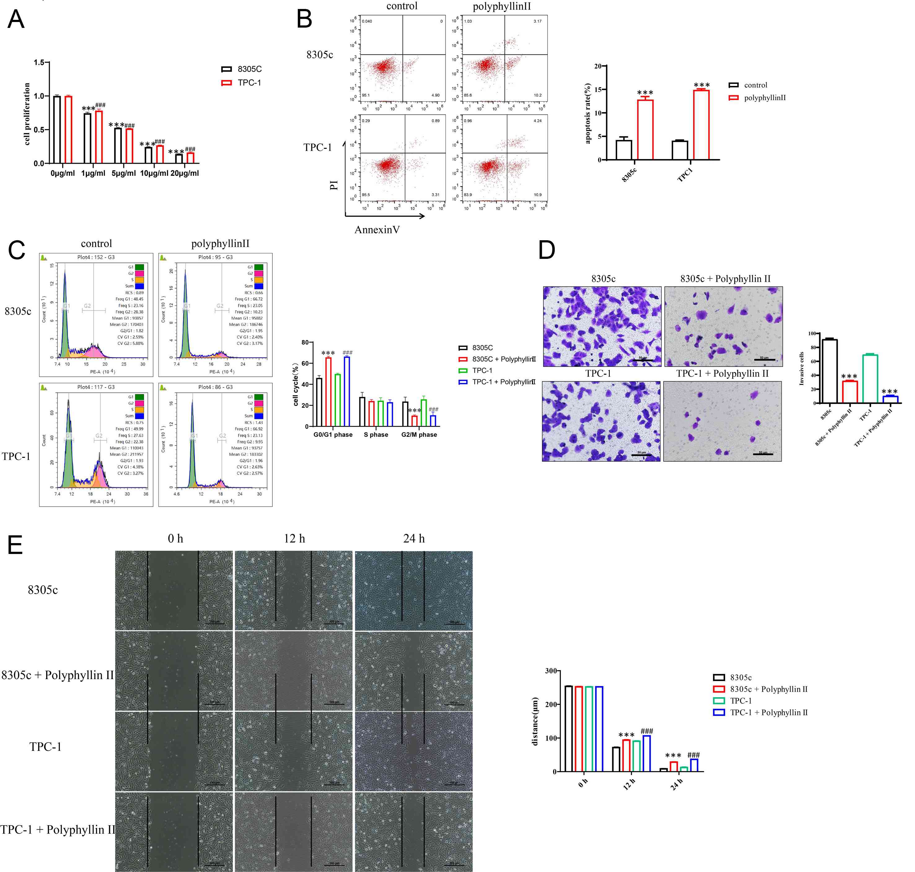
CCK-8 assays demonstrated that polyphyllin II
markedly reduced the viability of thyroid cancer cells in a
dose-dependent manner (Fig. 1A).
Based on the IC50 value, it was ultimately determined
that 5 µg/ml was the final experimental concentration. Flow
cytometry analysis further revealed a significant increase in
apoptotic cell populations following polyphyllin II treatment
(Fig. 1B). Cell cycle analysis
showed that polyphyllin II markedly increased the
G0/G1-phase population in both 8305C and
TPC-1 cells, while reducing the G2/M-phase population.
These results indicate that polyphyllin II induces
G0/G1-phase cell cycle arrest, which may
contribute to its anti-proliferative effect in thyroid cancer cells
(Fig. 1C). Transwell invasion
assays indicated that polyphyllin II attenuated the invasive
capacity of 8305C and TPC-1 cells, whereas wound healing assays
indicated impaired migratory ability following treatment (Fig. 1D and E). These findings indicate
that polyphyllin II effectively restrains thyroid cancer
progression by inhibiting proliferation and metastatic
behavior.
Identification of HRH1 as a potential
molecular target of polyphyllin II
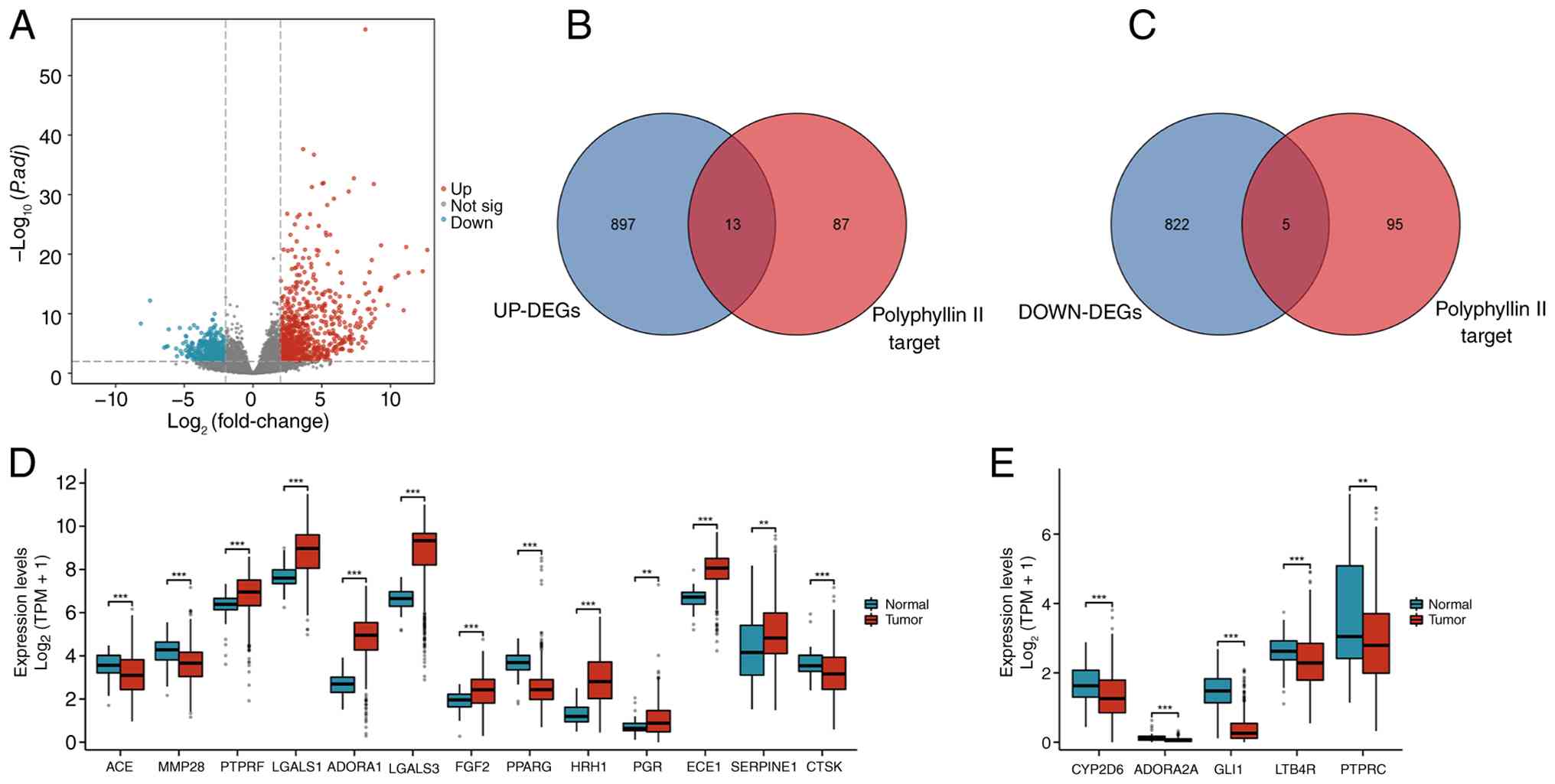
Differential gene expression analysis revealed
significant transcriptomic alterations in thyroid cancer, with 910
genes upregulated and 827 downregulated compared with non-tumor
tissues (Fig. 2A). Target
prediction using SwissTarget and intersection analysis identified
13 upregulated genes potentially associated with polyphyllin II
(Fig. 2B and C). TCGA and GEO
dataset validation confirmed that the expression levels of HRH1,
LGALS1, ADORA1 and ECE1 were significantly elevated in thyroid
cancer (Fig. 2D and E). Among these
genes, HRH1 has been reported as an oncogenic mediator in multiple
cancer types through regulation of cell survival, angiogenesis,
immune modulation and invasion (14,15),
suggesting its potential role in thyroid cancer progression.
HRH1 is overexpressed in thyroid
cancer cells
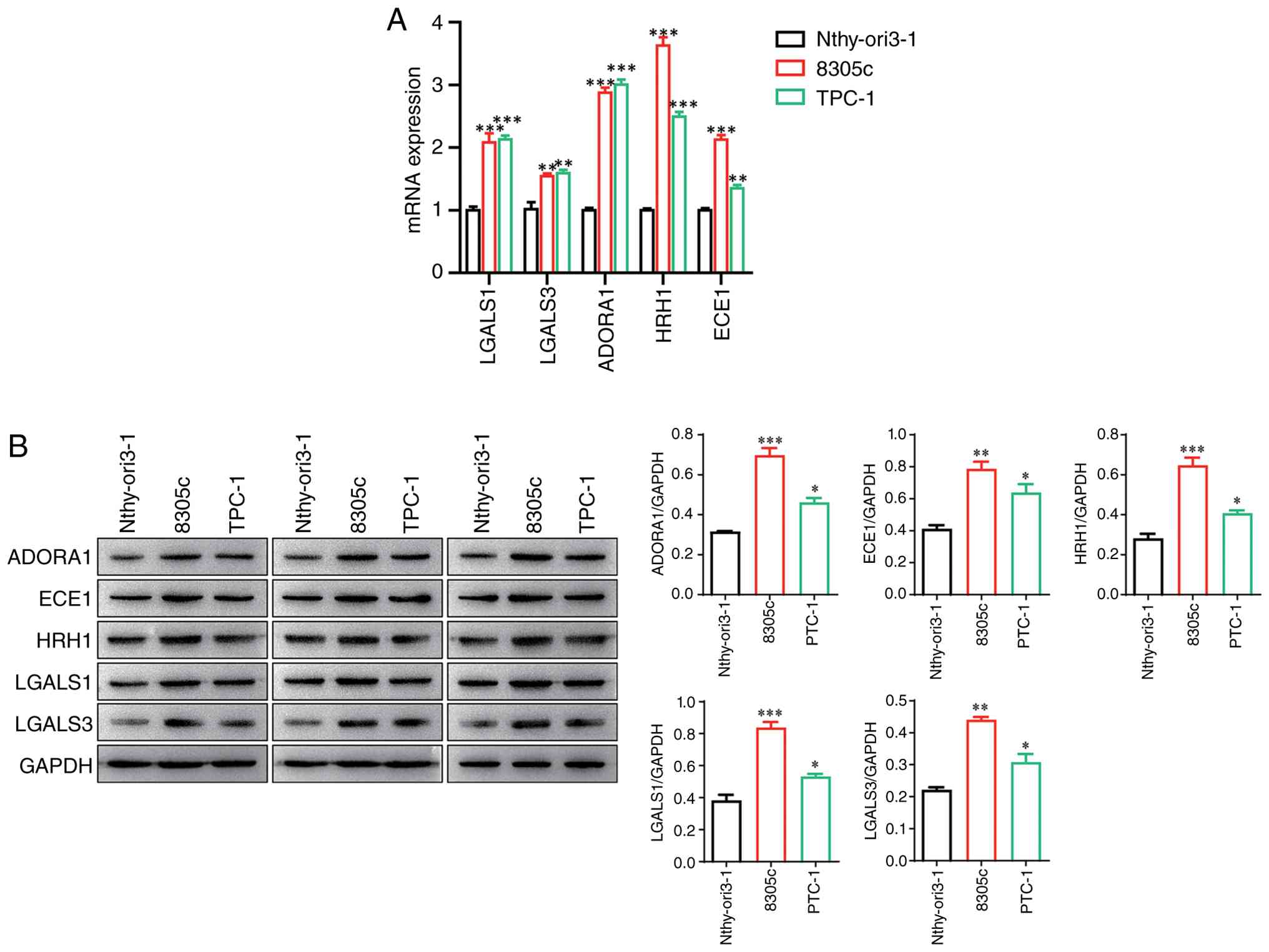
RT-qPCR analysis confirmed that the expression
levels of HRH1, LGALS1, ADORA1 and ECE1 were markedly upregulated
in thyroid cancer cells (Fig. 3A).
Western blotting further validated elevated protein levels of these
targets, with HRH1 indicating notably high expression in TPC-1 and
8305C cells (Fig. 3B). Given that
HRH1 signaling mediates histamine-induced tumor proliferation and
immune escape across multiple malignancies, HRH1 was selected as
the primary candidate for further investigation.
Polyphyllin II directly interacts with
HRH1 and regulates HRH1 expression
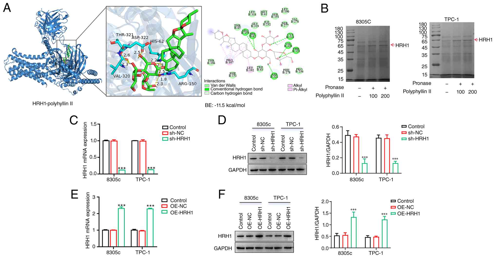
To determine whether HRH1 is a direct target of
polyphyllin II, molecular docking and DARTS assays were performed.
Molecular docking showed that polyphyllin II could fit into the
predicted binding pocket of HRH1, suggesting a potential
interaction between polyphyllin II and HRH1 (Fig. 4A). Consistently, DARTS assay showed
that polyphyllin II increased the resistance of HRH1 protein to
pronase digestion, further supporting the direct interaction
between polyphyllin II and HRH1 (Fig.
4B).
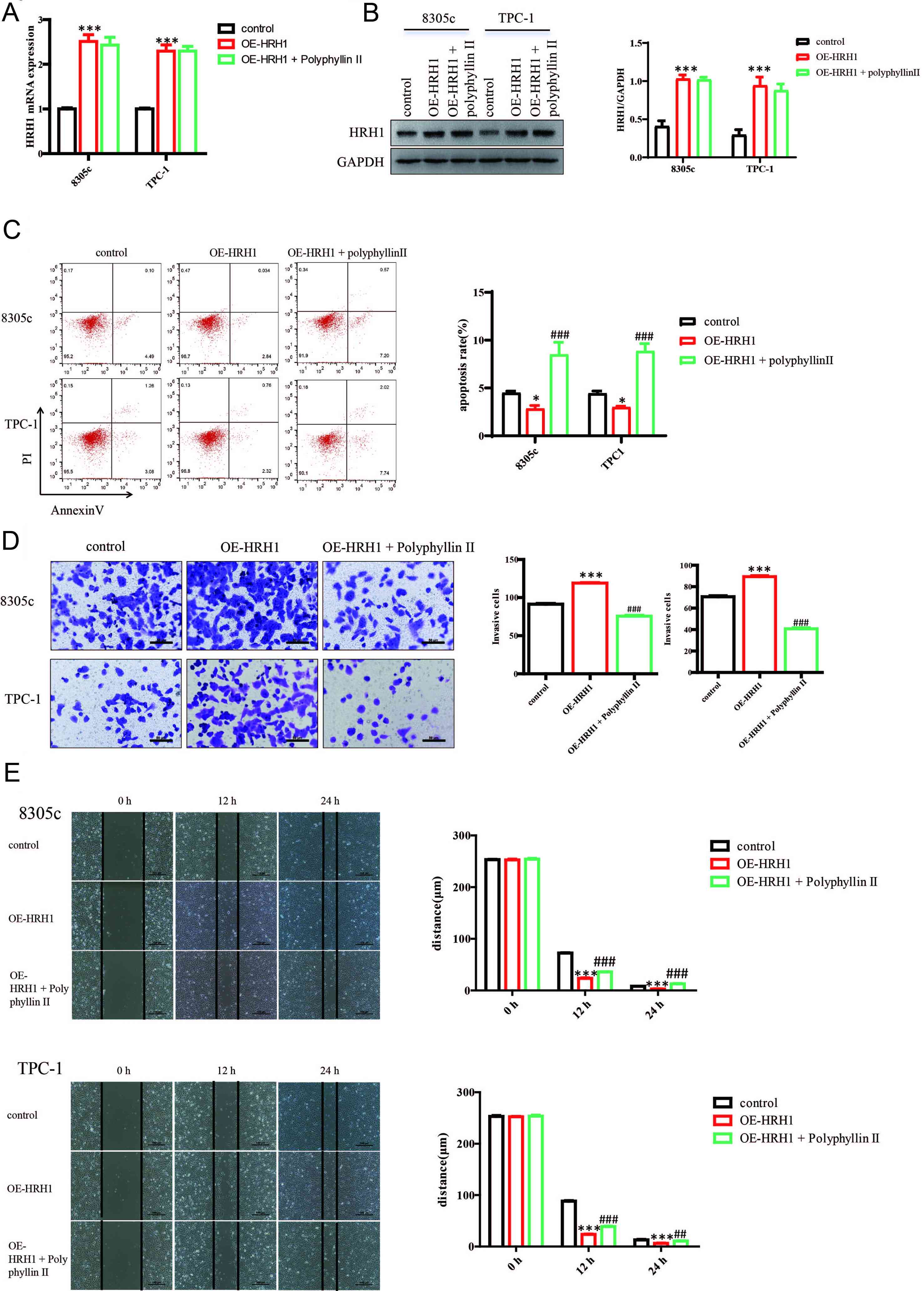
The present study next modulated HRH1 expression in
thyroid cancer cells. RT-qPCR and western blot results confirmed
that sh-HRH1 significantly reduced HRH1 expression, whereas OE-HRH1
markedly increased HRH1 expression (Fig. 4C-F). These results indicated that
HRH1 can be directly targeted by polyphyllin II and that the HRH1
knockdown and overexpression models were successfully established
for subsequent functional experiments.
HRH1 silencing attenuates the
malignant phenotype of thyroid cancer cells
To determine the functional role of HRH1 in thyroid
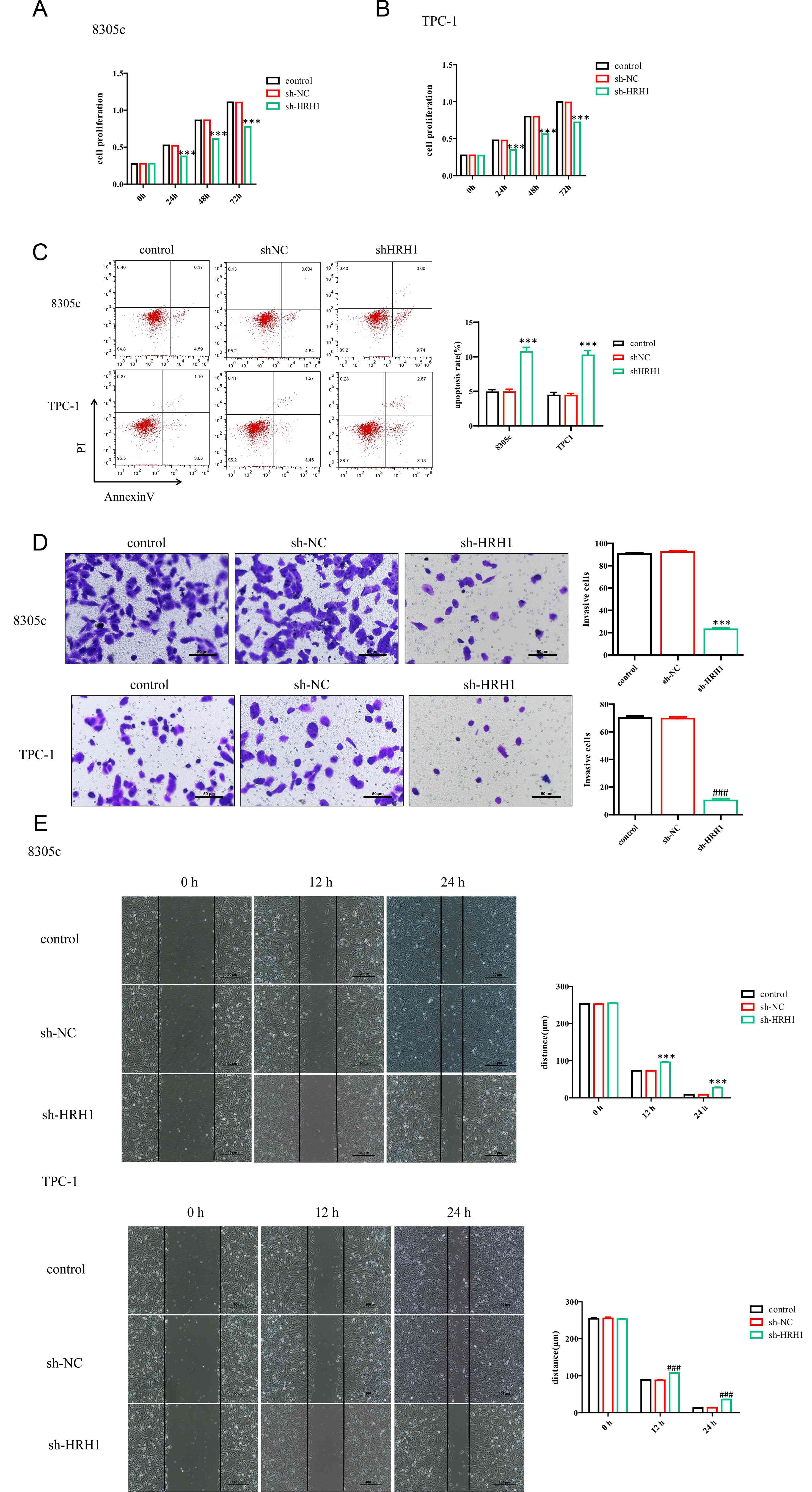
cancer cells, HRH1 was silenced using sh-HRH1. CCK-8 assays showed
that HRH1 knockdown significantly reduced the proliferation of both
8305C and TPC-1 cells compared with the control and sh-NC groups
(Fig. 5A and B). Flow cytometry
further showed that sh-HRH1 increased the apoptosis rate in both
cell lines (Fig. 5C). In addition,
Transwell assays demonstrated that HRH1 knockdown markedly
decreased the invasive ability of 8305C and TPC-1 cells (Fig. 5D). Wound-healing assays also showed
that sh-HRH1 delayed wound closure, indicating impaired migratory
capacity after HRH1 silencing (Fig.
5E). These results suggest that HRH1 promotes malignant
phenotypes in thyroid cancer cells.
Polyphyllin II exerts its antitumor
effects via HRH1 suppression
Overexpression of HRH1 counteracted apoptosis and
enhanced invasive and migratory abilities in TPC-1 and 8305C cells,
whereas polyphyllin II administration reversed these effects
(Fig. 6A-E). These results suggest
that polyphyllin II inhibits thyroid cancer malignancy in part by
attenuating HRH1 signaling. Given that HRH1 acts as a downstream
regulator of histamine signaling and oncogenic activity, these
findings provide a mechanistic basis for using polyphyllin II as a
potential HRH1-targeted therapeutic strategy.
HRH1 regulates Wnt/β-catenin signaling
and polyphyllin II reverses its activation
Previous evidence suggests that histamine receptor
H1-related signaling may activate canonical β-catenin signaling,
while Wnt/β-catenin activation is closely associated with thyroid
cancer progression (16–18). Consistently, TCGA-THCA single-gene
correlation analysis showed that HRH1 was positively correlated
with several Wnt/β-catenin-related genes, including MMP7, WNT7A,
CCND1, TCF7, CTNNB1 and MYC (Fig.
S1). These findings provided a rationale for subsequent
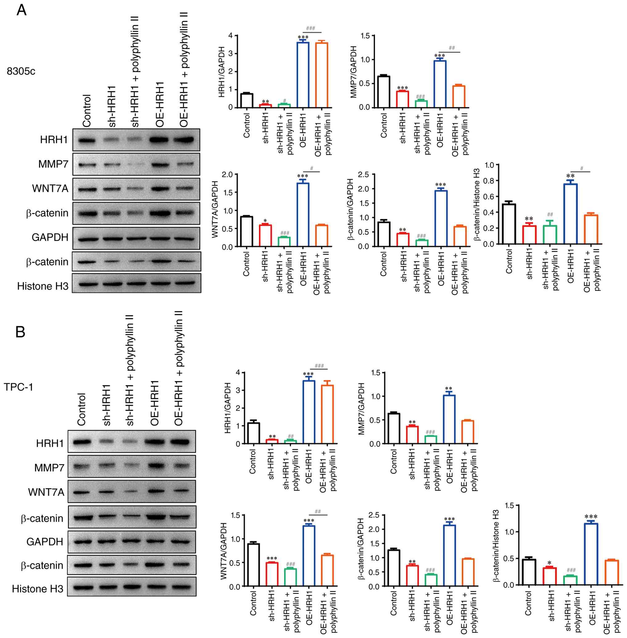
experimental validation. Knockdown of HRH1 expression significantly
reduced the expression levels of the β-catenin pathway-related
genes (MMP7, Wnt7A and β-catenin) and decreased β-catenin nuclear
translocation, whereas polyphyllin II treatment further suppressed
pathway activation (Fig. 7A and B).
Conversely, HRH1 overexpression enhanced β-catenin signaling, while
polyphyllin II blocked this activation, indicating an
HRH1-dependent regulatory mechanism. Therefore, polyphyllin II may
exert antitumor effects in thyroid cancer by suppressing
HRH1-mediated β-catenin activation.
Treatment of thyroid cancer cells with
polyphyllin II in vivo
To validate the in vivo findings, xenograft
models were established using 8305C cells in nude mice. The growth
transplanted 8305C xenograft tumors in mice was accelerated
significantly. During the in vivo treatment period, no
obvious treatment-related toxicity was observed. Mice in the
polyphyllin II-treated group showed no marked abnormalities in
general behavior, activity, food intake or water intake. Body
weight remained relatively stable throughout the experiment,
suggesting that polyphyllin II was generally tolerated under the
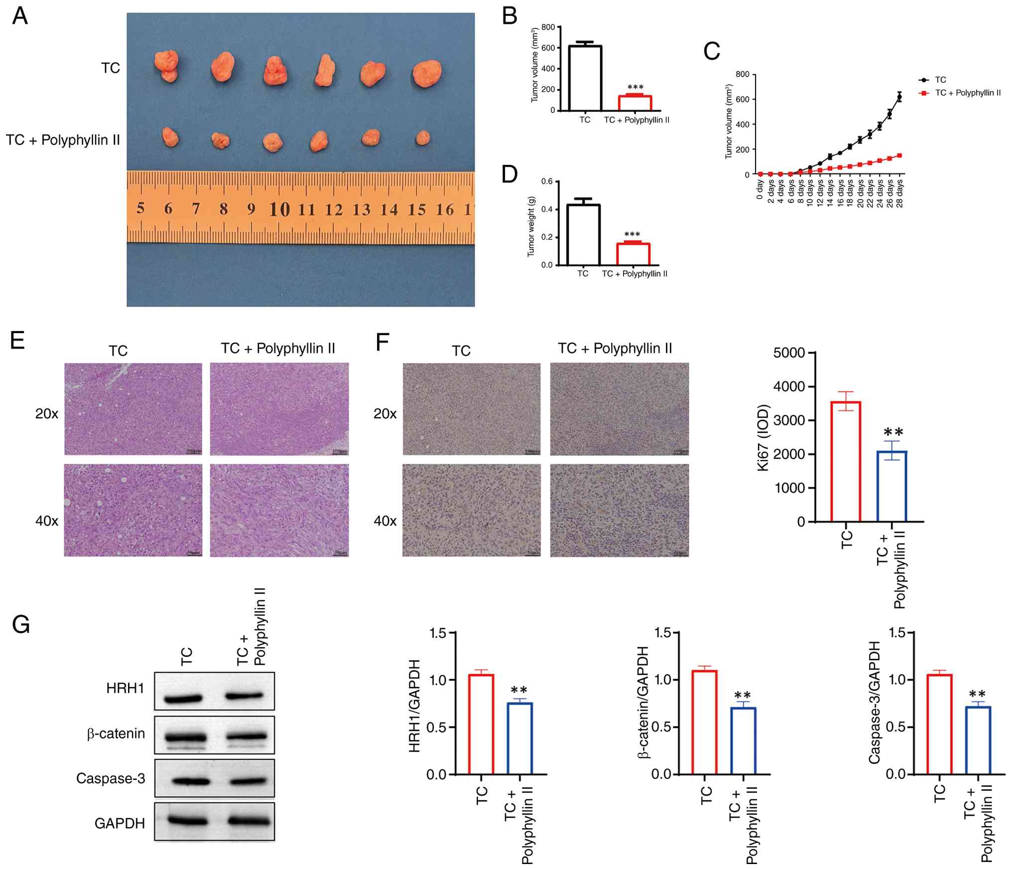
present treatment conditions. The mice that were treated with
polyphyllin II demonstrated a significant reduction in tumor size,
indicating that polyphyllin II exhibited an inhibitory effect on
tumors (Fig. 8A-D). The tumor
tissues were stained using IHC analysis for H&E and Ki67. The
results indicated that polyphyllin II could alleviate the
deterioration of the tumor tissues and reduce cancer cell
proliferation (Fig. 8E and F). WB
results indicated that the expression levels of the proteins of the
HRH1 signaling pathway were reduced following the use of
polyphyllin II (Fig. 8G).
To further confirm the effect of polyphyllin II on
the HRH1 signaling pathway, 8305C cells were divided into three
groups; NC, lentiviral sh-HRH1 and lentiviral OE-HRH1 combined with
polyphyllin II treatment. Compared with NC, following knockdown of
HRH1 expression, the tumor size decreased. Following IHC staining
with H&E and Ki67, the severity of tumor tissue deterioration
decreased and the proliferation of the cells was reduced (Fig. 9A-D). Following treatment of the
tumor-bearing mice expressing HRH1 with polyphyllin II, the volumes
of the tumors were reduced. The tissue staining results indicated
that the severity of tumor tissue deterioration and cellular
proliferation were reduced (Fig. 9E and
F). WB results also demonstrated that polyphyllin II inhibited
tumor growth by suppressing the HRH1 signaling pathway (Fig. 9G).
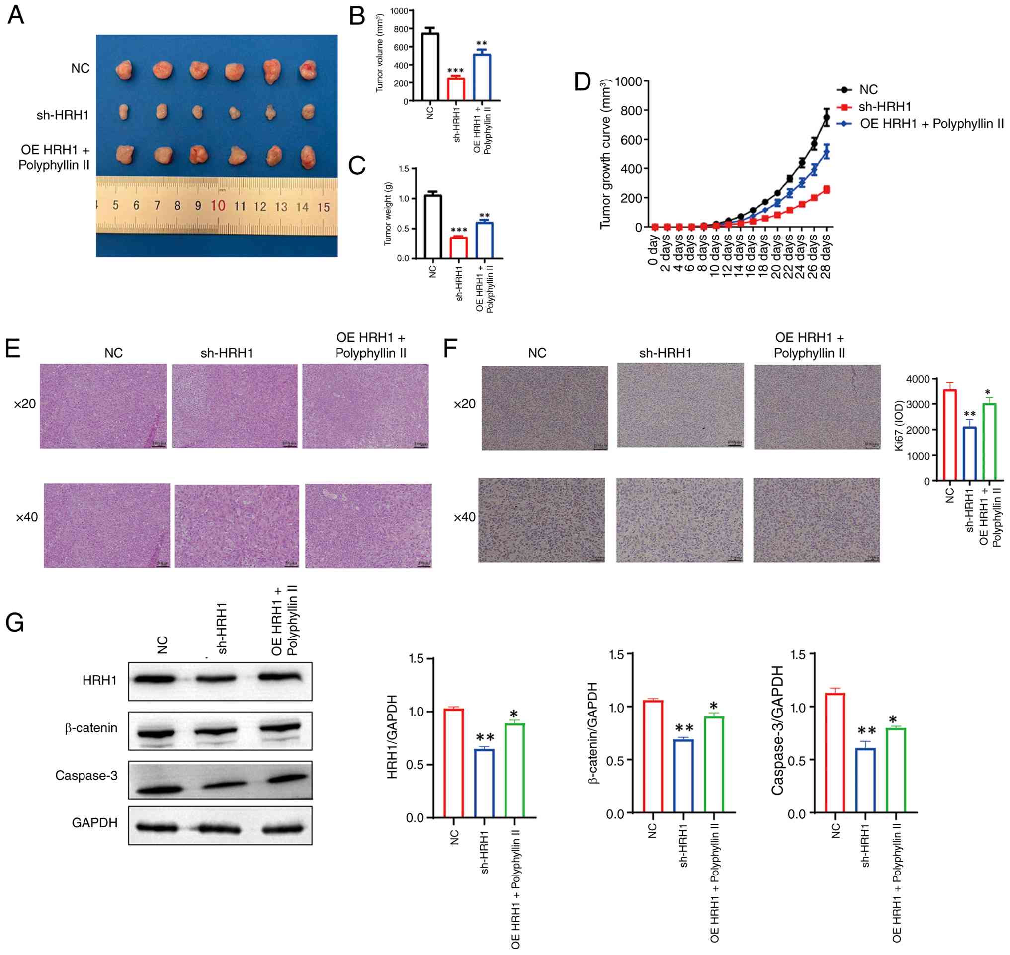 | Figure 9.HRH1 modulation affects xenograft
tumor growth and polyphyllin II suppresses HRH1-mediated tumor
progression in vivo. (A) Representative images of xenograft
tumors from the NC, sh-HRH1, and OE-HRH1 + polyphyllin II groups.
(B and C) Quantitative analysis of tumor volume and tumor weight
showing that HRH1 knockdown significantly reduced tumor burden,
while polyphyllin II treatment also suppressed tumor growth in
HRH1-overexpressing xenografts. (D) Tumor growth curves showing
reduced tumor progression in the sh-HRH1 group and the OE-HRH1 +
polyphyllin II group compared with the NC group. (E) H&E
staining of xenograft tumor tissues showing decreased tumor cell
density and improved histological morphology after HRH1 knockdown
or polyphyllin II treatment. (F) Immunohistochemical staining and
quantitative analysis of Ki67 showing reduced tumor cell
proliferation in the sh-HRH1 and OE-HRH1 + polyphyllin II groups.
Scale bar=100 µm for images acquired at ×20 magnification and 50 µm
for images acquired at ×40 magnification. (G) Western blotting and
quantification of HRH1, β-catenin and caspase-3 expression in
xenograft tumor tissues. GAPDH was used as the loading control.
Data are presented as the mean ± SD (n=6). *P<0.05, **P<0.01
and ***P<0.001 vs. NC group. HRH1, histamine receptor H1; sh,
short hairpin; OE, overexpression; NC, negative control; H&E,
hematoxylin-eosin. |
Discussion
Thyroid cancer remains a heterogeneous endocrine
malignancy and aggressive or treatment-resistant subtypes still
require more effective molecular therapeutic strategies. In the
present study, polyphyllin II inhibited proliferation, invasion and
migration, while promoting apoptosis in thyroid cancer cells.
Mechanistically, HRH1 was identified as a potential target of
polyphyllin II, and HRH1 suppression was associated with reduced
activation of the Wnt/β-catenin pathway. These findings suggested
that the HRH1/Wnt/β-catenin axis may contribute to thyroid cancer
progression and may serve as a potential therapeutic target.
HRH1 is a histamine receptor involved in
inflammatory signaling, cell proliferation and tumor progression.
Previous evidence has shown that histamine can activate the
canonical β-catenin pathway through H1 receptor-dependent
regulation of glycogen synthase kinase-3β activity and β-catenin
stabilization (19). To further
support this potential link in thyroid cancer, the present study
performed single-gene correlation analysis using the TCGA-THCA
dataset. The results showed that HRH1 expression was positively
correlated with several Wnt/β-catenin-related genes, including
MMP7, WNT7A, CCND1, TCF7, CTNNB1 and MYC. These literature and
bioinformatics findings provided a rationale for further
investigating whether HRH1 regulates Wnt/β-catenin signaling in
thyroid cancer cells.
Wnt/β-catenin signaling is an important oncogenic
pathway involved in thyroid cancer proliferation, invasion,
dedifferentiation and treatment resistance (20–22).
In the present study, HRH1 knockdown reduced the expression of
β-catenin pathway-related molecules, whereas HRH1 overexpression
enhanced pathway activation. Notably, polyphyllin II attenuated
HRH1-mediated activation of Wnt/β-catenin signaling, suggesting
that its antitumor effect may be partly dependent on inhibition of
the HRH1/Wnt/β-catenin axis. These findings indicated that
polyphyllin II may not act merely as a cytotoxic compound, but may
suppress thyroid cancer progression through a defined
HRH1/Wnt/β-catenin signaling axis. This also raises the possibility
that polyphyllin II could be further explored in combination with
agents targeting the Wnt/β-catenin pathway, particularly in
aggressive or treatment-resistant thyroid cancer.
From a clinical perspective, these findings may have
potential relevance for biomarker-based risk stratification in
thyroid cancer. Current endocrine surgical practice increasingly
emphasizes the integration of imaging, cytology, molecular markers
and systemic inflammatory indicators to improve the evaluation of
thyroid nodules and tumor aggressiveness (23,24).
Gambardella et al (7)
reported that inflammatory biomarkers, including
neutrophil-to-lymphocyte ratio, lymphocyte-to-monocyte ratio and
platelet-to-lymphocyte ratio, may help predict malignancy in
indeterminate thyroid nodules. As an inflammation-related receptor,
HRH1 may provide a molecular link between inflammatory regulation
and oncogenic signaling in thyroid cancer. Therefore, HRH1-related
signaling may have potential value for further evaluating tumor
behavior and identifying patients who may benefit from
pathway-oriented therapeutic strategies.
The translational relevance of the present study
should also be considered in the context of multimodal management
for aggressive thyroid cancer. For highly aggressive subtypes,
particularly anaplastic thyroid carcinoma, surgery alone is often
insufficient, and combined strategies involving surgery,
radiotherapy, chemotherapy and systemic treatment are frequently
required. Conzo et al (25)
emphasized the importance of multimodal treatment in improving
local control and clinical outcomes in selected patients with
aggressive thyroid cancer. In this context, the HRH1/Wnt/β-catenin
axis identified in the present study may provide a molecular basis
for future combination strategies. Although polyphyllin II remains
a preclinical candidate, its inhibitory effect on HRH1-mediated
Wnt/β-catenin activation suggests that it may have potential value
as part of pathway-oriented therapeutic exploration.
The broader surgical literature also indicates that
clinical outcomes are influenced not only by local disease control,
but also by systemic metabolic status and perioperative management.
Studies by Pizza et al (26–28) in
metabolic surgery showed that postoperative metabolic modulation,
antral size and biliopancreatic limb length may affect
postoperative complications, nutritional status and long-term
outcomes. Therefore, future translational studies of thyroid cancer
should not only focus on tumor-intrinsic molecular pathways, but
also consider inflammatory status, metabolic background, surgical
risk and postoperative recovery.
Several limitations should be acknowledged. First,
although xenograft experiments were performed in the present study,
the in vivo regulatory relationship among polyphyllin II,
HRH1 and Wnt/β-catenin signaling requires further validation using
more clinically relevant models, such as orthotopic thyroid cancer
models or patient-derived xenografts. Second, the upstream
mechanisms responsible for HRH1 overexpression in thyroid cancer
were not fully explored. Future studies should examine whether
inflammatory mediators, epigenetic regulation, histamine-related
metabolic changes or tumor microenvironmental signals contribute to
HRH1 activation. Moreover, it remains possible that polyphyllin II
may also affect other components of the Wnt/β-catenin pathway,
including Frizzled receptors, low-density lipoprotein
receptor-related protein 5/6 (LRP5/6), GSK3β, β-catenin and TCF/LEF
transcriptional complexes. Future studies should evaluate whether
polyphyllin II directly interacts with these Wnt/β-catenin-related
targets or indirectly suppresses the pathway through HRH1-dependent
regulation. Third, although molecular docking and DARTS assays
supported a potential interaction between polyphyllin II and HRH1,
additional target-engagement assays, such as surface plasmon
resonance or cellular thermal shift assays, would further
strengthen the evidence for direct binding. Finally, given the
important role of Wnt/β-catenin signaling in thyroid cancer
progression and treatment resistance, future studies should
investigate whether polyphyllin II can be combined with
Wnt/β-catenin pathway inhibitors, targeted therapies or
conventional treatments to enhance antitumor efficacy.
In conclusion, the present study demonstrated that
polyphyllin II suppresses thyroid cancer progression by targeting
HRH1 and inhibiting Wnt/β-catenin signaling. These findings
provided new insight into the role of HRH1-related signaling in
thyroid cancer and suggested that HRH1 may serve as a potential
therapeutic target. Further studies are needed to validate the
clinical relevance of HRH1 and to explore polyphyllin II-based
combination strategies for aggressive or treatment-resistant
thyroid cancer.
Supplementary Material
Supporting Data
Acknowledgements
Not applicable.
Funding
The present study was supported by funds from Yunnan Provincial
Department of Science and Technology, Key Project for Applied Basic
Research (Yunnan Fundamental Research Kunming Medical University
Projects; grant no. 202301AY070001-037), Yunnan Province ‘Xingdian
Talents’ Support Program for Healthcare Professionals (grant no.
XDYC-MY-2022-070) and China Health Promotion Foundation, Key
Project of the Research Fund for High-Quality Development of Public
Hospitals (grant no. GL-A013).
Availability of data and materials
The data generated in the present study may be
requested from the corresponding author.
Authors' contributions
JS was responsible for manuscript writing and
revision, cellular experiments and molecular mechanistic studies.
HL and YZ were responsible for cellular experiments. DD and QX were
responsible for manuscript revision. MZ was responsible for
clinical research. PZ was responsible for overall design and
direction of the dissertation. JS and PZ confirm the authenticity
of all the raw data. All authors read and approved the final
manuscript.
Ethics approval and consent to
participate
All animal procedures were conducted in accordance
with the ARRIVE guidelines and the AVMA Guidelines for the
Euthanasia of Animals: 2020 Edition and were approved by the Ethics
Committee of The Fifth Affiliated Hospital of Kunming Medical
University (approval no. kmmu20240805).
Patient consent for publication
Not applicable.
Competing interests
The authors declare that they have no competing
interests.
Glossary
Abbreviations
Abbreviations:
|
TC
|
Thyroid cancer
|
|
DTC
|
differentiated thyroid cancer
|
|
ATC
|
anaplastic thyroid carcinoma
|
|
HRH1
|
histamine receptor H1
|
|
Wnt/β-catenin
|
Wnt/β-catenin signaling pathway
|
|
MMP7
|
matrix metalloproteinase 7
|
|
WNT7A
|
Wingless-type MMTV integration site
family member 7A
|
|
CCK-8
|
Cell Counting Kit-8
|
|
PBS
|
phosphate-buffered saline
|
|
DMSO
|
dimethyl sulfoxide
|
|
RT-qPCR
|
reverse transcription quantitative
PCR
|
|
WB
|
western blotting
|
|
FITC
|
fluorescein isothiocyanate
|
|
GAPDH
|
glyceraldehyde-3-phosphate
dehydrogenase
|
References
|
1
|
Megwalu UC and Moon PK: Thyroid cancer
incidence and mortality trends in the United States: 2000–2018.
Thyroid. 32:560–570. 2022. View Article : Google Scholar : PubMed/NCBI
|
|
2
|
Cabanillas ME, McFadden DG and Durante C:
Thyroid cancer. Lancet. 388:2783–2795. 2016. View Article : Google Scholar : PubMed/NCBI
|
|
3
|
Haugen BR, Alexander EK, Bible KC, Doherty
GM, Mandel SJ, Nikiforov YE, Pacini F, Randolph GW, Sawka AM,
Schlumberger M, et al: 2015 American thyroid association management
guidelines for adult patients with thyroid nodules and
differentiated thyroid cancer: The American thyroid association
guidelines task force on thyroid nodules and differentiated thyroid
cancer. Thyroid. 26:1–133. 2016. View Article : Google Scholar : PubMed/NCBI
|
|
4
|
Parker WA, Edafe O and Balasubramanian SP:
Long-term treatment-related morbidity in differentiated thyroid
cancer: A systematic review of the literature. Pragmat Obs Res.
8:57–67. 2017.PubMed/NCBI
|
|
5
|
Rao SN and Smallridge RC: Anaplastic
thyroid cancer: An update. Best Pract Res Clin Endocrinol Metab.
37:1016782023. View Article : Google Scholar : PubMed/NCBI
|
|
6
|
Spitzweg C, Bible KC, Hofbauer LC and
Morris JC: Advanced radioiodine-refractory differentiated thyroid
cancer: The sodium iodide symporter and other emerging therapeutic
targets. Lancet Diabetes Endocrinol. 2:830–842. 2014. View Article : Google Scholar : PubMed/NCBI
|
|
7
|
Gambardella C, Mongardini FM, Paolicelli
M, Bentivoglio D, Cozzolino G, Ruggiero R, Pizza A, Tolone S, Del
Genio G, Parisi S, et al: Role of inflammatory biomarkers (NLR,
LMR, PLR) in the prognostication of malignancy in indeterminate
thyroid nodules. Int J Mol Sci. 24:64662023. View Article : Google Scholar : PubMed/NCBI
|
|
8
|
Cheong TY, Hong SD, Jung KW and So YK: The
diagnostic predictive value of neutrophil-to-lymphocyte ratio in
thyroid cancer adjusted for tumor size. PLoS One. 16:e02514462021.
View Article : Google Scholar : PubMed/NCBI
|
|
9
|
Ding YG, Zhao YL, Zhang J, Zuo ZT, Zhang
QZ and Wang YZ: The traditional uses, phytochemistry, and
pharmacological properties of Paris L. (Liliaceae): A review. J
Ethnopharmacol. 278:1142932021. View Article : Google Scholar : PubMed/NCBI
|
|
10
|
Carnazza M, Yang N, Tiwari RK, Geliebter J
and Li XM: Natural compounds targeting MAPK, PI3K/Akt, and JAK/STAT
signaling in papillary thyroid cancer. Int J Mol Sci. 26:104982025.
View Article : Google Scholar : PubMed/NCBI
|
|
11
|
Li JK, Sun HT, Jiang XL, Chen YF, Zhang Z,
Wang Y, Chen WQ, Zhang Z, Sze SCW, Zhu PL and Yung KKL: Polyphyllin
II induces protective autophagy and apoptosis via inhibiting
PI3K/AKT/mTOR and STAT3 signaling in colorectal cancer cells. Int J
Mol Sci. 23:118902022. View Article : Google Scholar : PubMed/NCBI
|
|
12
|
Miao W, Wang Z, Gao J and Ohno Y:
Polyphyllin II inhibits breast cancer cell proliferation via the
PI3K/Akt signaling pathway. Mol Med Rep. 30:2242024. View Article : Google Scholar : PubMed/NCBI
|
|
13
|
Livak KJ and Schmittgen TD: Analysis of
relative gene expression data using real-time quantitative PCR and
the 2(−Delta Delta C(T)) method. Methods. 25:402–408. 2001.
View Article : Google Scholar : PubMed/NCBI
|
|
14
|
Zhao J, Hou Y, Yin C, Hu J, Gao T, Huang
X, Zhang X, Xing J, An J, Wan S and Li J: Upregulation of histamine
receptor H1 promotes tumor progression and contributes to poor
prognosis in hepatocellular carcinoma. Oncogene. 39:1724–1738.
2020. View Article : Google Scholar : PubMed/NCBI
|
|
15
|
Ding YF, Ho KH, Lee WJ, Chen LH, Hsieh FK,
Tung MC, Lin SH, Hsiao M, Yang SF, Yang YC and Chien MH: Cyclic
increase in the histamine receptor H1-ADAM9-Snail/Slug axis as a
potential therapeutic target for EMT-mediated progression of oral
squamous cell carcinoma. Cell Death Dis. 16:1912025. View Article : Google Scholar : PubMed/NCBI
|
|
16
|
Diks SH, Hardwick JC, Diab RM, van Santen
MM, Versteeg HH, van Deventer SJ, Richel DJ and Peppelenbosch MP:
Activation of the canonical beta-Catenin pathway by histamine. J
Biol Chem. 278:52491–52496. 2003. View Article : Google Scholar : PubMed/NCBI
|
|
17
|
Sastre-Perona A and Santisteban P: Role of
the Wnt pathway in thyroid cancer. Front Endocrinol (Lausanne).
3:312012. View Article : Google Scholar : PubMed/NCBI
|
|
18
|
Gilbert-Sirieix M, Makoukji J, Kimura S,
Talbot M, Caillou B, Massaad C and Massaad-Massade L: Wnt/β-Catenin
signaling pathway is a direct enhancer of thyroid transcription
factor-1 in human papillary thyroid carcinoma cells. PLoS One.
6:e222802011. View Article : Google Scholar : PubMed/NCBI
|
|
19
|
Veeragoni D, Ghosh H, Ansari A,
Bhattacharyya S, Azaz A, Ommi O, Covarrubias-Zambrano O, Köhler
LHF, Ködel JF, Hamza A, et al: Novel N-chloroacetyl-2-pyrazoline
analogs with 2-naphthyl and ferrocenyl groups: targeting histamine
receptor H1 to overcome colorectal cancer drug resistance. Cancer
Drug Resist. 9:152026.PubMed/NCBI
|
|
20
|
Zhang W, Ruan X, Li Y, Zhi J, Hu L, Hou X,
Shi X, Wang X, Wang J, Ma W, et al: KDM1A promotes thyroid cancer
progression and maintains stemness through the Wnt/β-catenin
signaling pathway. Theranostics. 12:1500–1517. 2022. View Article : Google Scholar : PubMed/NCBI
|
|
21
|
Diaz D, Bergdorf K, Loberg MA, Phifer CJ,
Xu GJ, Sheng Q, Chen SC, Byrant JM, Tigue ML, Hartmann H, et al:
Wnt/β-catenin signaling is a therapeutic target in anaplastic
thyroid carcinoma. Endocrine. 86:114–118. 2024. View Article : Google Scholar : PubMed/NCBI
|
|
22
|
Xu Q, Yang H, Fan G, Zhang B, Yu J, Zhang
Z and Jia G: Clinical importance of PLA2R1 and RASSF9 in thyroid
cancer and their inhibitory roles on the Wnt/β-catenin pathway and
thyroid cancer cell malignant behaviors. Pathol Res Pract.
238:1540922022. View Article : Google Scholar : PubMed/NCBI
|
|
23
|
Xu Y, Pi J, Jinghu Y, Wang X, Xu D and Liu
J: Diagnostic efficiency of ACR-TIRADS score for differentiating
benign and malignant thyroid nodules of various pathological types.
Med Sci Monit. 30:e9432282024. View Article : Google Scholar : PubMed/NCBI
|
|
24
|
Zhao Z, Ren T, Zhao Y, Xu W, Xie R, Lin J,
Li H, Zheng L, Zhang C, Huo H, et al: Salivary biomarkers-assisted
ultrasound-based differentiation of malignant and benign thyroid
nodules. Gland Surg. 11:196–206. 2022. View Article : Google Scholar : PubMed/NCBI
|
|
25
|
Conzo G, Avenia N, Bellastella G, Candela
G, de Bellis A, Esposito K, Pasquali D, Polistena A, Santini L and
Sinisi AA: The role of surgery in the current management of
differentiated thyroid cancer. Endocrine. 47:380–388. 2014.
View Article : Google Scholar : PubMed/NCBI
|
|
26
|
Pizza F, Lucido FS, D'Antonio D, Tolone S,
Gambardella C, Dell'Isola C, Docimo L and Marvaso A:
Biliopancreatic limb length in one anastomosis gastric bypass:
Which is the best? Obes Surg. 30:3685–3694. 2020. View Article : Google Scholar : PubMed/NCBI
|
|
27
|
Pizza F, D'Antonio D, Lucido FS, Tolone S,
Del Genio G, Dell'Isola C, Docimo L and Gambardella C: The role of
ursodeoxycholic acid (UDCA) in cholelithiasis management after one
anastomosis gastric bypass (OAGB) for morbid obesity: Results of a
monocentric randomized controlled trial. Obes Surg. 30:4315–4324.
2020. View Article : Google Scholar : PubMed/NCBI
|
|
28
|
Pizza F, D'Antonio D, Lucido FS,
Gambardella C, Carbonell Asíns JA, Dell'Isola C and Tolone S: Does
antrum size matter in sleeve gastrectomy? A prospective randomized
study. Surg Endosc. 35:3524–3532. 2021. View Article : Google Scholar : PubMed/NCBI
|