Introduction
Bidirectional trafficking of biological
macromolecules between the nucleus and the cytoplasm in eukaryotic
cells is mediated by the nuclear pore complexes (NPCs). The NPCs
span the inner and outer nuclear membranes and act as gatekeepers
by regulating molecular transit. Each mammalian NPC is made up of a
large cylindrical hollow structure composed of multiple copies of
more than 30 distinct polypeptides, termed nucleoporins (Nups)
(1). Selective nuclear transport
via NPCs is regulated primarily by the meshwork formed in the
central pore of NPCs. About one-third of Nups contain phenylalanine
and glycine (FG) repeats, which comprise the inner central pore of
NPCs. The FG repeats serve as diffusion barriers for limiting the
passage of macromolecules as well as docking sites for nuclear
transport receptors that mediate cargo transport through NPCs
(2–4). In addition to their role in nuclear
transport, Nups are implicated in various cellular processes, such
as mitosis, gene regulation, DNA repair and development (5,6). As
such, dysregulation of Nups is associated with various human
disorders, including cancer (7).
Nucleoporin (Nup) 88 is a non-FG Nup located
primarily on the cytoplasmic side of NPCs where Nup88 interacts
with other Nups and participates in nuclear export (8–12).
Notably, high levels of Nup88 expression in the cytoplasm have been
reported in a variety of tumor tissues, such as endometrial cancer,
colorectal cancer, breast cancer, cervical cancer and
hepatocellular carcinoma (13–20).
As this aberrant expression was associated with the tumor grade,
Nup88 appears to be involved in cancer malignancy (21). We previously reported that Nup88
promotes cell migration by inducing matrix metalloproteinase-12
(22). However, its effect was
relatively modest, implying that other factors additionally
contribute to Nup88-dependent migration.
Elevated expression of glioma-associated oncogene
homolog (Gli) 1 is also associated with several malignant
phenotypes of cancer, including anti-apoptotic effects, metastasis,
angiogenesis, cancer drug resistance and maintenance of cancer stem
cells (23). In mammalian cells,
expression of Gli1 is regulated at the transcriptional level by the
Hedgehog (Hh) pathway and oncogenic signaling (23,24).
The Hh pathway regulates expression of Gli1 directly via the
effector transcription factors Gli2 and Gli3. Although Gli2 and
Gli3 function in a context-dependent manner, Gli2 and Gli3
generally act as a transcriptional activator and a transcriptional
repressor, respectively (25).
The activity of Gli2/3 is regulated primarily by
kinesin family member (Kif) 7, which controls their processing and
nuclear translocation in the Hh pathway (26,27).
In the absence of Hh ligands, Kif7-free Gli2/3 are processed into
transcriptional repressors, whereas Gli2/3 bound to Kif7 are
protected from processing, but their activation is inhibited. Upon
Hh pathway activation, Kif7 releases Gli2/3, allowing their
activation and nuclear translocation as transcriptional activators
(28). While Kif7 acts as a
negative regulator of Gli2/3, it can function as a tissue-specific
positive regulator during mouse embryonic development (27). The downregulation of Kif7 is linked
to malignant phenotypes in cancer cells. While the epigenetic
suppression of Kif7 has been reported in some prostate cancer cell
lines (29), the molecular
mechanism that triggers this decrease in expression has not been
elucidated. In addition to the Hh pathway, the expression of Gli1
is often driven by oncogenic signaling, such as Rat sarcoma-rapidly
accelerated fibrosarcoma-mitogen-activated protein kinase/ERK
kinase-extracellular signal-regulated kinase (Raf-Ras-MEK-ERK)
signaling, in cancer cells. However, the regulation of Gli1
expression by oncogenic signaling is complex and poorly
understood.
The present study focused on cervical cancer, which
remains one of the leading causes of cancer-related mortality in
women worldwide, largely due to its high potential for metastasis
and recurrence (30,31). Cervical cancer is known to be caused
by persistent infection with high-risk human papillomaviruses,
whose oncoproteins (E6 and E7) facilitate malignant transformation
by degrading tumor suppressors p53 and pRb (31). Given this clinical significance, we
have consistently utilized the HeLa cervical cancer cell line as a
robust model of aggressive malignancy to investigate the molecular
mechanisms of Nup88-driven progression. The present study found
that stable overexpression of Nup88 in HeLa cells induced not only
the downregulation of Kif7 but also the upregulation of Gli1.
Furthermore, it was found that changes in their expression markedly
impact the HeLa cell motility. Based on these findings, the present
study proposed a mechanistic model in which Nup88 promotes cell
migration through the independent modulation of Kif7 and Gli1
expression, providing new insight into Nup88-driven malignancy.
Materials and methods
Cell lines and cell culture
Stable cell lines that inducibly overexpress GFP and
GFP-tagged Nup88 in response to doxycycline were previously
established from HeLa R19 cells containing a single Flp
recombination site in the genome (32,33).
Cells were cultured in Dulbecco's modified Eagle's medium (DMEM;
FUJIFILM Wako Pure Chemical Corporation) supplemented with 10%
fetal bovine serum (HyClone™; Cytiva), penicillin/streptomycin
(FUJIFILM Wako Pure Chemical Corporation) and 1 µg/ml doxycycline
(Nacalai Tesque, Inc.) in a humidified atmosphere with 5%
CO2 at 37°C.
Plasmids and small interfering
(si)RNAs
pEGFP-N2-NUP88, a plasmid that overexpresses
GFP-fused Nup88, was constructed previously (32). pEGFP-N2-GLI1, a plasmid that
overexpresses GFP-tagged Gli1 was constructed as follows. The
GLI1 gene was obtained from pOTB7 coding GLI1 cDNA by
PCR using the following pair of oligonucleotide primers (Forward:
5′-CTCAAGCTTCGAATTCCGCCACCATGTTCAACTCGATGACCCCA-3′, Reverse:
5′-GTCGACTGCAGAATTCGGCACTAGAGTTGAGGAATTCTGT-3′). The underlined
sequences represent the regions complementary to the GLI1
gene. The amplified product was cloned into the EcoRI site
of pEGFP-N2 (Takara Bio, Inc.) using an In-Fusion HD Cloning Kit
(Takara Bio, Inc.). The plasmids were transformed into competent
cells via heat shock at 42°C for 30 sec. After the addition of LB
medium, the cells were incubated at 37°C for 30 min and then
cultured overnight on LB agar plate containing appropriate
antibiotics. A plasmid that overexpresses Flag-tagged GLI1
was constructed by replacing the eGFP gene in pEGFP-N2-GLI1
with the 3×FLAG gene. pDPNR223 containing the truncated KIF7
gene (ID100072702A), which encodes amino acid residues 514–1343 of
full-length Kif7 (1343 aa), was purchased from DNAFORM.
mCherry-tagged truncated KIF7 [pmCherry-KIF7(514–1343)] was
constructed as follows. The entire truncated KIF7 in
pDPNR223 was amplified by PCR using the following primer set
(Forward: 5′-gtcgacggtaccgcgggcccgggcgccaccatggagcagtacaaactgca-3′,
Reverse: 5′-tccttgtagtcttggatcccgggcagggggtttttccggac-3′) and
cloned into pEGFP-N2 using an In-Fusion HD Cloning Kit. The
underlined sequences represent the regions complementary to the
KIF7 gene. The eGFP gene in the plasmid was then swapped
with the mCherry gene. siRNAs for control (cat. no. 4390843),
KIF7 (cat. no. 4427037, ID: s51568), GLI1 (cat. no.
4427037, ID: s5816), GLI2 (cat. no. 4427037, ID: s5817),
GLI3 (cat. no. 4427037, ID: s533814), and KRAS (cat.
no. 4427038, ID: s7939) were purchased from Thermo Fisher
Scientific, Inc.
Transfection of plasmids and
siRNAs
Plasmids and siRNAs were transfected using
Lipofectamine® LTX and Lipofectamine® RNAiMax
reagent, respectively, according to the manufacturer's instructions
(Invitrogen; Thermo Fisher Scientific, Inc.). The transfection
complexes prepared were added to cells at room temperature, and the
cells were then incubated at 37°C in a CO2 incubator.
Subsequent experiments were performed 24 h after transfection for
plasmids. For the siRNAs, cells were analyzed 48 h after
transfection for KRAS, GLI1, GLI2, and GLI3-targeting
siRNAs and 24 h after transfection for KIF7-targeting
siRNA.
Reverse transcription-quantitative
(RT-q) PCR
Total RNA was prepared from ~1×10⁷ cells
using an RNeasy Kit (Qiagen GmbH). RT-qPCR was performed using One
Step TB® Green PrimeScript RT-PCR Kit II (Takara Bio,
Inc.) and StepOnePlus™ (Applied Biosystems; Thermo Fisher
Scientific, Inc.). All experiments using these kits and apparatus
were performed according to the manufacturer's instructions or
protocols. RT-qPCR conditions were as follows: Initial denaturation
at 95°C for 30 sec, followed by 40 cycles of 95°C for 5 sec and
60°C for 30 sec. The expression of genes of interest was assessed
using the 2−ΔΔCq method with human 18S rRNA
(RN18S1) as the internal control for normalization (34). Oligonucleotide primers employed in
this assay were as follows: RN18S1 Forward:
5′-CGGCTACCACATCCAAGGAA-3′; RN18S1 Reverse:
5′-GCTGGAATTACCGCGGCT-3′; KIF7 Forward:
5′-AGACGGAAGAGATCGCGGCATT-3′; KIF7 Reverse:
5′-CTGCTGTAGCACCTTCTCCATC-3′.
Antibodies and reagents
Antibodies used for immunoblotting were as follows:
Anti-GFP (cat. no. 598), anti-RFP (cat. no. PM005), anti-α-tubulin
(cat. no. PM054), and anti-β-actin (cat. no. PM053-7) from Medical
& Biological Laboratories Co., Ltd.; anti-Kif7 (cat. no.
ABS1458) and anti-FLAG® M2 (cat. no. F1804) from
MilliporeSigma; anti-DNMT1 (D63A4; cat. no. 5032), anti-Gli1
(C68H3; cat. no. 3538), anti-p44/42 MAPK (ERK1/2; 137F5; cat. no.
4695), anti-phospho-p44/42 MAPK (Thr202/Tyr204; D13.14.4E; cat. no.
4370P), anti-mTOR (cat. no. 2983), anti-p-mTOR (S2448) (cat. no.
2971), anti-AKT (cat. no. 9272), anti-phospho-AKT (Thr308) (cat.
no. 9275), anti-phospho-AKT (Ser473) (cat. no. 4060P), anti-Smad2/3
(cat. no. 8685T), and anti-Lamin A/C (cat. no. 4777S) from Cell
Signaling Technology, Inc.; anti-Nup88 (cat. no. 611896; BD
Biosciences); anti-Gli2 (cat. no. 18989-1-AP),
anti-phospho-ERK(1/2; Thr202/Tyr204; cat. no. 80031-1-RR), and
anti-KRAS (cat. no. 12063-1-AP) from Proteintech Group, Inc.;
anti-human/mouse Gli3 (cat. no. AF3690; R&D Systems);
anti-phospho-mTOR (Ser2481) (cat. no. 09-343SP) from Merck
Millipore. Peroxidase-conjugated goat anti-rabbit IgG (cat. no.
115-035-003) and anti-mouse IgG (cat. no. 115-035-062) were
purchased from Jackson Immuno Research Laboratory, Inc.
5-Aza-2′-deoxycytidine (cat. no. A2232) and U0126 (cat. no. S1102)
were purchased from Tokyo Chemical Industry and Selleck
Biotechnology, respectively.
Fluorescence microscopy
Cells were seeded into 24-well plates containing
coverslips (3×10⁴ cells/well). After 2 days of culture,
cells were washed once with PBS and fixed with 4% paraformaldehyde
at 4°C for 10 min. For indirect immunofluorescence imaging, the
cells were permeabilized with 0.5% Triton X-100 in PBS for 10 min
and then incubated with blocking buffer [3% bovine serum albumin
(FUJIFILM Wako Pure Chemical Corporation), 0.05% Triton X-100 in
PBS] for 1 h, followed by incubation with primary antibodies
diluted 1:1,000 in the same blocking buffer for 1 h at room
temperature. Following primary antibody incubation, the cells on
coverslips were incubated with wash buffer (1.5% bovine serum
albumin, 0.05% Triton X-100 in PBS) three times at 5 min intervals.
Secondary antibodies conjugated with Alexa Fluor 568 or 488 (Thermo
Fisher Scientific, Inc.) were diluted 1:1,000 in the same blocking
buffer and then added for 1 h at room temperature in dark
conditions. Following secondary antibody incubation, the cells on
coverslips were washed three times in the same wash buffer. For
direct fluorescence imaging, or after secondary antibody incubation
in indirect immunofluorescence, cells were stained with Hoechst
33342 in PBS for 10 min at room temperature. Coverslips were
mounted using ProLong Gold antifade reagent (Thermo Fisher
Scientific, Inc.), and fluorescence was detected using a 20×
objective of a FSX100 inverted fluorescence microscope (Olympus
Corporation).
Immunoblotting
Whole cell lysates prepared with
radioimmunoprecipitation assay (RIPA) buffer (FUJIFILM Wako Pure
Chemical Corporation) were quantified using the Bradford assay
reagent (Nacalai Tesque, Inc.) and then the lysates (~30 µg of
protein per lane) were subjected to sodium dodecyl
sulfate-polyacrylamide gel electrophoresis (SDS-PAGE) using 7, 10,
or 12% gels depending on the molecular weight of the target
proteins. Proteins separated in the gel were electroblotted onto an
Immobilon-P transfer membrane (MilliporeSigma). The membrane was
blocked with blocking buffer (5% skimmed milk and 0.05% Tween 20 in
PBS) for 30 min at room temperature, followed by incubation
overnight with primary antibody diluted 1:1,000 in dilution buffer
(1% skimmed milk and 0.05% Tween 20 in PBS). The membrane was then
washed three times with wash buffer (0.05% Tween 20 in PBS) for 5
min each and further incubated with peroxidase-conjugated secondary
antibodies at 4°C for 1 h. For each immunoblot, loading controls
were detected on the same membrane as the corresponding target
proteins. Chemiluminescence on the membrane produced by secondary
antibodies in the presence of Luminate Crescendo Western HRP
substrate (MilliporeSigma) was detected using a LAS2000 imaging
analyzer (FUJIFILM Corporation). Densitometric analysis of target
protein band intensities was performed using ImageJ version 1.54p
(National Institutes of Health).
Live-cell single-cell imaging
Conditions used for live-cell single-cell imaging
were established previously (22).
Briefly, cells were seeded with 0.7×10⁴ cells on 4-well
collagen-coated glass-bottom dishes (Matsunami Glass Ind., Ltd.)
and incubated at 37°C overnight with a phenol red-free culture
medium in a humidified atmosphere with 5% CO2. The
medium was then changed just before setting the dishes on a stage
top CO2 incubator of a confocal microscope. Fluorescence
signals from cells were acquired using a C2 confocal microscope
system (Nikon, Tokyo, Japan) at 10 min intervals for 24 h. The
migration velocity of each cell was analyzed using ImageJ version
1.54p (National Institutes of Health).
Chromatin immunoprecipitation (ChIP)
assay
ChIP assays were performed using a
SimpleChIP® Enzymatic Chromatin IP Kit (Cell Signaling
Technology, Inc.), according to the manufacturer's instructions.
Cells (4×10⁶ cells per reaction) were cross-linked,
lysed, and digested with Micrococcal Nuclease. Chromatin lysate (20
µg) was incubated with primary antibody (2 µg) overnight at 4°C
with rotation and 2% of the lysate was used as the Input control.
Immune complexes captured with protein G magnetic beads were washed
with low-salt and high-salt reagents. After cross-link reversal,
purified DNA was subjected to PCR using Taq DNA polymerase (Takara
Bio Inc.) under the following conditions: 95°C for 5 min; 30 cycles
of 95°C for 30 sec, 55°C for 30 sec, 72°C for 30 sec and 72°C for 1
min. PCR products were electrophoresed on a 2% agarose gel,
visualized with ethidium bromide, and evaluated solely by visual
inspection.
GFP pulldown assay
GFP-tagged proteins were retrieved from mammalian
cell lysates using GFP-Trap A beads (Proteintech Group, Inc.). Cell
lysates (500 µg; 1 mg/ml) containing GFP or GFP-tagged Gli1 and
FLAG-tagged truncated Kif7 were incubated with the beads at 4°C
overnight. Beads were then washed three times with lysis buffer at
4°C for 5 min and incubated with SDS sample buffer at 100°C for 1
min to elute the bound proteins.
Data utilization from previous DNA
microarray analysis
The expression data for KIF7 used in this
study were obtained from the DNA microarray analysis performed in
our previous study (22). The raw
dataset is accessible via GSE310626. KIF7 expression data
were extracted from this pre-existing dataset.
Statistical analysis
Statistical significance was evaluated using the
paired t-test for most of the immunoblotting data. For the analysis
of Fig. 3H, one-way analysis of
variance followed by Dunnett's test was applied. Migration assays
were analyzed using one-way ANOVA followed by the Tukey-Kramer
multiple-comparison test. P<0.05 was considered to indicate a
statistically significant difference.
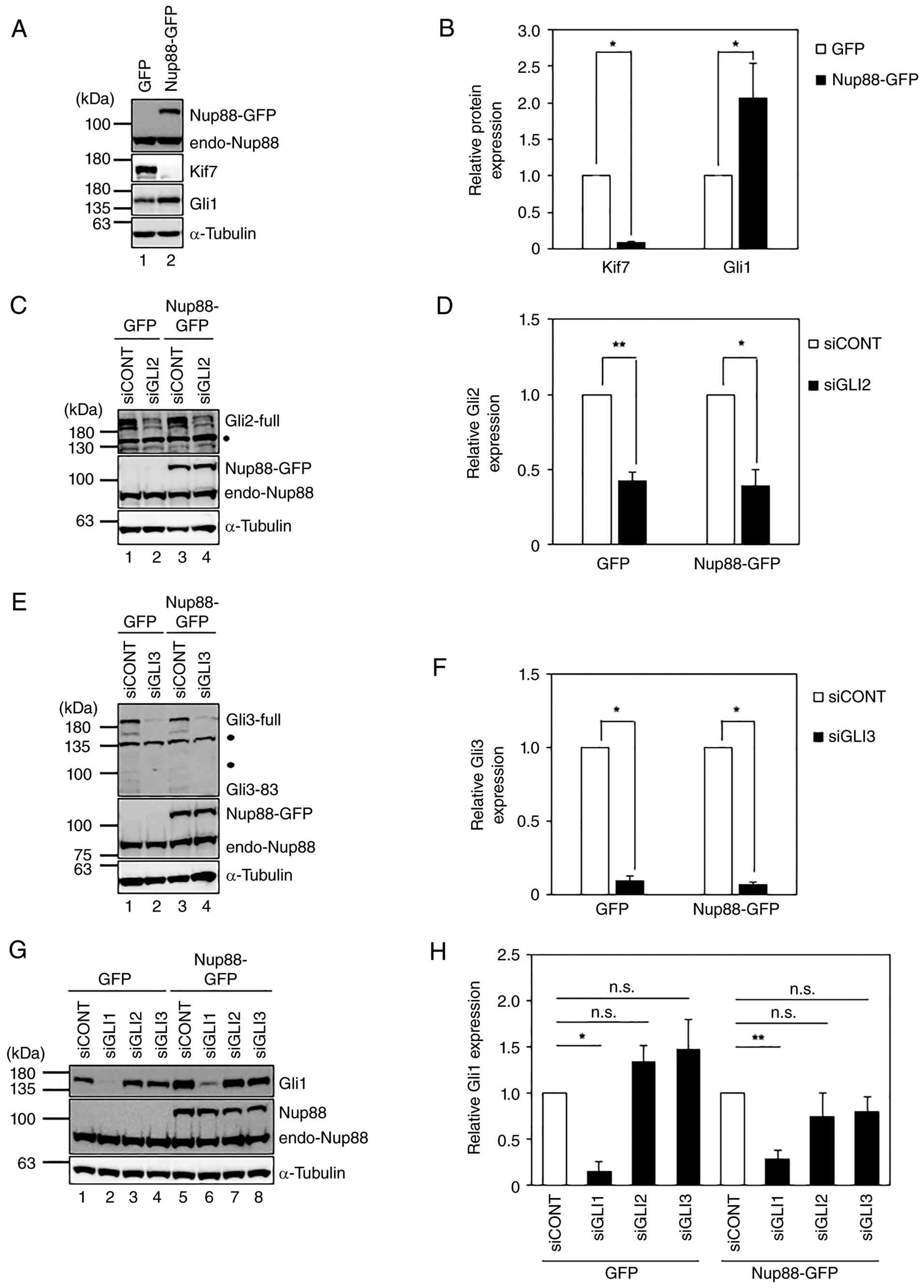 | Figure 3.Increased expression of Gli1 in
Nup88-overexpressing cells appears to be independent of the
Hedgehog pathway. (A) Expression of Gli1 in cells overexpressing
GFP or Nup88-GFP. (B) Statistical analysis of the expression of
Kif7 and Gli1 shown in (A). Data represent mean ± SD (n=3). (C)
Knockdown of Gli2. GLI2-specific siRNA was transfected into
cells overexpressing GFP or Nup88-GFP, and Gli2 levels were
analyzed by immunoblotting. (D) Statistical analysis of the
expression of Gli2 shown in (C). Data represent mean ± SD (n=3).
(E) Knockdown of Gli3. GLI3-specific siRNA was transfected
into cells overexpressing GFP or Nup88-GFP, and the level of Gli3
was analyzed by immunoblotting. The very faint band at an apparent
molecular mass of 83 kDa corresponds to the proteolytically
processed form of full-length Gli3 (Gli3-83) (27). (F) Statistical analysis of the
expression of Gli3 shown in (E). Data represent mean ± SD (n=3).
(G) Effect of GLI2 and GLI3 knockdown on the
expression of Gli1. The expression of Gli1 in both GFP- and
Nup88-overexpressing cells with GLI2 or GLI3
knockdown, as shown in (C) and (E), was analyzed by immunoblotting.
(H) Statistical analysis of the expression of Gli1 shown in (G).
Data represent mean ± SD (n=3). α-Tubulin was used as a loading
control. Closed circles in (C) and (E) indicate non-specific bands
detected by the anti-Gli2 or anti-Gli3 antibodies, respectively.
*P<0.05, **P<0.01, n.s., not significant. Gli1,
glioma-associated oncogene homolog 1; Nup88, nucleoporin 88. |
Results
Decreased expression of Kif7 enhances
the migration of HeLa cells overexpressing Nup88
We previously demonstrated by DNA microarray
analysis that overexpression of Nup88 alters gene expression, which
may influence cell motility (22).
The present study, to identify additional factors involved in this
Nup88-dependent migration, focused on KIF7, which showed a
marked decrease in expression in our previous microarray analysis
(22) (Table SI). Kif7 is a negative regulator of
the Hh pathway that contributes to cancer development. It has been
reported that in prostate cancer cell lines, the expression of
KIF7 is epigenetically repressed through hypermethylation of
CpGs within its promoter (5′ upstream) region, which promotes
formation of the malignant phenotype (29). The present study therefore
hypothesized that the downregulation of Kif7 could also contribute
to motility in HeLa cells. To verify the DNA microarray results,
previously established HeLa cell lines that stably overexpress GFP
or GFP-tagged Nup88 were utilized (32). As shown in Fig. 1A and C, both proteins were
overexpressed and exhibited the expected subcellular localization
described previously (32).
Specifically, immunoblotting with both anti-GFP and anti-Nup88
antibodies (Fig. 1C) allowed the
present study to identify the Nup88-GFP fusion protein and verify
its expression level relative to endogenous Nup88. Using these cell
lines, the present study performed RT-PCR. This analysis confirmed
that Kif7 expression at the RNA level was markedly decreased in the
Nup88-overexpressing cells (Fig.
1B). This decreased expression of Kif7 was subsequently
confirmed at the protein level (Fig.
1C). If the expression of Kif7 is epigenetically regulated,
demethylation of DNA should restore its expression. Thus, it was
examined whether the decreased expression of Kif7 was due to
epigenetic regulation. Expression of Kif7 in cells stably
overexpressing Nup88-GFP was monitored after treatment with a DNA
methylation inhibitor, 5-aza-2′-deoxycytidine, which not only
inhibits the activity of DNA methyltransferases (DNMTs) but also
induces selective degradation of DNMT1 (35). In the experiment, cells
overexpressing GFP or Nup88-GFP were cultured in standard medium
containing the inhibitor (0 days) and then cells were collected
every 2 days for up to 6 days to estimate the expression of Kif7 by
immunoblotting. As shown in Fig.
1D, the expression of DNMT1 in both cell lines was markedly
reduced 2 days after treatment with the inhibitor and the effect
persisted for at least 6 days, indicating that the inhibitor
remained effective throughout this time period. Under the given
experimental conditions, expression of Kif7 in control cells was
largely unaffected (Fig. 1D; lanes
1–4), while its expression in the Nup88-GFP overexpressing cells
markedly recovered by days 4 and 6 (Fig. 1D; lanes 7 and 8). These results
suggested that the expression of KIF7 is regulated by DNA
methylation in Nup88-overexpressing HeLa cells. It has been
reported that Drosophila Nup88 binds to silent chromatin
(36). Therefore, it was examined
whether Nup88 binds to the KIF7 promoter and contributes to
DNA methylation. However, ChIP assays revealed that Nup88 did not
specifically bind to the KIF7 promoter (Fig. 1E).
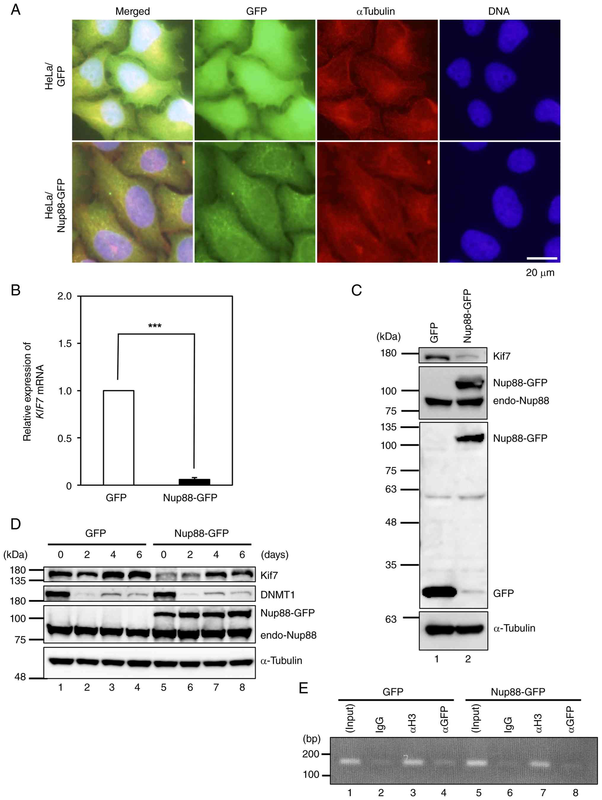 | Figure 1.Decreased expression of Kif7 in
Nup88-overexpressing HeLa cells. (A) Indirect immunofluorescence
imaging of HeLa cells stably overexpressing GFP or GFP-tagged Nup88
(Nup88-GFP). (B) RT-qPCR for the gene expression of Kif7 in cells
overexpressing GFP or Nup88-GFP. Data represent mean ± SD (n=3),
***P<0.001. (C) Immunoblotting for protein expression of Kif7 in
cells overexpressing GFP or Nup88-GFP. Endogenous Nup88 is labeled
as endo-Nup88. α-Tubulin was used as a loading control. Expression
of Nup88-GFP was confirmed by immunoblotting using both anti-GFP
and anti-Nup88 antibodies to verify the expression of the fusion
protein. (D) Expression of Kif7 in cells incubated in the presence
of a DNMT1 inhibitor. Cells overexpressing GFP (lanes 1–4) or
Nup88-GFP (lanes 5–8) were incubated in complete medium containing
1 µM 5-aza-2′-deoxycytidine for 6 days. Cells were collected every
2 days, and their lysates were analyzed by immunoblotting.
α-Tubulin was used as a loading control. (E) ChIP assay for Nup88
binding to the KIF7 promoter region. After crosslinking,
chromatin fractions were prepared from cells overexpressing GFP or
Nup88-GFP. DNA fragments crosslinked to the protein of interest
were immunoprecipitated using non-immune IgG (IgG; negative
control), anti-histone H3 (αH3; positive control), or anti-GFP
(αGFP). Crosslinking was then reversed, and the liberated DNA
fragments were amplified by PCR using KIF7 promoter-specific
primers (see Materials and methods). Kif7, kinesin family member 7;
Nup88, nucleoporin 88; RT-qPCR, reverse transcription-quantitative
PCR; DNMT, DNA methyltransferases; ChIP, chromatin
immunoprecipitation. |
Decreased expression of Kif7 has been reported to
induce malignant phenotypes in prostate cancer cell lines, which
can be suppressed by the overexpression of a Kif7 fragment
(Kif7-CC) comprising a discontinuous coiled-coil and a globular
C-terminal tail domain (29). Thus,
complementation assays were performed to investigate whether the
decreased expression of Kif7 also contributes to the malignant
phenotype in Nup88-overexpressing cells. In these experiments, the
migration activity of GFP- or Nup88-GFP-overexpressing cells was
assessed using co-overexpression of mCherry or mCherry-tagged
truncated Kif7 (514–1343 aa). The tagged protein was derived from
human KIF7 cDNA (BC112271) encoding a region largely
comparable to Kif7-CC but lacking a portion of the Gli2-binding
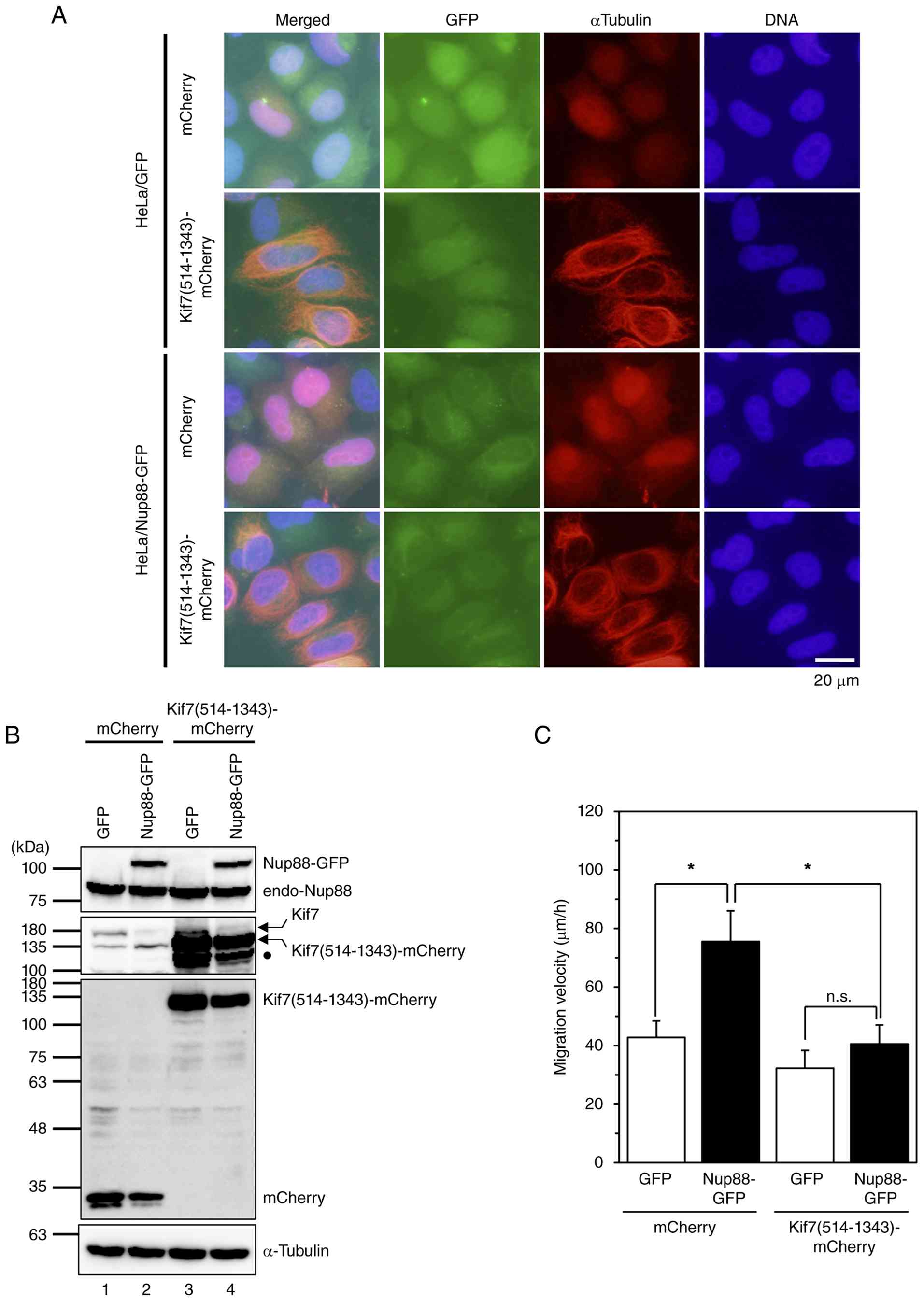
domain (481–541 aa). As shown in Fig.
2A and B, the expression of mCherry and mCherry-tagged
truncated Kif7 was confirmed. Immunoblotting using anti-mCherry and
anti-Kif7 antibodies confirmed that the transiently expressed
proteins were correctly produced as intended fusion proteins at the
expected molecular weights. Using these validated cells, live cell
imaging analysis was performed, and it was found that, in cells
expressing mCherry alone, the migration velocity of
Nup88-GFP-overexpressing cells was almost twice that of
GFP-overexpressing cells (Fig. 2C
and Video S1). By contrast, in
cells expressing mCherry-tagged truncated Kif7, the migration
velocity of Nup88-GFP-overexpressing cells was suppressed to a
level comparable to that of GFP-overexpressing cells (Fig. 2C and Video S1). These results indicate that
downregulation of Kif7 contributes to the enhanced cell
migration.
Expression of Gli1 is upregulated via
the MEK-ERK pathway but not the Hh pathway in Nup88-overexpressing
cells
Kif7 is a major negative regulator of the mammalian
Hh pathway. Therefore, decreased expression of Kif7 was expected to
activate the Hh pathway. Activation of the Hh pathway leads to
increased expression of Gli1, which can promote malignant
phenotypes in certain cancers (23). Thus, it was hypothesized that
decreased expression of Kif7 might lead to malignant phenotypes by
promoting expression of Gli1 via the Hh pathway in
Nup88-overexpressing cells. To examine this possibility,
immunoblotting was used to compare the expression of Gli1 between
GFP- and Nup88-GFP-overexpressing cells. As expected, the
expression of Gli1 was enhanced in Nup88-GFP-overexpressing cells
(Fig. 3A and B). However, Gli1
expression was unaffected when both Gli2 and Gli3, which are
involved in Gli1 transcription in the Hh pathway, were depleted by
RNA interference in either GFP- or Nup88-GFP-overexpressing HeLa
cells (Fig. 3C-H). These findings
suggest that the Nup88-induced increase in Gli1 expression is
independent of the Hh pathway. The expression of Gli1 is known to
be regulated by oncogenic signaling pathways, such as
phosphoinositide 3-kinase-Protein kinase B-mechanistic target of
rapamycin (PI3K-AKT-mTOR), Transforming Growth Factor-β (TGF-β),
and Ras-Rapidly accelerated Fibrosarcoma (Raf)-Mitogen-Activated
Protein Kinase/ERK Kinase (MEK-ERK) (Ras-Raf-MEK-ERK) pathways
(23,24). The present study therefore examined
the involvement of these pathways in the expression of Gli1.
Although PI3K-AKT-mTOR and TGF-β signaling were not specifically
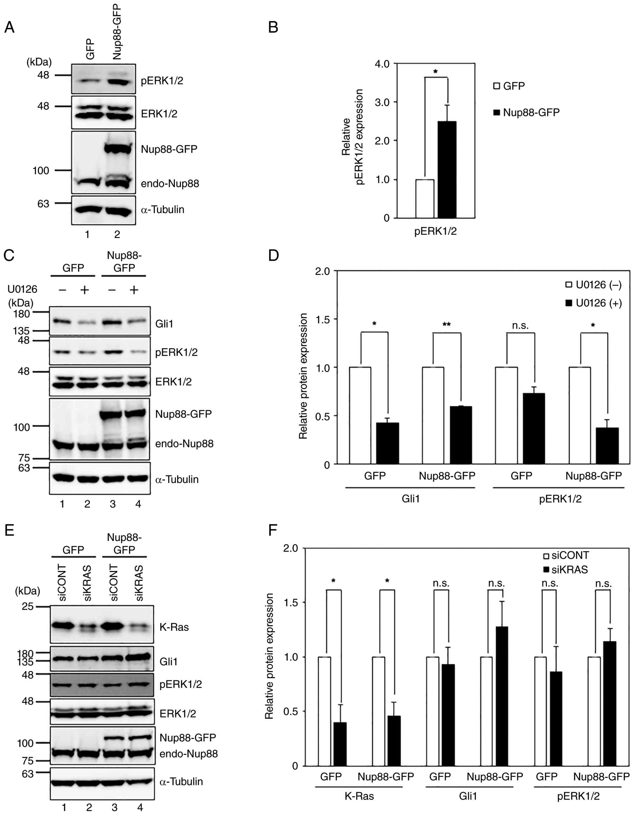
activated by the overexpression of Nup88 in HeLa cells (Fig. S1), the levels of phosphorylated (p)
ERK1/2 increased in Nup88-GFP-overexpressing cells (Fig. 4A and B). Furthermore, blocking the
phosphorylation of ERK with U0126, an inhibitor of MEK1/2, strongly
suppressed the expression of Gli1 (Fig.
4C and D). These results indicated the involvement of the
MEK-ERK pathway in Gli1 expression. Ras is a major upstream
regulator of the MEK-ERK pathway; the present study therefore
examined the involvement of Ras in the expression of Gli1. Among
three Ras isoforms, Kirsten's rat sarcoma virus (K-Ras) is the
predominant isoform in HeLa cells (37). When K-Ras was depleted by RNA
interference, the level of pERK1/2 remained unchanged (Fig. 4E and F), suggesting that Nup88
activates the MEK-ERK pathway to regulate the expression of Gli1
independently of K-Ras.
Gli1 is involved in Nup88-dependent
migration of HeLa cells
Aberrant expression of Gli1 induces various
malignant phenotypes in cancer (23). Therefore, the present study examined
whether increased expression of Gli1 in Nup88-overexpressing HeLa
cells contributes to malignant phenotypes. To investigate the
effect of Gli1 on motility, the migration velocity of
Nup88-overexpressing HeLa cells with or without knockdown of Gli1
was monitored using live-cell single-cell imaging analysis.
Knockdown of Gli1 in both cell lines was confirmed by
immunoblotting (Fig. 5A). When
control siRNA was transfected into both cell lines, the migration
velocity of Nup88-GFP-overexpressing cells was nearly double that
of GFP-overexpressing cells (Fig.
5B and Video S2). This finding
was consistent with the result shown in Fig. 2C. By contrast, when
GLI1-specific siRNA was transfected, the migration velocity
of cells overexpressing Nup88-GFP was suppressed to a level
comparable to that of cells overexpressing GFP (Fig. 5B and Video S2). These results indicate that the
increase in Gli1 expression contributes to cell motility.
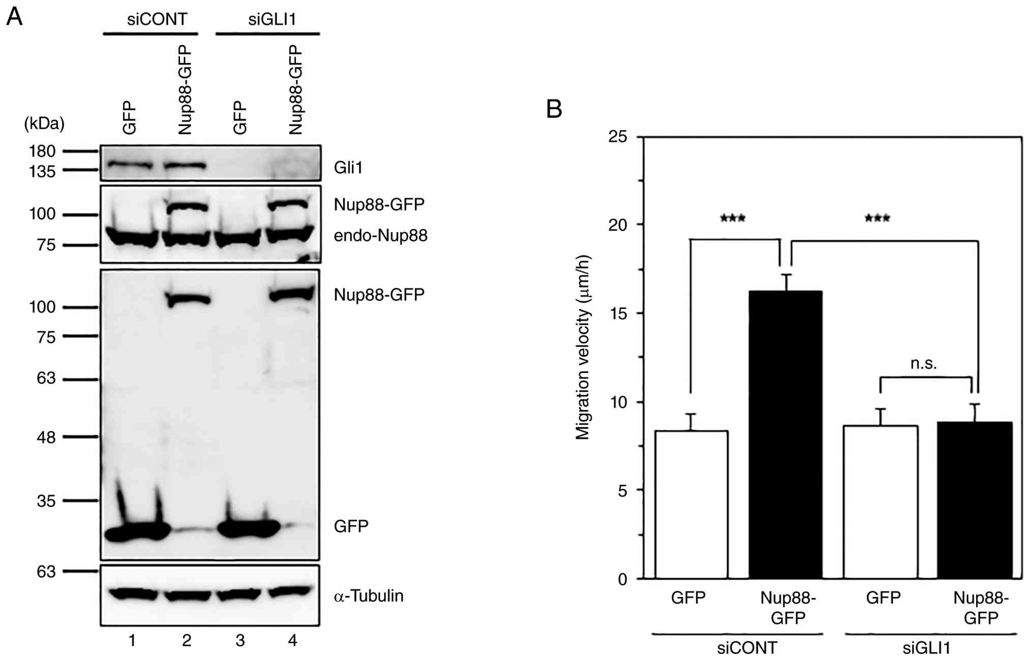 | Figure 5.Enhanced migration of
Nup88-overexpressing cells is Gli1-dependent. (A) Knockdown of
GLI1. HeLa cells overexpressing GFP or Nup88-GFP were
treated with control siRNA (siCONT) or GLI1-specific siRNA
(siGLI1) for 2 days. Protein expression was analyzed by
immunoblotting. Closed circle indicates a non-specific band
detected by the anti-GFP antibody. α-Tubulin was used as a loading
control. Endogenous Nup88 is labeled as endo-Nup88. Expression of
Nup88-GFP was confirmed by immunoblotting using both anti-GFP and
anti-Nup88 antibodies to verify the expression of the fusion
protein. (B) Migration activity of Gli1-depleted cells
overexpressing GFP or Nup88-GFP. Average migration velocity was
calculated after tracking the cells for 24 h. In GFP-overexpressing
cells, the average migration velocity of cells with control
depletion or Gli1 depletion was 8.3 µm/h (n=73) and 8.6 µm/h
(n=74), respectively. In Nup88-GFP-overexpressing cells, the
average migration velocity of cells with control depletion or Gli1
depletion was 16.2 µm/h (n=75) and 8.8 µm/h (n=74), respectively.
***P<0.001, n.s., not significant. The data in (B) are based on
the values obtained from Data S2. Nup88, nucleoporin 88; Gli1,
glioma-associated oncogene homolog 1; si, small interfering; p,
phosphorylated. |
Nup88 regulates Kif7 and Gli1 through
two independent pathways
The present study demonstrated that the
overexpression of Nup88 in HeLa cells promoted cell migration
through the epigenetic repression of Kif7 (Fig. 2) and the MEK-ERK pathway-dependent
increase of Gli1 (Fig. 5). Since
both Kif7 and Gli1 are components of the Hh signaling pathway, the
possibility that the expression of these two proteins might be
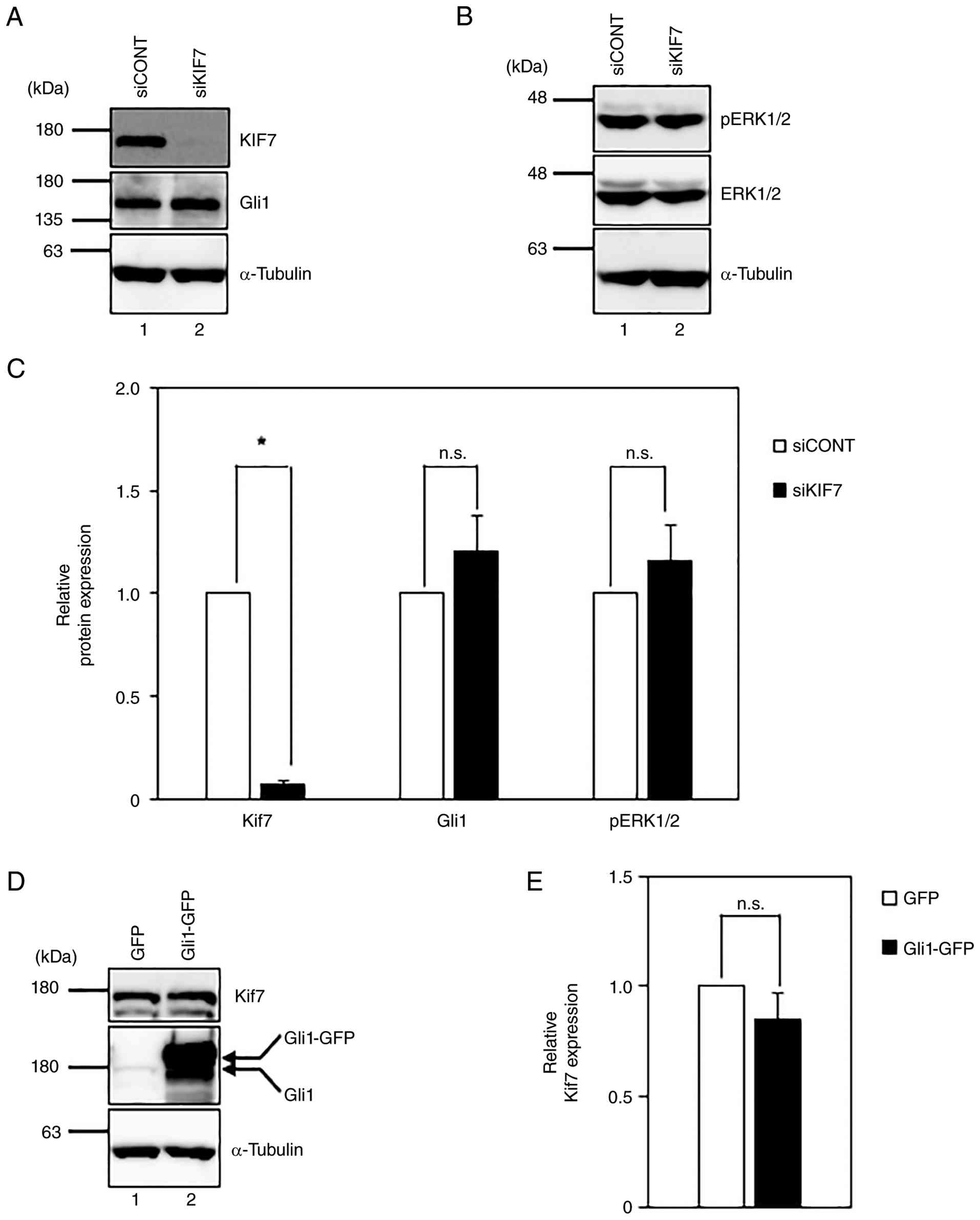
interdependently regulated was considered. Therefore, the present
study examined the effect of Kif7 knockdown on the expression of
Gli1, as well as the effect of the overexpression of Gli1 on the
expression of Kif7. However, neither the knockdown of Kif7
(Fig. 6A-C) nor the overexpression
of Gli1 (Fig. 6D and E) affected
the expression of the other protein. These results suggested that
the changes in expression of Kif7 and Gli1 are independent of each
other.
Truncated Kif7 interacts with
Gli1
The mechanism by which the truncated Kif7 suppresses
the migration activity of Nup88-overexpressing cells remains
unclear (Fig. 2). The present study
considered two plausible mechanisms. One possibility is that
co-overexpression of the truncated Kif7 reduces the expression
levels of phosphorylated ERK and/or Gli1. The other possibility is
that the truncated Kif7 binds to Gli1 and suppresses its activity,
as it does for Gli2/3, although the truncated Kif7 lacks a portion
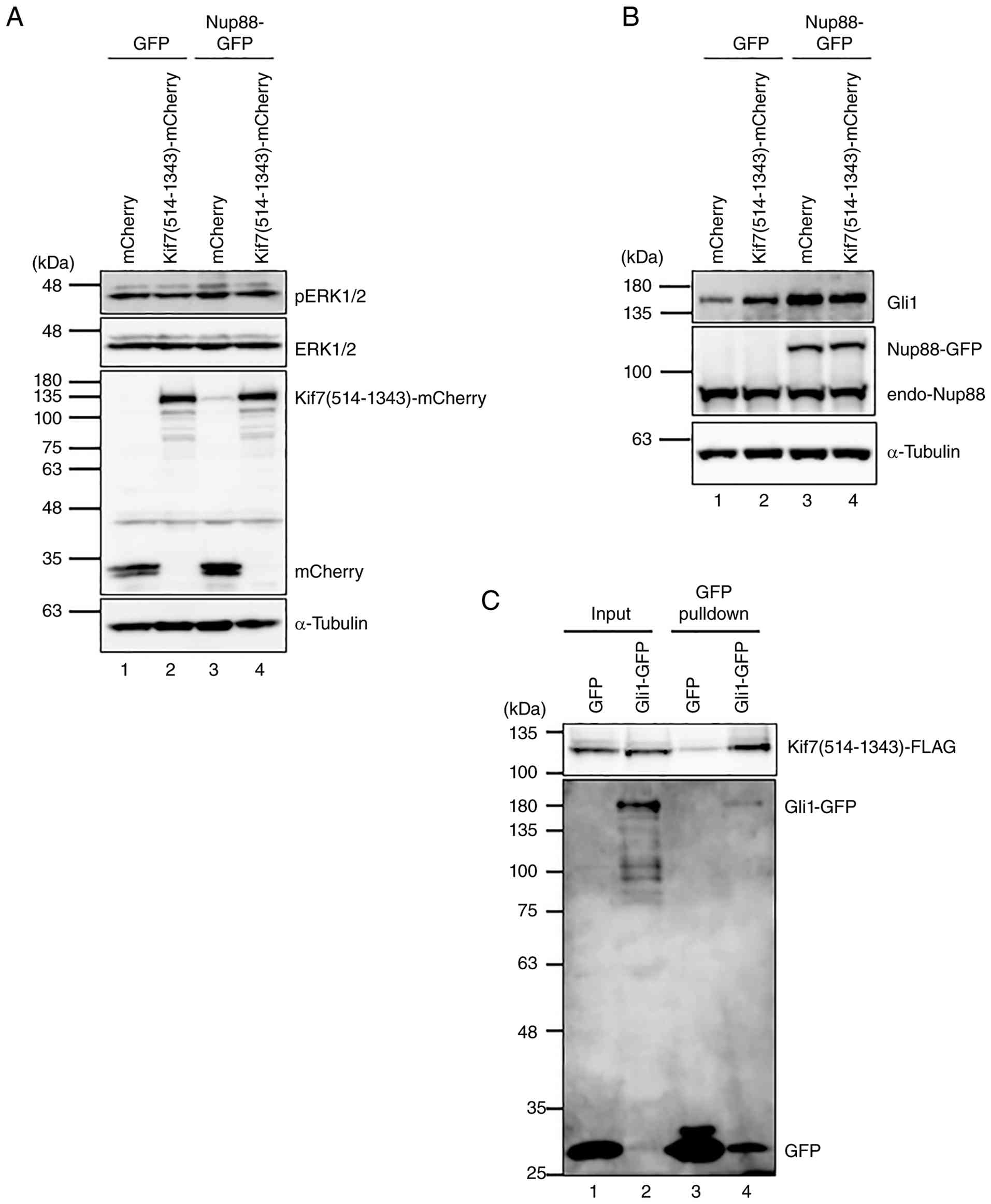
of the Gli2-binding domain. To address the former possibility,
expression of phosphorylated ERK and Gli1 was examined in cells
co-overexpressing Nup88-GFP and truncated Kif7. However,
overexpression of the truncated Kif7 did not affect the expression
of either phosphorylated ERK or Gli1 (Fig. 7A and B). For the latter possibility,
a GFP-pulldown assay using parental HeLa cells co-overexpressing
truncated Kif7 and GFP-tagged Gli1 was performed to test their
interaction. The pulldown assay showed that the truncated Kif7
specifically co-precipitated with Gli1 (Fig. 7C). Taken together, these results
suggested that the truncated Kif7 interacts with Gli1 and
potentially sequesters it, thereby inhibiting its function without
affecting its expression levels.
Discussion
The present study demonstrated that the migration of
HeLa cells overexpressing Nup88 is enhanced by decreased expression
of Kif7 and increased expression of Gli1. Moreover, the enhanced
migration is suppressed by the knockdown of Gli1 or by the
overexpression of the truncated Kif7 [Kif7(514–1343)], which
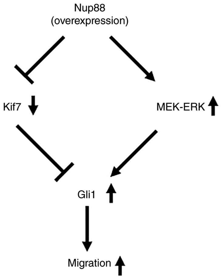
interacts with Gli1. Based on these findings, the present study
proposed a model to explain the mechanism by which overexpression
of Nup88 promotes cell migration (Fig.
8). In the Nup88-overexpressing cells the present study
established, Nup88 led to decreased expression of Kif7 and
activation of the MEK-ERK pathway, which is required for the
expression of Gli1. As knockdown of Kif7 did not activate ERK, Kif7
is unlikely to contribute to the increased expression of Gli1.
Reciprocally, overexpression of Gli1 did not decrease the
expression of Kif7. These findings suggested that the changes in
expression of Kif7 and Gli1 occur independently of each other upon
overexpression of Nup88. By contrast, migration induced by the
overexpression of Nup88 was almost completely suppressed by either
co-overexpression of the truncated Kif7 or knockdown of Gli1. Given
that Kif7 binds to Gli1, this interaction may negatively regulate
its function, as it does for Gli2 and Gli3. Thus, Kif7 likely
suppresses migration through inhibiting the activity of Gli1,
resulting in an effect similar to Gli1 knockdown.
In several prostate cancer cell lines, Kif7
expression appears to be, at least in part, epigenetically
regulated. Indeed, it has been reported that the KIF7
promoter region is hypermethylated in these cells and that
treatment with DNA methylation inhibitors restores KIF7 mRNA
expression (29). However, the
factor(s) that trigger epigenetic repression of KIF7 remain
unidentified. In the present study, HeLa cells stably
overexpressing Nup88 exhibited decreased expression of Kif7, which
was reversed by treatment with a DNA methylation inhibitor, as
observed in prostate cancer cell lines. Therefore, Nup88 may
function as an upstream factor that induces epigenetic repression
of Kif7.
Increased expression of Gli1 in cancer cells can
promote malignant phenotypes (23).
In Nup88-overexpressing cells, expression of Gli1 appeared to be
driven by the Ras-Raf-MEK-ERK pathway. However, knockdown of K-Ras
did not affect expression of phosphorylated ERK, suggesting that
there may be other upstream factors that activate the MEK-ERK
pathway to stimulate expression of Gli1. NF-κB is one such
candidate that can activate the MEK-ERK pathway (38). Specifically, overexpression of Nup88
in HeLa cells was reported to promote the nuclear accumulation of
p65, an essential subunit of NF-κB involved in transcriptional
activation (39). However, no
nuclear accumulation of p65 was detected in the
Nup88-overexpressing HeLa cells used in this study (32). Therefore, increased expression of
Gli1 in these cells may be controlled by signaling molecules other
than NF-κB, which connect with the MEK-ERK pathway.
Nup88-dependent migration was almost completely
suppressed not only by the knockdown of Gli1 but also by the
overexpression of the truncated Kif7 [Kif7(514–1343)]. Notably,
this truncated Kif7 was still capable of binding to Gli1 despite
lacking a portion of the coiled-coil domain (480–542 aa)
responsible for binding to the zinc finger domain of Gli2, which is
highly conserved among Gli proteins (40). Given that Kif7 is known to function
as a negative regulator of Gli2/3, it is plausible that the
interaction of the truncated Kif7 with Gli1 can inhibit Gli1
function. Indeed, Kif7 has additional predicted coiled-coil domains
(698–1057 aa and 1109–1211 aa). Gli1 also possesses a zinc finger
domain that is highly conserved among Gli proteins. Therefore, the
truncated Kif7 may interact with Gli1 through one of these domains
and thereby inhibit the function of Gli1. However, the possibility
that Gli1 binds to the incomplete coiled-coil domain responsible
for Gli2-binding cannot be ruled out.
In the present study, overexpression of Nup88 was
shown to result in decreased expression of Kif7 and increased
expression of Gli1, thereby enhancing cell migration. This
observation may support the hypothesis that Nup88 is actively
involved in the process of cancer progression. The clinical
significance of the elevated expression of Nup88 has been reported
in several types of cancers, where it primarily correlates with
tumor grade and poor prognosis (18–20).
For instance, in endometrial cancer, a positive correlation was
observed between Nup88 mRNA levels and the depth of myometrial
invasion (16). Similarly, in
colorectal cancer, elevated expression of Nup88 was often observed
at the invasive margin and in vascular-invaded areas of the primary
and metastatic tumors (14). The
finding of the present study that Nup88 modulates the expression
levels of Kif7 and Gli1 in HeLa cells may provide a molecular basis
for these clinical features. In cervical cancer, while early-stage
tumors are generally responsive to conventional treatments such as
surgery or radiochemotherapy, metastatic and recurrent cases
present significant therapeutic challenges due to limited treatment
options and poor prognosis. The findings of the present study
suggested that sustaining Kif7 expression or inhibiting Gli1 could
be an effective strategy to suppress Nup88-mediated migration.
Given that Gli1 inhibitors are already being investigated in
clinical trials, this approach may also help overcome resistance to
conventional therapies.
The findings of the present study offered key
insights into Nup88-mediated migration in cervical cancer, yet its
conservation in other cancers remains to be fully elucidated. Given
that low expression of Kif7 correlates with poor prognosis in
prostate, ovarian and breast cancers (41–43),
investigating these malignancies will confirm the functional
conservation of the Nup88 pathway and its viability as a
therapeutic target.
Supplementary Material
Supporting Data
Supporting Data
Supporting Data
Supporting Data
Acknowledgments
The authors thank Dr Katharine Ullman (University of
Utah, US) for providing HeLa cells.
Funding
The present study was supported by JSPS KAKENHI grant no.
19K07700.
Availability of data and materials
The data generated in the present study may be
requested from the corresponding author.
Authors' contributions
MM, RU and AS designed and performed the
experiments. MM and AK interpreted the results, wrote the
manuscript and confirm the authenticity of all the raw data. All
authors read and approved the final manuscript.
Ethics approval and consent to
participate
Not applicable.
Patient consent for publication
Not applicable.
Competing interests
The authors declare that they have no competing
interests.
Glossary
Abbreviations
Abbreviations:
|
FG
|
phenylalanine and glycine
|
|
Gli
|
glioma-associated oncogene homolog
|
|
Hh
|
Hedgehog
|
|
KIF
|
kinesin family member
|
|
NPCs
|
nuclear pore complexes
|
|
Nups
|
nucleoporins
|
References
|
1
|
Cronshaw JM, Krutchinsky AN, Zhang W,
Chait BT and Matunis MJ: Proteomic analysis of the mammalian
nuclear pore complex. J Cell Biol. 158:915–927. 2002. View Article : Google Scholar : PubMed/NCBI
|
|
2
|
Ribbeck K and Gorlich D: Kinetic analysis
of translocation through nuclear pore complexes. EMBO J.
20:1320–1330. 2001. View Article : Google Scholar : PubMed/NCBI
|
|
3
|
Rout MP and Aitchison JD: The nuclear pore
complex as a transport machine. J Biol Chem. 276:16593–16596. 2001.
View Article : Google Scholar : PubMed/NCBI
|
|
4
|
Dickmanns A, Kehlenbach RH and Fahrenkrog
B: Nuclear pore complexes and nucleocytoplasmic transport: From
structure to function to disease. Int Rev Cell Mol Biol.
320:171–233. 2015. View Article : Google Scholar : PubMed/NCBI
|
|
5
|
Simon DN and Rout MP: Cancer and the
nuclear pore complex. Adv Exp Med Biol. 773:285–307. 2014.
View Article : Google Scholar : PubMed/NCBI
|
|
6
|
Snow CJ and Paschal BM: Roles of the
nucleoporin Tpr in cancer and aging. Adv Exp Med Biol. 773:309–322.
2014. View Article : Google Scholar : PubMed/NCBI
|
|
7
|
Nofrini V, Di Giacomo D and Mecucci C:
Nucleoporin genes in human diseases. Eur J Hum Genet. 24:1388–1395.
2016. View Article : Google Scholar : PubMed/NCBI
|
|
8
|
Hutten S and Kehlenbach RH: Nup214 is
required for CRM1-dependent nuclear protein export in vivo. Mol
Cell Biol. 26:6772–6785. 2006. View Article : Google Scholar : PubMed/NCBI
|
|
9
|
Bernad R, Engelsma D, Sanderson H,
Pickersgill H and Fornerod M: Nup214-Nup88 nucleoporin subcomplex
is required for CRM1-mediated 60 S preribosomal nuclear export. J
Biol Chem. 281:19378–19386. 2006. View Article : Google Scholar : PubMed/NCBI
|
|
10
|
Bernad R, van der Velde H, Fornerod M and
Pickersgill H: Nup358/RanBP2 attaches to the nuclear pore complex
via association with Nup88 and Nup214/CAN and plays a supporting
role in CRM1-mediated nuclear protein export. Mol Cell Biol.
24:2373–2384. 2004. View Article : Google Scholar : PubMed/NCBI
|
|
11
|
Xylourgidis N, Roth P, Sabri N, Tsarouhas
V and Samakovlis C: The nucleoporin Nup214 sequesters CRM1 at the
nuclear rim and modulates NFκB activation in Drosophila. J Cell
Sci. 119:4409–4419. 2006. View Article : Google Scholar : PubMed/NCBI
|
|
12
|
Griffis ER, Xu S and Powers MA: Nup98
localizes to both nuclear and cytoplasmic sides of the nuclear pore
and binds to two distinct nucleoporin subcomplexes. Mol Biol Cell.
14:600–610. 2003. View Article : Google Scholar : PubMed/NCBI
|
|
13
|
Agudo D, Gomez-Esquer F, Martinez-Arribas
F, Nunez-Villar MJ, Pollan M and Schneider J: Nup88 mRNA
overexpression is associated with high aggressiveness of Breast
Cancer. Int J Cancer. 109:717–720. 2004. View Article : Google Scholar : PubMed/NCBI
|
|
14
|
Emterling A, Skoglund J, Arbman G,
Schneider J, Evertsson S, Carstensen J, Zhang H and Sun XF:
Clinicopathological significance of Nup88 expression in patients
with colorectal cancer. Oncology. 64:361–369. 2003. View Article : Google Scholar : PubMed/NCBI
|
|
15
|
Knoess M, Kurz AK, Goreva O, Bektas N,
Breuhahn K, Odenthal M, Schirmacher P, Dienes HP, Bock CT, Zentgraf
H and Hausen AZ: Nucleoporin 88 expression in hepatitis B and C
Virus-Related Liver Diseases. World J Gastroenterol. 12:5870–5874.
2006. View Article : Google Scholar : PubMed/NCBI
|
|
16
|
Schneider J, Martinez-Arribas F and
Torrejon R: Nup88 expression is associated with myometrial invasion
in endometrial carcinoma. Int J Gynecol Cancer. 20:804–808. 2010.
View Article : Google Scholar : PubMed/NCBI
|
|
17
|
Zhao ZR, Zhang LJ, Wang YY, Li F, Wang MW
and Sun XF: Increased serum level of Nup88 protein is associated
with the development of colorectal cancer. Med Oncol. 29:1789–1795.
2012. View Article : Google Scholar : PubMed/NCBI
|
|
18
|
Martinez N, Alonso A, Moragues MD, Ponton
J and Schneider J: The nuclear pore complex protein Nup88 is
overexpressed in tumor cells. Cancer Res. 59:5408–5411.
1999.PubMed/NCBI
|
|
19
|
Brustmann H and Hager M: Nucleoporin 88
expression in normal and neoplastic squamous epithelia of the
uterine cervix. Ann Diagn Pathol. 13:303–307. 2009. View Article : Google Scholar : PubMed/NCBI
|
|
20
|
Gould VE, Martinez N, Orucevic A,
Schneider J and Alonso A: A novel, nuclear pore-associated, widely
distributed molecule overexpressed in oncogenesis and development.
Am J Pathol. 157:1605–1613. 2000. View Article : Google Scholar : PubMed/NCBI
|
|
21
|
Xu S and Powers MA: Nuclear pore proteins
and cancer. Semin Cell Dev Biol. 20:620–630. 2009. View Article : Google Scholar : PubMed/NCBI
|
|
22
|
Makise M, Uchimura R, Higashi K, Mashiki
Y, Shiraishi R, Shutoku Y and Kuniyasu A: Overexpression of the
nucleoporin Nup88 stimulates migration and invasion of HeLa cells.
Histochem Cell Biol. 156:409–421. 2021. View Article : Google Scholar : PubMed/NCBI
|
|
23
|
Avery JT, Zhang R and Boohaker RJ: GLI1: A
therapeutic target for cancer. Front Oncol. 11:6731542021.
View Article : Google Scholar : PubMed/NCBI
|
|
24
|
Pietrobono S, Gagliardi S and Stecca B:
Non-canonical hedgehog signaling pathway in cancer: Activation of
GLI transcription factors beyond smoothened. Front Genet.
10:5562019. View Article : Google Scholar : PubMed/NCBI
|
|
25
|
Persson M, Stamataki D, Welscher P,
Andersson E, Böse J, Rüther U, Ericson J and Briscoe J:
Dorsal-ventral patterning of the spinal cord requires Gli3
transcriptional repressor activity. Genes Dev. 16:2865–2878. 2002.
View Article : Google Scholar : PubMed/NCBI
|
|
26
|
Stone DM, Murone M, Luoh S, Ye W, Armanini
MP, Gurney A, Phillips H, Brush J, Goddard A, de Sauvage FJ and
Rosenthal A: Characterization of the human suppressor of fused, a
negative regulator of the zinc-finger transcription factor Gli. J
Cell Sci. 112:4437–4448. 1999. View Article : Google Scholar : PubMed/NCBI
|
|
27
|
Cheung HO, Zhang X, Ribeiro A, Mo R,
Makino S, Puviindran V, Law KKL, Briscoe J and Hui CC: The kinesin
protein Kif7 is a critical regulator of Gli transcription factors
in mammalian hedgehog signaling. Sci Signal. 2:ra292009. View Article : Google Scholar : PubMed/NCBI
|
|
28
|
Niewiadomski P, Niedziolka SM, Markiewicz
L, Uspienski T, Baran B and Chojnowska K: Gli Proteins: Regulation
in development and cancer. Cells. 8:1472019. View Article : Google Scholar : PubMed/NCBI
|
|
29
|
Wong KY, Liu J and Chan KW: KIF7
attenuates prostate tumor growth through LKB1-mediated AKT
inhibition. Oncotarget. 8:54558–54571. 2017. View Article : Google Scholar : PubMed/NCBI
|
|
30
|
Sung H, Ferlay J, Siegel RL, Laversanne M,
Soerjomataram I, Jemal A and Bray F: Global cancer statistics 2020:
GLOBOCAN Estimates of Incidence and Mortality Worldwide for 36
Cancers in 185 Countries. CA Cancer J Clin. 71:209–249.
2021.PubMed/NCBI
|
|
31
|
Tewari KS: Cervical Cancer. N Engl J Med.
392:56–71. 2025. View Article : Google Scholar : PubMed/NCBI
|
|
32
|
Makise M, Nakamura H and Kuniyasu A: The
role of vimentin in the tumor marker Nup88-dependent multinucleated
phenotype. BMC Cancer. 18:5192018. View Article : Google Scholar : PubMed/NCBI
|
|
33
|
Kaiser C, Dobrikova EY, Bradrick SS,
Shveygert M, Herbert JT and Gromeier M: Activation of
cap-independent translation by variant eukaryotic initiation factor
4G in vivo. RNA. 14:2170–2182. 2008. View Article : Google Scholar : PubMed/NCBI
|
|
34
|
Livak KJ and Schmittgen TD: Analysis of
relative gene expression data using real-time quantitative PCR and
the 2−ΔΔCT method. Methods. 25:402–408. 2001.
View Article : Google Scholar : PubMed/NCBI
|
|
35
|
Patel K, Dickson J, Din S, Macleod K,
Jodrell D and Ramsahoye B: Targeting of 5-aza-2′-deoxycytidine
residues by chromatin-associated DNMT1 induces proteasomal
degradation of the free enzyme. Nucleic Acids Res. 38:4313–4324.
2010. View Article : Google Scholar : PubMed/NCBI
|
|
36
|
Capelson M, Liang Y, Schulte R, Mair W,
Wagner U and Hetzer MW: Chromatin-bound nuclear pore components
regulate gene expression in higher eukaryotes. Cell. 140:372–383.
2010. View Article : Google Scholar : PubMed/NCBI
|
|
37
|
Hood FE, Sahraoui YM, Jenkins RE and Prior
IA: Ras protein abundance correlates with Ras isoform mutation
patterns in cancer. Oncogene. 42:1224–1232. 2023. View Article : Google Scholar : PubMed/NCBI
|
|
38
|
Oeckinghaus A, Hayden MS and Ghosh S:
Crosstalk in NF-κB signaling pathways. Nat Immunol. 12:695–708.
2011. View Article : Google Scholar : PubMed/NCBI
|
|
39
|
Takahashi N, van Kilsdonk JW, Ostendorf B,
Smeets R, Bruggeman SWM, Alonso A, van de Loo F, Schneider M, van
den Berg WB and Swart GWM: Tumor marker nucleoporin 88 kDa
regulates nucleocytoplasmic transport of NF-κB. Biochem Biophys Res
Commun. 374:424–430. 2008. View Article : Google Scholar : PubMed/NCBI
|
|
40
|
Haque F, Freniere C, Ye Q, Mani N,
Wilson-Kubalek EM, Ku PI, Milligan RA and Subramanian R:
Cytoskeletal regulation of a transcription factor by DNA mimicry
via coiled-coil interactions. Nat Cell Biol. 24:1088–1098. 2022.
View Article : Google Scholar : PubMed/NCBI
|
|
41
|
Ho J, Du Y, Wong OG, Siu MK, Chan KK and
Cheung AN: Downregulation of the gli transcription factors
regulator Kif7 facilitates cell survival and migration of
choriocarcinoma cells. PLoS One. 9:e1082482014. View Article : Google Scholar : PubMed/NCBI
|
|
42
|
Shagufta Aftab M, Sisodiya S, Mishra S,
Priya K and Hussain S: Prognostic significance of the kinesin
superfamily in breast cancer: A systematic review &
meta-analysis. Indian J Med Res. 161:627–635. 2025. View Article : Google Scholar : PubMed/NCBI
|
|
43
|
Yao Y, Liu L, He W, Lin X, Zhang X, Lin Z,
Zeng Z and Guo S: Low expression of KIF7 indicates poor prognosis
in epithelial ovarian cancer. Cancer Biomark. 26:481–489. 2019.
View Article : Google Scholar : PubMed/NCBI
|