Introduction
Cervical cancer remains one of the leading causes of
cancer-related incidence and mortality among women worldwide.
According to GLOBOCAN 2022 estimates, ~662,000 new cases of
cervical cancer and 349,000 associated deaths were reported
globally in 2022 (1). Early-stage
cervical cancer is usually treated with conization; however, when
early detection fails and metastasis occurs, patients require
treatment with chemotherapy, with alkylating agents and antitumor
antibiotics commonly used (2).
However, various challenges, including the high in vivo
toxicity of anticancer drugs and the relatively high cost of
treatment, indicate that further research to improve anticancer
treatment strategies is still required (3). Cervical cancer, which is primarily
caused by a persistent infection with a high-risk strain of the
human papillomavirus (HPV), is characterized by HPV-mediated
alterations to, and functional inactivation of, tumor suppressor
proteins, including p53. Accordingly, strategies aimed at restoring
the activity and expression of tumor suppressor proteins may
represent a promising therapeutic approach for cervical cancer
(4). Notably, studies have
demonstrated that phytochemicals can modulate the expression of
tumor suppressor proteins in cancer cells (5,6); thus
highlighting the need to evaluate the therapeutic potential of
phytochemicals in the treatment of cervical cancer.
Phytochemicals are naturally occurring plant-derived
compounds that exhibit relatively low biological toxicity compared
with synthetic drugs (7). Because
of these benefits, phytochemicals are being explored for the
treatment of various diseases, including cancer and Alzheimer's
disease (8,9). Phytochemicals are broadly classified
into polyphenols, terpenoids, alkaloids and organosulfur compounds,
each with distinct chemical structures and biological activities.
Among these, polyphenols have attracted considerable attention due
to their potent antioxidant and anticancer properties, and include
subclasses such as flavonoids and phenolic acids (10). Flavonoids are a major class of
polyphenols with a C6-C3-C6 structure and include subclasses such
as flavones, flavanols, flavanones and isoflavones (11). Previous studies have reported that
flavonoids exhibit antioxidant and anticancer properties in various
cancer types, including breast, colorectal, lung and liver cancer,
and are generally associated with low toxicity toward normal cells
(12,13). However, a number of flavonoids have
limited bioavailability and polymethoxylated flavonoids (PMFs) have
emerged as a promising strategy to address this limitation
(14,15).
Tangeretin (TAN) is a PMF found predominantly in
citrus peels. It has been suggested that the multiple methoxy
(−OCH3) groups of TAN may contribute to its enhanced
chemical stability and resistance to oxidative degradation compared
with other flavonoids (16).
According to a previous pharmacokinetic study, TAN exhibits higher
bioavailability than other PMFs, including nobiletin and
sinensetin, thus suggesting that TAN may possess relatively
favorable pharmacokinetic properties compared with other PMFs
(17). These pharmacokinetic
characteristics could contribute to the biological activity of TAN
in cellular systems. TAN has been reported to exert anticancer
effects associated with increased intracellular reactive oxygen
species (ROS) levels, apoptosis and cell cycle arrest; these
effects have been observed in multiple cancer types, including
lung, colon and gastric cancer (18–20).
However, to the best of our knowledge, the anticancer effects of
TAN on cervical cancer have not yet been elucidated, highlighting
the need for further investigation. The present study aimed to
evaluate the effects of TAN on cervical cancer cell viability,
proliferation and migration. Furthermore, the ability of TAN to
induce mitochondrial ROS generation and apoptosis was investigated,
and changes in p53 protein expression following TAN treatment were
examined.
Materials and methods
Chemicals
TAN (purity ≥95%) was purchased from Aladdin
Scientific Corporation. A stock solution of TAN was prepared in
dimethyl sulfoxide (DMSO; Sigma-Aldrich; Merck KGaA) and was
subsequently diluted in culture medium to achieve the desired
concentrations, with the final DMSO concentration maintained at
<0.1% in all experiments.
Cell culture
CaSki cells were purchased from the Korean Cell Line
Bank; Korean Cell Line Research Foundation. The cells were cultured
in Roswell Park Memorial Institute (RPMI)-1640 medium (R&D
Systems, Inc.) supplemented with 10% fetal bovine serum (FBS;
R&D Systems, Inc.), 10 mM HEPES (Sigma-Aldrich; Merck KGaA) and
1% antibiotic-antimycotic solution (Sigma-Aldrich; Merck KGaA).
Subculturing was performed using 0.05% trypsin-EDTA (Thermo Fisher
Scientific, Inc.) at a split ratio of 1:5. The cells were
maintained at 37°C in a humidified incubator containing 5%
CO2.
Cell viability assays
CaSki cells were seeded in 96-well plates at a
density of 4×10³ cells/well. After 24 h of incubation,
the medium was replaced with RPMI-1640 supplemented with 5% FBS
containing either 0.1% DMSO (control) or TAN (2.5–40 µM). After 48
h of treatment at 37°C in a humidified incubator with 5%
CO2, the culture medium was removed and 5% EZ-Cytox cell
viability assay reagent (cat. no. EZ-3000; DoGenBio), a
water-soluble tetrazolium salt reagent, was added and incubated for
30 min at 37°C in a humidified incubator with 5% CO2.
Absorbance was measured at 450 nm using a Synergy Neo2 Hybrid
Multimode Reader (Agilent Technologies, Inc.).
Colony formation assay
CaSki cells were seeded in 6-well plates at a
density of 1×10³ cells/well and incubated for 24 h. The
cells were then treated with RPMI-1640 medium supplemented with 5%
FBS containing either 0.1% DMSO (control) or TAN (10–40 µM) for 48
h at 37°C in a humidified incubator with 5% CO2.
Thereafter, the culture medium was replaced every 3 days with
RPMI-1640 supplemented with 5% FBS, and the experiment was
continued for 2 weeks. For analysis, the colonies were fixed with
4% paraformaldehyde (PFA; GeneAll Biotechnology Co., Ltd.) for 15
min at room temperature and stained with 0.5% crystal violet
(Sigma-Aldrich; Merck KGaA) for 30 min at room temperature. Colony
formation was quantified by measuring the total colony area using
ImageJ software (version 1.53; National Institutes of Health).
Colonies with an area of ≥20 pixels2 were included in
the analysis. The colony formation rate was calculated by
normalizing the colony area of each treatment group to that of the
control group, which was set to 100%.
Wound-healing assay
CaSki cells were seeded in 6-well plates at a
density of 1×10⁵ cells/well and incubated at 37°C in a
humidified atmosphere containing 5% CO2 for 48 h until
they reached ~80% confluence. To minimize the contribution of cell
proliferation during wound closure analysis, the cells were
pretreated with mitomycin C (2 µg/ml; Roche Diagnostics) for 1.5 h
at 37°C in a humidified incubator containing 5% CO2
prior to scratch formation. A linear scratch was then generated in
each well using sterile surgical forceps. After removing the
culture medium and washing to eliminate cellular debris, the cells
were incubated in RPMI-1640 medium supplemented with 5% FBS,
containing either 0.1% DMSO (control) or TAN (1.25–5 µM). The FBS
concentration (5%) was selected to maintain cell viability during
the wound-healing assay (21).
After treatment, the cells were incubated at 37°C in a humidified
atmosphere containing 5% CO2. Images of the wound area
were captured at 0, 24 and 48 h at ×40 magnification using an
Olympus CKX41 inverted light microscope (Olympus Corporation), and
wound closure was semi-quantified using ImageJ software (version
1.53).
Transwell migration assay
CaSki cells (5×10⁵ cells/well) were
seeded into the upper chamber of 24-well Transwell inserts (pore
size, 8.0-µm). The upper chamber was filled with RPMI-1640 medium
supplemented with 1% FBS containing either 0.1% DMSO (control) or
TAN (10–40 µM). The lower chamber contained RPMI-1640 medium
supplemented with 10% FBS as a chemoattractant. After 48 h of
incubation at 37°C in a humidified atmosphere containing 5%
CO2, the non-migratory cells on the upper surface of the
membrane were removed, and cells that had migrated to the lower
surface were fixed with 4% PFA for 15 min at room temperature and
stained with 0.5% crystal violet for 30 min at room temperature.
Images were captured using an Olympus CKX41 microscope, and the
number of migrated cells was semi-quantified using ImageJ software
(version 1.53).
Cell cycle arrest assay
CaSki cells (3×10³ cells/well) were
seeded in 6-well plates and incubated for 24 h. The culture medium
was then replaced with RPMI-1640 supplemented with 5% FBS
containing either 0.1% DMSO (control) or TAN (10–40 µM). After 48 h
of treatment at 37°C in a humidified atmosphere containing 5%
CO2, the cells were harvested and fixed in 70% ethanol
at −20°C overnight. Fixed cells were subsequently stained with
Hoechst 33342 (10 µg/ml; cat. no. H3570; Thermo Fisher Scientific,
Inc.) for 30 min at 37°C in a humidified atmosphere containing 5%
CO2. Hoechst 33342, a DNA-binding fluorescent dye, was
used to determine cell cycle distribution based on cellular DNA
content, as previously described (22). Cell cycle distribution was analyzed
using a FACSymphony™ flow cytometer (BD Biosciences)
with the BV421 channel and data were analyzed using FlowJo software
(version 10; BD Biosciences).
Annexin V and propidium iodide (PI)
staining
CaSki cells (3×10³ cells/well) were
seeded in 6-well plates and incubated for 24 h. The culture medium
was then replaced with RPMI-1640 supplemented with 5% FBS
containing either 0.1% DMSO (control) or TAN (10–40 µM). After 48 h
of treatment at 37°C in a humidified atmosphere containing 5%
CO2, the cells were harvested and resuspended in 100 µl
1X Annexin-binding buffer. Subsequently, 5 µl Alexa
Fluor™ 488 Annexin V and 1 µl PI (100 µg/ml) from the
Alexa Fluor 488 Annexin V/Dead Cell Apoptosis Kit (Invitrogen;
Thermo Fisher Scientific, Inc.) were added to each sample and
incubated for 15 min at room temperature in the dark according to
the manufacturer's instructions. Following incubation, 400 µl 1X
Annexin-binding buffer was added and the samples were immediately
analyzed using a FACSymphony flow cytometer and the percentage of
apoptotic cells was quantified using FlowJo software (version
10).
MitoSOX™ staining
CaSki cells were seeded in 96-well plates at a
density of 6×10³ cells/well and incubated for 24 h at
37°C in a humidified atmosphere containing 5% CO2. The
culture medium was then replaced with RPMI-1640 supplemented with
5% FBS containing either 0.1% DMSO (control) or TAN (10–40 µM).
After 48 h of treatment at 37°C in a humidified atmosphere
containing 5% CO2, the medium was removed, and the cells
were incubated with MitoSOX™ Red Mitochondrial
Superoxide Indicator (5 µg/ml; cat. no. M36008; Thermo Fisher
Scientific, Inc.) and Hoechst 33342 (5 µg/ml; cat. no. H3570;
Thermo Fisher Scientific, Inc.) for 10 min at 37°C. Fluorescence
images were acquired using a Lionheart™ FX Automated
Imaging System (BioTek; Agilent Technologies, Inc.), with Hoechst
33342 detected in the DAPI channel and MitoSOX Red detected in the
RFP channel. Images were acquired using a Lionheart FX Automated
Microscope (BioTek; Agilent Technologies, Inc.) and analyzed using
Gen5 software (version 3.05; BioTek; Agilent Technologies, Inc.)
under identical image acquisition settings. Background fluorescence
was automatically corrected using the built-in background
subtraction function of the instrument. MitoSOX Red fluorescence
intensity was normalized to the number of Hoechst 33342-positive
nuclei in each field.
JC-1 staining
CaSki cells were seeded in black 96-well plates at a
density of 5×10³ cells/well and incubated for 24 h. The
culture medium was then replaced with RPMI-1640 supplemented with
5% FBS containing either 0.1% DMSO (control) or TAN (10–40 µM).
After 24 h of treatment at 37°C in a humidified atmosphere
containing 5% CO2, the medium was removed and JC-1 dye
(cat. no. ab113850; Abcam) was added to each well at a final
concentration of 3 µg/ml. The cells were incubated for 30 min at
37°C and fluorescence images were acquired using a Lionheart FX
Automated Imaging System, with JC-1 monomers detected in the GFP
channel and JC-1 aggregates detected in the RFP channel. Images
were acquired using a Lionheart FX Automated Microscope and
fluorescence intensity was quantified using Gen5 software (version
3.05) under identical image acquisition settings. Background
fluorescence was automatically corrected using the built-in
background subtraction function of the instrument. Mitochondrial
membrane potential (MMP) was evaluated based on the ratio of JC-1
aggregate (RFP) fluorescence to JC-1 monomer (GFP)
fluorescence.
Western blot analysis
Cells in the control group were cultured in
RPMI-1640 supplemented with 5% FBS containing 0.1% DMSO (vehicle
control), and TAN-treated cells were cultured in RPMI-1640
supplemented with 5% FBS containing TAN (20 or 40 µM) for 48 h.
Total cellular proteins were extracted using PRO-PREP™ protein
extraction solution (Intron Biotechnology, Inc.) supplemented with
a protease inhibitor cocktail (ATTO Corporation). Protein
concentrations were determined using a bicinchoninic acid (BCA)
protein assay following the Sigma-Aldrich BCA protein assay
protocol, using BCA solution (cat. no. B9643; Sigma-Aldrich; Merck
KGaA) and 4% copper(II) sulfate solution (cat. no. C2284;
Sigma-Aldrich; Merck KGaA), with bovine serum albumin (BSA; cat.
no. A9418; MilliporeSigma) used as the protein standard. Western
blot analysis was performed using 30 µg total protein per lane.
Total proteins were separated by SDS-PAGE on 8% gels for E-cadherin
and 10% gels for Bax and p53, followed by transfer onto PVDF
membranes (Amersham™ Hybond™ P; cat. no.
10600023; Cytiva). The membranes were blocked with 5% BSA in
Tris-buffered saline containing 0.1% Tween-20 for 1 h 30 min at
room temperature. After blocking, the membranes were incubated
overnight at 4°C with primary antibodies against Bax (1:1,000; cat.
no. 2772; Cell Signaling Technology, Inc.), E-cadherin (1:1,000;
cat. no. 3195; Cell Signaling Technology, Inc.), p53 (1:1,000; cat.
no. bs-2090R; BIOSS) and GAPDH (1:5,000; cat. no. ABS16;
MilliporeSigma) diluted in 5% BSA (MilliporeSigma) in Tris-buffered
saline containing 0.1% Tween-20 (cat. no. 11332465001; Roche
Diagnostics). The membranes were then incubated with horseradish
peroxidase (HRP)-conjugated goat anti-rabbit IgG secondary antibody
(1:3,000; cat. no. 1706515; Bio-Rad Laboratories, Inc.) for 1 h at
room temperature. Protein bands were visualized using an enhanced
chemiluminescence (ECL) detection reagent (Amersham™
ECL™ Select Western Blotting Detection Reagent; cat. no.
RPN2235; Cytiva) and images were captured using a LuminoGraph II
imaging system (Cytiva). Band intensities were semi-quantified
using ImageJ software (version 1.53).
Statistical analysis
All in vitro experiments were performed using
technical triplicates under identical experimental conditions
unless otherwise specified. Data are presented as the mean ±
standard deviation and were analyzed using GraphPad Prism software
(version 10; Dotmatics). Statistical significance was evaluated
using one-way analysis of variance followed by Dunnett's post hoc
test for multiple comparisons against the control group. As the
data were obtained from technical triplicates, the sample size was
insufficient for reliable assessment of normality and homogeneity
of variance; therefore, these tests were not consistently
performed. P<0.05 was considered to indicate a statistically
significant difference.
Results
TAN decreases the viability and
proliferation of CaSki cells
Previous studies have reported that TAN suppresses
cell viability in various types of cancer (23–25).
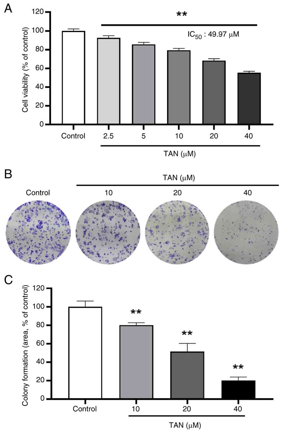
In the present study, TAN treatment reduced the viability of CaSki
cells (Fig. 1A). Based on the cell
viability assay results, 10, 20 and 40 µM TAN were selected for
subsequent experiments because these concentrations showed greater
inhibitory effects on cell viability than lower concentrations and
allowed the evaluation of concentration-dependent biological
responses. In addition, the anti-proliferative effect of TAN in
CaSki cells was evaluated using a colony formation assay (Fig. 1B), which revealed a significant
reduction in colony formation area in the TAN-treated group
compared with that in the control group (Fig. 1C). These results indicated that TAN
may reduce cell viability by inhibiting CaSki cell
proliferation.
TAN induces G1 phase arrest
in CaSki cells
TAN has been reported to suppress cancer cell
proliferation primarily through the induction of G1
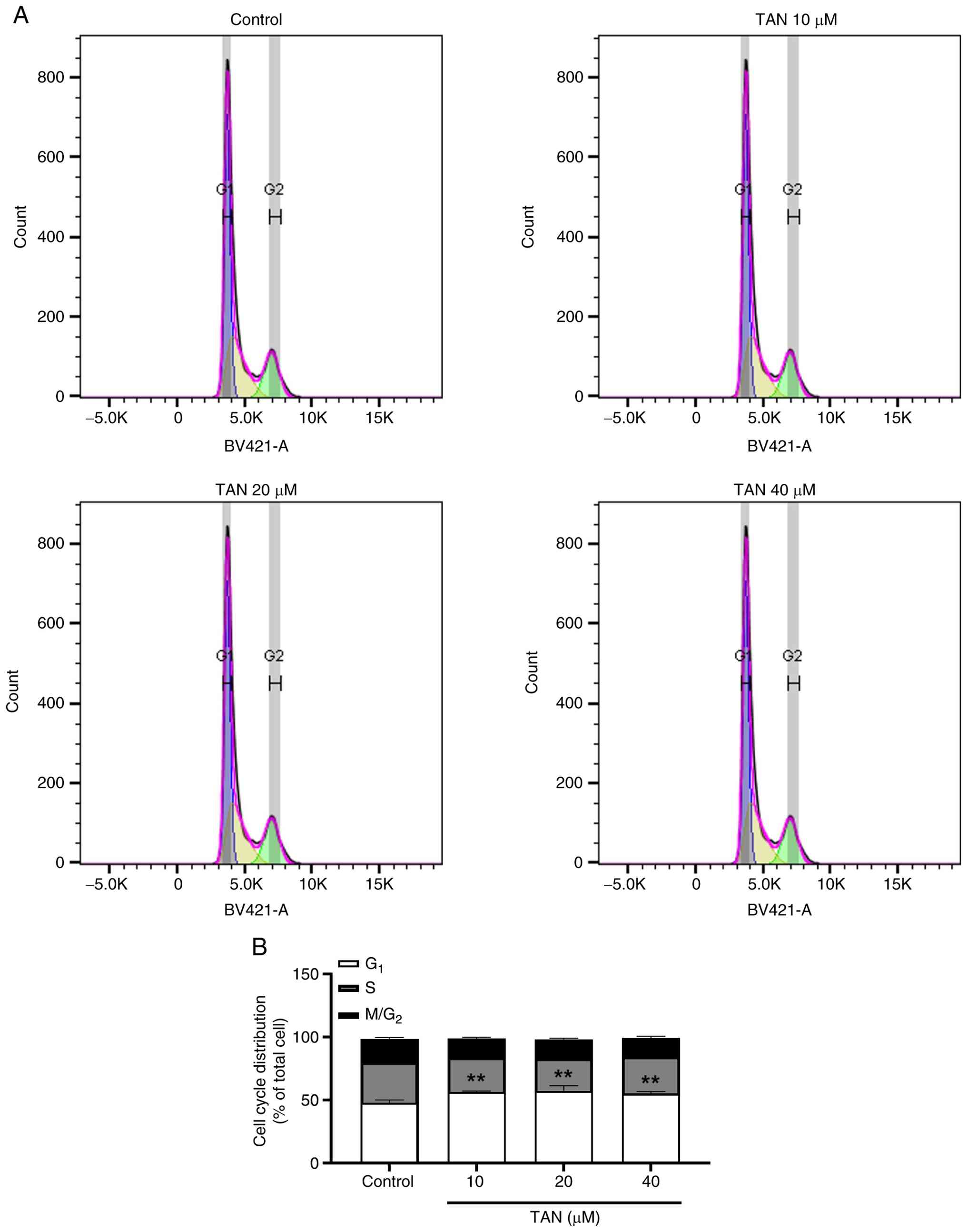
phase arrest (26,27). In the current study, Hoechst 33342
staining was employed to examine cell cycle alterations associated
with TAN-mediated inhibition of CaSki cell proliferation (Fig. 2A). The assay showed that TAN
treatment increased the proportion of cells in the G1
phase compared with that in the control group (Fig. 2B). These findings suggested that
TAN-associated inhibition of CaSki cell proliferation may be
related to G1 phase arrest.
TAN suppresses CaSki cell
migration
Effective cancer treatment requires not only the
inhibition of cancer cell proliferation but also the suppression of
cancer cell migration and metastasis (28). The effects of TAN on cell migration
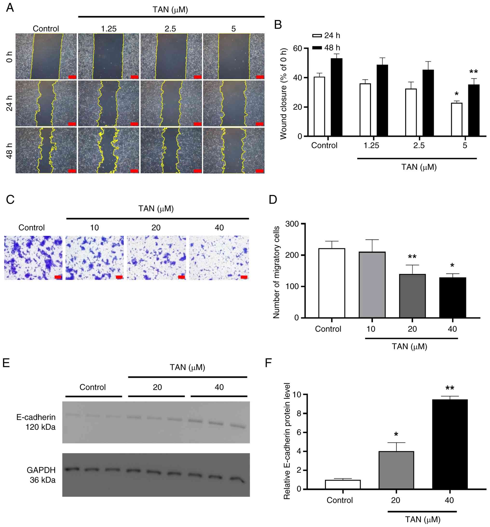
were first investigated using a wound-healing assay (Fig. 3A). Notably, TAN treatment at 5 µM
significantly reduced wound closure compared with the control group
at both 24 and 48 h (Fig. 3B). In
addition, a Transwell migration assay was conducted to evaluate the
effect of TAN on chemotactic migration (Fig. 3C). The number of migratory cells was
significantly decreased in cells treated with 20 and 40 µM TAN
compared with the control group (Fig.
3D). Western blot analysis was subsequently performed to detect
E-cadherin (Fig. 3E). The protein
expression levels of E-cadherin were increased in the TAN-treated
groups compared with those in the control group (Fig. 3F). These results suggested that TAN
not only inhibits the proliferation of CaSki cells, but is also
associated with reduced migratory capacity and migration-associated
phenotypic changes.
TAN induces intrinsic apoptosis of
CaSki cells
Previous studies have reported that TAN suppresses
cancer cell proliferation by inducing apoptosis, a form of
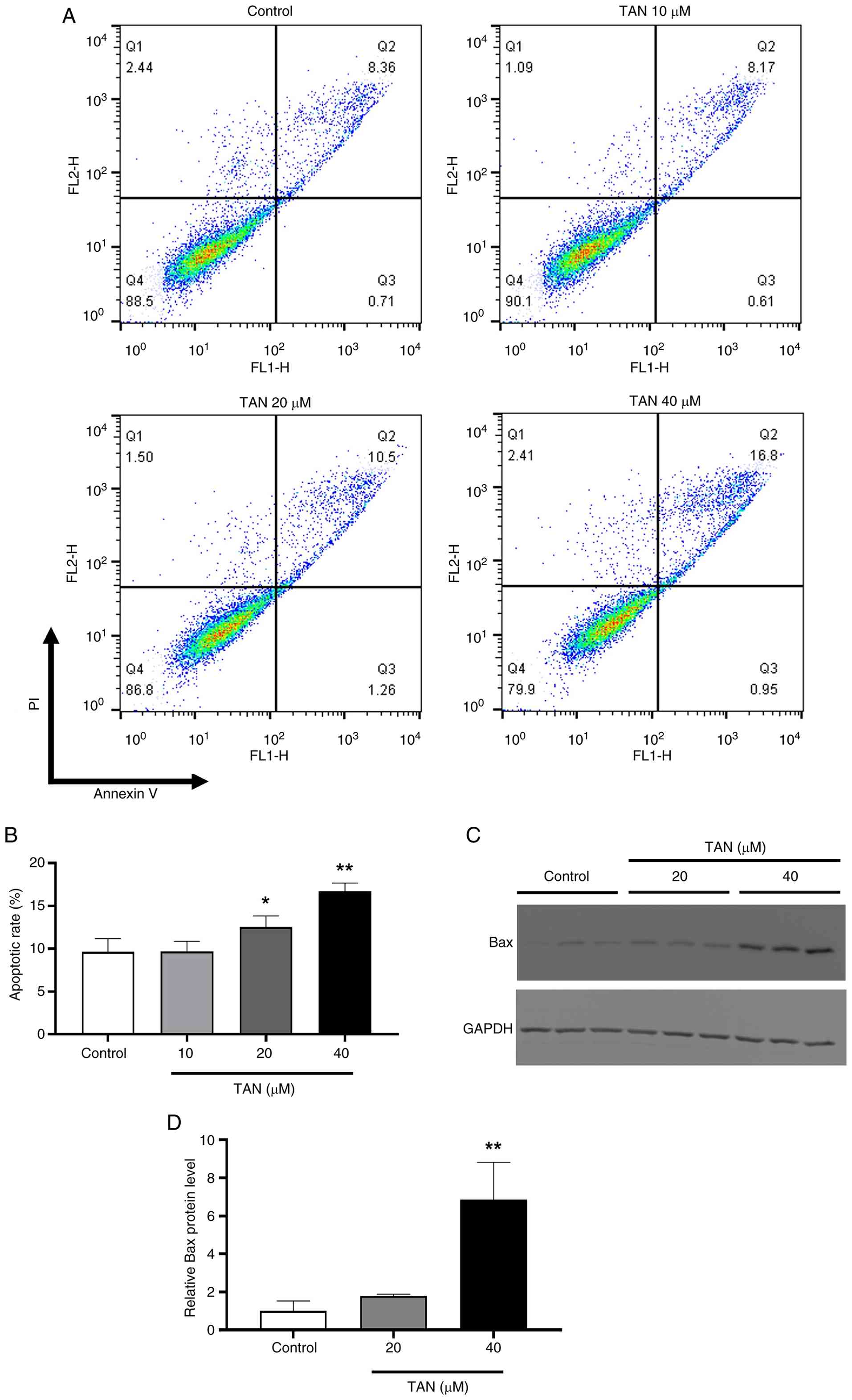
programmed cell death (19,26). In the present study, apoptotic cells
were analyzed by flow cytometry using Annexin V and PI staining
(Fig. 4A). The proportion of
apoptotic cells was significantly increased in the 20 and 40 µM
TAN-treated groups compared with in the control group (Fig. 4B). Subsequently, the expression
levels of the pro-apoptotic protein Bax were examined by western
blotting (Fig. 4C). Bax expression
was significantly upregulated in the 40 µM TAN-treated group
compared with in the control group (Fig. 4D). These results suggested that TAN
treatment may be associated with apoptosis-related changes and
increased Bax expression in CaSki cells, potentially involving the
intrinsic apoptosis-associated signaling pathway.
TAN increases mitochondrial oxidative
stress and decreases MMP in CaSki cells
TAN has been reported to exhibit various anticancer
effects by increasing ROS levels in various cancer cell lines
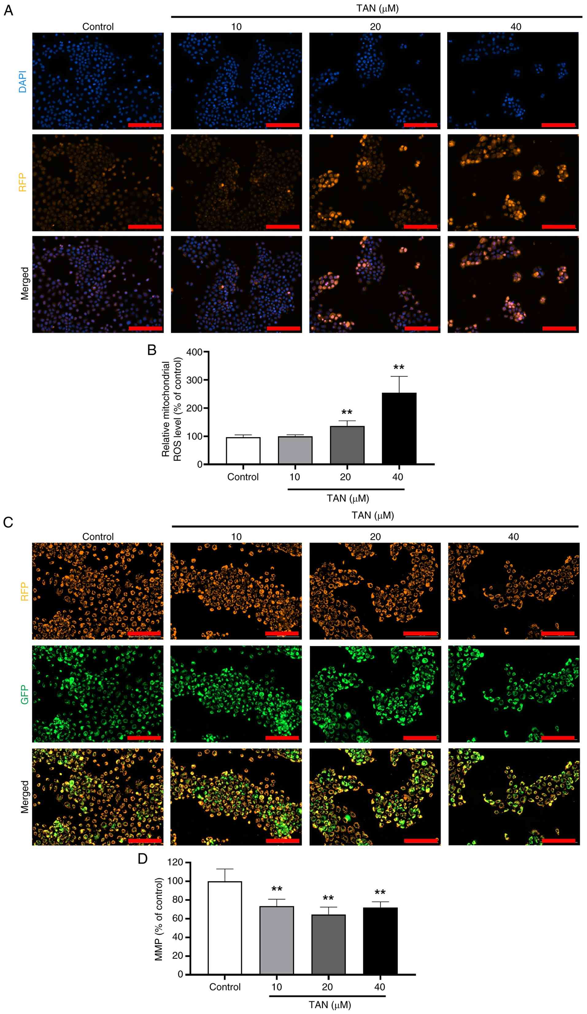
(18). The present study performed
MitoSOX staining to evaluate whether TAN increases mitochondrial
ROS (Fig. 5A), and observed
elevated intramitochondrial ROS levels in the 20 and 40 µM
TAN-treated groups compared with in the control group (Fig. 5B). Based on reports that excessive
mitochondrial ROS in cancer cells induces changes in MMP (29), the current study further evaluated
changes in MMP using JC-1 staining (Fig. 5C). The decrease in MMP was confirmed
in the TAN-treated groups compared with in the control group
(Fig. 5D). These findings suggested
that TAN treatment is associated with increased mitochondrial ROS
accumulation and decreased MMP in CaSki cells.
TAN upregulates the expression levels
of a tumor suppressor protein in CaSki cells
The tumor suppressor protein p53 is frequently
inactivated or dysregulated in various cancer cell lines, and
previous studies have reported that phytochemicals can modulate the
expression of tumor suppressor proteins, including p53 (30–32).
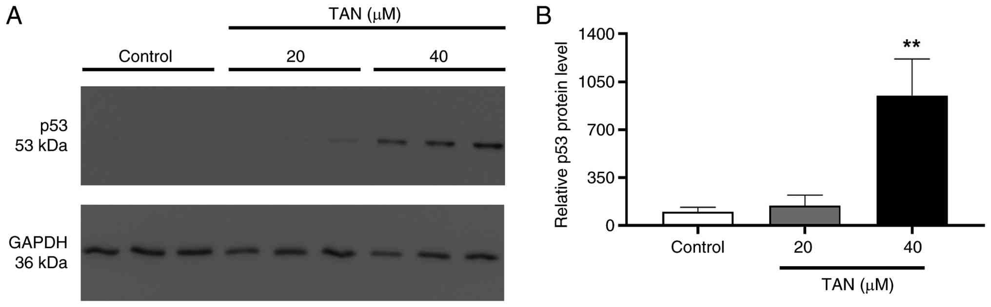
To evaluate the effect of TAN on p53 expression in CaSki cells, p53
protein levels were analyzed by western blotting (Fig. 6A). TAN treatment at 40 µM
significantly increased p53 protein expression compared with that
in the control group (Fig. 6B).
These findings suggested that TAN treatment may be associated with
increased p53 expression in CaSki cells.
Discussion
Phytochemicals, such as polyphenols, alkaloids and
flavonoids, are plant-derived metabolites reported to exhibit a
variety of bioactivities, including anticancer, antioxidant and
anti-inflammatory effects (23,33,34).
TAN is a methyl flavone-based phytochemical reported to have
anticancer effects in previous studies (19,35,36).
However, the anticancer effects of TAN in cervical cancer are still
unclear and require further research. Therefore, the current study
investigated the potential of TAN in the treatment of CaSki
cervical cancer cells.
The present study demonstrated that TAN
significantly reduced the viability and proliferation of CaSki
cells, suggesting the induction of physiological and biochemical
alterations in CaSki cells. The observed G1 phase arrest
induced by TAN was consistent with the findings of previous studies
conducted in colon and breast cancer cells (37,38),
suggesting that TAN may suppress the proliferation of CaSki cells
through regulation of cell cycle progression. In addition, TAN
upregulated the expression of the epithelial marker E-cadherin and
reduced the migratory capacity of CaSki cells. Increased E-cadherin
expression may be associated with suppression of
epithelial-mesenchymal transition (EMT) through modulation of
β-catenin-related signaling pathways, thereby reducing
migration-associated phenotypic changes and metastatic potential
(39,40). EMT is also associated with various
malignant characteristics, including cancer stem cell (CSC)
properties and resistance to apoptosis (41,42).
Therefore, further studies are required to determine whether TAN
influences additional EMT-associated processes, including CSC
properties and apoptosis resistance.
Previous studies have reported that TAN can induce
apoptosis in various cancer cell types through mitochondrial
dysfunction and ROS-associated signaling pathways (19,26).
Intrinsic apoptosis is triggered by mitochondrial dysfunction
resulting from an imbalance in Bax/Bcl-2 expression and loss of
MMP, leading to cytochrome c release (43,44).
Increased mitochondrial ROS levels have also been recognized as an
important factor associated with activation of the intrinsic
apoptotic pathway (45,46). In the present study, increased Bax
expression, elevated mitochondrial ROS levels and decreased MMP
were observed following TAN treatment, suggesting the involvement
of intrinsic apoptosis-associated mitochondrial dysfunction in
CaSki cells. However, additional studies investigating intrinsic
apoptosis-related signaling molecules, including caspase activation
and cytochrome c release, are required to further clarify
the underlying mechanisms. Furthermore, mitochondrial dysfunction
has been reported to be closely associated with impaired electron
transport chain activity, reduced adenosine triphosphate (ATP)
production and alterations in mitochondria-dependent respiratory
function (47). Therefore, further
studies are needed to elucidate the effects of TAN on ATP
production, mitochondrial metabolism and respiratory regulation in
cancer cells.
Tumor suppressor proteins are frequently inactivated
or functionally altered in numerous cancer cells and have therefore
been extensively investigated as therapeutic targets (48). Among them, p53 and retinoblastoma
proteins are known to be functionally suppressed by HPV
oncoproteins, which serve a central role in the initiation and
progression of cervical cancer (4,49,50).
Therefore, modulation of tumor suppressor proteins such as p53 has
been considered an important therapeutic target in cervical cancer
research (51,52). In the present study, TAN treatment
significantly increased p53 protein levels in CaSki cells; however,
the precise mechanisms responsible for p53 regulation were not
elucidated. In particular, changes in HPV16 E6/E7 mRNA and protein
expression levels were not evaluated, and cycloheximide chase
experiments were not performed to determine whether TAN influences
p53 protein stability through modulation of E6-mediated p53
degradation. Furthermore, p53 transcriptional activity and
downstream target genes associated with cell cycle arrest and
apoptosis, including p21, PUMA and NOXA, were not investigated.
Functional validation experiments using p53 inhibitors, such as
pifithrin-α or p53-specific small interfering RNA knockdown were
also not performed. Therefore, it remains unclear as to whether the
anticancer effects induced by TAN are directly dependent on p53
activation or whether TAN regulates HPV16 E6/E7 expression or
activity to promote p53 stabilization. Further mechanistic studies
are required to clarify the molecular mechanisms underlying
TAN-induced p53 upregulation and to determine the functional
contribution of p53 signaling to the anticancer effects of TAN.
Although TAN-induced G1 phase arrest was
observed in the present study, the molecular mechanisms underlying
this effect were not fully investigated. In particular, key
G1/S transition-related regulatory proteins, including
cyclin D1, CDK4/6, p21 and p27, were not evaluated. Therefore,
additional studies are required to clarify the detailed molecular
pathways associated with TAN-induced cell cycle arrest in cervical
cancer cells. In addition, although increased mitochondrial ROS
levels, Bax upregulation and decreased MMP were observed following
TAN treatment, the present study did not directly establish ROS as
a necessary upstream mediator of apoptosis. Rescue experiments
using ROS scavengers, such as N-acetylcysteine or Mito-TEMPO, were
not performed. Furthermore, apoptosis-related signaling molecules,
including Bcl-2, cleaved caspase-3, cleaved caspase-9 and
cytochrome c release, were not evaluated. Therefore,
additional mechanistic studies are required to further clarify the
ROS-associated intrinsic apoptotic pathways induced by TAN. In
addition, given that the present study was limited to a single
cervical cancer cell line, further investigations using multiple
cervical cancer models with distinct biological characteristics,
together with normal cervical epithelial cells, are warranted to
establish the broader applicability and therapeutic potential of
TAN. Moreover, although the present study demonstrated the
anticancer potential of TAN in cervical cancer cells, its
translational relevance remains to be further established. The
concentrations of TAN used in vitro may not be
physiologically achievable in vivo owing to pharmacokinetic
limitations, including low bioavailability and limited systemic
exposure (53). Therefore, further
studies using appropriate animal models are required to evaluate
the in vivo antitumor efficacy, systemic toxicity and
pharmacokinetic profile of TAN under physiologically relevant
conditions. A previous study suggested that TAN may exhibit
relatively improved solubility and pharmacokinetic properties
compared with other PMFs, including nobiletin, due to its
structural characteristics (54).
Nevertheless, TAN still possesses pharmacokinetic limitations,
including relatively low bioavailability, which may restrict its
clinical applicability. To overcome these limitations, studies have
investigated various drug delivery systems, including
self-microemulsifying drug delivery systems, nanoemulsions and
nanoparticle-based formulations, to improve the stability and
bioavailability of TAN, suggesting their potential utility for
enhancing the therapeutic efficacy of TAN (53,55,56).
Therefore, the incorporation of advanced drug delivery strategies
may represent a promising approach to improve the translational
potential and therapeutic applicability of TAN in cervical
cancer.
Although several limitations remain to be addressed,
the present study demonstrated that TAN treatment was associated
with reduced proliferation, apoptosis-related changes and
mitochondrial dysfunction in CaSki cervical cancer cells. These
findings provide preliminary evidence supporting the potential
anticancer activity of TAN, and may serve as a foundation for
future mechanistic, pharmacological and preclinical studies in
cervical cancer.
Acknowledgements
Not applicable.
Funding
This research was supported by the Regional Innovation System
& Education (RISE) program through the Chungbuk Regional
Innovation System & Education Center, funded by the Ministry of
Education and the provincial government of Chungcheongbuk-do,
Republic of Korea (grant no. 2025-RISE-11-014-03) and by Chungbuk
National University BK21 program (2026). In addition, this work was
supported by the National Research Foundation of Korea (NRF) grant
funded by the Korean government (grant no. RS-2026-25486352) and
the Sejong Fellowship through the NRF funded by the Ministry of
Science and ICT (grant no. RS-2025-00557567).
Availability of data and materials
The data generated in the present study may be
requested from the corresponding author.
Authors' contributions
HKL and KCC conceptualized the study. SHA, ZAB and
HN developed the experimental methodology. HKL, ZAB and HN
validated the experimental results. SHA performed the formal
analysis. SHA, ZAB and HN conducted the investigation. SHA, ZAB and
HN curated and organized the experimental data. SHA and ZAB wrote
the original draft of the manuscript. HKL and KCC reviewed and
edited the manuscript. SHA and ZAB prepared the figures and
visualized the data. KCC supervised the study. HKL and KCC
administered the project. KCC acquired the funding. SHA, ZAB and
KCC confirm the authenticity of all the raw data. All authors read
and approved the final manuscript.
Ethics approval and consent to
participate
Not applicable.
Patient consent for publication
Not applicable.
Competing interests
The authors declare that they have no competing
interests.
Glossary
Abbreviations
Abbreviations:
|
DMSO
|
dimethyl sulfoxide
|
|
EMT
|
epithelial-mesenchymal transition
|
|
FBS
|
fetal bovine serum
|
|
HPV
|
human papillomavirus
|
|
MMP
|
mitochondrial membrane potential
|
|
PI
|
propidium iodide
|
|
PFA
|
paraformaldehyde
|
|
RPMI
|
Roswell Park Memorial Institute
|
|
ROS
|
reactive oxygen species
|
|
TAN
|
tangeretin
|
References
|
1
|
Bray F, Laversanne M, Sung H, Ferlay J,
Siegel RL, Soerjomataram I and Jemal A: Global cancer statistics
2022: GLOBOCAN estimates of incidence and mortality worldwide for
36 cancers in 185 countries. CA Cancer J Clin. 74:229–263.
2024.PubMed/NCBI
|
|
2
|
Áyen Á, Jiménez Martínez Y and Boulaiz H:
Targeted gene delivery therapies for cervical cancer. Cancers
(Basel). 12:13012020. View Article : Google Scholar : PubMed/NCBI
|
|
3
|
Yan H, Wang P, Yang F, Cheng W, Chen C,
Zhai B and Zhou Y: Anticancer therapy-induced adverse drug
reactions in children and preventive and control measures. Front
Pharmacol. 15:13292202024. View Article : Google Scholar : PubMed/NCBI
|
|
4
|
Ruttkay-Nedecky B, Jimenez Jimenez AM,
Nejdl L, Chudobova D, Gumulec J, Masarik M, Adam V and Kizek R:
Relevance of infection with human papillomavirus: the role of the
p53 tumor suppressor protein and E6/E7 zinc finger proteins
(review). Int J Oncol. 43:1754–1762. 2013. View Article : Google Scholar : PubMed/NCBI
|
|
5
|
Chew YC, Adhikary G, Wilson GM, Xu W and
Eckert RL: Sulforaphane induction of p21(Cip1) cyclin-dependent
kinase inhibitor expression requires p53 and Sp1 transcription
factors and is p53-dependent. J Biol Chem. 287:16168–16178. 2012.
View Article : Google Scholar : PubMed/NCBI
|
|
6
|
Fu H, Wang C, Yang D, Wei Z, Xu J, Hu Z,
Zhang Y, Wang W, Yan R and Cai Q: Curcumin regulates proliferation,
autophagy, and apoptosis in gastric cancer cells by affecting PI3K
and P53 signaling. J Cell Physiol. 233:4634–4642. 2018. View Article : Google Scholar : PubMed/NCBI
|
|
7
|
Younas M, Hano C, Giglioli-Guivarc'h N and
Abbasi BH: Mechanistic evaluation of phytochemicals in breast
cancer remedy: Current understanding and future perspectives. RSC
Adv. 8:29714–29744. 2018. View Article : Google Scholar : PubMed/NCBI
|
|
8
|
Hamdan AME, Alharthi FHJ, Alanazi AH,
El-Emam SZ, Zaghlool SS, Metwally K, Albalawi SA, Abdu YS, Mansour
RE, Salem HA, et al: Neuroprotective effects of phytochemicals
against aluminum chloride-induced Alzheimer's disease through
ApoE4/LRP1, Wnt3/β-Catenin/GSK3β, and TLR4/NLRP3 pathways with
physical and mental activities in a rat model. Pharmaceuticals
(Basel). 15:10082022. View Article : Google Scholar : PubMed/NCBI
|
|
9
|
Sohel M, Aktar S, Biswas P, Amin MA,
Hossain MA, Ahmed N, Mim MIH, Islam F and Mamun AA: Exploring the
anti-cancer potential of dietary phytochemicals for the patients
with breast cancer: A comprehensive review. Cancer Med.
12:14556–14583. 2023. View Article : Google Scholar : PubMed/NCBI
|
|
10
|
Paudel S, Mishra N and Agarwal R:
Phytochemicals as immunomodulatory molecules in cancer
therapeutics. Pharmaceuticals (Basel). 16:16522023. View Article : Google Scholar : PubMed/NCBI
|
|
11
|
Chen S, Wang X, Cheng Y, Gao H and Chen X:
A review of classification, biosynthesis, biological activities and
potential applications of flavonoids. Molecules. 28:49822023.
View Article : Google Scholar : PubMed/NCBI
|
|
12
|
Hosseinzadeh A, Poursoleiman F, Biregani
AN and Esmailzadeh A: Flavonoids target different molecules of
autophagic and metastatic pathways in cancer cells. Cancer Cell
Int. 23:1142023. View Article : Google Scholar : PubMed/NCBI
|
|
13
|
Kopustinskiene DM, Jakstas V, Savickas A
and Bernatoniene J: Flavonoids as anticancer agents. Nutrients.
12:4572020. View Article : Google Scholar : PubMed/NCBI
|
|
14
|
An JP, Liu X, Kim D, Madden R and Wang Y:
Comprehensive analysis of polymethoxyflavone metabolism in orange
peel using an animal model. RSC Adv. 15:38189–38200. 2025.
View Article : Google Scholar : PubMed/NCBI
|
|
15
|
Wang Y, Mou Y, Lu S, Xia Y and Cheng B:
Polymethoxylated flavonoids in citrus fruits: Absorption,
metabolism, and anticancer mechanisms against breast cancer. PeerJ.
12:e167112024. View Article : Google Scholar : PubMed/NCBI
|
|
16
|
Raza W, Luqman S and Meena A: Prospects of
tangeretin as a modulator of cancer targets/pathways. Pharmacol
Res. 161:1052022020. View Article : Google Scholar : PubMed/NCBI
|
|
17
|
Hung WL, Chang WS, Lu WC, Wei GJ, Wang Y,
Ho CT and Hwang LS: Pharmacokinetics, bioavailability, tissue
distribution and excretion of tangeretin in rat. J Food Drug Anal.
26:849–857. 2018. View Article : Google Scholar : PubMed/NCBI
|
|
18
|
Dey DK, Chang SN, Vadlamudi Y, Park JG and
Kang SC: Synergistic therapy with tangeretin and 5-fluorouracil
accelerates the ROS/JNK mediated apoptotic pathway in human
colorectal cancer cell. Food Chem Toxicol. 143:1115292020.
View Article : Google Scholar : PubMed/NCBI
|
|
19
|
Dong Y, Cao A, Shi J, Yin P, Wang L, Ji G,
Xie J and Wu D: Tangeretin, a citrus polymethoxyflavonoid, induces
apoptosis of human gastric cancer AGS cells through extrinsic and
intrinsic signaling pathways. Oncol Rep. 31:1788–1794. 2014.
View Article : Google Scholar : PubMed/NCBI
|
|
20
|
Xie Y, Feng SL, He F, Yan PY, Yao XJ, Fan
XX, Leung EL and Zhou H: Down-regulating Nrf2 by tangeretin
reverses multiple drug resistance to both chemotherapy and EGFR
tyrosine kinase inhibitors in lung cancer. Pharmacol Res.
186:1065142022. View Article : Google Scholar : PubMed/NCBI
|
|
21
|
Varankar SS and Bapat SA: Migratory
metrics of wound healing: A quantification approach for in vitro
scratch assays. Front Oncol. 8:6332018. View Article : Google Scholar : PubMed/NCBI
|
|
22
|
Ibrahim SM, Bakhashab S, Ilyas AM,
Pushparaj PN, Karim S, Khan JA, Abuzenadah AM, Chaudhary AG,
Al-Qahtani MH and Ahmed F: WYE-354 restores Adriamycin sensitivity
in multidrug-resistant acute myeloid leukemia cell lines. Oncol
Rep. 41:3179–3188. 2019.PubMed/NCBI
|
|
23
|
Atchan APN, Monthe OC, Tchamgoue AD, Singh
Y, Shivashankara ST, Selvi MK, Agbor GA, Magni P, Piazza S,
Manjappara UV, et al: Anti-inflammatory, antioxidant activities,
and phytochemical characterization of edible plants exerting
synergistic effects in human gastric epithelial cells. Antioxidants
(Basel). 12:5912023. View Article : Google Scholar : PubMed/NCBI
|
|
24
|
Lin JJ, Huang CC, Su YL, Luo HL, Lee NL,
Sung MT and Wu YJ: Proteomics analysis of tangeretin-induced
apoptosis through mitochondrial dysfunction in bladder cancer
cells. Int J Mol Sci. 20:10172019. View Article : Google Scholar : PubMed/NCBI
|
|
25
|
Zhang X, Zheng L, Sun Y, Wang T and Wang
B: Tangeretin enhances radiosensitivity and inhibits the
radiation-induced epithelial-mesenchymal transition of gastric
cancer cells. Oncol Rep. 34:302–310. 2015. View Article : Google Scholar : PubMed/NCBI
|
|
26
|
Jeoung YH, Lee HK, Ahn SH, Bhutta ZA and
Choi KC: Tangeretin induces apoptosis and cell cycle arrest in
thyroid cancer cells. Toxicol Res. 41:477–488. 2025. View Article : Google Scholar : PubMed/NCBI
|
|
27
|
Surichan S, Arroo RR, Tsatsakis AM and
Androutsopoulos VP: Tangeretin inhibits the proliferation of human
breast cancer cells via CYP1A1/CYP1B1 enzyme induction and
CYP1A1/CYP1B1-mediated metabolism to the product 4′ hydroxy
tangeretin. Toxicol In Vitro. 50:274–284. 2018. View Article : Google Scholar : PubMed/NCBI
|
|
28
|
Schegoleva AA, Khozyainova AA,
Gerashchenko TS, Zhuikova LD and Denisov EV: Metastasis prevention:
Targeting causes and roots. Clin Exp Metastasis. 39:505–519. 2022.
View Article : Google Scholar : PubMed/NCBI
|
|
29
|
Ting YW, Chiou YS, Pan MH, Ho CT and Huang
QR: In vitro and in vivo anti-cancer activity of tangeretin against
colorectal cancer was enhanced by emulsion-based delivery system. J
Funct Foods. 15:264–273. 2015. View Article : Google Scholar
|
|
30
|
Li X, Li Q, Huang Y, Zhou H, Yang Q and
Kong L: Quercetin, a natural dietary flavonoid, emerges a novel
USP7 inhibitor with anti-colorectal cancer effects. Biol Pharm
Bull. 48:1485–1492. 2025. View Article : Google Scholar : PubMed/NCBI
|
|
31
|
Kang KA, Piao MJ, Hyun YJ, Zhen AX, Cho
SJ, Ahn MJ, Yi JM and Hyun JW: Luteolin promotes apoptotic cell
death via upregulation of Nrf2 expression by DNA demethylase and
the interaction of Nrf2 with p53 in human colon cancer cells. Exp
Mol Med. 51:1–14. 2019. View Article : Google Scholar
|
|
32
|
Jung EJ, Lee WS, Paramanantham A, Kim HJ,
Shin SC, Kim GS, Jung JM, Ryu CH, Hong SC, Chung KH and Kim CW: p53
Enhances Artemisia annua L. Polyphenols-induced cell death through
upregulation of p53-dependent targets and cleavage of PARP1 and
lamin A/C in HCT116 colorectal cancer cells. Int J Mol Sci.
21:93152020. View Article : Google Scholar : PubMed/NCBI
|
|
33
|
Alshehri A, Ahmad A, Tiwari RK, Ahmad I,
Alkhathami AG, Alshahrani MY, Asiri MA, Almeleebia TM, Saeed M,
Yadav DK and Ansari IA: In vitro evaluation of antioxidant,
anticancer, and anti-inflammatory activities of ethanolic leaf
extract of Adenium obesum. Front Pharmacol. 13:8475342022.
View Article : Google Scholar : PubMed/NCBI
|
|
34
|
Pandey BP, Adhikari K, Pradhan SP, Shin
HJ, Lee EK and Jung HJ: In-vitro antioxidant, anti-cancer, and
anti-inflammatory activities of selected medicinal plants from
western Nepal. Futur J Pharm Sci. 6:752020. View Article : Google Scholar
|
|
35
|
Ko YC, Choi HS, Liu R, Kim JH, Kim SL, Yun
BS and Lee DS: Inhibitory effects of tangeretin, A citrus
peel-derived flavonoid, on breast cancer stem cell formation
through suppression of Stat3 signaling. Molecules. 25:25992020.
View Article : Google Scholar : PubMed/NCBI
|
|
36
|
Zhu WB, Xiao N and Liu XJ: Dietary
flavonoid tangeretin induces reprogramming of epithelial to
mesenchymal transition in prostate cancer cells by targeting the
PI3K/Akt/mTOR signaling pathway. Oncol Lett. 15:433–440.
2018.PubMed/NCBI
|
|
37
|
Morley KL, Ferguson PJ and Koropatnick J:
Tangeretin and nobiletin induce G1 cell cycle arrest but not
apoptosis in human breast and colon cancer cells. Cancer Lett.
251:168–178. 2007. View Article : Google Scholar : PubMed/NCBI
|
|
38
|
Pan MH, Chen WJ, Lin-Shiau SY, Ho CT and
Lin JK: Tangeretin induces cell-cycle G1 arrest through inhibiting
cyclin-dependent kinases 2 and 4 activities as well as elevating
Cdk inhibitors p21 and p27 in human colorectal carcinoma cells.
Carcinogenesis. 23:1677–1684. 2002. View Article : Google Scholar : PubMed/NCBI
|
|
39
|
Hui San S and Ching Ngai S: E-cadherin
re-expression: Its potential in combating TRAIL resistance and
reversing epithelial-to-mesenchymal transition. Gene.
909:1482932024. View Article : Google Scholar : PubMed/NCBI
|
|
40
|
Lin WH, Cooper LM and Anastasiadis PZ:
Cadherins and catenins in cancer: Connecting cancer pathways and
tumor microenvironment. Front Cell Dev Biol. 11:11370132023.
View Article : Google Scholar : PubMed/NCBI
|
|
41
|
Mani SA, Guo W, Liao MJ, Eaton EN, Ayyanan
A, Zhou AY, Brooks M, Reinhard F, Zhang CC, Shipitsin M, et al: The
epithelial-mesenchymal transition generates cells with properties
of stem cells. Cell. 133:704–715. 2008. View Article : Google Scholar : PubMed/NCBI
|
|
42
|
Vega S, Morales AV, Ocaña OH, Valdés F,
Fabregat I and Nieto MA: Snail blocks the cell cycle and confers
resistance to cell death. Genes Dev. 18:1131–1143. 2004. View Article : Google Scholar : PubMed/NCBI
|
|
43
|
Mustafa M, Ahmad R, Tantry IQ, Ahmad W,
Siddiqui S, Alam M, Abbas K, Moinuddin Hassan MI, Habib S and Islam
S: Apoptosis: A comprehensive overview of signaling pathways,
morphological changes, and physiological significance and
therapeutic implications. Cells. 13:18382024. View Article : Google Scholar : PubMed/NCBI
|
|
44
|
Vogler M, Braun Y, Smith VM, Westhoff MA,
Pereira RS, Pieper NM, Anders M, Callens M, Vervliet T, Abbas M, et
al: The BCL2 family: From apoptosis mechanisms to new advances in
targeted therapy. Signal Transduct Target Ther. 10:912025.
View Article : Google Scholar : PubMed/NCBI
|
|
45
|
Heo JW, No MH, Park DH, Kang JH, Seo DY,
Han J, Neufer PD and Kwak HB: Effects of exercise on
obesity-induced mitochondrial dysfunction in skeletal muscle.
Korean J Physiol Pharmacol. 21:567–577. 2017. View Article : Google Scholar : PubMed/NCBI
|
|
46
|
Roy MJ, Vom A, Czabotar PE and Lessene G:
Cell death and the mitochondria: Therapeutic targeting of the BCL-2
family-driven pathway. Br J Pharmacol. 171:1973–1987. 2014.
View Article : Google Scholar : PubMed/NCBI
|
|
47
|
Khan T, Waseem R, Zehra Z, Aiman A,
Bhardwaj P, Ansari J, Hassan MI and Islam A: Mitochondrial
dysfunction: Pathophysiology and mitochondria-targeted drug
delivery approaches. Pharmaceutics. 14:26572022. View Article : Google Scholar : PubMed/NCBI
|
|
48
|
Wang H, Guo M, Wei H and Chen Y: Targeting
p53 pathways: Mechanisms, structures, and advances in therapy.
Signal Transduct Target Ther. 8:922023. View Article : Google Scholar : PubMed/NCBI
|
|
49
|
Rastogi N, Duggal S, Singh SK, Porwal K,
Srivastava VK, Maurya R, Bhatt ML and Mishra DP: Proteasome
inhibition mediates p53 reactivation and anti-cancer activity of
6-gingerol in cervical cancer cells. Oncotarget. 6:43310–43325.
2015. View Article : Google Scholar : PubMed/NCBI
|
|
50
|
Zhao X, Sun W, Ren Y and Lu Z: Therapeutic
potential of p53 reactivation in cervical cancer. Crit Rev Oncol
Hematol. 157:1031822021. View Article : Google Scholar : PubMed/NCBI
|
|
51
|
Wang JCK, Baddock HT, Mafi A, Foe IT,
Bratkowski M, Lin TY, Jensvold ZD, Preciado López M, Stokoe D,
Eaton D, et al: Structure of the p53 degradation complex from
HPV16. Nat Commun. 15:18422024. View Article : Google Scholar : PubMed/NCBI
|
|
52
|
Zou J, Li Y, Chen T and Zhu C: An
E7-retinoblastoma protein pathway mechanism may account for the
higher carcinogenic ability of HPV16 over HPV58 in cervical cancer.
Transl Cancer Res. 13:1876–1886. 2024. View Article : Google Scholar : PubMed/NCBI
|
|
53
|
Wang W, Jia T, Zhang Y and Zhou Y:
Progress of researches on pharmacological effects and
bioavailability of tangeretin. J Oleo Sci. 74:13–23. 2025.
View Article : Google Scholar : PubMed/NCBI
|
|
54
|
Woo SH, Park IC, Park MJ, Lee HC, Lee SJ,
Chun YJ, Lee SH, Hong SI and Rhee CH: Arsenic trioxide induces
apoptosis through a reactive oxygen species-dependent pathway and
loss of mitochondrial membrane potential in HeLa cells. Int J
Oncol. 21:57–63. 2002.PubMed/NCBI
|
|
55
|
Elhennawy MG and Lin HS: Determination of
tangeretin in rat plasma: Assessment of its clearance and absolute
oral bioavailability. Pharmaceutics. 10:32017. View Article : Google Scholar : PubMed/NCBI
|
|
56
|
Hu Y, Liu F, Pang J, McClements DJ, Zhou
Z, Li B and Li Y: Biopolymer additives enhance tangeretin
bioavailability in emulsion-based delivery systems: an in vitro and
in vivo study. J Agric Food Chem. 69:730–740. 2021. View Article : Google Scholar : PubMed/NCBI
|