Introduction
Currently, the incidence and mortality of malignant
tumors continue to rise globally (1,2),
imposing a substantial burden on the global healthcare systems
(1,3). Immunity refers to a physiological
surveillance mechanism whereby organisms distinguish self from
non-self antigens, eliminate exogenous pathogens and aberrant
cells, and maintain internal environmental homeostasis via immune
responses. The immune microenvironment constitutes a dynamic
regulatory network composed of immune cells, non-immune stromal
cells, extracellular matrix, and soluble mediators. This network
not only mediates canonical immune reactions but also orchestrates
immune homeostasis through immunosuppressive cell populations and
paracrine factors. Under physiological conditions, the immune
microenvironment recognizes and eliminates nascent tumor cells. In
tumor-bearing tissues, however, malignant cells upregulate
immunosuppressive mediators, reprogram immune cell lineages and
metabolic circuits, and remodel the local niche into a tumor immune
microenvironment (TIME) characterized by functional dysfunction and
dominant immune suppression. This transformation enables tumor
cells to evade immune surveillance and clearance (4,5). The
biological features of the TIME display substantial heterogeneity,
and the regulatory functions of each constituent dynamically shift
throughout disease progression. This highly complex
microenvironment plays a pivotal role in shaping tumor therapeutic
responses to immunotherapies.
Connective tissue growth factor (CTGF), also termed
cellular communication network factor 2 (CCN2), is a core member of
the cellular communication network (CCN) matricellular protein
family. Mounting evidence implicates CTGF in the recruitment and
phenotypic polarization of immune cells, including driving M1-to-M2
macrophage polarization (6,7). Moreover, CTGF drives fibroblast
activation and extracellular matrix (ECM) remodeling, enabling
these stromal populations to assemble physical tissue barriers
(8,9). In addition, CTGF collaborates with
regulatory cytokines, including interleukin (IL)-6 (10), IL-8 (11), tumor necrosis factor (TNF)-α
(7), and transforming growth factor
(TGF)-β (12), to collectively
modulate the reprogramming of the immune microenvironment,
facilitate tumor immune evasion, and contribute to the initiation
and progression of malignant tumors. Thus, the pleiotropic effects
of CTGF within the TIME establish CTGF as a central modulator of
tumor immune escape and immunotherapy resistance. Several recent
reviews have summarized CTGF/CCN2 from the perspectives of
molecular structure, gene regulation, disease-associated signaling,
drug discovery, broad therapeutic targeting, or cancer progression
in specific tumor types (13–16).
However, these studies have not systematically distinguished how
CTGF may coordinate the structural, signaling, and immune
dimensions of the tumor immune microenvironment. In particular, the
potential links among CTGF-driven ECM remodeling, stromal
stiffening, vascular aberrancy, cancer-associated fibroblast (CAF)
activation, immune-cell spatial exclusion, cytokine-network
remodeling, and immunotherapy resistance remain insufficiently
integrated.
In the present review, the core signaling axes and
intercellular crosstalk mechanisms governing CTGF-dependent TIME
remodeling are systematically summarized, translatable therapeutic
strategies and unresolved research gaps are elaborated, and the
cell-type-specific molecular and cellular cascades through which
CTGF synergistically promotes tumor immune evasion are dissected,
with the aim of identifying novel combinatorial therapeutic targets
to enhance the efficacy of cancer immunotherapies. Compared with
previous CTGF/CCN2 reviews (13–16),
the major incremental contribution of the present review is the
construction of a TIME-centered ‘structure-signal-immunity’
framework. This framework organizes available evidence into four
interconnected layers: Tumor-cell phenotypic remodeling, ECM and
vascular remodeling, immune-cell functional reprogramming, and
cytokine-network remodeling. By doing so, this review shifts the
discussion of CTGF from a general profibrotic or tumor-promoting
molecule toward a context-dependent regulatory node that may shape
immune exclusion and therapeutic resistance within the TIME. The
mechanisms that are experimentally supported, those that remain
inferential, and those that should be prioritized for spatially
resolved, tumor-type-specific, and immunotherapy-oriented
validation are further highlighted.
CTGF regulates tumor cell phenotype in
TIME
Tumor cells are central contributors to the
formation and remodeling of the TIME. Tumor stem cells with
stem-like properties contribute to tumor initiation, recurrence,
and metastasis by secreting immunosuppressive factors, recruiting
suppressive immune cells, and expressing immune checkpoint
molecules, thereby limiting effector immune-cell infiltration and
promoting an immunosuppressive TIME (17). As a multifunctional matricellular
protein, CTGF is associated with malignant tumor-cell phenotypes in
several settings (18) and may
contribute to TIME remodeling during tumor progression (Fig. 1).
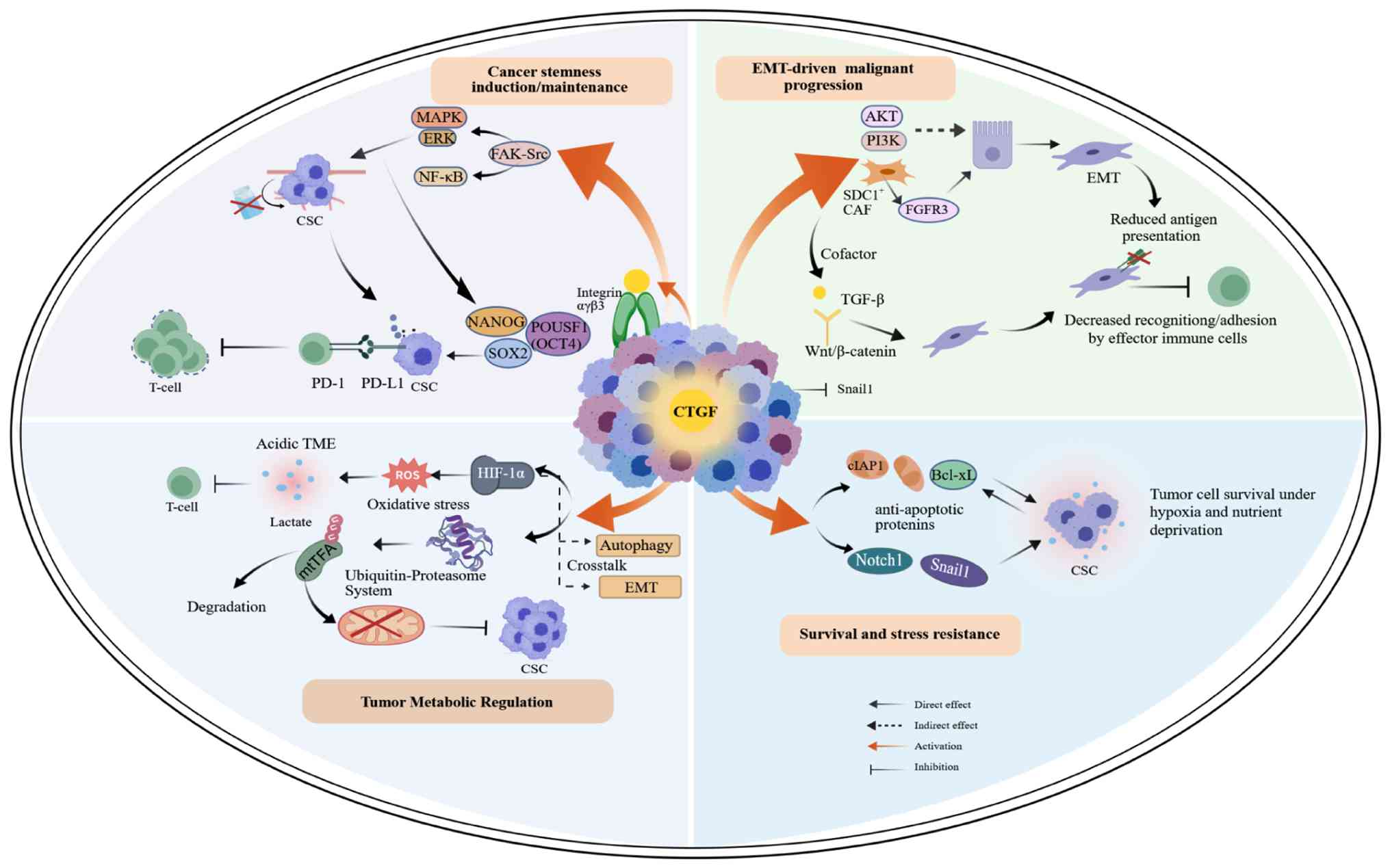 | Figure 1.CTGF modulates tumor-cell phenotypes
associated with CSC maintenance, EMT plasticity, metabolic
reprogramming, and stress resistance, thereby contributing to an
immunosuppressive TIME. CTGF regulates CSC properties through
integrin αvβ3/FAK-Src-NF-κB and ERK/MAPK signaling; EMT through
PI3K/AKT and CAF-derived CTGF-FGFR3 signaling; metabolism through
two opposing regulatory axes: HIF-1α-driven glycolysis and
mtTFA-mediated mitochondrial suppression; and cell survival through
Bcl-xL, cIAP1, Notch1, and Snail1. CSC, cancer stem cell; EMT,
epithelial-mesenchymal transition; TIME, tumor immune
microenvironment; ERK/MAPK, extracellular signal-regulated
kinase/mitogen-activated protein kinase; PI3K/AKT,
phosphatidylinositol 3-kinase/protein kinase B; HIF-1α,
hypoxia-inducible factor-1α; mtTFA, mitochondrial transcription
factor A; Notch1/Snail1, Notch homolog 1/Snail family
transcriptional repressor 1; FAK, focal adhesion kinase; Src, SRC
proto-oncogene, non-receptor tyrosine kinase; NF-κB, nuclear
factor-κB; FGFR3, fibroblast growth factor receptor 3; OSCC, oral
squamous cell carcinoma; Bcl-xL, B-cell lymphoma-extra large;
cIAP1, cellular inhibitor of apoptosis protein 1; SOX2, SRY-box
transcription factor 2; POUSF1, POU class 5 homeobox 1; PD-1,
programmed cell death protein 1; PD-L1, programmed death-ligand 1;
ROS, reactive oxygen species; CAF, cancer-associated fibroblast;
SDC1, syndecan-1. |
CTGF-mediated induction and
maintenance of cancer stem cell (CSC) properties
CSCs, characterized by self-renewal capacity and
therapeutic resistance, are considered major contributors to tumor
recurrence and metastasis (19). In
addition, CSCs highly express immune checkpoint molecules such as
programmed death-ligand 1 (PD-L1) and actively secrete
immunosuppressive factors, which directly inhibit T-cell function
or mediate immune tolerance. Thus, CSCs participate in TIME shaping
and act as a core driver of tumor immune evasion (20–23).
It has been demonstrated that CTGF binds to its
receptor integrin αvβ3 to activate the downstream focal adhesion
kinase-SRC proto-oncogene, non-receptor tyrosine kinase
(SRC)-nuclear factor-κB (NF-κB) and extracellular signal-regulated
kinase/mitogen-activated protein kinase (ERK/MAPK) signaling
pathways (14,20,21).
CTGF may contribute to the upregulation of stemness-associated
transcription factors, including Nanog homeobox, SRY-box
transcription factor 2, and POU class 5 homeobox 1 (22), which may maintain CSC-like
properties and enhance tumor invasive potential under specific
microenvironmental conditions.
This mechanism is supported by functional in
vitro pathway activation research (24). However, the extent to which CTGF
directly maintains CSC stemness in vivo remains incompletely
validated. Further clarification is needed to determine whether
CTGF regulates CSC properties directly or through secondary changes
in the TIME.
CTGF-driven epithelial-mesenchymal
transition (EMT) mediates malignant progression of tumor cells
CTGF has been implicated in the regulation of EMT
(25–27). However, most evidence is
context-dependent and derived from pathway activation studies,
rather than direct lineage-tracing or EMT-reversal experiments
(28,29). Rather than acting as a uniformly
pro-EMT factor, CTGF may modulate EMT through both tumor
cell-intrinsic signaling and stromal paracrine mechanisms.
Indirect evidence suggests that CTGF may promote EMT
through phosphatidylinositol 3-kinase (PI3K)/protein kinase B (AKT)
signaling. CTGF-mediated PI3K/AKT activation has been validated in
retinal pigment epithelial cells (30). Although PI3K/AKT-dependent EMT has
been widely reported, CTGF-specific causal induction of EMT across
tumor types remains largely inferential (31–34).
In addition, syndecan-1 (SDC1)+ CAF-derived CTGF has been shown to
activate fibroblast growth factor receptor 3 (FGFR3) signaling in
tumor cells, thereby driving EMT progression (26). Following EMT, tumor cells have been
reported to exhibit reduced antigen-presentation capacity and
diminished recognition or adhesion by effector immune cells. These
changes may enhance phenotypic plasticity and immune evasion
potential, while altered secretory profiles may contribute to TIME
remodeling (35).
Notably, emerging evidence indicates that CTGF may
exert context-dependent or even opposing roles in EMT regulation.
In epithelial ovarian cancer cells, CTGF expression was enriched in
epithelial-like cells but was reduced or undetectable in
mesenchymal-like cells. CTGF knockout induced early EMT-associated
changes, including Snail upregulation, cytoskeletal reorganization,
ECM remodeling, increased cell stiffness, anoikis resistance, and
enhanced invasiveness, while Twist1 remained inactive, suggesting a
partial or intermediate epithelial-mesenchymal state rather than
complete EMT transition (28).
Consistently, recombinant CTGF partially reversed several
CTGF-loss-associated phenotypes, supporting the notion that CTGF is
required, at least in part, for maintaining epithelial architecture
and restraining early mesenchymal transformation in this context
(28). Therefore, CTGF may function
as either a pro-EMT mediator or an epithelial phenotype maintainer
depending on tumor type, cellular state, stromal context, and
upstream signaling background. These divergent findings indicate
that CTGF-related EMT regulation remains incompletely defined
across malignancies. Such bidirectional and context-specific
regulatory behavior may even lead to opposing phenotypes within the
same tumor entity. To better illustrate the heterogeneity of CTGF
functions across tumor types, key tumor-specific mechanisms covered
in the present review are summarized in Table I.
 | Table I.Context-dependent regulatory roles
and molecular mechanisms of CTGF across distinct tumor types. |
Table I.
Context-dependent regulatory roles
and molecular mechanisms of CTGF across distinct tumor types.
| Tumor type | Core mechanism and
signaling axis | Regulated
biological functions | Evidence type and
experimental design | (Refs.) |
|---|
| Epithelioid
hemangioendothelioma | CTGF acts as an
oncogenic transcriptional target of TAZ-CAMTA1, directly binds
tointegrin αIIbβ3, and activates the Ras-MAPK signaling
cascade | Maintains
anchorage-independent proliferation, mediates cell malignant
transformation and tumorigenic phenotype | In vitro
molecular and cellular functional assays (gene knockdown, pathway
activity detection, and drug inhibition assay) | (81) |
| Breast cancer
(including triple- negative subtype) | i) TNBC:
Extracellular CTGF binds to integrin αvβ3, activates the FAK/Src/
NFκB p65-Glut3 axis; ii) luminal subtype: CTGF activates the
integrin αvβ3-ERK1/2 pathway, upregulates S100A4, Bcl-xL and cIAP1;
and iii) stromal regulation: Cav1 deficiency activates the TGF-β
pathway, upregulates CTGF, induces HIF-1α-dependent metabolic
reprogramming. | i) Promotes TNBC
cell proliferation, migration, invasion, adhesion and glycolytic
metabolic reprogramming; ii) enhances breast cancer cell motility,
induces EMT and chemoresistance to doxorubicin/ paclitaxel; and
iii) drives CAF autophagy, glycolysis and senescence, metabolically
promoting tumor growth | Clinical sample
prognostic correlation analysis, in vivo tumorigenesis
assay, in vitro cell functional assay, neutralizing antibody
experiment, and gene overexpression/knockdown validation | (18,24,36,38) |
| Colorectal
cancer | CSF1R inhibitor
activates the PI3K/AKT pathway, and promotes CTGF release; CTGF
mediates CAF activation and inhibits T-cell infiltration and
function (PI3K/AKT- CTGF-CAF activation axis) | Induces
immunotherapy resistance; anti-CTGF combined with CSF1R inhibitor
and immune checkpoint therapy achieves complete tumor
regression | Imaging mass
cytometry, in vivo animal model, and combination therapy
validation experiment | (9) |
| Ovarian cancer | CTGF
deficiency/inhibition triggers cytoskeleton remodeling, ECM
reconstruction, and drives EMT initiation (CTGF
deficiency-LAMC2-FAK-PI3K-Akt-Snail-partial EMT axis) | Induces partial
EMT, gains anoikis resistance, tumorigenesis and migration
capacity, and promotes ovarian cancer metastasis | In vitro
cell functional assay, cell phenotype and ECM detection, and EMT
phenotype validation | (28) |
| Oral squamous cell
carcinoma | CTGF promotes mtTFA
ubiquitination and proteasomal degradation, and downregulates
mitochondrial function-related indicators (CTGF-mtTFA
ubiquitin-proteasome-mitochondrial function axis) | Inhibits
mitochondrial metabolism, suppresses tumor cell migration and
invasion; and mtTFA overexpression reverses the pro-metastatic
effect of CTGF |
Immunoprecipitation, proteasome inhibitor
experiment, overexpression rescue experiment, and mitochondrial
functional assay | (37) |
| Glioma/glioblastoma
multiforme | i) Cell
proliferation/drug resistance: CTGF upregulates anti-apoptotic
proteins Bcl-xL, Survivin, and Flip, and activates multiple
oncogenic pathways; ii) immune regulation: GBM cell-derived EVs
secrete hsa-miR-27a-3p, activate CTGF transcription via H3K27ac
modification (hsa-miR-27a-3p/EZH1/KDM3A/CTGF axis); and iii)
clinical correlation: CTGF is overexpressed in a MES-like glioma
subpopulation, associated with SPP1-CD44 pathway activation | i) Promotes glioma
cell proliferation, migration, in vivo tumor formation and
temozolomide resistance; ii) induces M2 polarization of TAMs,
constructs immunosuppressive TIME; and iii) associated with poor
clinical prognosis and high-risk glioma subtypes | In vitro
cell functional assay, in vivo nude mouse tumorigenesis
assay, molecular interaction validation, multi-omics analysis, and
clinical sample correlation analysis | (6,7,39) |
| HCC | i) Metastasis
regulation: CAF-derived EVs secrete CTGF, and CTGF activates the
Notch1/Snail1 signaling pathway; ii) immune regulation:
Mesenchymal-like HCC cells secrete CTGF, induce M2-TAM
polarization, and form a CTGF-M2-TAM-CCL18 positive feedback loop;
and iii) stromal activation: HCC cell-derived CTGF activates HSCs,
and forms an IL-6/STAT3 positive feedback loop | i) Promotes HCC
cell proliferation, invasion and metastasis; ii) induces M2
polarization of TAMs, and constructs an immunosuppressive TIME; and
iii) activates HSCs, promotes liver fibrosis and HCC
progression | In vitro
cell functional assay, clinical sample correlation analysis, in
vivo xenograft model, and neutralizing antibody blocking
experiment | (40,53,76) |
| Osteosarcoma | i) Angiogenesis:
CTGF activates the PLC/PKCδ pathway, upregulates Angpt2, and
negatively regulates miR-543 (CTGF-PLC/PKCδ-Angpt2/miR-543 axis);
and ii) metastasis: CTGF activates MEK/ERK pathway, downregulates
miR-519d, upregulates MMP2/3 (CTGF-MEK/ERK-miR-519d-MMP2/3
axis) | i) Promotes tumor
angiogenesis; and ii) enhances cell migration, invasion and in
vivo lung metastasis; and CTGF expression is correlated with
patient clinical stage and tumor metastasis | In vivo/in
vitro angiogenesis assay, in vitro cell functional
assay, in vivo lung metastasis model, and clinical sample
correlation analysis | (42,44) |
| GC | i) Stromal CTGF
induction: GC cell-derived TGF-β1 activates SRC kinase in CAFs,
induces CTGF transcription via the ERK/Smad2/3 pathway GC-derived
(TGF-β1 → SRC in CAFs → ERK/Smad2/3 → CTGF transcription axis); and
ii) EMT induction: CAF-secreted CTGF acts on GC cells, inhibits
epithelial markers E-cadherin/ZO-1, and disrupts intercellular
adhesion | i) Induces CTGF
secretion by CAFs, provides paracrine signals for GC cell EMT and
migration; and ii) promotes GC cell EMT progression, and enhances
migration and invasion capacities | Pathway inhibitor
experiment, CAF-GC co-culture experiment, CTGF targeting inhibition
experiment, and EMT marker detection | (8) |
| Pancreatic cancer
(including colon/bladder cancer subtype analysis) | i)
Colon/pancreatic/bladder cancer: Transcription factor KLF6
activates CTGF expression in SDC1+ CAFs, paracrine CTGF binds to
FGFR3 receptor, and induces EMT (SDC1+ CAF-KLF6-CTGF-FGFR3-EMT
axis); and ii) pancreatic fibrosis: CTGF binds to α5β1 integrin on
PSCs, activates the NF-κB pathway, induces pro-inflammatory
cytokines IL-1β/IL-6, promotes PSC proliferation and collagen
synthesis (CTGF-α5β1 integrin-NF-κB-IL-6/IL-1β axis) | i) Promotes tumor
EMT, invasion and lymphatic metastasis, and associated with poor
tumor prognosis; and ii) induces PSC activation, proliferation and
collagen synthesis, mediates pancreatic fibrosis and chronic
inflammation, indirectly promotes pancreatic cancer
progression | Clinical sample
correlation analysis, in vivo lymphatic metastasis model,
in vitro cell functional assay, siRNA knockdown experiment,
qPCR, and ChIP | (26,75) |
CTGF-mediated metabolic reprogramming
of tumor cells
CTGF contributes to oxidative stress and
hypoxia-inducible factor-1α (HIF-1α) expression, thereby supporting
a shift toward glycolysis-dominant (Warburg-like) metabolic
reprogramming (18,36). Lactate accumulation resulting from
this process contributes to an acidic TME and impaired immune-cell
function (33)
Notably, CTGF may also suppress tumor metabolism in
specific contexts. In oral squamous cell carcinoma (OSCC), CTGF was
shown to decrease the extracellular acidification rate, oxygen
consumption rate, ATP production and mitochondrial DNA copy number,
and was demonstrated to promote ubiquitin-proteasome-mediated
degradation of mitochondrial transcription factor A (mtTFA).
Restoration of mtTFA rescued CTGF-suppressed glycolysis, oxidative
phosphorylation, migration, and invasion. These findings suggest
that CTGF may inhibit OSCC progression by disrupting
mtTFA-dependent metabolic activity rather than promoting
Warburg-like reprogramming (37).
Therefore, CTGF may exert context-dependent
bidirectional effects on tumor metabolism. Its role depends not
only on tumor type and microenvironmental context but also on
whether CTGF preferentially activates HIF-1α-driven glycolytic
programs or suppresses mitochondrial biogenesis via mtTFA
degradation. The metabolic consequences of CTGF signaling,
therefore, require tumor-type-specific validation. This
bidirectional metabolic regulation further highlights the
context-dependent activity of CTGF. Representative tumor-specific
regulatory mechanisms covered in the present review are summarized
in Table I.
CTGF-mediated enhancement of tumor
cell survival and stress resistance
In the process of shaping the TIME, CTGF may
contribute to tumor cell survival and stress resistance under
adverse microenvironmental conditions. It contributes to the tumor
cell survival by upregulating Bcl-xL and cellular inhibitor of
apoptosis protein 1 (38), and by
activating pro-survival signaling pathways such as Notch1 and
Snail1. These pathways collectively enhance tumor-cell fitness
under stress conditions, including hypoxia and nutrient deprivation
(39,40). This CTGF-associated survival
advantage may enable subsets of tumor cells to persist under
hostile microenvironment conditions. Consequently, it may help
sustain the immunosuppressive state of the TIME and contribute to
tumor progression.
CTGF modulates ECM remodeling
ECM remodeling is a dynamic process that is
characterized by alterations in the content, activity, assembly,
and cross-linking of ECM components, which in turn induce changes
in cellular signaling transduction (41). In tumors, this process is primarily
manifested as ECM stiffening, CAF activation, and vascular
abnormalities. CTGF is associated with and contributes to these
processes. Collectively, CTGF may participate in the establishment
of physical and mechanical barriers that sustain the TIME (8,40,42)
(Fig. 2).
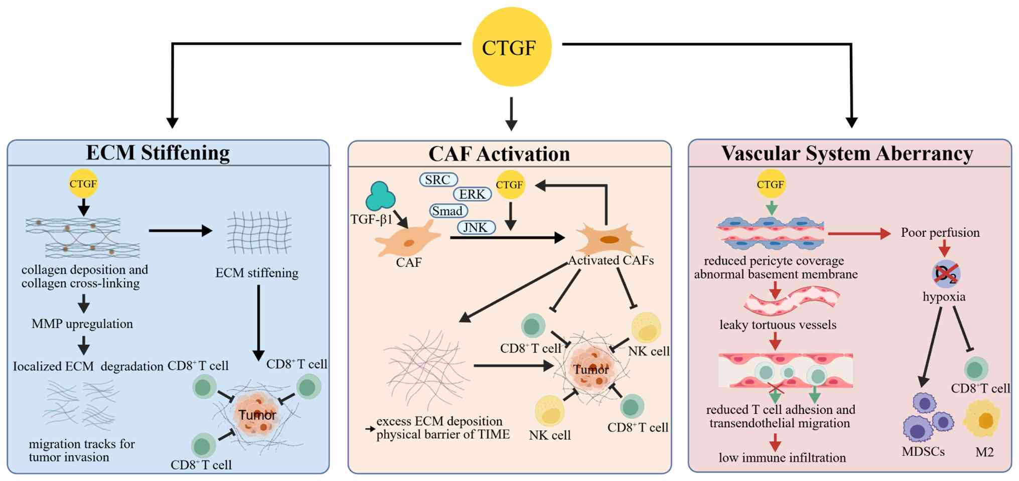 | Figure 2.CTGF modulates ECM remodeling to
establish physical and mechanical barriers in the tumor
microenvironment. CTGF may promote ECM stiffening through collagen
deposition, cross-linking, and MMP induction; activate CAFs through
tumor-derived TGF-β1/SRC-dependent ERK/Smad/JNK signaling; and
impair vascular integrity through reduced pericyte coverage and
abnormal basement membrane formation. CTGF, connective tissue
growth factor; ECM, extracellular matrix; MMP, matrix
metalloproteinase; CAFs, cancer-associated fibroblasts; TGF-β1,
transforming growth factor-β1; SRC, SRC proto-oncogene,
non-receptor tyrosine kinase; ERK, extracellular signal-regulated
kinase; JNK, c-Jun N-terminal kinase; Smad, mothers against
decapentaplegic; TIME, tumor immune microenvironment; NK, natural
killer; MDSCs, myeloid-derived suppressor cells. |
CTGF-mediated enhancement of
extracellular matrix stiffening
ECM stiffening is predominantly driven by the
excessive deposition and aberrant cross-linking of fibrous proteins
such as collagen. CTGF promotes collagen deposition or
cross-linking, thereby remodeling ECM architecture and increasing
tissue stiffness, which provides structural support for tumor
progression (43); furthermore,
elevated CTGF expression is associated with, and in selected models
contributes to, matrix metalloproteinase (MMP) induction. This
process enables localized ECM degradation and may generate
permissive tracks for tumor cell migration and invasion (44–46).
The interplay between collagen accumulation and
matrix degradation creates a heterogeneous ECM landscape that
facilitates tumor invasion. Increased ECM stiffness can also
function as a physical barrier that restricts effector immune-cell
infiltration, particularly CD8+ T cells, into the tumor core. This
represents a mechanobiological mode of immune suppression that is
partially independent of canonical cytokine signaling (43,47).
However, whether CTGF-driven ECM stiffening directly impairs
immune-cell penetration in specific tumor contexts remains to be
fully validated.
CTGF-mediated enhancement of CAF
activation
CAFs are key effector cells that drive ECM
remodeling and shape the TIME. CTGF contributes to CAF activation
and ECM remodeling, thereby promoting the formation of dense matrix
networks and reinforcing physical barriers within the TIME
(9). This conclusion is supported
by functional knockdown experiments in selected tumor models and
correlative clinical observations (8,26).
Current evidence supports this effect based on a combination of
functional knockdown studies and correlative analyses (8,26,48).
However, CTGF alone is unlikely to be sufficient to define lineage
commitment or full functional reprogramming.
Activated CAFs can further secrete immunosuppressive
mediators that impair CD4+ and CD8+ T-cell function, thereby
attenuating antitumor immune responses at the cellular level
(49). In gastric cancer, tumor
cell-derived TGF-β1 was found to induce CTGF expression in CAFs
through SRC-dependent ERK/Smad/JNK signaling pathways. CTGF
targeting has been shown to reduce CAF-mediated tumor cell
migration and invasion in this context (8). Notably, a conserved SDC1+ CAF subset
associated with advanced tumor stage and poor prognosis drives
tumor cell EMT, invasion, and lymphatic metastasis via Krüppel-like
factor 6-regulated CTGF secretion and activation of tumor FGFR3
signaling, highlighting that CTGF and FGFR3 may serve as actionable
stromal targets with anti-metastatic therapeutic implications
across multiple malignancies (26).
Furthermore, an anti-CTGF/programmed cell death
protein 1 (PD-1) bispecific antibody Y126S was shown to mediate
suppression of CAF activation, reduction of collagen deposition,
and downregulation of PD-L1 expression on CAFs by targeting CTGF in
a pancreatic cancer model. This dual targeting strategy was found
to enhance CD8+ T-cell-mediated antitumor immunity and to improve
the efficacy of PD-1 blockade (48). However, the functional heterogeneity
of CTGF across CAF subsets remains incompletely characterized.
CTGF-mediated vascular system
aberrancy
The vascular system is a highly organized tubular
network responsible for oxygen and nutrient delivery and metabolic
waste removal. As a functional unit embedded within the ECM, its
structure and function are directly regulated by the
physicochemical properties of the ECM. Accordingly, vascular
abnormalities represent a key feature and functional consequence of
ECM remodeling in tumors. As a critical factor involved in vascular
homeostasis, dysregulated CTGF signaling is associated with reduced
pericyte coverage and abnormal basement membrane formation, as
shown in vascular modeling systems and tumor-associated vascular
analyses. These alterations contribute to vascular destabilization
and structural disorganization (50).
CTGF-associated vascular dysfunction may impair
effector T-cell adhesion and transendothelial migration, resulting
in reduced immune-cell infiltration into tumor tissues (51). In addition, CTGF-associated vascular
abnormalities may compromise tissue perfusion, promote hypoxia,
suppress T-cell cytotoxic function, and facilitate the recruitment
of immunosuppressive cell populations. Collectively, these effects
further reinforce the immunosuppressive and immune-excluded state
of the TIME.
CTGF orchestrates immune cell
remodeling
In the TIME, the functional state of immune cells is
a key determinant of antitumor immune efficacy (52). As an important signaling molecule,
CTGF may directly or indirectly modulate multiple immune cell
populations and may promote their polarization toward pro-tumor
phenotypes, thereby contributing to the establishment of a
tumor-promoting TIME (Fig. 3).
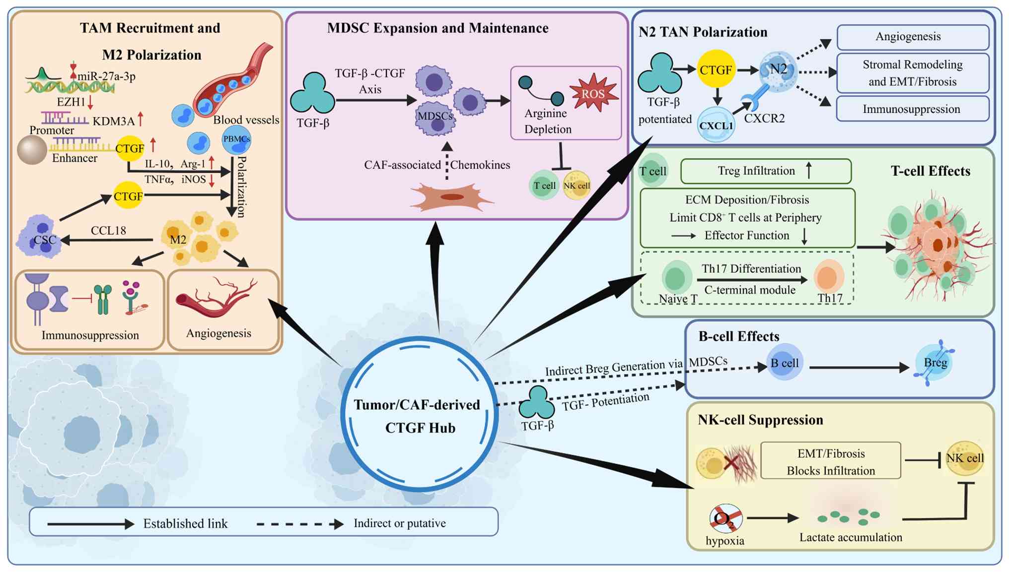 | Figure 3.CTGF promotes M2-type TAM
polarization via the miR-27a-3p/EZH1/KDM3A axis and forms a CTGF-M2
TAM-CCL18 positive feedback loop; potentiates TGF-β-dependent MDSC
expansion, with MDSCs suppressing T and NK cell function through
arginine depletion and ROS accumulation; amplifies TGF-β-induced N2
TAN polarization via upregulating CXCL1-CXCR2 chemokine signaling
to mediate angiogenesis, stromal fibrosis and immune suppression;
remodels T-cell responses by driving C-terminal module-dependent
Th17 differentiation, boosting Treg infiltration, and restricting
CD8+ T-cell effector function via ECM physical barriers; indirectly
induces Breg generation through MDSC intermediates; and inhibits NK
cell antitumor activity via dual mechanisms: ECM/EMT-mediated
infiltration blockage and hypoxia-lactate metabolic dysfunction.
Solid lines represent established regulatory links; dashed lines
represent indirect or putative regulatory axes. CTGF, connective
tissue growth factor; TAM, tumor-associated macrophage; miR-,
microRNA; EZH1, enhancer of zeste homolog 1; KDM3A, lysine
demethylase 3A; CCL18, C-C motif chemokine ligand 18; TGF-β,
transforming growth factor-β; MDSC, myeloid-derived suppressor
cell; NK, natural killer; ROS, reactive oxygen species; TAN,
tumor-associated neutrophil; CXCL1, C-X-C motif chemokine ligand 1;
CXCR2, C-X-C motif chemokine receptor 2; Th17, T helper 17 cell;
Treg, regulatory T cell; ECM, extracellular matrix; Breg,
regulatory B cell; IL-10, interleukin 10; Arg-1, Arginase-1; TNF-α,
tumor necrosis factor-α; iNOS, inducible nitric oxide synthase;
PBMCs, peripheral blood mononuclear cells; CAF, cancer-associated
fibroblast. |
Tumor-associated macrophages
(TAMs)
TAMs are broadly classified into M1 and M2 subsets,
which exert antitumor and pro-tumor activities, respectively. CTGF
may contribute to pro-tumorigenic M2 polarization in solid tumors.
Correlative findings from clinical and omics analysis indicate that
high CTGF expression is associated with increased infiltration of
M2-type TAMs (6). In addition,
functional research suggests that CTGF contributes to macrophage
polarization signaling via the hsa-miR-27a-3p/enhancer of zeste
homolog 1 (EZH1)/lysine demethylase 3A (KDM3A) axis (7). However, direct in vivo causal
validation across tumor types remains limited. In hepatocellular
carcinoma (HCC), experimental evidence demonstrates that CTGF
derived from mesenchymal-like tumor cells promotes M2 macrophage
polarization. These M2-like TAMs further enhance tumor progression
by secreting C-C motif chemokine ligand 18 (CCL18), forming a
CTGF-M2 TAM-CCL18 positive feedback loop (53). Whether this regulatory axis is
broadly conserved across solid tumors remains unclear.
Myeloid-derived suppressor cells
(MDSCs)
MDSCs, as key immunosuppressive cells, inhibit the
functions of T cells and natural killer (NK) cells by depleting
arginine and generating reactive oxygen species (ROS), thereby
mediating tumor immune evasion (54,55).
As a critical downstream molecule of the TGF-β signaling pathway,
CTGF may potentiate TGF-β-driven MDSC expansion and functional
maintenance (14,56). However, this mechanism is mainly
supported by pathway-level evidence and indirect inference rather
than direct CTGF-MDSC functional or depletion studies. High CTGF
expression is associated with increased secretion of
MDSC-recruiting chemokines derived from CAFs and with MDSC
accumulation in the TIME (57),
suggesting a correlative link between CTGF-enriched stromal
environments and MDSC infiltration rather than a fully established
causal relationship. Current evidence is largely limited to
correlative findings, and it remains unclear whether MDSCs and
CTGF+ CAFs are spatially and functionally coupled within the TIME.
The subtype-specific regulatory effects of CTGF on
polymorphonuclear MDSCs and monocytic MDSCs, including their
proliferation, apoptosis, metabolic programs, and suppressive
effector molecule expression, remain largely undefined.
Tumor-associated neutrophils
(TANs)
TANs predominantly exhibit the pro-tumor N2-like
phenotype in the TME, contributing to angiogenesis, stromal
remodeling, and immunosuppression (58). CTGF may contribute to the
establishment of a pro-tumor TAN niche by reinforcing
TGF-β-associated stromal remodeling and neutrophil-recruiting
inflammatory programs. However, current evidence remains largely
indirect, based on TGF-β-dependent TAN polarization models and
CTGF-associated stromal signatures, rather than direct experimental
validation of CTGF in TAN lineage commitment. TGF-β is a key
regulator of TAN functional polarization, promoting pro-tumor
N2-like pro-tumor phenotypes, whereas TGF-β blockade can reprogram
TANs toward an antitumor N1-like state (59,60).
As a downstream effector and functional amplifier of TGF-β
signaling, CTGF contributes to CAF activation, collagen deposition,
ECM remodeling, and tissue fibrosis (14). These stromal alterations may
collectively facilitate the recruitment and functional polarization
of N2-like TANs, which are associated with angiogenesis, matrix
remodeling, chronic inflammation, immune suppression, and tumor
progression (61).
In addition to stromal remodeling, CTGF may regulate
TAN infiltration through chemokine-mediated neutrophil recruitment.
TAN trafficking is largely controlled by neutrophil-attracting
C-X-C motif chemokine receptor (CXCR)2 ligands, including C-X-C
motif chemokine ligand (CXCL)1, CXCL2, CXCL5, and CXCL8/IL-8
(61). Evidence from inflammatory
stromal models suggests that CTGF contributes to CXCL1 induction,
indicating that CTGF-rich stromal environments may enhance
neutrophil recruitment and sustain a pro-tumor TAN-enriched niche
(62). Collectively, CTGF may
contribute to tumor progression by a TGF-β/ECM-chemokine axis that
supports TAN recruitment, N2 polarization, angiogenesis, matrix
remodeling, and immunosuppression.
T cells
Research in chronic inflammatory disorders, such as
kidney disease, have demonstrated that CTGF and its C-terminal
module mediate human CD4+ T-cell polarization by driving the
differentiation of proinflammatory Th17 polarization and local
inflammatory responses (63).
Although this mechanism was characterized in non-neoplastic
inflammatory settings, given that the tumor microenvironment, such
as in pancreatic cancer, shares key features with chronic
inflammatory tissues, including persistent protease activity that
drives ECM remodeling and tumor progression (64), it is plausible that the
proinflammatory effects of CTGF fragments observed in inflammatory
contexts may also contribute to shaping the T-cell landscape in
cancer. In addition, CTGF contributes to CAF activation and ECM
deposition, which may physically restrict infiltration of CD8+ T
cells into tumor cores and impair effector function (9). CTGF expression was also shown to be
positively correlated with regulatory T-cell infiltration in the
TIME (6). These findings suggest
that CTGF may promote immunosuppressive T-cell polarization while
simultaneously limiting effector T-cell infiltration and activity
through stromal remodeling. However, the precise molecular
mechanisms underlying CTGF-induced T-cell dysfunction remain
incompletely understood. Its potential role in regulating T-cell
exhaustion, metabolic reprogramming, and spatial distribution
within tumors remains to be elucidated.
B cells
B cells exert dual functions in the tumor immune
microenvironment, exerting both antitumor effects and
immunosuppressive functions through differentiation into regulatory
B cells (Bregs) (65,66). Direct evidence for CTGF-mediated
regulation of B cells remains limited. However, studies in
hematologic malignancies have indicated that high CTGF expression
is associated with poor prognosis in precursor B-cell leukemia
(67,68). In solid tumors, CTGF-associated
fibrotic and inflammatory microenvironments may promote the
accumulation of MDSCs and other immunosuppressive cell populations,
and MDSCs may contribute to Breg generation (69). This suggests that CTGF may
indirectly regulate B-cell immunosuppressive programs through an
MDSC-dependent axis.
NK cells
NK cells represent the first-line effector
population in antitumor immunity (70). CTGF may suppress NK-cell function
primarily through indirect mechanisms. Structurally, CTGF-driven
EMT and ECM remodeling may restrict NK-cell infiltration into the
tumor parenchyma. Metabolically, CTGF-mediated hypoxia and lactic
acid accumulation may further impair NK-cell effector functions
(36,71).
CTGF shapes cytokine network remodeling
The immunosuppressive state of the TIME is closely
associated with aberrations in the cytokine signaling network. By
regulating multiple key cytokines, CTGF may reshape the cytokine
landscape and thereby contribute to the establishment and
maintenance of the TIME (Fig.
4).
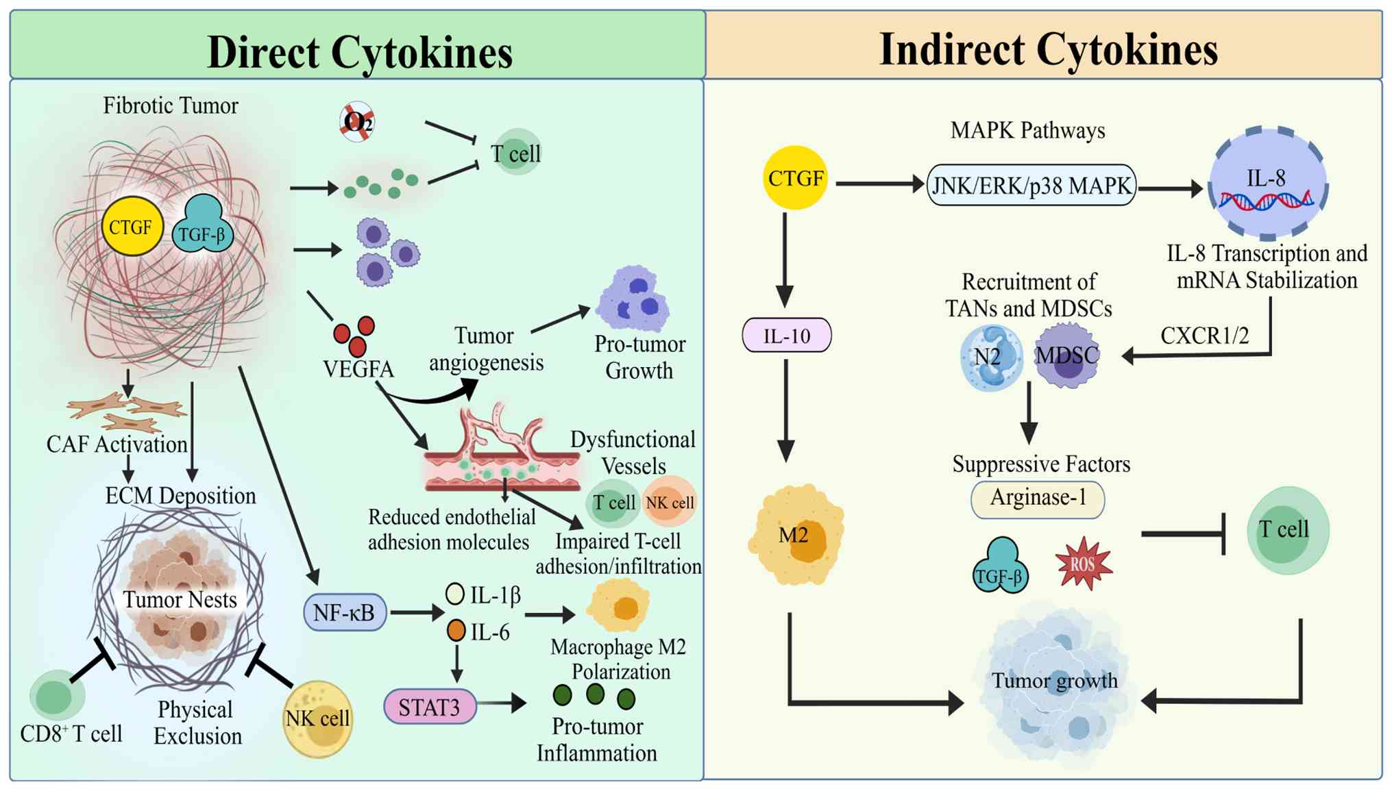 | Figure 4.CTGF remodels the TIME cytokine
network via two distinct regulatory branches: Direct cytokine
modulation and indirect chemokine/cytokine cascades. In fibrotic
tumors, CTGF synergizes with TGF-β to drive CAF activation, ECM
deposition and physical immune exclusion of CD8+ T cells and NK
cells; CTGF upregulates VEGFA to induce dysfunctional angiogenesis
and limit T-cell endothelial adhesion and infiltration; CTGF
activates NF-κB/STAT3 signaling to induce IL-1β and IL-6, promoting
M2 macrophage polarization and pro-tumor inflammation; hypoxic
oxidative stress further suppresses effector T-cell function within
fibrotic tumor nests. For indirect cytokine signaling, CTGF
activates JNK/ERK/p38 MAPK pathways to induce IL-8 transcription
and mRNA stabilization, recruiting TANs and MDSCs via CXCR1/2; CTGF
also upregulates IL-10 to foster M2 TAM polarization. Myeloid
populations recruited by IL-8 secrete arginase-1, ROS and TGF-β to
suppress CD8+ T cell antitumor function, forming a self-amplifying
immunosuppressive loop that accelerates tumor progression. CTGF,
connective tissue growth factor; TIME, tumor immune
microenvironment; TGF-β, transforming growth factor-β; CAF,
cancer-associated fibroblast; ECM, extracellular matrix; NK,
natural killer; VEGFA, vascular endothelial growth factor A; NF-κB,
nuclear factor-κB; STAT3, signal transducer and activator of
transcription 3; IL, interleukin; JNK, c-Jun N-terminal kinase;
ERK, extracellular signal-regulated kinase; MAPK, mitogen-activated
protein kinase; TANs, tumor-associated neutrophils; MDSCs,
myeloid-derived suppressor cells; CXCR1/2, C-X-C motif chemokine
receptor 1/2; TAM, tumor-associated macrophage; ROS, reactive
oxygen species. |
CTGF-mediated regulation of direct
cytokines
Direct cytokines within the TIME mainly include
TGF-β, VEGF, IL-6, IL-35, IL-1β, IL-12, TNF-α, and interferon
(IFN)-γ. In tumors characterized by prominent fibrosis, such as
breast cancer, CTGF and TGF-β are frequently co-localized and
highly expressed (12). This
co-activation can synergistically amplify profibrotic signaling,
promoting CAF activation and ECM deposition (8,14),
enhancing MDSC expansion and functional maintenance (14), and increasing oxidative stress
(36,69,72).
Accordingly, CTGF may function as a downstream effector or
transcriptional target of TGF-β signaling in fibrotic and
metabolically stressed tumor contexts (14,18,36,56).
In these settings, CTGF can further amplify TGF-β-driven stromal
and immunosuppressive programs by reinforcing CAF activation, ECM
deposition, and MDSC accumulation (8,14,56).
However, CTGF may also act as a relatively independent paracrine
regulator when produced by specific stromal or tumor-cell subsets,
such as SDC1+ CAFs or mesenchymal-like HCC cells (31,53).
This may contribute to spatial segregation between tumor cells and
immune effector cells, thereby impairing immune-cell function and
promoting tumor progression (73).
In acute myeloid leukemia, CTGF was shown to mediate
enhanced tumor angiogenic capacity through the CTGF-VEGFA axis.
This axis promotes aberrant angiogenesis and suppresses endothelial
adhesion molecule expression, thereby restricting T-cell adhesion
and infiltration. Collectively, these effects further consolidate
an immunosuppressive TIME and support tumor progression and
metastasis (74). CTGF activates
and upregulates the expression of IL-1β and IL-6 by activating the
NF-κB signaling pathway, thereby triggering STAT3 activation,
exacerbating local inflammation (10,75),
and driving inflammation-cancer transformation (76). In addition, the
miR-27a-3p/EZH1/KDM3A/CTGF axis was also shown to contribute to
TNF-α downregulation in vivo (7). Reduced TNF-α may facilitate M2
macrophage polarization, weaken antitumor immunity, and ultimately
promote tumor progression (7).
However, most evidence is derived from pathway activation studies
(7,10,74,75)
rather than direct cytokine perturbation experiments that isolate
CTGF as a primary upstream regulator. Therefore, the cytokine
regulatory network governed by CTGF remains to be further validated
in tumor-specific experimental models.
CTGF-mediated regulation of indirect
cytokines
Indirect cytokines in the TIME mainly include IL-8
(CXCL8), granulocyte-macrophage colony-stimulating factor (GM-CSF),
IL-10, and IL-4. As a key chemokine, IL-8 has been shown to promote
the recruitment of TANs and MDSCs to tumor sites via the CXCR1/2
axis, and was revealed to be closely associated with angiogenesis
and the amplification of local inflammation (77,78).
These myeloid populations produce immunosuppressive mediators,
including arginase-1, ROS, and TGF-β, within the TIME. This
suppresses CD8+ T cells, driving the transition of inflammatory
signals to an immunosuppressive phenotype, and facilitating the
establishment and maintenance of the TIME (79,80).
CTGF can induce IL-8 expression by activating the JNK, ERK, and p38
MAPK signaling pathways and by enhancing IL-8 mRNA stability
(11). These findings support the
existence of a CTGF-IL-8-myeloid cell axis, which may form an
inflammation-immunosuppression feedback loop within the TIME. This
loop may further amplify the immunosuppressive state and contribute
to the aberrant remodeling of the TIME. Beyond IL-8, emerging
evidence suggests that CTGF may regulate IL-10, another key
immunosuppressive cytokine in the TIME. In glioblastoma, the
miR-27a-3p/EZH1/KDM3A/CTGF axis was found to contribute to IL-10
upregulation, which was associated with enhanced M2 macrophage
polarization and tumor progression (7). The proposed CTGF-GM-CSF relationship
remains inferential and requires direct mechanistic validation.
Conclusion
CTGF has emerged as a context-dependent
matricellular regulator that integrates tumor cell plasticity,
extracellular matrix remodeling, vascular dysfunction, and immune
modulation to shape the TIME. Rather than functioning as a linear
signaling effector, accumulating evidence supports CTGF as a
potential structural-signaling interface that couples stromal
mechanics with immune exclusion programs, thereby contributing to
spatially constrained antitumor immunity.
Across tumor contexts, CTGF is implicated in ECM
remodeling, CAF activation, vascular abnormality, and myeloid- and
lymphoid-cell reprogramming, collectively converging on an
immunosuppressive and immune-excluded niche. Notably, these effects
appear highly context-dependent, with CTGF exerting divergent or
even opposing roles depending on tumor type, cellular source, and
microenvironmental state, underscoring its non-canonical and
non-linear biology. Representative tumor-specific regulatory
mechanisms summarized throughout this review are compiled in
Table I for cross-cancer
comparison.
Functionally, CTGF-associated stromal and cytokine
networks may reinforce resistance to immune checkpoint blockade by
promoting physical immune barriers and sustaining immunosuppressive
feedback loops involving key inflammatory and fibrotic mediators.
This positions CTGF as a potential complementary axis to classical
immune checkpoints in the regulation of therapeutic response.
However, most current evidence remains correlative
or derived from pathway-level inference, and definitive causal
validation of CTGF as a central driver of immune exclusion across
tumor types is still lacking. In particular, its context-specific
role in defining immune cell spatial organization and functional
states requires rigorous in vivo and spatially resolved
experimental confirmation.
From a translational perspective, CTGF represents a
promising candidate for combined stromal-immune targeting
strategies aimed at remodeling the tumor microenvironment rather
than inhibiting single signaling nodes. Future efforts integrating
spatial profiling, functional perturbation, and clinical validation
will be essential to determine whether CTGF-directed interventions
can effectively convert immune-excluded tumors into
immune-permissive states and enhance responsiveness to
immunotherapy.
Acknowledgements
Not applicable.
Funding
This work was supported by the Intramural Research Fund Project
of Gansu Provincial Hospital (grant no. 25GSSYE-2).
Availability of data and materials
Not applicable.
Authors' contributions
YZ conceived the review framework, collected and
organized literature extensively, and drafted the full manuscript
as the primary writer. LH conceptualized figures, designed and
constructed key mechanism diagrams (Figs. 1 and 2) for the review, and optimized the visual
presentation of core research mechanisms. QW assisted in the
visualization of research findings, refined the drawing details of
the review mechanism diagrams (Figs.
3 and 4), and ensured the
accuracy of graphical data expression. HL and FD supervised the
overall research and writing process, provided in-depth academic
guidance for the manuscript structure and content, revised the
manuscript critically for important intellectual content, and
finalized the final version of the manuscript. All authors read and
approved the final version of the manuscript. Data authentication
is not applicable.
Ethics approval and consent to
participate
Not applicable.
Patient consent for publication
Not applicable.
Competing interests
The authors declare that they have no competing
interests.
Glossary
Abbreviations
Abbreviations:
|
AKT
|
protein kinase B (PKB)
|
|
ATP
|
adenosine triphosphate
|
|
Bcl-xL
|
B-cell lymphoma-extra large
|
|
Bregs
|
regulatory B cells
|
|
CAF
|
cancer-associated fibroblasts
|
|
CCL18
|
C-C motif chemokine ligand 18
|
|
CCN
|
cellular communication network
|
|
CTGF
|
connective tissue growth factor,
cellular communication network factor 2 (CCN2)
|
|
CD4
|
cluster of differentiation 4
|
|
CD8
|
cluster of differentiation 8
|
|
cIAP1
|
cellular inhibitor of apoptosis
protein 1
|
|
CSC
|
cancer stem cell
|
|
CXCL1
|
C-X-C motif chemokine ligand 1
|
|
CXCL2
|
C-X-C motif chemokine ligand 2
|
|
CXCL5
|
C-X-C motif chemokine ligand 5
|
|
CXCL8
|
C-X-C motif chemokine ligand 8
|
|
CXCR1
|
C-X-C motif chemokine receptor 1
|
|
CXCR2
|
C-X-C motif chemokine receptor 2
|
|
ECM
|
extracellular matrix
|
|
EMT
|
epithelial-mesenchymal transition
|
|
ERK
|
extracellular signal-regulated
kinase
|
|
ERK/MAPK
|
extracellular signal-regulated
kinase/mitogen-activated protein kinase
|
|
EZH1
|
enhancer of zeste homolog 1
|
|
FGFR3
|
fibroblast growth factor receptor
3
|
|
GM-CSF
|
granulocyte-macrophage
colony-stimulating factor
|
|
HCC
|
hepatocellular carcinoma
|
|
HIF-1α
|
hypoxia-inducible factor 1-α
|
|
hsa-miR-27a-3p
|
Homo sapiens microRNA-27a-3p
|
|
IFN-γ
|
interferon-γ
|
|
IL-1β
|
interleukin-1β
|
|
IL-4
|
interleukin-4
|
|
IL-6
|
interleukin-6
|
|
IL-8
|
interleukin-8
|
|
IL-10
|
interleukin-10
|
|
IL-12
|
interleukin-12
|
|
IL-35
|
interleukin-35
|
|
JNK
|
c-Jun N-terminal kinase
|
|
KDM3A
|
lysine demethylase 3A
|
|
M1
|
classically activated macrophage
phenotype
|
|
M2
|
alternatively activated macrophage
phenotype
|
|
MAPK
|
mitogen-activated protein kinase
|
|
MDSC
|
myeloid-derived suppressor cell
|
|
MDSCs
|
myeloid-derived suppressor cells
|
|
MMP
|
matrix metalloproteinase
|
|
mtDNA
|
mitochondrial DNA
|
|
mtTFA
|
mitochondrial transcription factor
A
|
|
N1
|
anti-tumor tumor-associated
neutrophil phenotype
|
|
N2
|
pro-tumor tumor-associated neutrophil
phenotype
|
|
NF-κB
|
nuclear factor-κB
|
|
NK
|
natural killer cell
|
|
Notch1
|
Notch receptor 1
|
|
OSCC
|
oral squamous cell carcinoma
|
|
p38 MAPK
|
p38 mitogen-activated protein
kinase
|
|
PD-1
|
programmed cell death protein 1
|
|
PD-L1
|
programmed death-ligand 1
|
|
PI3K
|
phosphatidylinositol 3-kinase
|
|
PI3K/AKT
|
phosphatidylinositol 3-kinase/protein
kinase B
|
|
ROS
|
reactive oxygen species
|
|
SDC1
|
syndecan-1
|
|
Snail1
|
Snail family transcriptional
repressor 1
|
|
SRC
|
SRC proto-oncogene, non-receptor
tyrosine kinase
|
|
STAT3
|
signal transducer and activator of
transcription 3
|
|
TAM
|
tumor-associated macrophage
|
|
TAN
|
tumor-associated neutrophil
|
|
TGF-β
|
transforming growth factor-β
|
|
Th17
|
T helper 17 cell
|
|
TIME
|
tumor immune microenvironment
|
|
TME
|
tumor microenvironment
|
|
TNF-α
|
tumor necrosis factor-α
|
|
Twist1
|
twist family bHLH transcription
factor 1
|
|
VEGF
|
vascular endothelial growth
factor
|
References
|
1
|
Duan Y, Zhang J and Cheng X: Comments on:
Global statistics and risk factors of neoplasms in the elderly, and
the impact of aging on neoplasms from 1990–2021. Int J Surg.
111:10011–10012. 2025. View Article : Google Scholar : PubMed/NCBI
|
|
2
|
Bray F, Laversanne M, Sung H, Ferlay J,
Siegel RL, Soerjomataram I and Jemal A: Global cancer statistics
2022: GLOBOCAN estimates of incidence and mortality worldwide for
36 cancers in 185 countries. CA Cancer J Clin. 74:229–263.
2024.PubMed/NCBI
|
|
3
|
Chen S, Cao Z, Prettner K, Kuhn M, Yang J,
Jiao L, Wang Z, Li W, Geldsetzer P, Bärnighausen T, et al:
Estimates and projections of the global economic cost of 29 cancers
in 204 countries and territories from 2020 to 2050. JAMA Oncol.
9:465–472. 2023. View Article : Google Scholar : PubMed/NCBI
|
|
4
|
Bhat IP, Rather TB, Maqbool I, Rashid G,
Akhtar K, Bhat GA, Parray FQ, Syed B, Khan IY, Kazi M, et al:
Connective tissue growth factor expression hints at aggressive
nature of colorectal cancer. World J Gastroenterol. 28:547–569.
2022. View Article : Google Scholar : PubMed/NCBI
|
|
5
|
Lv B, Wang Y, Ma D, Cheng W, Liu J, Yong
T, Chen H and Wang C: Immunotherapy: Reshape the tumor immune
microenvironment. Front Immunol. 13:8441422022. View Article : Google Scholar : PubMed/NCBI
|
|
6
|
Gu J, Tong W, Wang X, Gu L, Wang W, Zang
T, Lou M and Liu Y: Multi-omics analysis revealed that the CCN
family regulates cell crosstalk, extracellular matrix, and immune
escape, leading to a poor prognosis of glioma. Cell Biochem
Biophys. 82:2157–2170. 2024. View Article : Google Scholar : PubMed/NCBI
|
|
7
|
Zhao G, Yu H, Ding L, Wang W, Wang H, Hu
Y, Qin L, Deng G, Xie B, Li G and Qi L: microRNA-27a-3p delivered
by extracellular vesicles from glioblastoma cells induces M2
macrophage polarization via the EZH1/KDM3A/CTGF axis. Cell Death
Discov. 8:2602022. View Article : Google Scholar : PubMed/NCBI
|
|
8
|
Choi KM, Kim B, Lee SM, Han J, Bae HS, Han
SB, Lee D, Ham IH, Hur H, Kim E and Kim JY: Characterization of
gastric cancer-stimulated signaling pathways and function of CTGF
in cancer-associated fibroblasts. Cell Commun Signal. 22:82024.
View Article : Google Scholar : PubMed/NCBI
|
|
9
|
Wang B, Li Y, Shi M, Liao Y, Ruan Y, Han
S, Ma Y, Guan X, Yang R, Dang T, et al: Targeting CTGF overcomes
resistance to CSF1R inhibitors by preventing CAF activation in
colorectal cancer. Cell Rep Med. 6:1024802025. View Article : Google Scholar : PubMed/NCBI
|
|
10
|
Makino Y, Hikita H, Kato S, Sugiyama M,
Shigekawa M, Sakamoto T, Sasaki Y, Murai K, Sakane S, Kodama T, et
al: STAT3 is activated by CTGF-mediated tumor-stroma cross talk to
promote HCC progression. Cell Mol Gastroenterol Hepatol. 15:99–119.
2023. View Article : Google Scholar : PubMed/NCBI
|
|
11
|
Lin CH, Wang YH, Chen YW, Lin YL, Chen BC
and Chen MC: Transcriptional and posttranscriptional regulation of
CXCL8/IL-8 gene expression induced by connective tissue growth
factor. Immunol Res. 64:369–384. 2016. View Article : Google Scholar : PubMed/NCBI
|
|
12
|
Hellinger JW, Schömel F, Buse JV, Lenz C,
Bauerschmitz G, Emons G and Gründker C: Identification of drivers
of breast cancer invasion by secretome analysis: Insight into CTGF
signaling. Sci Rep. 10:178892020. View Article : Google Scholar : PubMed/NCBI
|
|
13
|
Ren M, Yao S, Chen T, Luo H, Tao X, Jiang
H, Yang X, Zhang H, Yu S, Wang Y, et al: Connective tissue growth
factor: Regulation, diseases, and drug discovery. Int J Mol Sci.
25:46922024. View Article : Google Scholar : PubMed/NCBI
|
|
14
|
Fu M, Peng D, Lan T, Wei Y and Wei X:
Multifunctional regulatory protein connective tissue growth factor
(CTGF): A potential therapeutic target for diverse diseases. Acta
Pharm Sin B. 12:1740–1760. 2022. View Article : Google Scholar : PubMed/NCBI
|
|
15
|
Ghosh P, Dey A, Nandi S, Majumder R, Das S
and Mandal M: CTGF (CCN2): A multifaceted mediator in breast cancer
progression and therapeutic targeting. Cancer Metastasis Rev.
44:322025. View Article : Google Scholar : PubMed/NCBI
|
|
16
|
Jia Q, Xu B, Zhang Y, Ali A and Liao X:
CCN family proteins in cancer: Insight into their structures and
coordination role in tumor microenvironment. Front Genet.
12:6493872021. View Article : Google Scholar : PubMed/NCBI
|
|
17
|
Pérez-González A, Bévant K and Blanpain C:
Cancer cell plasticity during tumor progression, metastasis and
response to therapy. Nat Cancer. 4:1063–1082. 2023. View Article : Google Scholar : PubMed/NCBI
|
|
18
|
Kim H, Son S, Ko Y and Shin I: CTGF
regulates cell proliferation, migration, and glucose metabolism
through activation of FAK signaling in triple-negative breast
cancer. Oncogene. 40:2667–2681. 2021. View Article : Google Scholar : PubMed/NCBI
|
|
19
|
Chu X, Tian W, Ning J, Xiao G, Zhou Y,
Wang Z, Zhai Z, Tanzhu G, Yang J and Zhou R: Cancer stem cells:
Advances in knowledge and implications for cancer therapy. Signal
Transduct Target Ther. 9:1702024. View Article : Google Scholar : PubMed/NCBI
|
|
20
|
Banchereau R, Chitre AS, Scherl A, Wu TD,
Patil NS, de Almeida P, Kadel Iii EE, Madireddi S, Au-Yeung A,
Takahashi C, et al: Intratumoral CD103+ CD8+ T cells predict
response to PD-L1 blockade. J Immunother Cancer. 9:e0022312021.
View Article : Google Scholar : PubMed/NCBI
|
|
21
|
Holm JS, Funt SA, Borch A, Munk KK,
Bjerregaard AM, Reading JL, Maher C, Regazzi A, Wong P, Al-Ahmadie
H, et al: Neoantigen-specific CD8 T cell responses in the
peripheral blood following PD-L1 blockade might predict therapy
outcome in metastatic urothelial carcinoma. Nat Commun.
13:19352022. View Article : Google Scholar : PubMed/NCBI
|
|
22
|
Kong X, Peng H, Liu P, Fu X, Wang N and
Zhang D: Programmed death ligand 1 regulates epithelial-mesenchymal
transition and cancer stem cell phenotypes in hepatocellular
carcinoma through the serum and glucocorticoid kinase 2/β-catenin
signaling pathway. Cancer Sci. 114:2265–2276. 2023. View Article : Google Scholar : PubMed/NCBI
|
|
23
|
Peña-Asensio J, Calvo H, Torralba M,
Miquel J, Sanz-de-Villalobos E and Larrubia JR: Anti-PD-1/PD-L1
based combination immunotherapy to boost Antigen-specific CD8+ T
cell response in hepatocellular carcinoma. Cancers (Basel).
13:19222021. View Article : Google Scholar : PubMed/NCBI
|
|
24
|
Chen PS, Wang MY, Wu SN, Su JL, Hong CC,
Chuang SE, Chen MW, Hua KT, Wu YL, Cha ST, et al: CTGF enhances the
motility of breast cancer cells via an
integrin-alphavbeta3-ERK1/2-dependent S100A4-upregulated pathway. J
Cell Sci. 120:2053–2065. 2007. View Article : Google Scholar : PubMed/NCBI
|
|
25
|
Zhu X, Zhong J, Zhao Z, Sheng J, Wang J,
Liu J, Cui K, Chang J, Zhao H and Wong S: Epithelial derived CTGF
promotes breast tumor progression via inducing EMT and collagen I
fibers deposition. Oncotarget. 6:25320–25338. 2015. View Article : Google Scholar : PubMed/NCBI
|
|
26
|
Gao GF, Ruan ZH, Zhang SB, He S, Li YQ,
Lyu JL, Liu Y, Tan XL, Wang YJ, Liu ZW, et al: SDC1+ CAFs secreting
CTGF drive tumour metastasis via FGFR3 signalling in cancers. Gut.
Feb 10–2026.doi: 10.1136/gutjnl-2025-337606 (Epub ahead of print).
View Article : Google Scholar
|
|
27
|
Shafieian M, Chen S and Wu S:
Integrin-linked kinase mediates CTGF-induced epithelial to
mesenchymal transition in alveolar type II epithelial cells.
Pediatr Res. 77:520–527. 2015. View Article : Google Scholar : PubMed/NCBI
|
|
28
|
Gogoi RP, Galoforo S, Fox A, Morris C,
Ramos H, Gogoi VK, Chehade H, Adzibolosu NK, Shi C, Zhang J, et al:
A novel role of connective tissue growth factor in the regulation
of the epithelial phenotype. Cancers (Basel). 15:48342023.
View Article : Google Scholar : PubMed/NCBI
|
|
29
|
Chen Z, Zhang N, Chu HY, Yu Y, Zhang ZK,
Zhang G and Zhang BT: Connective tissue growth factor: From
molecular understandings to drug discovery. Front Cell Dev Biol.
8:5932692020. View Article : Google Scholar : PubMed/NCBI
|
|
30
|
Wang Y, Chang T, Wu T, Ye W, Wang Y, Dou
G, Du H, Hui Y and Guo C: Connective tissue growth factor promotes
retinal pigment epithelium mesenchymal transition via the PI3K/AKT
signaling pathway. Mol Med Rep. 23:3892021. View Article : Google Scholar : PubMed/NCBI
|
|
31
|
Moghbeli M: PI3K/AKT pathway as a pivotal
regulator of epithelial-mesenchymal transition in lung tumor cells.
Cancer Cell Int. 24:1652024. View Article : Google Scholar : PubMed/NCBI
|
|
32
|
Tong H, Zhang A, Shi Y, Zhu S and Liu L:
TSPAN31 Activates EMT through the PI3 K/AKT signaling pathway to
promote glioma progression. Neurochem Res. 50:1922025. View Article : Google Scholar : PubMed/NCBI
|
|
33
|
Chi M, Liu J, Mei C, Shi Y, Liu N, Jiang
X, Liu C, Xue N, Hong H, Xie J, et al: TEAD4 functions as a
prognostic biomarker and triggers EMT via PI3K/AKT pathway in
bladder cancer. J Exp Clin Cancer Res. 41:1752022. View Article : Google Scholar : PubMed/NCBI
|
|
34
|
Wei N, Wu X, Yu Y, Zhou H, Cui K, Zhao X
and Zhang X: CD146 Promotes EMT-Mediated migration and invasion of
NSCLC via PI3K/Akt signaling pathway. Front Biosci (Landmark Ed).
29:1402024. View Article : Google Scholar : PubMed/NCBI
|
|
35
|
Ray I, Michael A, Meira LB and Ellis PE:
The role of cytokines in Epithelial-mesenchymal transition in
gynaecological cancers: A systematic review. Cells. 12:4162023.
View Article : Google Scholar : PubMed/NCBI
|
|
36
|
Capparelli C, Whitaker-Menezes D, Guido C,
Balliet R, Pestell TG, Howell A, Sneddon S, Pestell RG,
Martinez-Outschoorn U, Lisanti MP and Sotgia F: CTGF drives
autophagy, glycolysis and senescence in cancer-associated
fibroblasts via HIF1 activation, metabolically promoting tumor
growth. Cell Cycle. 11:2272–2284. 2012. View Article : Google Scholar : PubMed/NCBI
|
|
37
|
Lai WT, Li YJ, Wu SB, Yang CN, Wu TS, Wei
YH and Deng YT: Connective tissue growth factor decreases
mitochondrial metabolism through ubiquitin-mediated degradation of
mitochondrial transcription factor A in oral squamous cell
carcinoma. J Formos Med Assoc. 117:212–219. 2018. View Article : Google Scholar : PubMed/NCBI
|
|
38
|
Wang MY, Chen PS, Prakash E, Hsu HC, Huang
HY, Lin MT, Chang KJ and Kuo ML: Connective tissue growth factor
confers drug resistance in breast cancer through concomitant
up-regulation of Bcl-xL and cIAP1. Cancer Res. 69:3482–3491. 2009.
View Article : Google Scholar : PubMed/NCBI
|
|
39
|
Yin D, Chen W, O'Kelly J, Lu D, Ham M,
Doan NB, Xie D, Wang C, Vadgama J, Said JW, et al: Connective
tissue growth factor associated with oncogenic activities and drug
resistance in glioblastoma multiforme. Int J Cancer. 127:2257–2267.
2010. View Article : Google Scholar : PubMed/NCBI
|
|
40
|
Zheng M, Liu L, Cui H, Zhao Y, Chen W, Bai
S, Peng W, Wang Y, Li Y, Wang R, et al: Cancer-associated
fibroblast-derived extracellular vesicles facilitate metastasis in
hepatocellular carcinoma by delivering CTGF. Cell Oncol (Dordr).
48:1413–1432. 2025. View Article : Google Scholar : PubMed/NCBI
|
|
41
|
Yuan Z, Li Y, Zhang S, Wang X, Dou H, Yu
X, Zhang Z, Yang S and Xiao M: Extracellular matrix remodeling in
tumor progression and immune escape: From mechanisms to treatments.
Mol Cancer. 22:482023. View Article : Google Scholar : PubMed/NCBI
|
|
42
|
Wang LH, Tsai HC, Cheng YC, Lin CY, Huang
YL, Tsai CH, Xu GH, Wang SW, Fong YC and Tang CH: CTGF promotes
osteosarcoma angiogenesis by regulating miR-543/angiopoietin 2
signaling. Cancer Lett. 391:28–37. 2017. View Article : Google Scholar : PubMed/NCBI
|
|
43
|
Wu J, Zhang Q, Yang Z, Xu Y, Liu X, Wang
X, Peng J, Xiao J, Wang Y, Shang Z, et al: CD248-expressing
cancer-associated fibroblasts induce non-small cell lung cancer
metastasis via Hippo pathway-mediated extracellular matrix
stiffness. J Cell Mol Med. 28:e700252024. View Article : Google Scholar : PubMed/NCBI
|
|
44
|
Tsai HC, Su HL, Huang CY, Fong YC, Hsu CJ
and Tang CH: CTGF increases matrix metalloproteinases expression
and subsequently promotes tumor metastasis in human osteosarcoma
through down-regulating miR-519d. Oncotarget. 5:3800–3812. 2014.
View Article : Google Scholar : PubMed/NCBI
|
|
45
|
Lee JH, Kim J, Kim HS and Kang YJ:
Unraveling connective tissue growth factor as a therapeutic target
and assessing kahweol as a potential drug candidate in
Triple-negative breast cancer treatment. Int J Mol Sci.
24:163072023. View Article : Google Scholar : PubMed/NCBI
|
|
46
|
Trampuž SR, van Riet S, Nordling Å and
Ingelman-Sundberg M: The role of CTGF in liver fibrosis induced in
3D human liver spheroids. Cells. 12:3022023. View Article : Google Scholar : PubMed/NCBI
|
|
47
|
Chen E, Zeng Z and Zhou W: The key role of
matrix stiffness in colorectal cancer immunotherapy: Mechanisms and
therapeutic strategies. Biochim Biophys Acta Rev Cancer.
1879:1891982024. View Article : Google Scholar : PubMed/NCBI
|
|
48
|
Chen S, Zhao Y, Cao M, Peng W, Huang H,
Yang Y, Liang J, Chen W, Bai S, Zhou Q, et al: Anti-CTGF/PD-1
bispecific antibody Y126S restrains desmoplastic and
immunosuppressive microenvironment in pancreatic cancer. J
ImmunoTher Cancer. 13:e0121442025. View Article : Google Scholar : PubMed/NCBI
|
|
49
|
Lee H, Hwang M, Jang S and Um SW: Immune
regulatory function of cancer-Associated fibroblasts in Non-small
cell lung cancer. Tuberc Respir Dis (Seoul). 86:304–318. 2023.
View Article : Google Scholar : PubMed/NCBI
|
|
50
|
Romeo SG, Secco I, Schneider E, Reumiller
CM, Santos CXC, Zoccarato A, Musale V, Pooni A, Yin X, Theofilatos
K, et al: Human blood vessel organoids reveal a critical role for
CTGF in maintaining microvascular integrity. Nat Commun.
14:55522023. View Article : Google Scholar : PubMed/NCBI
|
|
51
|
Ma Q, Fu S, Xia Y, Gao S, Xia Z, Gu P,
Yuan S, Liu J, Liang S, Li N, et al: NO-driven Janus nanomotor
enhances T-cell infiltration by reconstructing tumor-associated
blood and lymphatic vessels. Adv Sci (Weinh). 12:e120902025.
View Article : Google Scholar : PubMed/NCBI
|
|
52
|
Gray JI and Farber DL: Tissue-resident
immune cells in humans. Annu Rev Immunol. 40:195–220. 2022.
View Article : Google Scholar : PubMed/NCBI
|
|
53
|
Wang TT, Yuan JH, Ma JZ, Yang WJ, Liu XN,
Yin YP, Liu Y, Pan W and Sun SH: CTGF secreted by mesenchymal-like
hepatocellular carcinoma cells plays a role in the polarization of
macrophages in hepatocellular carcinoma progression. Biomed
Pharmacother. 95:111–119. 2017. View Article : Google Scholar : PubMed/NCBI
|
|
54
|
Barry ST, Gabrilovich DI, Sansom OJ,
Campbell AD and Morton JP: Therapeutic targeting of tumour myeloid
cells. Nat Rev Cancer. 23:216–237. 2023. View Article : Google Scholar : PubMed/NCBI
|
|
55
|
Lasser SA, Ozbay Kurt FG, Arkhypov I,
Utikal J and Umansky V: Myeloid-derived suppressor cells in cancer
and cancer therapy. Nat Rev Clin Oncol. 21:147–164. 2024.
View Article : Google Scholar : PubMed/NCBI
|
|
56
|
Wang L, Wang H, Zhu M, Ni X, Sun L, Wang
W, Xie J, Li Y, Xu Y, Wang R, et al: Platelet-derived TGF-β1
induces functional reprogramming of myeloid-derived suppressor
cells in immune thrombocytopenia. Blood. 144:99–112. 2024.
View Article : Google Scholar : PubMed/NCBI
|
|
57
|
Liang T, Tao T, Wu K, Liu L, Xu W, Zhou D,
Fang H, Ding Q, Huang G and Wu S: Cancer-associated
Fibroblast-induced remodeling of tumor microenvironment in
recurrent bladder cancer. Adv Sci (Weinh). 10:e23032302023.
View Article : Google Scholar : PubMed/NCBI
|
|
58
|
Geh D, Leslie J, Rumney R, Reeves HL, Bird
TG and Mann DA: Neutrophils as potential therapeutic targets in
hepatocellular carcinoma. Nat Rev Gastroenterol Hepatol.
19:257–273. 2022. View Article : Google Scholar : PubMed/NCBI
|
|
59
|
Fridlender ZG, Sun J, Kim S, Kapoor V,
Cheng G, Ling L, Worthen GS and Albelda SM: Polarization of
tumor-associated neutrophil phenotype by TGF-beta: ‘N1’ versus ‘N2’
TAN. Cancer Cell. 16:183–194. 2009. View Article : Google Scholar : PubMed/NCBI
|
|
60
|
Qin F, Liu X, Chen J, Huang S, Wei W, Zou
Y, Liu X, Deng K, Mo S, Chen J, et al: Anti-TGF-β attenuates tumor
growth via polarization of tumor associated neutrophils towards an
anti-tumor phenotype in colorectal cancer. J Cancer. 11:2580–2592.
2020. View Article : Google Scholar : PubMed/NCBI
|
|
61
|
Mukaida N, Sasaki SI and Baba T: Two-Faced
roles of Tumor-associated neutrophils in cancer development and
progression. Int J Mol Sci. 21:34572020. View Article : Google Scholar : PubMed/NCBI
|
|
62
|
Lu M, Yan XF, Si Y and Chen XZ: CTGF
triggers rat astrocyte activation and Astrocyte-mediated
inflammatory response in culture conditions. Inflammation.
42:1693–1704. 2019. View Article : Google Scholar : PubMed/NCBI
|
|
63
|
Rodrigues-Díez R, Rodrigues-Díez RR,
Rayego-Mateos S, Suarez-Alvarez B, Lavoz C, Stark Aroeira L,
Sánchez-López E, Orejudo M, Alique M, Lopez-Larrea C, et al: The
C-terminal module IV of connective tissue growth factor is a novel
immune modulator of the Th17 response. Lab Invest. 93:812–824.
2013. View Article : Google Scholar : PubMed/NCBI
|
|
64
|
Yuan Y, Zhang H, Wang Z, Huang L,
Kabacaoglu D, Zhang B, Song L and Ai J: The role of inflammatory
factors in the tumor microenvironment of pancreatic cancer. Front
Immunol. 16:16251142025. View Article : Google Scholar : PubMed/NCBI
|
|
65
|
Bod L, Kye YC, Shi J, Torlai Triglia E,
Schnell A, Fessler J, Ostrowski SM, Von-Franque MY, Kuchroo JR,
Barilla RM, et al: B-cell-specific checkpoint molecules that
regulate anti-tumour immunity. Nature. 619:348–356. 2023.
View Article : Google Scholar : PubMed/NCBI
|
|
66
|
Kang N, Duan Q, Min X, Li T, Li Y, Gao J
and Liu W: Multifaceted function of B cells in tumorigenesis. Front
Med. 19:297–317. 2025. View Article : Google Scholar : PubMed/NCBI
|
|
67
|
Lu H, Kojima K, Battula VL, Korchin B, Shi
Y, Chen Y, Spong S, Thomas DA, Kantarjian H, Lock RB, et al:
Targeting connective tissue growth factor (CTGF) in acute
lymphoblastic leukemia preclinical models: Anti-CTGF monoclonal
antibody attenuates leukemia growth. Ann Hematol. 93:485–492. 2014.
View Article : Google Scholar : PubMed/NCBI
|
|
68
|
Wang LL, Tang X, Zhou G, Liu S, Wang Y,
Chen F, Li T, Wen F, Liu S and Mai H: PROM1 and CTGF expression in
childhood MLL-Rearrangement acute lymphoblastic leukemia. J Oncol.
2022:58960222022. View Article : Google Scholar : PubMed/NCBI
|
|
69
|
Chen W, Ning X, Liu Y, Shen T, Liu M, Yin
H, Ding Y, Zhou J, Yin R, Cai L, et al: Myeloid-derived suppressor
cells from tumour-bearing mice induce the population expansion of
CD19hiFcγRIIbhi regulatory B cells via PD-L1. Immunology.
172:127–143. 2024. View Article : Google Scholar : PubMed/NCBI
|
|
70
|
Masmoudi D, Villalba M and Alix-Panabières
C: Natural killer cells: The immune frontline against circulating
tumor cells. J Exp Clin Cancer Res. 44:1182025. View Article : Google Scholar : PubMed/NCBI
|
|
71
|
Ielpo S, Barberini F, Gaiba A, Baronti C,
Greppi M, Obino V, Ravera S, Bussola N, De Ninno A, Businaro L, et
al: Lactate-mediated NK cell dysfunction as a prognostic marker and
therapeutic target in breast cancer. Cell Death Discov. 12:2002026.
View Article : Google Scholar : PubMed/NCBI
|
|
72
|
Dong S, Li X, Chen Z, Shi H, Wang Z and
Zhou W: MMP28 recruits M2-type tumor-associated macrophages through
MAPK/JNK signaling pathway-dependent cytokine secretion to promote
the malignant progression of pancreatic cancer. J Exp Clin Cancer
Res. 44:602025. View Article : Google Scholar : PubMed/NCBI
|
|
73
|
Sabit H, Arneth B, Pawlik TM, Abdel-Ghany
S, Ghazy A, Abdelazeem RM, Alqosaibi A, Al-Dhuayan IS, Almulhim J,
Alrabiah NA and Hashash A: Leveraging Single-cell Multi-Omics to
decode tumor microenvironment diversity and therapeutic resistance.
Pharmaceuticals (Basel). 18:752025. View Article : Google Scholar : PubMed/NCBI
|
|
74
|
Li ZJ, Cheng J, Song Y, Li HH and Zheng
JF: LncRNA SNHG5 upregulation induced by YY1 contributes to
angiogenesis via miR-26b/CTGF/VEGFA axis in acute myelogenous
leukemia. Lab Invest. 101:341–352. 2021. View Article : Google Scholar : PubMed/NCBI
|
|
75
|
Karger A, Fitzner B, Brock P, Sparmann G,
Emmrich J, Liebe S and Jaster R: Molecular insights into connective
tissue growth factor action in rat pancreatic stellate cells. Cell
Signal. 20:1865–1872. 2008. View Article : Google Scholar : PubMed/NCBI
|
|
76
|
Makino Y, Hikita H, Kodama T, Shigekawa M,
Yamada R, Sakamori R, Eguchi H, Morii E, Yokoi H, Mukoyama M, et
al: CTGF mediates Tumor-stroma interactions between hepatoma cells
and hepatic stellate cells to accelerate HCC progression. Cancer
Res. 78:4902–4914. 2018. View Article : Google Scholar : PubMed/NCBI
|
|
77
|
Han ZJ, Li YB, Yang LX, Cheng HJ, Liu X
and Chen H: Roles of the CXCL8-CXCR1/2 axis in the tumor
microenvironment and immunotherapy. Molecules. 27:1372021.
View Article : Google Scholar : PubMed/NCBI
|
|
78
|
Liu H, Zhao Q, Tan L, Wu X, Huang R, Zuo
Y, Chen L, Yang J, Zhang ZX, Ruan W, et al: Neutralizing IL-8
potentiates immune checkpoint blockade efficacy for glioma. Cancer
Cell. 41:693–710.e8. 2023. View Article : Google Scholar : PubMed/NCBI
|
|
79
|
Gao X, Xu M, Xiao H, Han Z, Wang Z, Sun G,
Zhang D, Shuangjian Q, Ren N, Zhou C and Yi Y: Tumor-associated
neutrophils: A complex role in cancer. Clin Immunol.
280:1105582025. View Article : Google Scholar : PubMed/NCBI
|
|
80
|
Najjar YG, Rayman P, Jia X, Pavicic PG Jr,
Rini BI, Tannenbaum C, Ko J, Haywood S, Cohen P, Hamilton T, et al:
Myeloid-derived suppressor cell subset accumulation in renal cell
carcinoma parenchyma is associated with intratumoral expression of
IL1β, IL8, CXCL5, and Mip-1α. Clin Cancer Res. 23:2346–2355. 2017.
View Article : Google Scholar : PubMed/NCBI
|
|
81
|
Ma S, Kanai R, Pobbati AV, Li S, Che K,
Seavey CN, Hallett A, Burtscher A, Lamar JM and Rubin BP: The
TAZ-CAMTA1 fusion protein promotes tumorigenesis via connective
tissue growth factor and Ras-MAPK signaling in epithelioid
hemangioendothelioma. Clin Cancer Res. 28:3116–3126. 2022.
View Article : Google Scholar : PubMed/NCBI
|

















