Introduction
Non-small cell lung cancer (NSCLC) accounts for ~85%
of all lung cancers and is a major cause of cancer-related death
(1). Despite advances in diagnosis
and therapy, most patients with NSCLC still present with advanced
disease and overall survival remains poor (2,3).
Therefore, there is an urgent need to identify novel biomarkers and
therapeutic targets for NSCLC as well as to elucidate the molecular
mechanisms driving its progression. Such advances would enable
early detection and lead to the development of more effective
treatment strategies.
Histone deacetylases (HDACs) are pivotal epigenetic
regulators that catalyze the deacetylation of histones and various
non-histone proteins, leading to chromatin condensation and
transcriptional repression (4). A
total of 18 mammalian HDACs have been identified and are classified
into four groups: Class I (HDAC1, 2, 3 and 8), Class II (IIa:
HDAC4, 5, 7 and 9; IIb: HDAC6 and 10), Class III (NAD+-dependent
sirtuins) and Class IV (HDAC11) (5). Among these, HDAC1 is frequently
overexpressed in NSCLC (6).
Elevated HDAC1 levels correlate with higher tumor grade, advanced
disease stage and worse survival outcomes (7–9). HDAC1
overexpression has also been shown to enhance tumor growth,
invasiveness and resistance to therapies including epidermal growth
factor receptor-tyrosine kinase inhibitors and chemotherapy
(10–12). However, the precise molecular
mechanisms by which HDAC1 contributes to NSCLC progression remain
to be fully elucidated.
HDAC1 has been implicated in multiple oncogenic
signaling pathways, including the wingless-type (Wnt)/β-catenin,
phosphoinositide 3-kinase (PI3K)/protein kinase B (AKT) and
mitogen-activated protein kinase (MAPK)/extracellular
signal-regulated kinase (ERK) pathways. Emerging evidence indicates
that HDAC1 expression is associated with activation of the PI3K/Akt
and Wnt/β-catenin signaling pathways in NSCLC (13). In addition, the plant homeodomain
finger protein 12-HDAC1 axis has been shown to promote EGFR/AKT
signaling in NSCLC progression (14). Furthermore, the HDAC1-Smad3-mSin3A
complex inhibits c-Met transcription, thereby suppressing
epithelial-mesenchymal transition (EMT), angiogenesis, invasion and
adhesion in lung cancer cells, which are mediated via the ERK1/2
and PI3K/Akt pathways (15).
Therefore, to elucidate the mechanistic role of HDAC1 in NSCLC, the
present study investigated the activities of Wnt/β-catenin,
PI3K/AKT and MAPK pathways by modulating HDAC1 expression in
vitro and in vivo.
Materials and methods
Patients and tissue specimens
This study was approved by the Institutional Review
Committee of the Affiliated Cancer Hospital of Hainan Medical
University (approval no. 2024.39). A total of 157 patients with
pathologically confirmed primary lung adenocarcinoma (LUAD) who
underwent surgical resection at Hainan Cancer Hospital between
January 2016 and December 2024 were enrolled. The cohort comprised
69 male and 88 female patients (male-to-female ratio, 1:1.28), with
a mean age of 63.1±8.5 years (range, 37–80 years). Paired tumor
tissues and adjacent non-tumorous lung tissues (obtained ≥5 cm from
the tumor margin) were collected at the time of surgery. Clinical
and pathological data, including age at diagnosis, TNM stage
(16), pathological subtype and
histological grade, were retrieved from medical records. Written
informed consent was obtained from all participants prior to
enrollment.
Tissue microarray (TMA) construction
and immunohistochemical (IHC) analyses
TMAs were constructed using paired tumor and
adjacent non-tumorous tissues. Specimens were fixed in 10%
neutral-buffered formalin at room temperature for 24 h, dehydrated
through a graded ethanol series (50–100%; 1 h each), cleared in
xylene (three changes, 30 min each), infiltrated with paraffin
(three changes, 1 h each) and embedded in paraffin blocks (melting
point, 58°C). Cylindrical cores (2 mm in diameter) were extracted
from representative regions of each donor block and transferred to
recipient paraffin blocks using a tissue arrayer. Sections (4 µm
thick) were cut from the TMA blocks, baked at 72°C for 30 min and
deparaffinized by sequential immersion in xylene (three changes)
and a descending ethanol series (100, 90, 80, 70 and 50%), 5 min
each. Antigen retrieval was performed in EDTA buffer (pH 9.0; cat.
no. K8004; Quantum Analytics, Inc.) using an automated IHC
pretreatment device at 97°C for 18 min, followed by gradual cooling
to 65°C. Endogenous peroxidase activity was quenched with 3%
hydrogen peroxide for 10 min at room temperature. The sections were
then incubated with an anti-HDAC1 primary antibody (dilution,
1:100; cat. no. 34589; Cell Signaling Technology, Inc.) at room
temperature for 1 h. A two-step horseradish peroxidase-conjugated
detection system (cat. no. PV-9000; ZSGB-Bio, Inc.) was
subsequently applied. The sections were incubated with reaction
enhancement solution (Reagent 2) at room temperature for 20 min,
followed by enhanced enzyme-labeled secondary antibody (Reagent 3)
at 37°C for 20 min. Immunoreactivity was visualized using
3,3′-diaminobenzidine (dilution, 1:20; cat. no. ZLI-9018; ZSGB-Bio,
Inc.), and the sections were counterstained with hematoxylin at
room temperature for 10–15 sec. After dehydration, clearing and
mounting, images were captured under a light microscope
(magnification, ×40 and ×400; Olympus BX53; Olympus Corporation).
All slides were evaluated independently by two pathologists blinded
to the clinical data.
HDAC1 expression was scored semi-quantitatively
based on the staining intensity (0, negative; 1, weak; 2, moderate;
3, strong) and the proportion of positively stained cells (0, 0%;
1, <10%; 2, 10–50%; 3, 51–80%; 4, >80%). The final
immunoreactive score (IRS) was calculated by multiplying the
staining intensity score by the proportion score, yielding a range
of 0–12. The cases were dichotomized into low-(IRS, 0–4) and
high-expression (IRS, 6–12) group.
Cell culture
Human NSCLC cell lines A549, H1299 and H1975 were
obtained from the Cell Bank of the Type Culture Collection of the
Chinese Academy of Sciences. Cells were maintained in Roswell Park
Memorial Institute-1640 (RPMI-1640) medium (cat. no. C11875500BT;
Gibco; Thermo Fisher Scientific, Inc.) supplemented with 10% fetal
bovine serum (FBS; cat. no. ST30-3302; PAN-Biotech GmbH) and 1%
penicillin-streptomycin (cat. no. 15140-122; Gibco; Thermo Fisher
Scientific, Inc.) at 37°C in a humidified 5% CO2
incubator.
Lentiviral packaging and stable cell
line establishment
HDAC1-knockdown (HDAC1 K/D), HDAC1-overexpressing
(HDAC1 O/E) and β-catenin-overexpressing (β-catenin O/E) stable
cell lines were generated in A549, H1299 and H1975 cells using
lentiviral transduction. For HDAC1 K/D, validated HDAC1-targeting
short hairpin RNA (shRNA) sequences were cloned into the
pLKO.1-GFP-puro lentiviral vector (Generay Biotech Co., Ltd.), with
the empty pLKO.1 vector serving as the negative control. For HDAC1
O/E, a full-length human HDAC1 coding sequence (GenBank Accession
No. NM_004964) was amplified and cloned into the pCDH-GFP-puro
lentiviral vector (Generay Biotech Co., Ltd.), with the empty pCDH
vector serving as the corresponding control. For β-catenin O/E, a
full-length human CTNNB1 coding sequence (GenBank Accession No.
NM_001904) was cloned into the same vector to generate
pCDH-β-catenin, again with the empty pCDH vector as the control.
The identity of each insert was verified by Sanger sequencing prior
to virus production. Recombinant lentiviruses were produced using a
second-generation packaging system. Briefly, 1 µg pMD2.G and 3 µg
psPAX2 were cotransfected with 4 µg pCDH-HDAC1, pLKO.1-HDAC1 or
pCDH-β-catenin, or with 4 µg of the corresponding empty vector
(pCDH or pLKO.1), into 293T cells (cat. no. NCL-10003; NCM Biotech)
using Nano293T Transfection Reagent (cat. no. C500T-1; NCM Biotech)
at 37°C for 12 h according to the manufacturer's instructions. The
culture medium was replaced 12 h following transfection and
virus-containing supernatants were harvested at 24, 48 and 72 h
post-transfection. The supernatants were filtered through 0.45-µm
polyvinylidene fluoride membranes to remove cell debris, pooled and
stored at −80°C until use. Target cells were seeded in six-well
plates and transduced with the harvested lentivirus in the presence
of 8 µg/ml polybrene (MilliporeSigma). The multiplicity of
infection in A549, H1299 and H1975 cells was 20, 40 and 20 for
pCDH-HDAC1; 40, 40 and 20 for pLKO.1-HDAC1; and 20, 40 and 10 for
pCDH-β-catenin. After 24 h, the medium was replaced with fresh
complete medium. Transduced cells were selected with 6 µg/ml
puromycin (Beijing Solarbio Science & Technology Co., Ltd.)
over 3 weeks, with the medium refreshed every 2–3 days. The
resulting stable lines were designated A549-HDAC1 K/D, H1299-HDAC1
K/D and H1975-HDAC1 K/D for the knockdown groups, and A549-HDAC1
O/E, H1299-HDAC1 O/E and H1975-HDAC1 O/E for the overexpression
groups, alongside their respective empty vector controls. All
subsequent experiments were performed using the stable cell lines
obtained after this 3-week selection period. Knockdown and
overexpression efficiencies were validated by western blotting. All
shRNA, overexpression and control sequences used in the present
study are listed in Table SI.
To investigate whether the oncogenic effects of
HDAC1 in NSCLC are mediated through β-catenin signaling, rescue
experiments were performed by overexpressing β-catenin in HDAC1 K/D
stable cell lines. Lentivirus production, transduction and
puromycin selection were performed as described above. The
resulting double-modified stable lines were designated A549-HDAC1
K/D + β-catenin O/E, H1299-HDAC1 K/D + β-catenin O/E and
H1975-HDAC1 K/D + β-catenin O/E. In parallel, β-catenin was
overexpressed in parental cells to generate β-catenin O/E-alone
groups (A549-β-catenin O/E, H1299-β-catenin O/E and H1975-β-catenin
O/E), which served as the reference groups for the rescue
experiments. β-catenin overexpression efficiency was confirmed by
western blotting.
Cell proliferation assay
Cell proliferation was measured using Cell Counting
Kit-8 (CCK-8; Vazyme Biotech Co., Ltd.) and colony formation
assays. For the CCK-8 assay, cells (1.5×10³ cells/well)
were seeded into 96-well plates in 200 µl culture medium. After
incubation for 0, 24, 48, 72 and 96 h at 37°C with 5%
CO2, 10 µl of CCK-8 reagent was added to each well. The
absorbance was then measured at 450 nm. Cell proliferation was
calculated as the optical density (OD) of the experimental well/OD
of control well (Day 1). For the colony assay, cells
(1×10⁴ cells/well) were plated in 6-well plates in 2 ml
complete medium and cultured for 2 weeks. The cells were fixed with
4% paraformaldehyde (MilliporeSigma) at room temperature for 30 min
and stained with 0.05% crystal violet (MilliporeSigma) at room
temperature for 30 min. Subsequently, colonies >1 mm in diameter
were manually counted. Colony formation efficiency was calculated
as the number of colonies divided by the number of cells seeded
initially and the results were normalized to the control
conditions. Each condition in both assays was tested in triplicate
and each experiment was independently repeated at least three
times.
Migration assay
Cell migration was assessed using a Transwell assay.
Transwell inserts with 8-µm pore membranes (Corning Inc.) were
placed in 24-well culture plates. Cells (5×10⁴) suspended in 200 µl
of serum-free culture medium were added to the upper chamber,
whereas the lower chamber was filled with 600 µl of medium
supplemented with 10% FBS as a chemoattractant. After 24 h of
incubation at 37°C, the non-migrated cells in the chamber were
removed with a cotton swab. The migrated cells were fixed with 20%
methanol for 30 min and stained with 0.1% crystal violet for 30 min
(both at room temperature). Migratory cells in four randomly
selected fields per insert were counted under a light microscope
(magnification, ×200; Olympus BX53; Olympus Corporation). Each
experiment was performed in triplicate.
Invasion assay
Cell invasion was evaluated using a Matrigel-coated
Transwell chamber. Transwell inserts with 8-µm pores were
pre-coated with Matrigel (3 mg/ml) and the gel was allowed to
solidify at 37°C for 30 min. Then, 1×10⁵ cells suspended
in 200 µl of serum-free medium were added to the upper chamber and
600 µl of medium containing 10% FBS was added to the lower chamber.
After 24 h of incubation at 37°C, non-invading cells in the upper
chamber were removed with a cotton swab. The invading cells were
fixed with 4% paraformaldehyde for 30 min and stained with 0.1%
crystal violet for 30 min (both at room temperature). Invading
cells were counted under a light microscope (magnification, ×200;
Olympus BX53; Olympus Corporation) across four randomly chosen
fields per insert. Each experiment was performed in triplicate.
Nuclear/cytoplasmic protein separation
and extraction
Nuclear and cytosolic proteins were extracted using
a nucleocytoplasmic separation and extraction kit (Thermo Fisher
Scientific Inc.) according to the manufacturer's instructions. The
supernatants were collected and analyzed by western blotting. To
verify the effectiveness of the fractionation, western blotting was
performed using Lamin A as a nuclear marker and GAPDH as a
cytoplasmic marker.
Western blotting
Cells were lysed in radioimmunoprecipitation assay
buffer (Beyotime Biotechnology) containing protease inhibitors for
15 min. Lysates were clarified by centrifugation at 16,000 × g for
15 min at 4°C. Protein concentrations were quantified using the
Bradford assay (Bio-Rad Laboratories, Inc.). Samples were mixed
with 5X Laemmli sample buffer and boiled. Proteins (30–40 µg/well)
were separated by 8% sodium dodecyl sulfate-polyacrylamide gel
electrophoresis (SDS-PAGE) and then transferred onto nitrocellulose
membranes (Whatman; Cytiva). The membranes were blocked with 5%
non-fat milk in Tris-buffered saline containing 0.1% Tween-20 for 1
h at room temperature, and then incubated overnight at 4°C with
primary antibodies against HDAC1 (dilution, 1:1,000),
phosphorylated (p-)ERK1/2 (dilution, 1:2,000), total ERK1/2
(dilution, 1:2,000), p-AKT (T308) (dilution, 1:2,000), p-AKT (S473)
(dilution, 1:2,000), total AKT (dilution, 1:2,000), c-Myc
(dilution, 1:2,000), cyclin D1 (dilution, 1:2,000), E-cadherin
(dilution, 1:2,000), vimentin (dilution, 1:2,000), β-catenin
(dilution, 1:2,000), GAPDH (dilution, 1:3,000) and Lamin A
(dilution, 1:1,000), all obtained from Cell Signaling Technology,
Inc. The membranes were subsequently incubated with HRP-conjugated
goat anti-rabbit IgG (cat. no. L3012; dilution, 1:5,000; SAB
Biotherapeutics, Inc.) or HRP-conjugated goat anti-mouse IgG (cat.
no. L3032; dilution, 1:5,000; SAB Biotherapeutics, Inc.) for 1 h at
room temperature. Signals were detected using an enhanced
chemiluminescence substrate (Thermo Fisher Scientific, Inc.) and
images were captured using an Amersham Imager 600 (GE Healthcare).
Band intensities were quantified using ImageJ software (version
1.8.0; National Institutes of Health).
Xenograft tumor model
All animal experiments were approved by the
Institutional Animal Care and Use Committee of Hainan Medical
University (approval no. 2024.331) and were performed at the
Medical Research Center, Hainan Cancer Hospital. A total of 20
six-week-old female BALB/c nude mice (nu/nu; body weight, 15–20 g;
SJA Laboratory Animal Center) were maintained under specific
pathogen-free conditions at a temperature of 22±2°C and a relative
humidity of 50±10%, with a 12-h light/dark cycle and ad
libitum access to food and water. Prior to tumor cell
inoculation, mice were allowed to acclimate for one week. A549
cells were selected as a representative lung adenocarcinoma model
for the in vivo rescue experiments, as the in vitro
effects of HDAC1 knockdown and β-catenin re-expression were
consistent across all three cell lines (A549, H1299 and H1975).
Mice were randomly divided into four groups and inoculated with
stable A549 cells: empty-vector control, HDAC1 K/D, β-catenin O/E,
or HDAC1 K/D + β-catenin O/E. For each mouse, 5×10⁵
cells were resuspended in 100 µl of a 1:1 mixture of PBS and
Matrigel and injected subcutaneously into the right flank. Tumor
diameter was measured every three days and tumor volume was
calculated as (length × width2)/2. Mice were sacrificed
when any individual tumor reached ~2,000 mm3 or when an
animal lost 20% of its initial body weight, in accordance with
humane endpoint guidelines. No mouse met these criteria during the
study. At the experimental endpoint, four weeks after inoculation,
all mice were sacrificed by carbon dioxide (CO2)
inhalation in accordance with institutional guidelines.
CO2 was introduced using a gradual-fill method at a rate
displacing 30–70% of the chamber volume per minute. Tumor tissues
were snap-frozen or lysed for western blotting analysis.
Statistical analyses
Data are expressed as mean ± standard deviation (SD)
of at least three independent experiments. Associations between
HDAC1 expression and clinicopathological characteristics were
evaluated using the χ2 test or Fisher's exact test, as
appropriate. Comparisons between two groups were performed using a
two-tailed Student's t-test (paired for matched tumor and adjacent
tissue specimens; unpaired for independent cell-line experiments),
whereas multigroup comparisons were analyzed using one-way analysis
of variance followed by Tukey's post hoc test. All analyses were
performed using SPSS for Windows (version 22.0; IBM Corp.).
P<0.05 was considered to indicate a statistically significant
difference.
Results
HDAC1 is highly expressed in LUAD
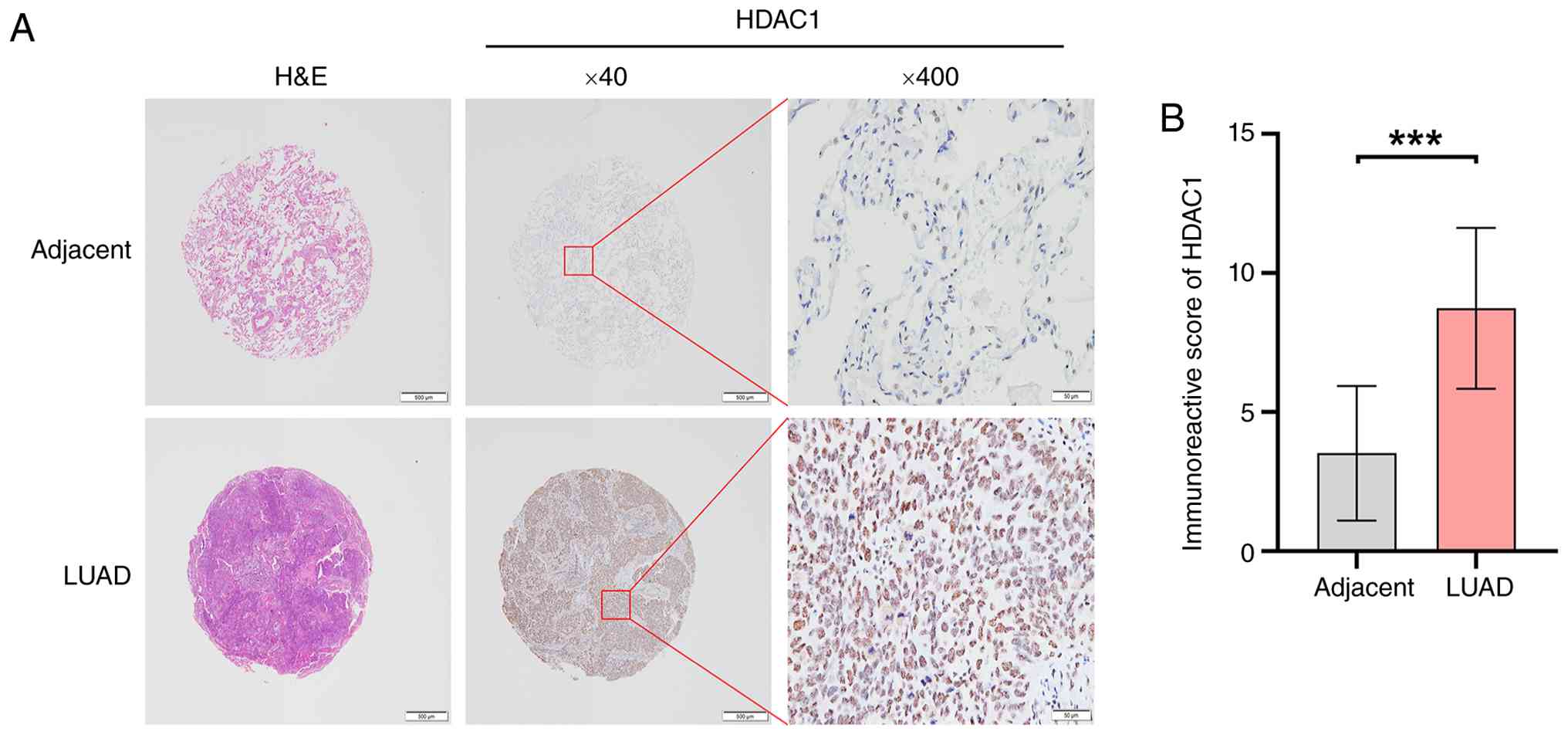
To evaluate the expression of HDAC1 in LUAD, IHC
staining was performed using tissue microarrays comprising 157
paired tumor and adjacent non-neoplastic lung tissues. As shown in
Fig. 1A, HDAC1 immunoreactivity was
weak or absent in adjacent non-neoplastic tissues, whereas LUAD
tissues exhibited strong, diffuse nuclear staining. Consistent with
these observations, the mean IRS was significantly higher in LUAD
tissues compared with that in the matched adjacent tissues
(P<0.001; Fig. 1B). When cases
were dichotomized by IRS, high HDAC1 expression was detected in 143
of 157 (91.1%) LUAD specimens, compared with only 37 of 157 (23.6%)
adjacent non-neoplastic tissues (χ2=146.273, P<0.001;
Table I). These results
demonstrated that HDAC1 is frequently overexpressed in LUAD.
 | Table I.Expression of HDAC1 in lung
adenocarcinoma tissues and adjacent non-neoplastic tissues
(n=157). |
Table I.
Expression of HDAC1 in lung
adenocarcinoma tissues and adjacent non-neoplastic tissues
(n=157).
| HDAC1 IHC
score | Lung adenocarcinoma
tissues n (%) | Adjacent
non-neoplastic tissues n (%) | χ2 | P-value |
|---|
| High | 143 (91.1) | 37 (23.6) | 146.273 | <0.001 |
| Low | 14 (8.9) | 120 (76.4) |
|
|
HDAC1 expression is associated with
clinicopathological features of LUAD patients
The association between HDAC1 expression and the
clinicopathological characteristics was further analyzed (Table II). High HDAC1 expression was
significantly associated with lymph node metastasis (P=0.027) and
the histological differentiation grade (P=0.006). Notably, all 46
patients with lymph node metastasis (100%) exhibited high HDAC1
expression compared with 97 of 111 (87.4%) patients without lymph
node involvement. Regarding tumor differentiation, the proportion
of high HDAC1 expression was greatest in poorly differentiated
tumors (64/65, 98.5%), followed by well to moderately
differentiated tumors (79/92, 85.9%), suggesting that HDAC1
upregulation is accompanied by a less differentiated and more
aggressive phenotype. By contrast, no significant association was
observed between HDAC1 expression and patient age, sex, maximum
tumor diameter, T stage, vascular invasion, perineural invasion,
necrotic components, or Ki-67 proliferation index (all P>0.05).
Collectively, these findings indicate that elevated HDAC1
expression is closely linked to lymph node metastasis and poor
tumor differentiation in LUAD.
| Table II.Relationship between HDAC1 expression
and clinicopathological features (n=157). |
Table II.
Relationship between HDAC1 expression
and clinicopathological features (n=157).
|
| HDAC1
Expression |
|
|
|---|
|
|
|
|
|
|---|
| Clinicopathological
features | High, n (%) | Low, n (%) | Total | P-value |
|---|
| Age (years) |
|
|
| 0.220 |
|
≤60 | 43 (86.0) | 7 (14.0) | 50 |
|
|
>60 | 100 (93.5) | 7 (6.5) | 107 |
|
| Sex |
|
|
| 0.515 |
|
Male | 64 (92.8) | 5 (7.2) | 69 |
|
|
Female | 79 (89.8) | 9 (10.2) | 88 |
|
| Maximum tumor
diameter (mm) |
|
|
| 1.000 |
|
≤30 | 110 (90.9) | 11 (9.1) | 121 |
|
|
>30 | 33 (91.7) | 3 (8.3) | 36 |
|
| T stage |
|
|
| 0.375 |
| T1 | 64 (88.9) | 8 (11.1) | 72 |
|
|
T2-T4 | 79 (92.9) | 6 (7.1) | 85 |
|
| Lymph node
metastasis |
|
|
| 0.027a |
|
Yes | 46 (100.0) | 0 (0.0) | 46 |
|
| No | 97 (87.4) | 14 (12.6) | 111 |
|
| Vascular
invasion |
|
|
| 0.461 |
|
Yes | 39 (95.1) | 2 (4.9) | 41 |
|
| No | 104 (89.7) | 12 (10.3) | 116 |
|
| Perineural
invasion |
|
|
| 1.000 |
|
Yes | 9 (90.0) | 1 (10.0) | 10 |
|
| No | 134 (91.2) | 13 (8.8) | 147 |
|
| Necrotic
component |
|
|
| 0.577 |
|
Yes | 24 (96.0) | 1 (4.0) | 25 |
|
| No | 119 (90.2) | 13 (9.8) | 132 |
|
| Degree of
differentiation |
|
|
| 0.006b |
|
Well-Moderate | 79 (85.9) | 13 (14.1) | 92 |
|
|
Poor | 64 (98.5) | 1 (1.5) | 65 |
|
| Ki-67 |
|
|
| 0.531 |
|
≤10% | 95 (89.6) | 11 (10.4) | 106 |
|
|
>10% | 48 (94.1) | 3 (5.9) | 51 |
|
HDAC1 K/D suppresses proliferation,
migration and invasion while reducing nuclear β-catenin
accumulation in NSCLC cells
To examine the functional role of HDAC1 in NSCLC,
the present study established stable HDAC1-K/D cell lines using two
independent shRNA constructs (K/D-1 and K/D-2) in A549, H1299 and
H1975 cells. CCK-8 assay revealed that HDAC1 knockdown
significantly reduced the proliferative capacity of all three cell
lines over a 96-h observation period compared with the empty vector
controls (P<0.001; Fig. 2A-C).
Colony formation assays further confirmed these findings; HDAC1-K/D
cells exhibited significantly reduced clonogenic ability compared
with control cells (P<0.001; Fig. 2D
and E). Next, it was examined whether HDAC1 depletion affects
the migratory and invasive potential of NSCLC cells using Transwell
assays. HDAC1 knockdown led to a significant reduction in both the
migration (Fig. 2F and G) and
invasiveness (Fig. 2H and I) of all
three cell lines relative to their respective controls
(P<0.001). Collectively, these data indicated that HDAC1 is
required for the proliferative, migratory and invasive capacities
of NSCLC cells.
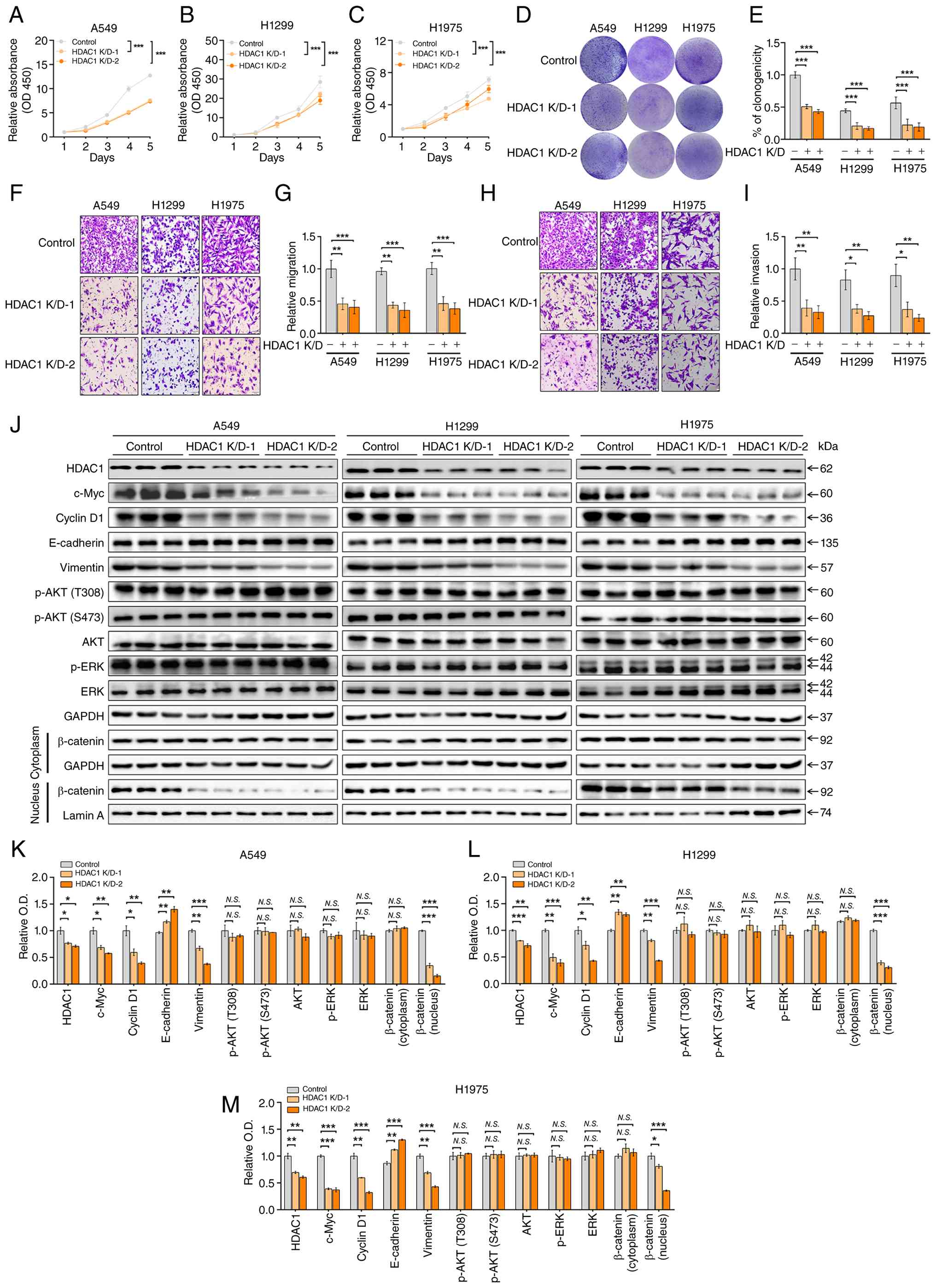 | Figure 2.Effects of HDAC1 knockdown on
proliferation, migration, invasion and downstream signaling in
NSCLC cells. A549, H1299 and H1975 cells were stably transduced
with HDAC1-targeting shRNA (K/D-1 and K/D-2) or empty vector
(Control). Cell proliferation was measured using CCK-8 assay over a
96-h period in (A) A549, (B) H1299 and (C) H1975 cells. (D)
Representative images of colony formation assays in A549, H1299 and
H1975 cells. (E) Quantification of clonogenicity from colony
formation assays. (F) Representative images of Transwell migration
assays in A549, H1299 and H1975 cells. Crystal violet staining;
original magnification, ×200. (G) Quantification of relative
migration. (H) Representative images of Transwell invasion assays
performed using Matrigel-coated chambers. Crystal violet staining;
original magnification, ×200. (I) Quantification of relative
invasion. (J) Western blotting of HDAC1, c-Myc, cyclin D1,
E-cadherin, vimentin, p-AKT (Thr308), p-AKT (Ser473), total AKT,
p-ERK and total ERK in whole-cell lysates and β-catenin in
cytoplasmic and nuclear fractions of A549, H1299 and H1975 cells.
GAPDH and Lamin A served as loading controls for whole
cell/cytoplasmic and nuclear fractions, respectively. Densitometric
quantification of protein levels normalized to the respective
loading controls in (K) A549, (L) H1299 and (M) H1975 cells. Data
are presented as mean ± SD from three independent experiments and
were analyzed using two-tailed unpaired Student's t-test.
*P<0.05, **P<0.01 and ***P<0.001. N.S., not significant;
HDAC1, histone deacetylase 1; NSCLC, non-small cell lung cancer;
sh, short hairpin; CCK-8, Cell Counting Kit-8; p-, phosphorylated;
AKT, protein kinase B; ERK, extracellular signal-regulated
kinase. |
Previous studies have demonstrated that HDAC1
expression is associated with activation of the PI3K/Akt, MAPK/ERK
and Wnt/β-catenin signaling pathways in lung cancer (13–15).
To determine which of these pathways mediated the observed
phenotypes, the present study examined the expression of key
signaling molecules by western blotting and nuclear-cytoplasmic
fractionation (Fig. 2J-M). HDAC1
K/D significantly decreased c-Myc and cyclin D1 protein levels in
all three cell lines (P<0.05), indicating impaired cell cycle
progression following HDAC1 depletion. With respect to EMT markers,
HDAC1 silencing led to a marked increase in the epithelial marker
E-cadherin (P<0.01) and a concomitant decrease in the
mesenchymal marker vimentin (P<0.01), indicating a reversal of
the mesenchymal phenotype. Subcellular fractionation analysis
further revealed that nuclear β-catenin levels were substantially
reduced in HDAC1-K/D cells (P<0.001), whereas cytoplasmic
β-catenin remained largely unchanged (P>0.05), suggesting that
HDAC1 promoted the nuclear accumulation of β-catenin. By contrast,
the phosphorylation of AKT (at both Thr308 and Ser473) and ERK as
well as the total AKT and ERK protein levels showed no significant
alterations following HDAC1 knockdown (P>0.05), indicating that
the PI3K/AKT and MAPK/ERK pathways are not the primary downstream
effectors of HDAC1 in this context. Thus, these results
demonstrated that HDAC1 knockdown suppresses the proliferation,
migration and invasion of NSCLC cells, accompanied by
downregulation of c-Myc and cyclin D1, reversal of EMT and reduced
nuclear accumulation of β-catenin.
HDAC1 O/E promotes proliferation,
migration and invasion and enhances nuclear β-catenin accumulation
in NSCLC cells
The present study performed reciprocal
gain-of-function experiments by stably overexpressing HDAC1 in
A549, H1299 and H1975 cells. CCK-8 assays demonstrated that HDAC1
O/E significantly enhanced the proliferative capacity of all three
cell lines over a 96-h period compared with the empty-vector
controls (P<0.001; Fig. 3A-C).
Consistent with these results, colony formation assays revealed
that HDAC1-O/E cells exhibited significantly greater clonogenic
ability than the control cells (P<0.001; Fig. 3D and E). Transwell assays further
showed that HDAC1 O/E augmented both the migratory (Fig. 3F and G) and invasive (Fig. 3H and I) abilities of all three cell
lines (P<0.001 for all comparisons). These gain-of-function data
mirror the knockdown results and collectively established that
HDAC1 promotes the proliferation, migration and invasion of NSCLC
cells.
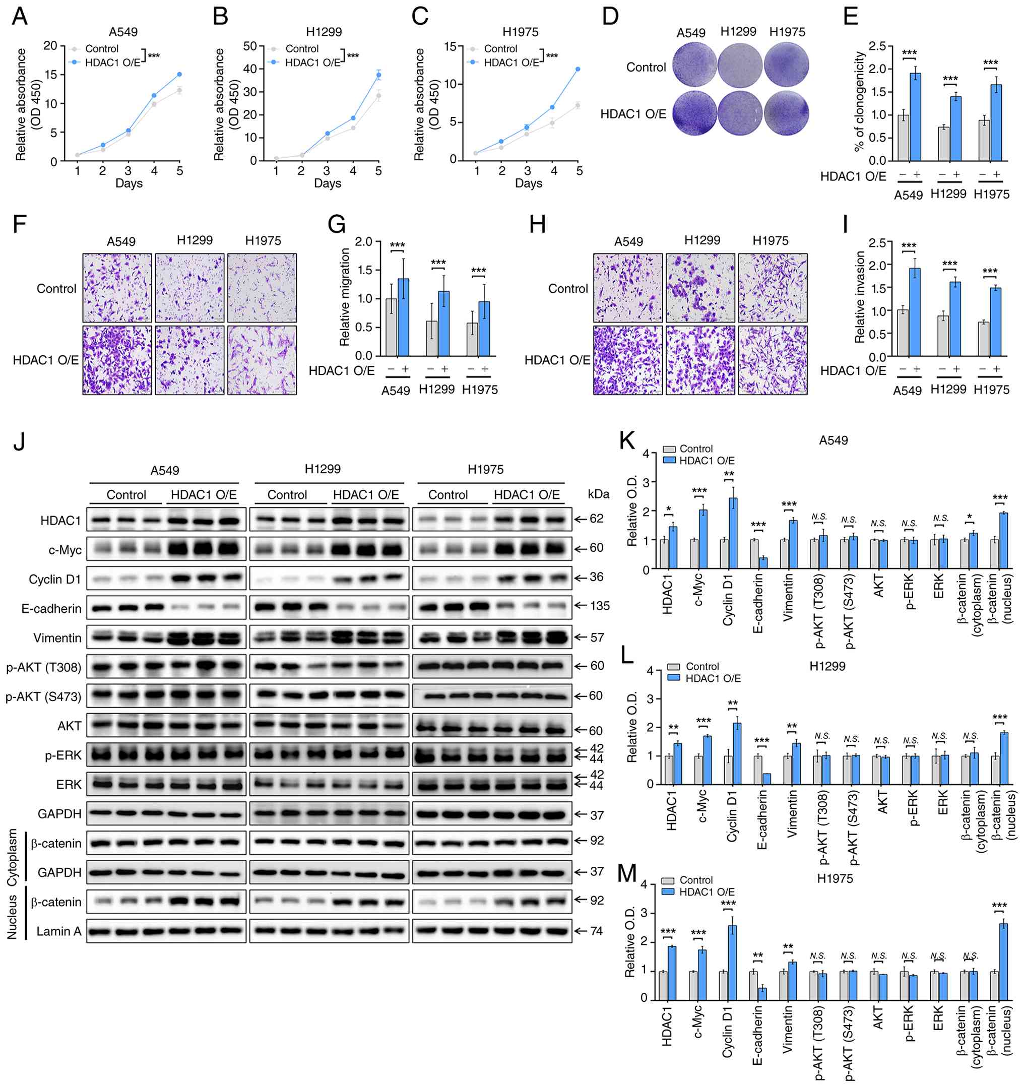 | Figure 3.Effects of HDAC1 overexpression on
proliferation, migration, invasion and downstream signaling in
NSCLC cells. A549, H1299 and H1975 cells were stably transduced
with HDAC1-overexpressing lentivirus (HDAC1 O/E) or empty vector
(Control). Cell proliferation was measured using CCK-8 assay over a
96-h period in (A) A549, (B) H1299 and (C) H1975 cells. (D)
Representative images of colony formation assays in A549, H1299 and
H1975 cells. (E) Quantification of clonogenicity from colony
formation assays. (F) Representative images of Transwell migration
assays in A549, H1299 and H1975 cells. Crystal violet staining;
original magnification, ×200. (G) Quantification of relative
migration. (H) Representative images of Transwell invasion assays
performed using Matrigel-coated chambers. Crystal violet staining;
original magnification, ×200. (I) Quantification of relative
invasion. (J) Western blotting of HDAC1, c-Myc, cyclin D1,
E-cadherin, vimentin, p-AKT (Thr308), p-AKT (Ser473), total AKT,
p-ERK and total ERK in whole-cell lysates and β-catenin in
cytoplasmic and nuclear fractions of A549, H1299 and H1975 cells.
GAPDH and Lamin A served as loading controls for whole
cell/cytoplasmic and nuclear fractions, respectively. Densitometric
quantification of protein levels normalized to the respective
loading controls in (K) A549, (L) H1299 and (M) H1975 cells. Data
are presented as mean ± SD from three independent experiments and
were analyzed using two-tailed unpaired Student's t-test.
*P<0.05, **P<0.01 and ***P<0.001. N.S., not significant;
HDAC1, histone deacetylase 1; NSCLC, non-small cell lung cancer;
O/E, overexpressing. |
Next, the present study assessed the effect of HDAC1
O/E on downstream signaling molecules using western blotting and
nuclear-cytoplasmic fractionation (Fig.
3J-M). Consistent with the knockdown data, HDAC1 O/E led to
significant upregulation of c-Myc (P<0.01) and cyclin D1
(P<0.01) protein levels across all three cell lines, further
supporting the role of HDAC1 in driving cell cycle progression.
Regarding EMT-related markers, HDAC1 O/E resulted in a marked
decrease in E-cadherin expression (P<0.01) and a corresponding
increase in vimentin expression (P<0.01), indicating a shift
toward a mesenchymal phenotype. Subcellular fractionation analysis
demonstrated that nuclear β-catenin levels were substantially
elevated in HDAC1-O/E cells (P<0.001), whereas cytoplasmic
β-catenin remained largely unchanged (P>0.05), reinforcing the
idea that HDAC1 facilitates nuclear accumulation of β-catenin.
Consistent with the knockdown data, neither the phosphorylation of
AKT (Thr308 and Ser473) nor ERK or total AKT and ERK protein levels
were significantly affected by HDAC1 O/E (P>0.05 for all
comparisons). Collectively, the complementary knockdown and
overexpression results demonstrated that HDAC1 drives NSCLC cell
proliferation, migration and invasion, at least in part through
upregulation of c-Myc and cyclin D1, induction of EMT and promotion
of nuclear β-catenin accumulation, independent of the PI3K/AKT and
MAPK/ERK pathways.
β-catenin O/E reverses the
tumor-suppressive effects of HDAC1 K/D in NSCLC cells
To determine whether β-catenin signaling is
functionally required for HDAC1-mediated oncogenic effects, the
present study overexpressed β-catenin in HDAC1-K/D A549, H1299 and
H1975 cells. Four experimental groups were established:
Empty-vector control, HDAC1 K/D alone, β-catenin O/E alone and
HDAC1 K/D + β-catenin O/E. As shown in Fig. 4A-C, CCK-8 assays revealed that HDAC1
K/D alone significantly suppressed the proliferative capacity of
all three cell lines (P<0.001), whereas β-catenin O/E alone
enhanced cell proliferation (P<0.001). Notably, co-expression of
β-catenin in HDAC1-K/D cells restored proliferation to levels
comparable to those of the β-catenin O/E group (P>0.05, HDAC1
K/D + β-catenin O/E vs. β-catenin O/E), indicating that β-catenin
overexpression effectively abrogates the growth-inhibitory effect
of HDAC1 silencing. Colony formation assays yielded concordant
results; the reduced clonogenic ability caused by HDAC1 knockdown
was effectively rescued by concomitant β-catenin overexpression
(P>0.05, HDAC1 K/D + β-catenin O/E vs. β-catenin O/E; Fig. 4D and E). Similarly, Transwell assays
demonstrated that β-catenin O/E largely reversed the inhibitory
effects of HDAC1 K/D on both migration (P>0.05; Fig. 4F and G) and invasion (P>0.05;
Fig. 4H and I) in all three cell
lines. The double-modified group showed no significant difference
compared with the β-catenin O/E-alone group for either endpoint.
These results indicated that HDAC1 promotes NSCLC cell
proliferation, migration and invasion, at least in part through
β-catenin signaling.
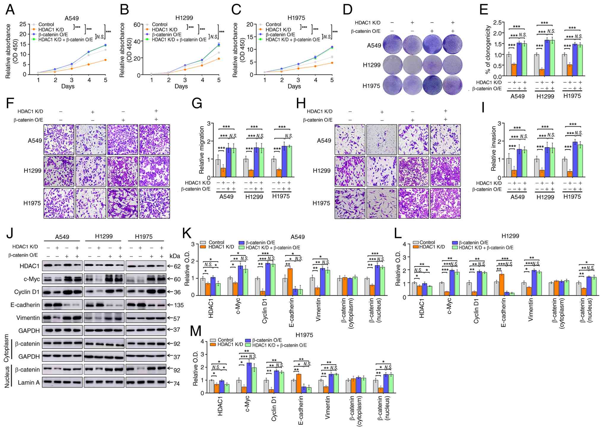 | Figure 4.Effects of combined HDAC1 knockdown
and β-catenin overexpression on malignant phenotypes and downstream
signaling in NSCLC cells. A549, H1299 and H1975 cells were stably
transduced to generate four experimental groups: empty-vector
control, HDAC1 K/D, β-catenin O/E and HDAC1 K/D + β-catenin O/E.
(A-C) Cell proliferation was measured using CCK-8 assay over a 96-h
period in (A) A549, (B) H1299 and (C) H1975 cells. (D)
Representative images of colony formation assays in A549, H1299 and
H1975 cells. (E) Quantification of clonogenicity from colony
formation assays. (F) Representative images of Transwell migration
assays in A549, H1299 and H1975 cells. Crystal violet staining;
original magnification, ×200. (G) Quantification of relative
migration. (H) Representative images of Transwell invasion assays
performed using Matrigel-coated chambers. Crystal violet staining;
original magnification, ×200. (I) Quantification of relative
invasion. (J) Western blotting of HDAC1, c-Myc, cyclin D1,
E-cadherin and vimentin in whole-cell lysates and β-catenin in
cytoplasmic and nuclear fractions of A549, H1299 and H1975 cells.
GAPDH and Lamin A served as loading controls for whole
cell/cytoplasmic and nuclear fractions, respectively. (K-M)
Densitometric quantification of protein levels normalized to the
respective loading controls in (K) A549, (L) H1299 and (M) H1975
cells. Data are presented as mean ± SD from three independent
experiments and were analyzed using one-way ANOVA followed by
Tukey's post hoc test. *P<0.05, **P<0.01 and ***P<0.001.
N.S., not significant; HDAC1, histone deacetylase 1; NSCLC,
non-small cell lung cancer; K/D, knockdown; O/E,
overexpression. |
Western blotting and nuclear-cytoplasmic
fractionation further corroborated these functional observations at
the molecular level (Fig. 4J-M).
Consistent with earlier findings, HDAC1 K/D alone reduced c-Myc,
cyclin D1 and vimentin protein levels while upregulating
E-cadherin. Simultaneous overexpression of β-catenin in HDAC1-K/D
cells effectively reversed these molecular changes: c-Myc, cyclin
D1 and vimentin levels were restored and E-cadherin was suppressed
to levels comparable with that of the β-catenin O/E-alone group
(P>0.05, HDAC1 K/D + β-catenin O/E vs. β-catenin O/E).
Subcellular fractionation revealed that nuclear β-catenin level,
which was reduced following HDAC1 K/D (P<0.01), was
substantially restored following β-catenin O/E (P>0.05), whereas
cytoplasmic β-catenin levels remained relatively stable across all
groups (P>0.05). Taken together, these results demonstrated that
β-catenin functions as a critical downstream effector of HDAC1 in
NSCLC cells. The tumor-promoting activities of HDAC1, including
enhanced proliferation, migration, invasion and EMT, are largely
dependent on β-catenin nuclear accumulation and its downstream
transcriptional activity.
β-catenin O/E inhibits HDAC1
K/D-mediated tumor growth inhibition in a xenograft tumor
model
To validate the proposed molecular mechanism in
vivo, the present study established a subcutaneous xenograft
tumor model. A549 cells stably expressing empty vector (control),
HDAC1 K/D, β-catenin O/E, or HDAC1 K/D + β-catenin O/E were
injected subcutaneously into the flanks of nude mice (n=5 per
group; Fig. 5A) and tumor growth
was monitored over a 28-day period. HDAC1 K/D significantly
suppressed xenograft tumor growth; both the final tumor volume and
tumor weight in the HDAC1 K/D group were significantly reduced
compared with those in the control group (P<0.001; Fig. 5B-D). Conversely, β-catenin O/E alone
yielded significantly larger and heavier tumors than controls
(P<0.001). Critically, simultaneous overexpression of β-catenin
in HDAC1-K/D cells restored tumor volume and weight to levels
comparable to those in the control group (P>0.05, HDAC1 K/D +
β-catenin O/E vs. control; Fig.
5C-E), effectively abolishing the growth-inhibitory effect of
HDAC1 silencing. Body weight remained stable in all four groups
throughout the experimental period, indicating no overt systemic
toxicity (Fig. 5F).
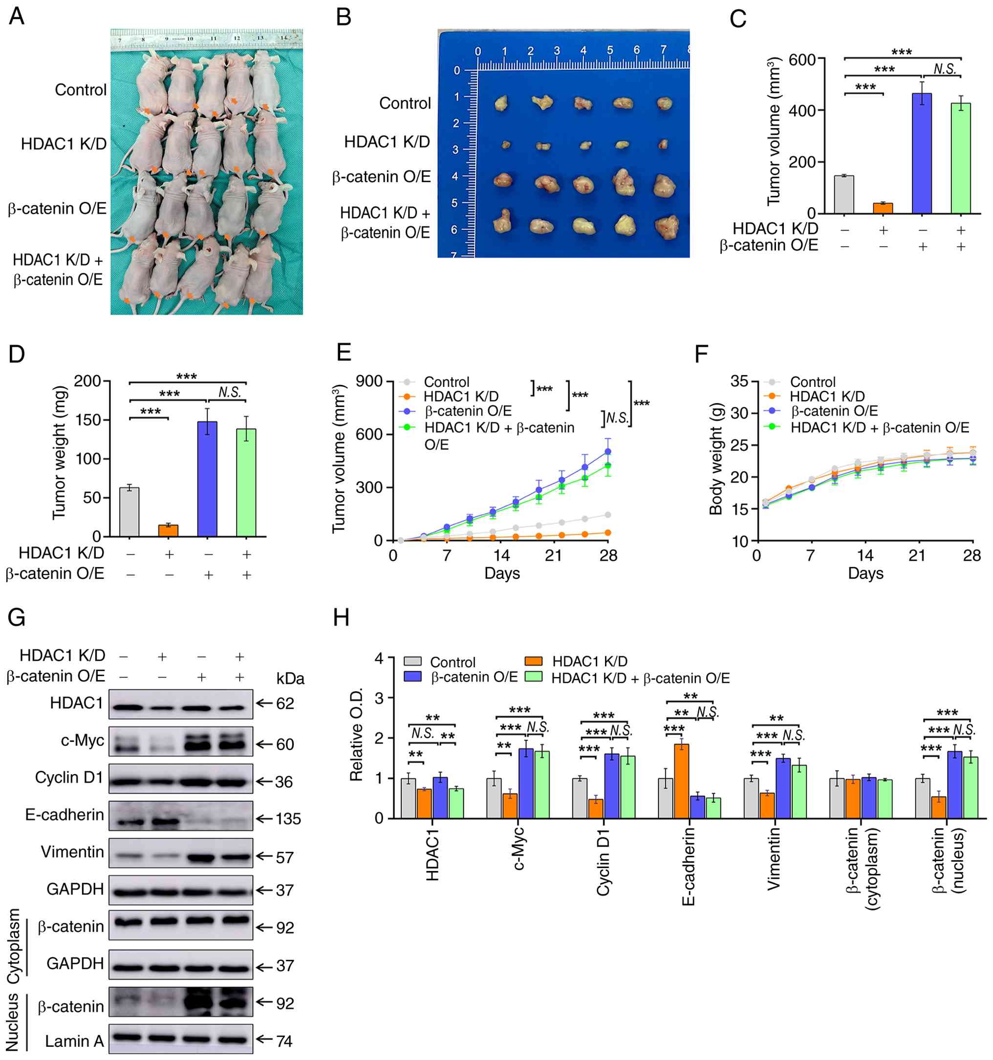 | Figure 5.In vivo effects of combined
HDAC1 knockdown and β-catenin overexpression on tumor growth and
downstream signaling in a xenograft tumor model. A549 cells stably
expressing empty vector (Control), HDAC1 K/D, β-catenin O/E, or
HDAC1 K/D + β-catenin O/E were injected subcutaneously into the
flanks of BALB/c nude mice (n=5 per group). (A) Representative
image of tumor-bearing mice at the experimental endpoint (day 28).
(B) Gross appearance of excised tumors from each group. (C) Final
tumor volume (mm3) and (D) Final tumor weight (mg) at
the experimental endpoint. (E) Tumor growth curves measured over a
28-day observation period. Tumor volume was calculated using the
formula V=0.5 × length × width2. (F) Body weight of mice
was monitored over the 28-day experimental period. (G) Western
blotting of HDAC1, c-Myc, cyclin D1, E-cadherin and vimentin in
whole-cell lysates and β-catenin in cytoplasmic and nuclear
fractions of harvested tumor tissues. GAPDH and Lamin A served as
loading controls for whole cell/cytoplasmic and nuclear fractions,
respectively. (H) Densitometric quantification of protein levels
normalized to the respective loading controls. Data are presented
as mean ± SD (n=5 per group) and were analyzed using one-way ANOVA
followed by Tukey's post hoc test. **P<0.01, ***P<0.001.
N.S., not significant; HDAC1, histone deacetylase 1. |
To determine whether the molecular changes observed
in vitro are recapitulated in vivo, the present study
performed western blotting of harvested tumor tissues (Fig. 5G and H). HDAC1 K/D reduced c-Myc,
cyclin D1 and vimentin protein levels (P<0.01) and upregulated
E-cadherin expression in xenograft tissues (P<0.01), which was
consistent with the in vitro data. Importantly, concomitant
β-catenin O/E in HDAC1-K/D cells effectively reversed these
alterations: c-Myc, cyclin D1 and vimentin levels were restored and
E-cadherin was suppressed to levels comparable to those in the
β-catenin O/E-alone group (P>0.05, HDAC1 K/D + β-catenin O/E vs.
β-catenin O/E). Subcellular fractionation of tumor lysates
demonstrated that nuclear β-catenin, which was reduced following
HDAC1 K/D (P<0.01), was substantially restored following
β-catenin O/E (P>0.05, HDAC1 K/D + β-catenin O/E vs. β-catenin
O/E), whereas cytoplasmic β-catenin levels remained relatively
stable across all groups (P>0.05). Notably, β-catenin O/E did
not alter HDAC1 protein levels (P>0.05), confirming that the
observed rescue effect was mediated by β-catenin reactivation
rather than by restoration of HDAC1 expression. In summary, these
in vivo data are in full agreement with the in vitro
observations and provide compelling evidence that HDAC1 promotes
LUAD tumorigenesis through a β-catenin-dependent mechanism.
Discussion
The main purpose of the present study was to
elucidate the molecular mechanisms by which HDAC1 influences LUAD
progression. The present study found that inhibition of HDAC1
markedly reduced nuclear β-catenin levels, whereas HDAC1 O/E
produced the opposite effect, increasing nuclear β-catenin
accumulation. In addition, the β-catenin O/E blocked the
tumor-suppressive effect of HDAC1 inhibition in NSCLC, both in
vitro and in vivo. These findings indicate that HDAC1
promotes LUAD tumorigenesis, at least in part, through modulation
of the β-catenin signaling pathway. HDAC1 functions primarily as a
transcriptional repressor. It has also been reported to interact
directly with β-catenin and to catalyze deacetylation that triggers
ubiquitin-mediated degradation (17,18).
On the basis of this mechanism, HDAC1 inhibition would be expected
to stabilize β-catenin, promote its nuclear accumulation (19) and thereby enhance transcription of
proliferation-related genes such as c-Myc and cyclin D1 (20–22).
The present data showed the opposite relationship in LUAD,
indicating that the effect of HDAC1 on β-catenin is
context-dependent. One explanation is that HDAC1 overexpression
selectively silences negative regulators of the Wnt/β-catenin
pathway, thereby relieving inhibition of β-catenin signaling.
Consistent with this possibility, HDAC inhibitor treatment
increased histone H3 acetylation at the promoters of the
Wnt/β-catenin repressors DKK1, SFRP1 and WIF1 and restored their
expression (23). HDAC1 inhibition
may thus reactivate tumor suppressor genes and attenuate tumor
progression (24). Elevated HDAC1
levels in LUAD may therefore drive aberrant Wnt/β-catenin signaling
through epigenetic silencing of endogenous β-catenin
inhibitors.
HDAC1 modulation altered the nuclear pool of
β-catenin without affecting the cytoplasmic pool, a pattern
consistent with the compartmentalized regulation of this protein.
Within the cell, β-catenin is distributed across three distinct
pools: Membranous, cytoplasmic and nuclear (25–27).
The nuclear pool constitutes only a minor fraction of the total
protein (28). A change large
enough to alter nuclear levels may therefore produce only a small,
statistically undetectable shift in the much larger cytoplasmic
pool. Importantly, the transcriptional activity of β-catenin
depends on the nuclear fraction and its binding to TCF/LEF factors
rather than on total abundance (29). Thus, these findings indicated that
HDAC1 acts principally on the nuclear accumulation and
transcriptional activity of β-catenin. The stability of the
cytoplasmic pool favors this interpretation over a
degradation-based mechanism, although altered β-catenin stability
cannot be entirely excluded.
The AKT and ERK signaling cascades serve as critical
regulators of proliferation and survival in NSCLC cells (30–32). A
previous study demonstrated that the HDAC1-Smad3-mSin3A
transcriptional complex coordinately suppresses c-Met expression,
subsequently reducing phosphorylation of both ERK and AKT, while
simultaneously inhibiting EMT and invasion in lung cancer (15); HDAC1 activity was associated with
reduced phosphorylation of AKT and ERK1/2. However, in the present
study, altering HDAC1 expression did not markedly affect the
phosphorylation of either kinase in NSCLC cells. This discrepancy
likely stems from key methodological differences between the
studies. Previous work has suggested that Smad3, rather than HDAC1,
directly mediates AKT/ERK regulation, implying that Smad3 may
engage in alternative mechanisms independent of HDAC1 (15). Accumulating evidence indicates that
Smad3 confers radiotherapy resistance in NSCLC cells through the
ITGA6/PI3K/AKT axis (33).
Furthermore, phosphorylated ERK directly interacts with and
modulates Smad3 function (34).
Thus, the present study indicated that HDAC1 does not independently
regulate AKT or ERK1/2 activity in NSCLC cell lines, highlighting
the inherent complexity of these signaling networks and
underscoring the importance of examining multiple regulatory nodes
when identifying therapeutic targets (11,12,35).
HDAC1 overexpression is associated with tumor
progression and poor prognosis in gastric cancer (36,37),
colorectal cancer (38,39) and LUAD (9). A meta-analysis further confirmed that
HDAC1 expression was associated with the histological
differentiation grade and worse overall survival in lung cancer,
although its association with lymph node metastasis remains
inconclusive (8). In the present
study, HDAC1 protein was highly expressed in 91.1% of the LUAD
tissues and was significantly associated with poor histological
differentiation (P=0.006) and lymph node metastasis (P=0.027).
Enrichment of HDAC1 in poorly differentiated tumors is biologically
plausible because HDAC1 is specifically overexpressed in cancer
stem cells and is essential for maintaining stem cell properties
(40). Mechanistically, HDAC1
promotes cellular dedifferentiation by forming transcriptional
repressor complexes that silence the differentiation-associated
genes via histone deacetylation (41,42).
Furthermore, the association between HDAC1 and lymph node
metastasis is consistent with the functional data of the present
study showing that HDAC1 drives EMT, enhances migratory and
invasive capacity and promotes nuclear β-catenin accumulation.
Collectively, these findings supported HDAC1 as both a functional
driver of aggressive LUAD behavior and a potential biomarker for
identifying high-risk patients.
The present study has several limitations. First,
although survival data would further strengthen the clinical
significance of the findings, a reliable survival analysis was not
feasible in the present cohort. The low-HDAC1 expression group was
small (n=14), the follow-up was relatively short and no mortality
occurred in this group. Consequently, no significant difference in
survival was detected between the high- and low-expression groups.
This imbalance reflects the high prevalence of HDAC1 expression in
lung adenocarcinoma, which leaves few low-expression cases for
comparison, rather than indicating a sampling bias. Future studies
are needed to clarify the prognostic value of HDAC1 in larger
cohorts with longer follow-up. Second, only the PI3K/AKT and
MAPK/ERK pathways were examined alongside Wnt/β-catenin. Other
HDAC1-regulated pathways that may contribute to tumor progression
were not assessed. The present study therefore established
β-catenin as a necessary downstream effector without excluding
additional mechanisms.
In conclusion, the present study demonstrated that
HDAC1 inhibition markedly suppressed the proliferation, migration
and invasion of NSCLC cells, accompanied by reduced nuclear
β-catenin levels. Conversely, HDAC1 overexpression promoted these
malignant behaviors by enhancing β-catenin nuclear localization.
Moreover, restoration of β-catenin expression effectively abolished
the anti-tumor effects of HDAC1 inhibition both in vitro and
in vivo, indicating that HDAC1 promotes LUAD progression, at
least in part, through β-catenin signaling. Furthermore,
HDAC1-mediated regulation occurred independently of the major
proliferative pathways, as neither AKT nor ERK1/2 phosphorylation
was affected by HDAC1 modulation alone. These findings provided a
strong rationale for developing HDAC1 inhibitor-based therapeutic
strategies for LUAD.
Supplementary Material
Supporting Data
Acknowledgments
Not applicable.
Funding
The present study was supported by the Nanhai Xinxing project of
Hainan Province (grant no. NHXXRCXM202350) and the National Natural
Science Foundation of China (grant nos. 82460608 and 82260275).
Support was also provided by the Joint Program on Health Science
& Technology Innovation of Hainan Province (grant nos.
WSJK2026MS254 and WSJK2026QN184).
Availability of data and materials
The data generated in the present study may be
requested from the corresponding author.
Authors' contributions
SX and XX designed the study and acquired funding.
SX, XC, FW, JX and CL performed the experiments and analyzed the
data. XW and HZ collected the clinical samples and provided
materials. JL provided materials and supervised the study. SX
drafted the manuscript. JL and XX reviewed and edited the
manuscript. SX, JL and XX confirm the authenticity of all the raw
data. All authors read and approved the final manuscript.
Ethics approval and consent to
participate
The animal study was approved by Institutional
Animal Care and Use Committee (IACUC) at Hainan Medical University
(approval no. 2024.331). Human subject studies were approved by the
Institutional Review Committee of the Affiliated Cancer Hospital of
Hainan Medical University (approval no. 2024.39). Written informed
consent was obtained from all participants prior to surgery.
Patient consent for publication
Not applicable.
Competing interests
The authors declare that they have no competing
interests.
References
|
1
|
Sung H, Ferlay J, Siegel RL, Laversanne M,
Soerjomataram I, Jemal A and Bray F: Global cancer statistics 2020:
GLOBOCAN estimates of incidence and mortality worldwide for 36
cancers in 185 countries. CA Cancer J Clin. 71:209–249.
2021.PubMed/NCBI
|
|
2
|
Majeed U, Manochakian R, Zhao Y and Lou Y:
Targeted therapy in advanced non-small cell lung cancer: Current
advances and future trends. J Hematol Oncol. 14:1082021. View Article : Google Scholar : PubMed/NCBI
|
|
3
|
Lu T, Yang X, Huang Y, Zhao M, Li M, Ma K,
Yin J, Zhan C and Wang Q: Trends in the incidence, treatment and
survival of patients with lung cancer in the last four decades.
Cancer Manag Res. 11:943–953. 2019. View Article : Google Scholar : PubMed/NCBI
|
|
4
|
Gallinari P, Di Marco S, Jones P, Pallaoro
M and Steinkühler C: HDACs, histone deacetylation and gene
transcription: From molecular biology to cancer therapeutics. Cell
Res. 17:195–211. 2007. View Article : Google Scholar : PubMed/NCBI
|
|
5
|
Seto E and Yoshida M: Erasers of histone
acetylation: The histone deacetylase enzymes. Cold Spring Harb
Perspect Biol. 6:a0187132014. View Article : Google Scholar : PubMed/NCBI
|
|
6
|
Zhang LB, Bu L, Hu J, Xu ZY, Ruan LB, Fang
Y and Wang P: HDAC1 knockdown inhibits invasion and induces
apoptosis in non-small cell lung cancer cells. Biol Chem.
399:603–610. 2018. View Article : Google Scholar : PubMed/NCBI
|
|
7
|
Sasaki H, Moriyama S, Nakashima Y,
Kobayashi Y, Kiriyama M, Fukai I, Yamakawa Y and Fujii Y: Histone
deacetylase 1 mRNA expression in lung cancer. Lung Cancer.
46:171–178. 2004. View Article : Google Scholar : PubMed/NCBI
|
|
8
|
Cao LL, Song XX, Pei L, Liu LH, Wang H and
Jia M: Histone deacetylase HDAC1 expression correlates with the
progression and prognosis of lung cancer: A meta-analysis. Medicine
(Baltimore). 96:e76632017. View Article : Google Scholar : PubMed/NCBI
|
|
9
|
Minamiya Y, Ono T, Saito H, Takahashi N,
Ito M, Mitsui M, Motoyama S and Ogawa J: Expression of histone
deacetylase 1 correlates with a poor prognosis in patients with
adenocarcinoma of the lung. Lung Cancer. 74:300–304. 2011.
View Article : Google Scholar : PubMed/NCBI
|
|
10
|
Wang L, Li H, Ren Y, Zou S, Fang W, Jiang
X, Jia L, Li M, Liu X, Yuan X, et al: Targeting HDAC with a novel
inhibitor effectively reverses paclitaxel resistance in non-small
cell lung cancer via multiple mechanisms. Cell Death Dis.
7:e20632016. View Article : Google Scholar : PubMed/NCBI
|
|
11
|
Lin YC, Lin YC, Shih JY, Huang WJ, Chao
SW, Chang YL and Chen CC: DUSP1 expression induced by HDAC1
inhibition mediates gefitinib sensitivity in non-small cell lung
cancers. Clin Cancer Res. 21:428–438. 2015. View Article : Google Scholar : PubMed/NCBI
|
|
12
|
Zhang LL, Li Q, Zhong DS, Zhang WJ, Sun XJ
and Zhu Y: MCM5 aggravates the HDAC1-mediated malignant progression
of lung cancer. Front Cell Dev Biol. 9:6691322021. View Article : Google Scholar : PubMed/NCBI
|
|
13
|
Fan Y, Ji X, Yuan K, Wu Q and Lou M: HDAC1
Mediates immunosuppression of the tumor microenvironment in
non-small cell lung cancer. J Inflamm Res. 18:3333–3347. 2025.
View Article : Google Scholar : PubMed/NCBI
|
|
14
|
Kong Y, Jiang R, Zhou H, Ge M, Lin H, Wang
Y, Yao R, Wang Q, Liang X, Li J and Zhou X: PHF12 regulates HDAC1
to promote tumorigenesis via EGFR/AKT signaling pathway in
non-small cell lung cancer. J Transl Med. 22:6892024. View Article : Google Scholar : PubMed/NCBI
|
|
15
|
Gui HX, Peng J, Yang ZP, Chen LY, Zeng H,
Shao YT, Mu X, Hao Q, Yang Y, An S, et al: HDAC1-Smad3-mSin3A
complex is required for Smad3-induced transcriptional inhibition of
hepatocyte growth factor receptor in human lung cancers.
Carcinogenesis. 42:587–600. 2021. View Article : Google Scholar : PubMed/NCBI
|
|
16
|
Amin MB, Edge SB, Greene FL, Byrd DR,
Brookland RK, Washington MK, Gershenwald JE, Compton CC, Hess KR,
Sullivan DC, et al: AJCC Cancer Staging Manual. (8th edition).
Springer; New York, NY: 2017
|
|
17
|
Liu E, Zhou Q, Xie AJ, Li X, Li M, Ye J,
Li S, Ke D, Wang Q, Xu ZP, et al: Tau acetylates and stabilizes
β-catenin thereby promoting cell survival. EMBO Rep. 21:e483282020.
View Article : Google Scholar : PubMed/NCBI
|
|
18
|
You H, Li Q, Kong D, Liu X, Kong F, Zheng
K and Tang R: The interaction of canonical Wnt/β-catenin signaling
with protein lysine acetylation. Cell Mol Biol Lett. 27:72022.
View Article : Google Scholar : PubMed/NCBI
|
|
19
|
Huang X, Xu J, Huang M, Li J, Dai L, Dai K
and Zhang X: Histone deacetylase1 promotes TGF-β1-mediated early
chondrogenesis through down-regulating canonical Wnt signaling.
Biochem Biophys Res Commun. 453:810–816. 2014. View Article : Google Scholar : PubMed/NCBI
|
|
20
|
Xu HT, Wang L, Lin D, Liu Y, Liu N, Yuan
XM and Wang EH: Abnormal β-catenin and reduced axin expression are
associated with poor differentiation and progression in non-small
cell lung cancer. Am J Clin Pathol. 125:534–541. 2006. View Article : Google Scholar : PubMed/NCBI
|
|
21
|
He TC, Sparks AB, Rago C, Hermeking H,
Zawel L, Da Costa LT, Morin PJ, Vogelstein B and Kinzler KW:
Identification of c-MYC as a target of the APC pathway. Science.
281:1509–1512. 1998. View Article : Google Scholar : PubMed/NCBI
|
|
22
|
Shtutman M, Zhurinsky J, Simcha I,
Albanese C, D'Amico M, Pestell R and Ben-Ze'ev A: The cyclin D1
gene is a target of the β-catenin/LEF-1 pathway. Proc Natl Acad Sci
USA. 96:5522–5527. 1999. View Article : Google Scholar : PubMed/NCBI
|
|
23
|
Foltz G, Yoon JG, Lee H, Ma L, Tian Q,
Hood L and Madan A: Epigenetic regulation of wnt pathway
antagonists in human glioblastoma multiforme. Genes Cancer.
1:81–90. 2010. View Article : Google Scholar : PubMed/NCBI
|
|
24
|
West AC and Johnstone RW: New and emerging
HDAC inhibitors for cancer treatment. J Clin Invest. 124:30–39.
2014. View
Article : Google Scholar : PubMed/NCBI
|
|
25
|
Kumar R and Bashyam MD: Multiple oncogenic
roles of nuclear beta-catenin. J Biosci. 42:695–707. 2017.
View Article : Google Scholar : PubMed/NCBI
|
|
26
|
Nelson WJ and Nusse R: Convergence of Wnt,
beta-catenin and cadherin pathways. Science. 303:1483–1487. 2004.
View Article : Google Scholar : PubMed/NCBI
|
|
27
|
Valenta T, Hausmann G and Basler K: The
many faces and functions of β-catenin. EMBO J. 31:2714–2736. 2012.
View Article : Google Scholar : PubMed/NCBI
|
|
28
|
Maher MT, Mo R, Flozak AS, Peled ON and
Gottardi CJ: Beta-catenin phosphorylated at serine 45 is spatially
uncoupled from beta-catenin phosphorylated in the GSK3 domain:
Implications for signaling. PLoS One. 5:e101842010. View Article : Google Scholar : PubMed/NCBI
|
|
29
|
MacDonald BT, Tamai K and He X:
Wnt/beta-catenin signaling: Components, mechanisms and diseases.
Dev Cell. 17:9–26. 2009. View Article : Google Scholar : PubMed/NCBI
|
|
30
|
Scrima M, De Marco C, Fabiani F, Franco R,
Pirozzi G, Rocco G, Ravo M, Weisz A, Zoppi P, Ceccarelli M, et al:
Signaling networks associated with AKT activation in non-small cell
lung cancer (NSCLC): New insights on the role of
phosphatydil-inositol-3 kinase. PLoS One. 7:e304272012. View Article : Google Scholar : PubMed/NCBI
|
|
31
|
Ciuffreda L, Cesta Incani U, Steelman LS,
Abrams SL, Falcone I, Del Curatolo A, Chappell WH, Franklin RA,
Vari S, Cognetti F, et al: Signaling intermediates (MAPK and PI3K)
as therapeutic targets in NSCLC. Curr Pharm Des. 20:3944–3957.
2014. View Article : Google Scholar : PubMed/NCBI
|
|
32
|
Vicent S, López-Picazo JM, Toledo G,
Lozano MD, Torre W, Garcia-Corchón C, Quero C, Soria JC,
Martín-Algarra S, Manzano RG and Montuenga LM: ERK1/2 is activated
in non-small-cell lung cancer and associated with advanced tumours.
Br J Cancer. 90:1047–1052. 2004. View Article : Google Scholar : PubMed/NCBI
|
|
33
|
Han F, Chen S, Zhang K, Zhang K, Wang M
and Wang P: Single-cell transcriptomic sequencing data reveal
aberrant DNA methylation in SMAD3 promoter region in
tumor-associated fibroblasts affecting molecular mechanism of
radiosensitivity in non-small cell lung cancer. J Transl Med.
22:2882024. View Article : Google Scholar : PubMed/NCBI
|
|
34
|
Matsuura I, Wang G, He D and Liu F:
Identification and characterization of ERK MAP kinase
phosphorylation sites in Smad3. Biochemistry. 44:12546–12553. 2005.
View Article : Google Scholar : PubMed/NCBI
|
|
35
|
Ma T, Yan B, Hu Y and Zhang Q: HOXA10
promotion of HDAC1 underpins the development of lung adenocarcinoma
through the DNMT1-KLF4 axis. J Exp Clin Cancer Res. 40:712021.
View Article : Google Scholar : PubMed/NCBI
|
|
36
|
Jiang Z, Yang H, Zhang X, Wang Z, Rong R
and Wang X: Histone deacetylase-1 as a prognostic factor and
mediator of gastric cancer progression by enhancing glycolysis. Hum
Pathol. 85:194–201. 2019. View Article : Google Scholar : PubMed/NCBI
|
|
37
|
Cao LL, Yue ZH, Liu LH, Pei L, Yin Y, Qin
L, Zhao J, Liu HX, Wang H and Jia M: The expression of histone
deacetylase HDAC1 correlates with the progression and prognosis of
gastrointestinal malignancy. Oncotarget. 8:39241–39253. 2017.
View Article : Google Scholar : PubMed/NCBI
|
|
38
|
Weichert W, Röske A, Niesporek S, Noske A,
Buckendahl AC, Dietel M, Gekeler V, Boehm M, Beckers T and Denkert
C: Class I histone deacetylase expression has independent
prognostic impact in human colorectal cancer: Specific role of
class I histone deacetylases in vitro and in vivo. Clin Cancer Res.
14:1669–1677. 2008. View Article : Google Scholar : PubMed/NCBI
|
|
39
|
Ishihama K, Yamakawa M, Semba S, Takeda H,
Kawata S, Kimura S and Kimura W: Expression of HDAC1 and CBP/p300
in human colorectal carcinomas. J Clin Pathol. 60:1205–1210. 2007.
View Article : Google Scholar : PubMed/NCBI
|
|
40
|
Witt AE, Lee CW, Lee TI, Azzam DJ, Wang B,
Caslini C, Petrocca F, Grosso J, Jones M, Cohick EB, et al:
Identification of a cancer stem cell-specific function for the
histone deacetylases, HDAC1 and HDAC7, in breast and ovarian
cancer. Oncogene. 36:1707–1720. 2017. View Article : Google Scholar : PubMed/NCBI
|
|
41
|
Rivas M, Johnston ME II, Gulati R, Kumbaji
M, Aguiar TFM, Timchenko L, Krepischi A, Shin S, Bondoc A, Tiao G,
et al: HDAC1-dependent repression of markers of hepatocytes and P21
is involved in development of pediatric liver cancer. Cell Mol
Gastroenterol Hepatol. 12:669–682. 2021.PubMed/NCBI
|
|
42
|
Xie J, Wang Z, Fan W, Liu Y, Liu F, Wan X,
Liu M, Wang X, Zeng D, Wang Y, et al: Targeting cancer cell
plasticity by HDAC inhibition to reverse EBV-induced
dedifferentiation in nasopharyngeal carcinoma. Signal Transduct
Target Ther. 6:3332021. View Article : Google Scholar : PubMed/NCBI
|