Introduction
Thyroid cancer is one of the most common
malignancies that accounts for ~2.5% of all cancer cases (1). Based on the latest cancer statistics
reports, the global number of new cases and mortality from thyroid
cancer per year reached 821,173 and 47,485, respectively, among
which China makes up 466,118 of new cases and 11,564 of mortalities
(2,3). Thyroid cancer is histologically
classified into three main subtypes: i) Differentiated [primarily
comprising papillary thyroid cancer (PTC) and follicular thyroid
cancer]; ii) poorly differentiated; and iii) undifferentiated
(anaplastic) thyroid cancer (4); of
these, PTC is the most prevalent accounting for 80–90% of thyroid
cancer cases (5). After standard
treatment involving radical resection followed by adjuvant
endocrine therapy or radioactive iodine ablation, the majority of
patients with PTC are able to obtain long-term survival. However,
for some patients, particularly those with metastasis or high
recurrent risk, the prognosis is still unsatisfactory (6,7). Thus,
deep investigation into the pathogenesis underlying PTC initiation
and progress remains important to further improve the outcomes.
Histone lactylation is a recently discovered
post-translational modification (PTM) of lysine (K) residues,
characterized by the covalent attachment of lactate to the ε-amino
group (−NH₂) of histones via an ester bond, forming lactylated
lysine (8,9). Although the concept of histone
lactylation was recently proposed, its relation to carcinogenesis
has attracted attention. Several recent reviews have preliminarily
summarized its implications in the pathogenesis of cancers via
multiples mechanisms, including metabolic reconstruction, immune
infiltration, angiogenesis and tumor invasiveness (10–12).
Among the histone lactylated sites, histone H3 lysine 18
lactylation (H3K18la) is identified in numerous types of tissues
and cells (13) and is the most
extensively studied site for histone lactylation (14). Moreover, the function of H3K18la is
largely investigated in a number of complex diseases such as
cancer, respiratory disease, renal disease and neurodegenerative
disease by recent studies (15–18).
Particularly in the cancer field, H3K18la is able to promote the
progression of gastric, lung, breast and ovarian cancer (15,19–21).
However, the role of H3K18la in PTC pathogenesis is still not
reported. Considering the general functions of histone lactylation
and the specific effects of H3K18la in cancers (10–12,15,19–21),
it is hypothesized that H3K18la is likely involved in the
pathogenesis of PTC.
Oxidized low density lipoprotein receptor 1 (OLR1),
the gene encoding lectin-like oxidized low density lipoprotein
receptor-1 (LOX-1), belongs to the C-type lectin superfamily and is
a 50 kDa transmembrane glycoprotein that recognizes various ligands
including oxidized low-density lipoprotein (oxLDL) and participates
in various metabolic activities (22–24).
Notably, OLR1 is able to facilitate the progression of cancers
through multiple mechanisms such as regulating proliferation,
epithelial-mesenchymal transition (EMT), angiogenesis, immune
microenvironment and drug resistance (23,25).
For example, one study indicated that OLR1 promoted the migration,
invasion and EMT of gastric cancer through the PI3K/AKT/GSK3β
pathway (26). Another study
reported that the activation of OLR1 by oxLDL elevated the
expressions of MMP2, MMP9 and VEGF, thereby inducing tumor
angiogenesis and growth in prostate cancer (27). Moreover, OLR1 knockdown represses
colon cancer growth and chemoresistance by inhibiting glycolytic
metabolism and lactate production (28). However, the relation between OLR1
and PTC is rarely investigated. Therefore, in the present study, a
preliminary investigation was performed and it was found that OLR1
was upregulated in thyroid cancer tissues versus non-tumor tissues
and worse overall survival (OS) was predicted in patients with
thyroid cancer via the Gene Expression Profiling Interactive
Analysis (GEPIA) database. Moreover, it was also observed that
LOX-1 (OLR1 coding protein) and H3K18la were upregulated in PTC
tissues compared with non-tumor tissues and LOX-1 was positively
associated with H3K18la in PTC tissues. Based on the prior studies
and our preliminary investigation, a possible connection between
OLR1 and H3K18la in PTC was hypothesized and they might be closely
implicated in the PTC pathogenesis.
Based on the aforementioned information, the present
study aimed to investigate the effect of OLR1 modification on PTC
proliferation, apoptosis and invasion and its interaction with
H3K18la and downstream molecular mechanisms.
Materials and methods
GEPIA public database analyses
The ‘Expression Boxplot’, ‘Expression Stage Plot’
and ‘Survival Plots’ modules on the GEPIA database (http://gepia.cancer-pku.cn/index.html)
were used to analyze the expression of OLR1 in tumor and adjacent
tissues and its correlation with tumor stage and survival in
thyroid carcinoma.
Immunohistochemistry (IHC)
A total of 20 PTC patients (between April 2024 and
July 2025) were included, with median age of 46.5 years (range:
29–58 years). Among them 3 (15%) were males and 17 (85%) were
females. Paired tumor and adjacent tumor tissues (~1 cm from the
tumor margin) were obtained both retrospectively using stored
samples and prospectively. The tissues were fixed in 4%
paraformaldehyde for 24 h, embedded in paraffin and cut into 4-µm
slices. The slides were deparaffinized, rehydrated and
antigen-restored, successively. After removing the endogenous
peroxidase in 3% H2O2, the slices were
blocked by 5% goat serum (Wuhan Servicebio Technology Co., Ltd.)
for 30 minutes (min) at room temperature and cultivated in
anti-LOX-1 antibody (1:200; cat. no. DF6522, Affinity Biosciences)
or anti-H3K18la (1:500; cat. no. PTM-1406RM, PTM BIO) at 4°C
overnight. Subsequently, the slices were incubated with
anti-rabbit-HRP secondary antibody (1:2,000; cat. no. GB23303,
Wuhan Servicebio Technology Co., Ltd.) at room temperature for 30
min. Finally, a DAB kit (Wuhan Servicebio Technology Co., Ltd.) was
used to develop the color. The slices were counterstained by
hematoxylin (Wuhan Servicebio Technology Co., Ltd.) at room
temperature for 3 min. The images were captured under an inverted
microscope (Motic Incorporation, Ltd.). The slices were analyzed by
two pathologists, independently. The IHC score was the result of
the staining intensity (score of 0, 1, 2 or 3) multiplied by the
staining percentage (score of 1, 2, 3 or 4), of which the highest
was 12. The final score was the average assessment by two
pathologists. The present study was approved by the Ethics
Committee of Harbin Medical University Cancer Hospital with
approval number YD2024-13 on November 4, 2024. The Ethics Committee
formally waived the requirement for additional informed consent for
the study. Moreover, participants had provided written consent for
the secondary use of biological specimens and medical data as part
of routine clinical procedures at our institution.
Cell culture
The human PTC cell lines TPC-1 (Cellverse Bioscience
Technology Co., Ltd.) and IHH-4 (Cellverse Bioscience Technology
Co., Ltd.) were obtained. RPMI-1640 (Procell Life Science &
Technology Co., Ltd.) with 10% fetal bovine serum (Procell Life
Science & Technology Co., Ltd.) was used to culture the cells
in 5% CO2 and at 37°C.
OLR1 plasmid and siRNA
transfection
Negative control (NC) overexpression plasmid (oeNC;
0.8 µg; Shanghai GenePharma Co., Ltd.), OLR1 overexpression plasmid
(oeOLR1; 0.8 µg; Shanghai GenePharma Co., Ltd.), NC siRNA (siNC; 50
nM; Shanghai GenePharma Co., Ltd.) and OLR1 siRNA (siOLR1; 50 nM;
Shanghai GenePharma Co., Ltd.) were transfected into TPC-1 and
IHH-4 cells at 37°C for 6 h. Transfect-mate (Shanghai GenePharma
Co., Ltd.) was applied to complete the transfection. At 48 h after
transfection, LOX-1 (OLR1 coding protein) expression, LDHA
expression, cell proliferation, cell apoptosis, cell invasion,
lactate concentration, H3K18la expression and LEM domain containing
1 (LEMD1) expression were assessed. The siRNA sequences were:
siOLR1 (sense, 5′-CAGCUGAUCUGGACUUCAUTT-3′, antisense,
5′-AUGAAGUCCAGAUCAGCUGTT-3′) and siNC (sense,
5′-UUCUCCGAACGUGUCACGUTT-3′, antisense,
5′-ACGUGACACGUUCGGAGAATT-3′).
Lactate and oxamate incubation
The TPC-1 and IHH-4 cells were incubated with 10 mM
L-sodium lactate (Shanghai Aladdin Biochemical Technology Co.,
Ltd.) and 10 mM sodium oxamate (Shanghai Aladdin Biochemical
Technology Co., Ltd.), respectively. H3K18la expression and H3K18la
enrich level on the LEMD1 promoter were evaluated.
Oxamate incubation and OLR1 plasmid
transfection
oeNC and oeOLR1 were transfected into TPC-1 and
IHH-4 cells. Meanwhile, 10 mM sodium oxamate was added and
incubated with the cells. Afterwards, H3K18la expression, LEMD1
expression, cell proliferation, cell apoptosis and cell invasion
were measured.
LEMD1 siRNA and OLR1 plasmid
transfection
oeNC (0.8 µg) + siNC (50 nM), oeOLR1 (0.8 µg) + siNC
(50 nM), oeNC (0.8 µg) + LEMD1 siRNA (siLEMD1; 50 nM; Shanghai
GenePharma Co., Ltd.) and oeOLR1 + siLEMD1 were transfected into
TPC-1 and IHH-4 cells in the presence of Transfect-mate (Shanghai
GenePharma Co., Ltd.) at 37°C for 6 h. LOX-1 (OLR1 coding protein)
expression, LEMD1 expression, cell proliferation, cell apoptosis
and cell invasion were analyzed at 48 h after transfection. The
siRNA sequences of siLEMD1 were listed: siLEMD1 (sense,
5′-GUGUCUGAGUGACUGUAAATT-3′, antisense,
5′-UUUACAGUCACUCAGACACTT-3′).
Cell proliferation, cell apoptosis and
lactate assessment
Cell counting kit-8 reagent (Wuhan Servicebio
Technology Co., Ltd.) was incubated with the cells at 0, 24, 48 and
72 h after transfection for 2 h. The OD value was read at 450 nm
and a cell apoptosis kit (Wuhan Servicebio Technology Co., Ltd.)
was used to analyze cell apoptosis 48 h after treatment. The cells
were incubated in Annexin V and propidium iodide working solution
for 15 min in darkness at room temperature. A BD FACSCalibur flow
cytometer (BD Biosciences) was used to analyze the cells. The data
was analyzed by FlowJo X (BD Biosciences). The percentage of early
+ late apoptotic cells was calculated. The concentration of lactate
was analyzed by a Lactic Acid Content Assay Kit (Abbkine Scientific
Co., Ltd.) according to the manufacturer's instructions 48 h after
transfection.
Cell invasion
After transfection for 48 h, the cells were
harvested. Transwell inserts (0.8 µm; Wuhan NEST Biotechnology Co.,
Ltd.) were cultured with Matrigel Matrix (BD Biosciences) at 37°C
for 1 h. After which, the cells (2×10⁴) in phosphate
buffer saline were loaded into the upper chamber of Transwell
inserts and the lower chamber was filled with 10% FBS-containing
RPMI-1640. The inserts were incubated for 24 h at 37°C. Afterwards,
the cells were stained in crystal violet solution (Wuhan Servicebio
Technology Co., Ltd.) at room temperature for 5 min after being
incubated with 4% paraformaldehyde at room temperature for 15 min.
Finally, the invasive cells were counted with a microscope (Motic
Incorporation, Ltd.).
Western blotting
The cells were lysed to extract total protein using
RIPA buffer (Wuhan Servicebio Technology Co., Ltd.) 48 h after
transfection or lactate/oxamate incubation. Total protein was
quantified using a BCA kit (Wuhan Servicebio Technology Co., Ltd.).
A total of 10 µg thermally denatured protein was loaded on 4–20%
pre-cast gels, separated and transferred to a nitrocellulose
membrane (Whatman plc; Cytiva). The membrane was then blocked in 5%
skimmed milk (Wuhan Servicebio Technology Co., Ltd.) at 37°C for 60
min. Primary antibodies, including anti-LOX-1 antibody (1:2,000;
cat. no. DF6522, Affinity Biosciences), anti-LEMD1 antibody
(1:1,000; cat. no. CSB-PA715035LA01HU, Cusabio Technology, LLC)
anti-H3K18la (1:2,000; cat. no. PTM-1406RM, PTM BIO), anti-H3
antibody (1:5,000; cat. no. AF0863, Affinity Biosciences), LDHA
antibody (1:2,000; cat. no. DF6280, Affinity Biosciences) and
anti-GAPDH antibody (1:3,000; cat. no. GB15004, Wuhan Servicebio
Technology Co., Ltd.), were cultivated with the membrane overnight
at 4°C. After which, anti-rabbit-HRP secondary antibody (1:20,000;
cat. no. GB23303, Wuhan Servicebio Technology Co., Ltd.) was
incubated with the membrane for 90 min. Finally, the protein bands
were illuminated with an ECL kit (MeilunBio). The western blotting
images were quantified using ImageJ software (v1.8; National
Institutes of Health).
Chromatin immunoprecipitation (ChIP)
assay
The cells (1×10⁷) were crosslinked with
formaldehyde for 10 min at 37°C and suspended in sodium dodecyl
sulfate buffer. After which, the cells were ultrasonically lysed
(25% frequency; 30 sec on, 30 sec off, 20 cycles) on ice and
centrifuged. The lysate (100 µl) was incubated with IgG (1:50) or
H3K18la (1:50) antibody at 4°C overnight, followed by incubation
with protein A+G Agarose (Beyotime Biotechnology) for 1 h, and the
1 µl lysate was used as input. After incubation, the lysate was
centrifuged (1,000 × g) at 4°C for 10 min and washed with specific
buffer (IP Elution Buffer), successively. After which, the
precipitate was reverse crosslinked and the DNA was analyzed with
Taq DNA Polymerase containing-SYBR Green qPCR Master Mix (Wuhan
Servicebio Technology Co., Ltd.). The thermal cycle procedures were
as follows: 95°C for 2 min, 1 cycle; 95°C for 15 sec, 61°C for 30
sec, for 40 cycles. The data was calculated using the %Input
method.
Xenograft experiment
The GV492 plasmid (Shanghai Genechem Co., Ltd.) and
GV872 plasmid (Shanghai Genechem Co., Ltd.) were used to establish
OLR1 overexpression plasmid and LEMD1 knockdown plasmid,
respectively. The lentivirus was constructed through second
generation system. The 20 µg OLR1 overexpression or LEMD1 knockdown
plasmid, 15 µg pHelper 1.0 plasmid and 10 µg pHelper 2.0 plasmid
were transfected into 293T cells (Cellverse Bioscience Technology
Co., Ltd) at 37°C for 6 h. The supernatant was collected to harvest
lentivirus at 48 h after transfection. The TPC-1 cells were
infected with OLR1 overexpression lentivirus, LEMD1 knock-down
lentivirus alone or in combination (Shanghai Genechem Co., Ltd.).
The multiplicity of infection (MOI) was 20. Moreover, control
lentivirus (Shanghai Genechem Co., Ltd.) was infected the TPC-1
cells. The stably infected TPC-1 cells were selected with 1.5 µg/ml
puromycin for 7 days. BALB/c nude mice with 4–6 weeks old and 18–22
g (Shanghai SLAC Laboratory Animal Co, Ltd.) were housed in
specific pathogen-free facility and randomly assigned into four
groups (three mice per group). The stably infected TPC-1 cells were
inoculated on the right flank of the mice (4×10⁶ cells
per mouse). A caliper was used to measure tumor volume every week.
The mice were sacrificed by cervical dislocation at four weeks. The
tumors were harvested, weighted, fixed and cut into 4 µm sections.
The IHC was performed as aforementioned, with the application of
anti-LOX-1 antibody (1:200), anti-H3K18la antibody (1:500) and
anti-LEMD1 antibody (1:100). The animal experiments were approved
by the Ethics Committee of Harbin Medical University Cancer
Hospital with approval number YD2024-13 and Wuhan Servicebio
Institutional Animal Care and Use Committee (IACUC) with approval
number 2025079-2.
Statistical analysis
All data in the present study were analyzed by
GraphPad 9.0 software (Dotmatics). The experiments were performed
in triplicate (n=3 in each group). Prior to conducting parametric
tests, the normality and homogeneity of variance were analyzed by
Shapiro-Wilk test and Brown-Forsythe test, respectively. If these
assumptions were met, one-way analysis of variance with Tukey's or
Dunnett's test was used for comparison among groups. A paired
t-test was applied for comparison between tumor and paired-adjacent
tumor groups. Pearson correlation coefficient test was adopted to
analyze the correlation of LOX-1 and H3K18la expression. P<0.05
was considered to indicate a statistically significant
difference.
Results
OLR1 and H3K18la expression in
PTC
Referring to the analyses based on the GEPIA public
database, OLR1 expression was upregulated in thyroid cancer tissues
compared with non-tumor tissues (Fig.
1A) and its expression differed among patients with thyroid
cancer at different TNM stages (Fig.
1B). Moreover, GEPIA also revealed that patients with thyroid
cancer with high OLR1 expression presented a worse OS compared with
those with low OLR1 expression (Fig.
1C). LOX-1 expression (OLR1 coding protein) was detected by IHC
assay in 20 pairs of PTC tumor and adjacent non-tumor tissues for
verification, which verified that LOX-1 expression was higher in
PTC tumor tissues compared with adjacent non-tumor tissues
(Fig. 1D). H3K18la expression was
also detected by IHC assay and it showed a higher expression in PTC
tumor tissues compared with adjacent non-tumor tissues (Fig. 1E). Moreover, LOX-1 expression was
positively associated with H3K18la expression in PTC tumor tissues
(Fig. 1F). LOX-1 expression was
also positively associated with tumor size of PTC (Fig. S1).
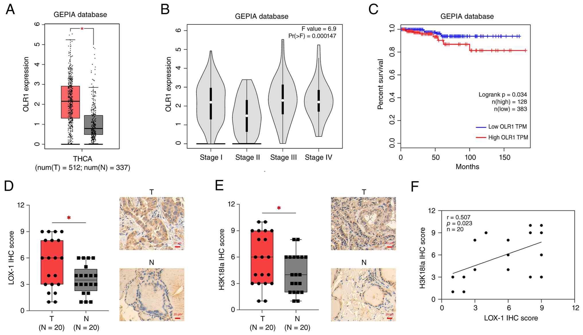 | Figure 1.Expression of OLR1 and H3K18la in
clinical samples. (A) OLR1 expression between thyroid cancer
tissues and non-tumor tissues, (B) its correlation with TNM stage
and (C) its correlation with OS in patients with thyroid cancer,
via the GEPIA database. Expressions and IHC image examples of (D)
LOX-1 and (E) H3K18la between PTC tissues and adjacent non-tumor
tissues and (F) the correlation between OLR1 and H3K18la expression
in PTC tissues. Magnification, ×200. *P<0.05. OLR1, oxidized low
density lipoprotein receptor 1; H3K18la, histone H3 lysine 18
lactylation; OS, overall survival; GEPIA, Gene Expression Profiling
Interactive Analysis; IHC, immunohistochemistry; LOX-1, lectin-like
oxidized low density lipoprotein receptor-1; PTC, papillary thyroid
cancer. |
OLR1 enhances PTC cell proliferation
and invasion, but repressed cell apoptosis
LOX-1 expression was upregulated after oeOLR1
transfection, but downregulated after siOLR1 transfection in TPC-1
and IHH-4 cells (Fig. 2A and B),
indicating the success of transfection. After which, cell
proliferation, apoptosis and invasion were detected after
transfection. oeOLR1 promoted cell proliferation, while siOLR1
inhibited cell proliferation in TPC-1 and IHH-4 cells (Fig. 2C). By contrast, oeOLR1 repressed the
cell apoptosis rate but siOLR1 enhanced the cell apoptosis rate in
TPC-1 and IHH-4 cells (Fig. 2D and
E). Moreover, oeOLR1 increased the invasive cell count while
siOLR1 reduced the invasive cell count in TPC-1 and IHH-4 cells
(Fig. 2F and G).
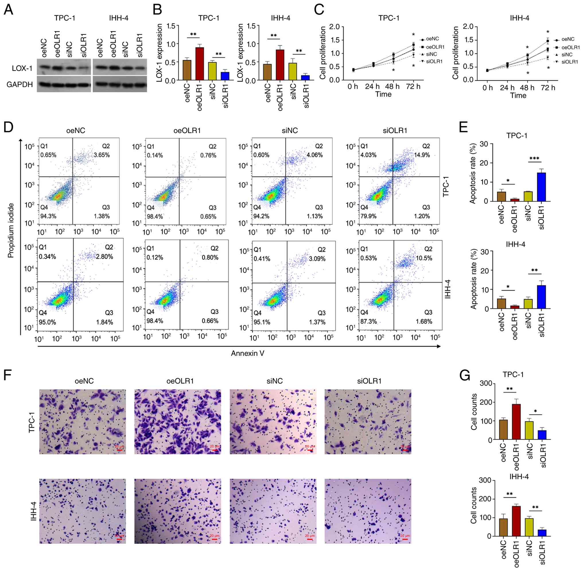 | Figure 2.Effect of OLR1 modification on PTC
proliferation, apoptosis and invasion. LOX-1 (A) western blotting
image example and (B) its expression among oeNC, oeOLR1, siNC and
siOLR1 groups. (C) Cell proliferation, (D) Annexin V/propidium
iodide image example, (E) cell apoptosis rate, (F) Transwell image
example and (G) invasive cell count among oeNC, oeOLR1, siNC and
siOLR1 groups. Magnification, ×200. *P<0.05; **P<0.01;
***P<0.001. OLR1, oxidized low density lipoprotein receptor 1;
PTC, papillary thyroid cancer; LOX-1, lectin-like oxidized low
density lipoprotein receptor-1; NC, negative control; oe,
overexpression; si, siRNA. |
OLR1 promotes H3K18la-mediated LEMD1
in PTC cells
oeOLR1 accelerated the production of lactate in
TPC-1 and IHH-4 cells, but siOLR1 weakened it (Fig. S2). oeOLR1 also upregulated while
siOLR1 downregulated LDHA expression in TPC-1 and IHH-4 cells
(Fig. S3A and B). Moreover, oeOLR1
upregulated H3K18la expression, while siOLR1 downregulated H3K18la
expression in TPC-1 and IHH-4 cells (Fig. 3A and B). Notably, a previous study
reported that H3K18la is directly bound to the promoter of LEMD1
(29). Another study confirmed the
oncogene role of LEMD1 in PTC (30). Therefore, the expression of LEMD1
and its relation to H3K18la was subsequently explored in the
present study. oeOLR1 increased LEMD1 expression but siOLR1
decreased LEMD1 expression in TPC-1 and IHH-4 cells (Fig. 3A and C). In addition, oxamate
reduced H3K18la expression, while lactate elevated H3K18la
expression in TPC-1 and IHH-4 cells (Fig. 3D and E). Finally, the ChIP assay
verified that H3K18la is bound to the LEMD1 promoter in TPC-1 and
IHH-4 cells (Fig. 3F).
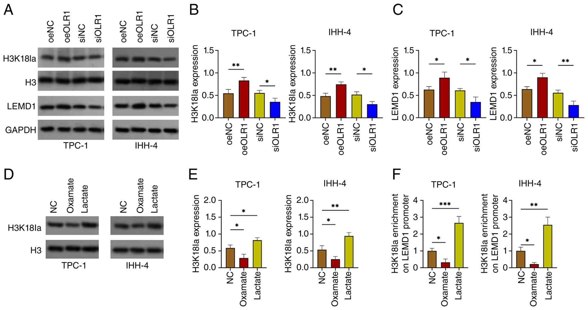 | Figure 3.Effect of OLR1 modification on
H3K18la and LEMD1. (A) Western blotting image example, (B) H3K18la
expression and (C) LEMD1 expression among oeNC, oeOLR1, siNC and
siOLR1 groups. (D) Western blotting image example and (E) H3K18la
expression among NC, oxamate and lactate groups. (F) ChIP assay
evaluating H3K18la enrichment on the LEMD1 promoter. *P<0.05;
**P<0.01; ***P<0.001. OLR1, oxidized low density lipoprotein
receptor 1; NC, negative control; oe, overexpression; si, siRNA;
H3K18la, histone H3 lysine 18 lactylation; LEMD1, LEM domain
containing 1. |
Lactate-mediated H3K18la may be
implicated in the function of OLR1 in PTC cells
Oxamate was used to reduce lactate production along
with plasmid transfection. Oxamate lowered H3K18la expression and
attenuated the effect of oeOLR1 on H3K18la expression in TPC-1 and
IHH-4 cells (Fig. 4A and B). In
addition, oxamate downregulated LEMD1 expression and weakened the
effect of oeOLR1 on LEMD1 expression in TPC-1 and IHH-4 cells
(Fig. 4A and C). Afterwards,
oxamate decreased cell proliferation and increased the cell
apoptosis rate in TPC-1 and IHH-4 cells, which also attenuated the
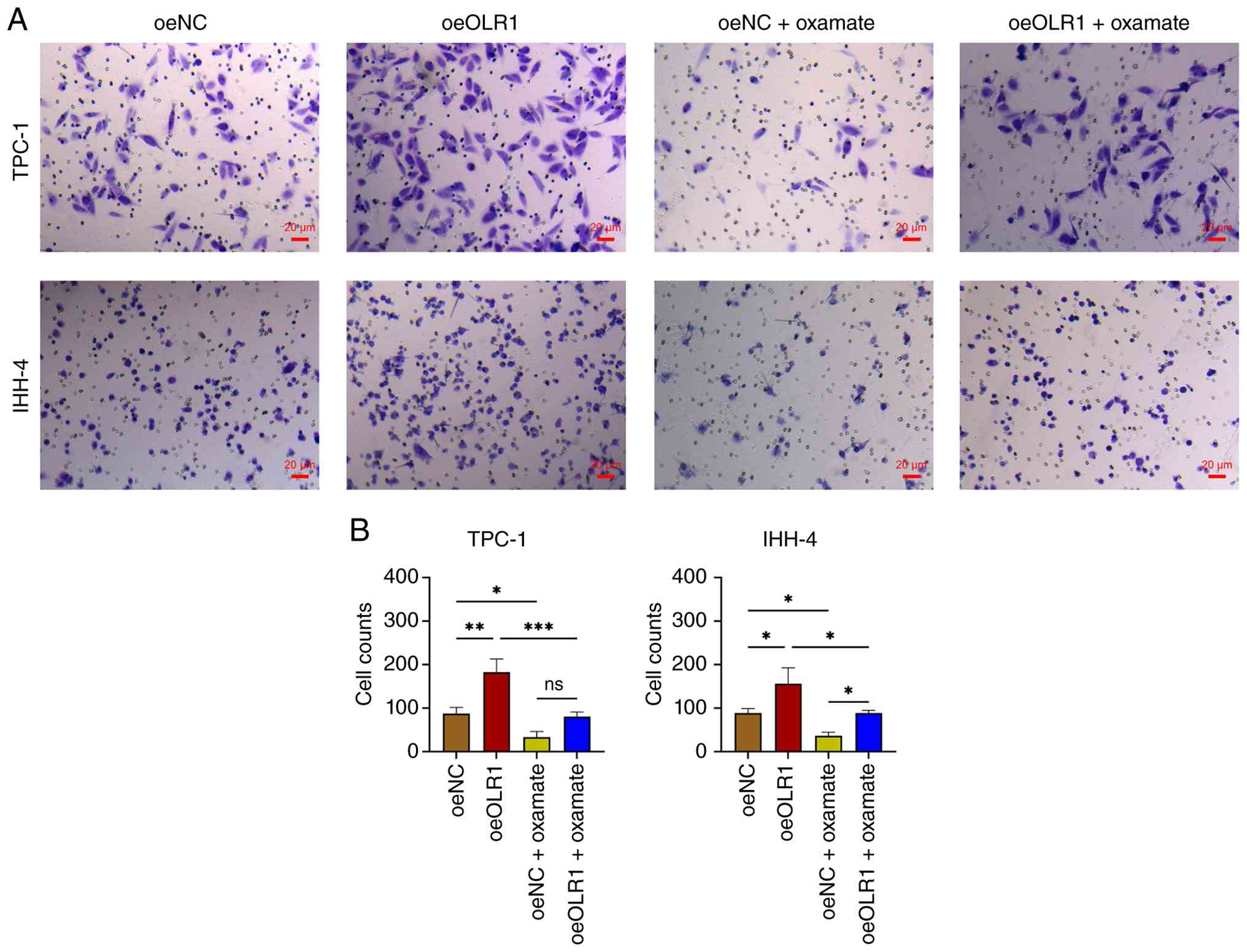
effect of oeOLR1 on cell proliferation and apoptosis (Fig. 4D-F). Moreover, oxamate reduced the
invasive cell count and attenuated the effect of oeOLR1 on the
invasive cell count in TPC-1 and IHH-4 cells (Fig. 5A and B).
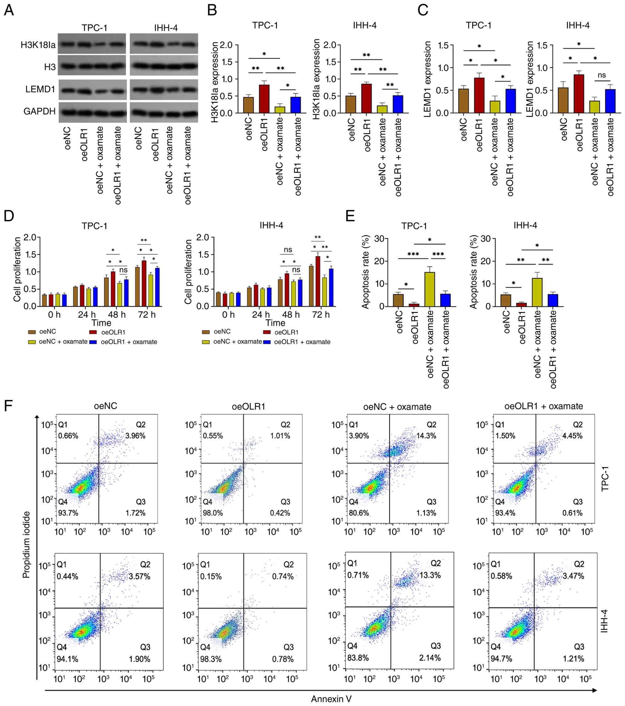 | Figure 4.Effect of oxamate on H3K18la, LEMD1
and PTC proliferation and apoptosis. (A) Western blotting image
example, (B) H3K18la expression and (C) LEMD1 expression among
oeNC, oeOLR1, oeNC + oxamate and oeOLR1 + oxamate groups. (D) Cell
proliferation, (E) cell apoptosis rate and (F) Annexin V/propidium
iodide image example among oeNC, oeOLR1, oeNC + oxamate and oeOLR1
+ oxamate groups. *P<0.05; **P<0.01; ***P<0.001. H3K18la,
histone H3 lysine 18 lactylation; LEMD1, LEM domain containing 1;
PTC, papillary thyroid cancer; oe, overexpression; NC, negative
control; OLR1, oxidized low density lipoprotein receptor 1. |
siLEMD1 attenuates the function of
OLR1 in PTC cells
LEMD1 expression was reduced and the effect of
oeOLR1 on LEMD1 expression was weakened by siLEMD1 transfection in
TPC-1 and IHH-4 cells. However, LOX-1 expression was not affected
by siLEMD1 transfection (Fig.
6A-C). siLEMD1 decreased cell proliferation but increased the
cell apoptosis rate in TPC-1 and IHH-4 cells, which attenuated the
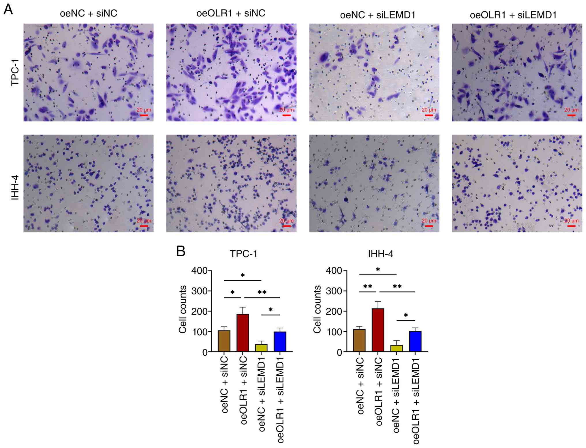
effect of oeOLR1 on these functions (Fig. 6D-F). Furthermore, siLEMD1 repressed
the invasive cell count and weakened the effect of oeOLR1 on the
invasive cell count in TPC-1 and IHH-4 cells (Fig. 7A and B).
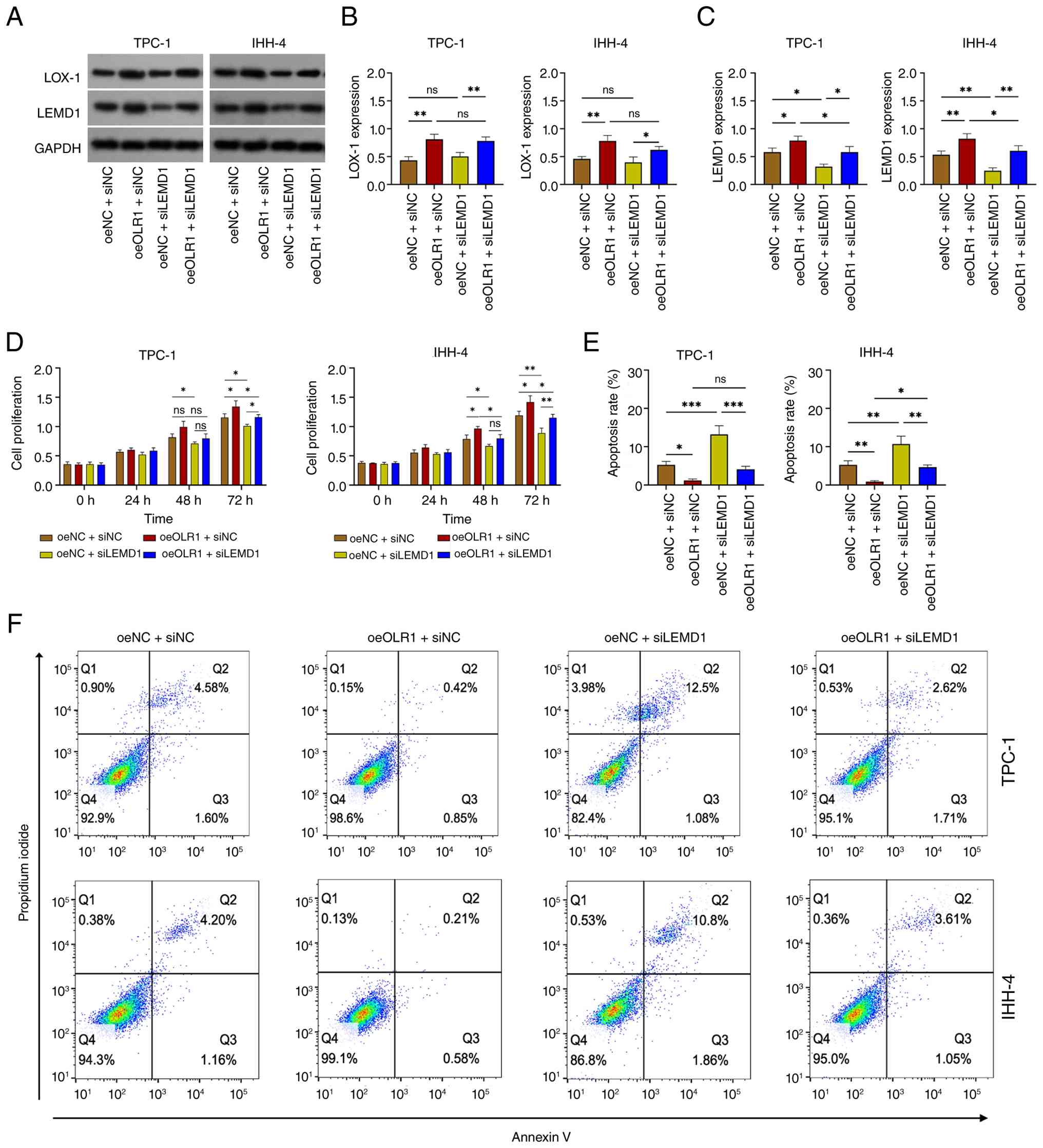 | Figure 6.Effect of LEMD1 modification on PTC
proliferation and apoptosis. (A) Western blotting image example,
(B) LOX-1 and (C) LEMD1 expression among oeNC + siNC, oeOLR1 +
siNC, oeNC + siLEMD1 and oeOLR1 + siLEMD1 groups. Cell
proliferation (D), cell apoptosis rate (E) and Annexin V/propidium
iodide image example (F) among oeNC + siNC, oeOLR1 + siNC, oeNC +
siLEMD1 and oeOLR1 + siLEMD1 groups. *P<0.05; **P<0.01;
***P<0.001. LEMD1, LEM domain containing 1; PTC, papillary
thyroid cancer; LOX-1, lectin-like oxidized low density lipoprotein
receptor-1; OLR1, oxidized low density lipoprotein receptor 1; NC,
negative control; oe, overexpression; si, siRNA. |
OLR1 promotes PTC tumor growth via
LEMD1 in xenograft mice
oeOLR1 elevated tumor volume during observational
period, maximum tumor volume and maximum tumor diameter (Fig. 8A and B). Moreover, oeOLR1 increased
tumor weight (Fig. 8C) and
upregulated the expressions of LEMD1 and H3K18la (Fig. 8D). By contrast, shLEMD1 reduced
tumor volume during observational period, maximum tumor volume,
maximum tumor diameter and weight, which also attenuated the effect
of oeOLR1 in xenograft mice, but it did not affect the expressions
of LOX-1 (OLR1 coding protein) and H3K18la. Moreover, the LOX-1
expression was upregulated in oeOLR1 group compared to Control
group, indicating oeOLR1 transfection success; the LEMD1 expression
was downregulated in shLEMD1 group compared with Control group,
indicating shLEMD1 transfection success (Fig. 8D). Regarding the detailed values of
maximum tumor volume and diameter: the maximum tumor volumes were
561±117 mm3, 1136±171 mm3, 241±18
mm3, 674±45 mm3 in the Control, oeOLR1,
shLEMD1 and oeOLR1+shLEMD1 groups, respectively; and the maximum
tumor diameters were 13.2±0.3 mm, 16.2±1.1 mm, 9.2±0.9 mm and
13.5±1.1 mm in the Control, oeOLR1, shLEMD1 and oeOLR1+shLEMD1
groups, respectively.
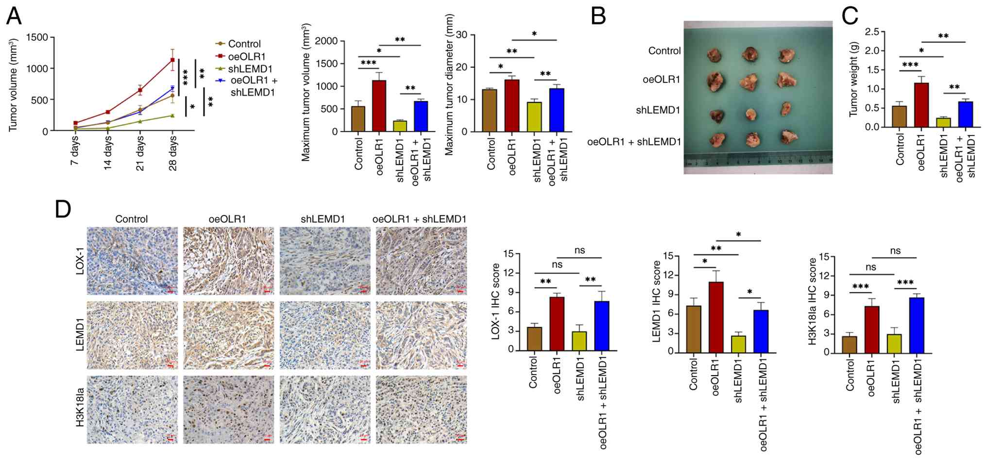 | Figure 8.Effect of OLR1 and LEMD1 modification
in PTC xenograft mice. (A) Tumor volume during observational
period, maximum tumor volume and maximum tumor diameter, (B) tumor
images, (C) tumor weight and (D) expressions of LOX-1, LEMD1 and
H3K18la among Control, oeOLR1, shLEMD1 and oeOLR1 + shLEMD1 groups
in PTC xenograft mice. Magnification, ×200. *P<0.05;
**P<0.01; ***P<0.001. OLR1, oxidized low density lipoprotein
receptor 1; LEMD1, LEM domain containing 1; PTC, papillary thyroid
cancer; LOX-1, lectin-like oxidized low density lipoprotein
receptor-1; oe, overexpression; sh, short hairpin; H3K18la, histone
H3 lysine 18 lactylation. |
Discussion
OLR1 is aberrantly expressed and presents with
prognostic value in several cancers (31–33).
For example, OLR1 is largely overexpressed in head and neck
squamous cell carcinoma (HNSCC) tissues compared with non-tumor
tissues and its high expression is associated with worse OS in
patients with HNSCC (31). OLR1 is
also upregulated in ovarian cancer tissues and cell lines compared
with non-tumor control tissues and normal cell lines, whose high
expression is associated with elevated risk of recurrence and
mortality in patients with ovarian cancer (32). Moreover, an upregulation of OLR1 in
tumor tissues and a correlation between OLR1 upregulation and
survival prognosis were discovered in patients with breast cancer
(33). However, the related
information is lacking in PTC. The present study preliminarily
employed the GEPIA public database and found that OLR1 was
upregulated in tumor tissues and associated with shorter OS in
patients with thyroid cancer. Subsequently, via the IHC assay, the
present study further confirmed that LOX-1 was upregulated in PTC
tissues compared with adjacent non-tumor tissues. The explanation
for this finding may be as follows: i) OLR1 serves as an oncogene
via the regulation of tumor proliferation, EMT, angiogenesis,
immune microenvironment and drug resistance (23,25)
and thus, it is upregulated in PTC tumor tissues; and ii) referring
to the function of OLR1 on regulating metabolic activities and
their relation to carcinogenesis (22–24,28)
and the finding that OLR1 promoted PTC proliferation and invasion,
while repressed apoptosis in the subsequent experiments, OLR1 and
its coding protein LOX-1 are considered to be upregulated in PTC
tumor tissues. However, the present study did not assess the
soluble level of LOX-1 in PTC patients, which could be explored in
future studies.
In carcinogenesis, OLR1 promotes the progression of
several types of cancer via multiple mechanisms (26–28,34–36).
For example, OLR1 facilitates lung cancer growth, mobility and
immune escape by activating the cancer-associated fibroblasts
(34) and stimulates esophageal
cancer tumorigenesis by regulating the RACK1-mediated
MAP2K/MEK/MAPK/ERK pathway and autophagy (35). Furthermore, targeting OLR1 reduces
oral cancer migration, invasion and stemness (36). However, data regarding OLR1
regulation during PTC pathogenesis are limited. Based on the
preliminary findings of the present study that demonstrated that
OLR1 was upregulated in tumor tissues and predicted a worse
prognosis in patients with PTC, the present study further
investigated the effect of OLR1 modification on PTC cellular
functions. It found that OLR1 overexpression enhanced PTC
proliferation and invasion but reduced PTC apoptosis and OLR1
knockdown revealed an opposite function. The explanation for this
finding may be as follows: i) OLR1 positively regulates cancer
glycolytic metabolism (28); ii)
OLR1 controls several oncogene pathways such as PI3K/AKT,
MAPK/MEK/ERK and Wnt/β-catenin (26,35,37);
and iii) OLR1 activated H3K18la-mediated LEMD1 as discovered in
subsequent experiments of the present study. Therefore, OLR1 is
able to facilitate PTC proliferation and invasion but reduce its
apoptosis.
The present study also found that OLR1 positively
regulated H3K18la in PTC and lactate-mediated H3K18la possibly
attenuated the effect of OLR1 on PTC proliferation, apoptosis and
invasion. The explanations for this finding may be as follows: i)
OLR1 regulates glycolytic metabolism and lactate production
(28) and further upregulates
H3K18la; therefore, OLR1 positively regulates H3K18la; ii) H3K18la
promotes the progression of other cancers apart from PTC (15,19–21);
therefore, it may facilitate PTC progression as that in other
cancers; and iii) H3K18la may bind to LEMD1 and the latter is able
to promote PTC progression (29,30);
this is further verified in subsequent experiments.
LEMD1, also known as CT50 or LEMP-1, has been
recognized as a newly identified member of the cancer-testis
antigen family (38). Its
expression pattern, primarily restricted to the testis under normal
conditions but frequently reactivated in various tumor types,
endows it with multiple attributes of an ideal biological target,
holding significant promise for both cancer immunotherapy and
diagnosis (38,39). Clinically, LEMD1 is upregulated in
prostate cancer tissues compared to benign prostate hyperplasia
tissues (40) and its associated
with lymph node metastasis, advanced TNM stage and worse survival
outcomes in patients with oral squamous cell carcinoma and
non-small cell lung cancer (41,42).
Moreover, LEMD1 is an independent factor predicting the recurrence
risk of colorectal cancer (43). In
the aspect of biological functions, LEMD1 has been previously
reported to accelerate the growth, migration and invasion of
pancreatic cancer via activating p53 and mTOR pathways (39). Moreover, LEMD1 knockdown suppresses
proliferation, migration and colony formation, while restoring
chemosensitivity via inactivating the ERK pathway in breast cancer
(44). Particularly in PTC, LEMD1
promotes PTC proliferation, migration, invasion and EMT via the
Wnt/β-catenin pathway (30). The
present study found that LEMD1 knockdown repressed PTC
proliferation and invasion but enhanced its apoptosis, which
attenuated the effect of OLR1 overexpression on these cellular
functions in PTC. The explanation for this finding may be as
follows: i) LEMD1 facilitates PTC progression via the Wnt/β-catenin
pathway (30); and ii) LEMD1
regulates several other oncogene pathways such as p53, mTOR, ERK
and PI3K/AKT (39,44,45).
The present study possessed some limitations. First,
the BRAF V600E status of the PTC patients was not detected and the
patients are still not recurrent at the time of the present study,
therefore the correlation of OLR1/LOX-1 with BRAF V600E status and
recurrent risk was not analyzed. Second, rescue experiments were
not performed to validate the findings. Third, only the effect of
OLR1 modification on LDHA expression was performed, while its
effect on other glycolytic enzymes such as HK2 and PKM2 was not
detected, which needs to be explored in the future studies.
In summary, OLR1 promoted proliferation and invasion
while repressed apoptosis via regulating H3K18la-mediated LEMD1 in
PTC. This may provide a novel insight regarding the involvement of
OLR1 and histone lactylation in the pathogenesis of PTC. However,
further validation is still required.
Supplementary Material
Supporting Data
Acknowledgements
Not applicable.
Funding
Funding: No funding was received.
Availability of data and materials
The data generated in the present study may be
requested from the corresponding author.
Authors' contributions
JZ conceived and designed the study. JY collected
the data. RP conducted the data analysis. BL and LC contributed to
the interpretation of the data. LC drafted the manuscript, while
all authors provided critical revisions. JZ and JY confirm the
authenticity of all the raw data. All authors read and approved the
final manuscript.
Ethics approval and consent to
participate
The present study was approved by the Ethics
Committee of Harbin Medical University Cancer Hospital with
approval number YD2024-13 on November 4, 2024. The Ethics Committee
formally waived the requirement for additional informed consent for
this study. Moreover, participants had provided written consent for
the secondary use of biological specimens and medical data as part
of routine clinical procedures at the Harbin Medical University
Cancer Hospital. The animal experiments were approved by the Ethics
Committee of Harbin Medical University Cancer Hospital with
approval number YD2024-13 and Wuhan Servicebio Institutional Animal
Care and Use Committee with approval number 2025079-2.
Patient consent for publication
The Ethics Committee formally waived the requirement
for additional informed consent for this study. Moreover,
participants had provided written consent for the secondary use of
biological specimens and medical data as part of routine clinical
procedures at our institution.
Competing interests
The authors declare that they have no competing
interests.
References
|
1
|
Hu J, Yuan IJ, Mirshahidi S, Simental A,
Lee SC and Yuan X: Thyroid carcinoma: Phenotypic features,
underlying biology and potential relevance for targeting therapy.
Int J Mol Sci. 22:19502021. View Article : Google Scholar : PubMed/NCBI
|
|
2
|
Bray F, Laversanne M, Sung H, Ferlay J,
Siegel RL, Soerjomataram I and Jemal A: Global cancer statistics
2022: GLOBOCAN estimates of incidence and mortality worldwide for
36 cancers in 185 countries. CA Cancer J Clin. 74:229–263.
2024.PubMed/NCBI
|
|
3
|
Lyu Z, Zhang Y, Sheng C, Huang Y, Zhang Q
and Chen K: Global burden of thyroid cancer in 2022: Incidence and
mortality estimates from GLOBOCAN. Chin Med J (Engl).
137:2567–2576. 2024. View Article : Google Scholar : PubMed/NCBI
|
|
4
|
Chen DW, Lang BHH, McLeod DSA, Newbold K
and Haymart MR: Thyroid cancer. Lancet. 401:1531–1544. 2023.
View Article : Google Scholar : PubMed/NCBI
|
|
5
|
Lam AK: Papillary thyroid carcinoma:
Current position in epidemiology, genomics and classification.
Methods Mol Biol. 2534:1–15. 2022. View Article : Google Scholar : PubMed/NCBI
|
|
6
|
Masui T, Adachi S, Uemura H, Kimura T and
Kitahara T: Clinical study on the risk factors for the recurrence
of papillary thyroid carcinoma. ORL J Otorhinolaryngol Relat Spec.
85:104–108. 2023. View Article : Google Scholar : PubMed/NCBI
|
|
7
|
Ulisse S, Baldini E, Lauro A, Pironi D,
Tripodi D, Lori E, Ferent IC, Amabile MI, Catania A, Di Matteo FM,
et al: Papillary thyroid cancer prognosis: An evolving field.
Cancers (Basel). 13:55672021. View Article : Google Scholar : PubMed/NCBI
|
|
8
|
Hu Y, He Z, Li Z, Wang Y, Wu N, Sun H,
Zhou Z, Hu Q and Cong X: Lactylation: The novel histone
modification influence on gene expression, protein function and
disease. Clin Epigenetics. 16:722024. View Article : Google Scholar : PubMed/NCBI
|
|
9
|
Xu K, Zhang K, Wang Y and Gu Y:
Comprehensive review of histone lactylation: Structure, function
and therapeutic targets. Biochem Pharmacol. 225:1163312024.
View Article : Google Scholar : PubMed/NCBI
|
|
10
|
Yu X, Yang J, Xu J, Pan H, Wang W, Yu X
and Shi S: Histone lactylation: From tumor lactate metabolism to
epigenetic regulation. Int J Biol Sci. 20:1833–1854. 2024.
View Article : Google Scholar : PubMed/NCBI
|
|
11
|
Zhang Y, Song H, Li M and Lu P: Histone
lactylation bridges metabolic reprogramming and epigenetic rewiring
in driving carcinogenesis: Oncometabolite fuels oncogenic
transcription. Clin Transl Med. 14:e16142024. View Article : Google Scholar : PubMed/NCBI
|
|
12
|
Rong Y, Dong F, Zhang G, Tang M, Zhao X,
Zhang Y, Tao P and Cai H: The crosstalking of lactate-histone
lactylation and tumor. Proteomics Clin Appl. 17:e22001022023.
View Article : Google Scholar : PubMed/NCBI
|
|
13
|
Galle E, Wong CW, Ghosh A, Desgeorges T,
Melrose K, Hinte LC, Castellano-Castillo D, Engl M, de Sousa JA,
Ruiz-Ojeda FJ, et al: H3K18 lactylation marks tissue-specific
active enhancers. Genome Biol. 23:2072022. View Article : Google Scholar : PubMed/NCBI
|
|
14
|
Yang Y, Luo N, Gong Z, Zhou W, Ku Y and
Chen Y: Lactate and lysine lactylation of histone regulate
transcription in cancer. Heliyon. 10:e384262024. View Article : Google Scholar : PubMed/NCBI
|
|
15
|
Zhao Y, Jiang J, Zhou P, Deng K, Liu Z,
Yang M, Yang X, Li J, Li R and Xia J: H3K18 lactylation-mediated
VCAM1 expression promotes gastric cancer progression and metastasis
via AKT-mTOR-CXCL1 axis. Biochem Pharmacol. 222:1161202024.
View Article : Google Scholar : PubMed/NCBI
|
|
16
|
Wang P, Xie D, Xiao T, Cheng C, Wang D,
Sun J, Wu M, Yang Y, Zhang A and Liu Q: H3K18 lactylation promotes
the progression of arsenite-related idiopathic pulmonary fibrosis
via YTHDF1/m6A/NREP. J Hazard Mater. 461:1325822024. View Article : Google Scholar : PubMed/NCBI
|
|
17
|
Qiao J, Tan Y, Liu H, Yang B, Zhang Q, Liu
Q, Sun W, Li Z, Wang Q, Feng W, et al: Histone H3K18 and ezrin
lactylation promote renal dysfunction in sepsis-associated acute
kidney injury. Adv Sci (Weinh). 11:e23072162024. View Article : Google Scholar : PubMed/NCBI
|
|
18
|
Wei L, Yang X, Wang J, Wang Z, Wang Q,
Ding Y and Yu A: H3K18 lactylation of senescent microglia
potentiates brain aging and Alzheimer's disease through the NFκB
signaling pathway. J Neuroinflammation. 20:2082023. View Article : Google Scholar : PubMed/NCBI
|
|
19
|
Zhang C, Zhou L, Zhang M, Du Y, Li C, Ren
H and Zheng L: H3K18 lactylation potentiates immune escape of
non-small cell lung cancer. Cancer Res. 84:3589–3601. 2024.
View Article : Google Scholar : PubMed/NCBI
|
|
20
|
Hou X, Ouyang J, Tang L, Wu P, Deng X, Yan
Q, Shi L, Fan S, Fan C, Guo C, et al: KCNK1 promotes proliferation
and metastasis of breast cancer cells by activating lactate
dehydrogenase A (LDHA) and up-regulating H3K18 lactylation. PLoS
Biol. 22:e30026662024. View Article : Google Scholar : PubMed/NCBI
|
|
21
|
Sun J, Feng Q, He Y, Wang M and Wu Y:
Lactate activates CCL18 expression via H3K18 lactylation in
macrophages to promote tumorigenesis of ovarian cancer. Acta
Biochim Biophys Sin (Shanghai). 56:1373–1386. 2024. View Article : Google Scholar : PubMed/NCBI
|
|
22
|
Sawamura T, Kume N, Aoyama T, Moriwaki H,
Hoshikawa H, Aiba Y, Tanaka T, Miwa S, Katsura Y, Kita T and Masaki
T: An endothelial receptor for oxidized low-density lipoprotein.
Nature. 386:73–77. 1997. View
Article : Google Scholar : PubMed/NCBI
|
|
23
|
Murdocca M, De Masi C, Pucci S, Mango R,
Novelli G, Di Natale C and Sangiuolo F: LOX-1 and cancer: An
indissoluble liaison. Cancer Gene Ther. 28:1088–1098. 2021.
View Article : Google Scholar : PubMed/NCBI
|
|
24
|
Sánchez-León ME, Loaeza-Reyes KJ,
Matias-Cervantes CA, Mayoral-Andrade G, Pérez-Campos EL,
Pérez-Campos-Mayoral L, Hernández-Huerta MT, Zenteno E,
Pérez-Cervera Y and Pina-Canseco S: LOX-1 in cardiovascular
disease: A comprehensive molecular and clinical review. Int J Mol
Sci. 25:52762024. View Article : Google Scholar : PubMed/NCBI
|
|
25
|
Balzan S and Lubrano V: LOX-1 receptor: A
potential link in atherosclerosis and cancer. Life Sci. 198:79–86.
2018. View Article : Google Scholar : PubMed/NCBI
|
|
26
|
Li C, Zhang J, Wu H, Li L, Yang C, Song S,
Peng P, Shao M, Zhang M, Zhao J, et al: Lectin-like oxidized
low-density lipoprotein receptor-1 facilitates metastasis of
gastric cancer through driving epithelial-mesenchymal transition
and PI3K/Akt/GSK3β activation. Sci Rep. 7:452752017. View Article : Google Scholar : PubMed/NCBI
|
|
27
|
Gonzalez-Chavarria I, Cerro RP, Parra NP,
Sandoval FA, Zuñiga FA, Omazábal VA, Lamperti LI, Jiménez SP,
Fernandez EA, Gutiérrez NA, et al: Lectin-like oxidized LDL
receptor-1 is an enhancer of tumor angiogenesis in human prostate
cancer cells. PLoS One. 9:e1062192014. View Article : Google Scholar : PubMed/NCBI
|
|
28
|
Zhao T, Li Y, Shen K, Wang Q and Zhang J:
Knockdown of OLR1 weakens glycolytic metabolism to repress colon
cancer cell proliferation and chemoresistance by downregulating
SULT2B1 via c-MYC. Cell Death Dis. 13:42021. View Article : Google Scholar : PubMed/NCBI
|
|
29
|
Li W, Zhou C, Yu L, Hou Z, Liu H, Kong L,
Xu Y, He J, Lan J, Ou Q, et al: Tumor-derived lactate promotes
resistance to bevacizumab treatment by facilitating autophagy
enhancer protein RUBCNL expression through histone H3 lysine 18
lactylation (H3K18la) in colorectal cancer. Autophagy. 20:114–130.
2024. View Article : Google Scholar : PubMed/NCBI
|
|
30
|
Xu M, Lin B, Zheng D, Wen J, Hu W, Li C,
Zhang X, Zhang X and Qu J: LEM domain containing 1 promotes thyroid
cancer cell proliferation and migration by activating the
Wnt/β-catenin signaling pathway and epithelial-mesenchymal
transition. Oncol Lett. 21:4422021. View Article : Google Scholar : PubMed/NCBI
|
|
31
|
Zhang P, Zhao Y, Xia X, Mei S, Huang Y,
Zhu Y, Yu S and Chen X: Expression of OLR1 gene on
tumor-associated macrophages of head and neck squamous cell
carcinoma, and its correlation with clinical outcome.
Oncoimmunology. 12:22030732023. View Article : Google Scholar : PubMed/NCBI
|
|
32
|
Liu J, Zeng Y and Zheng L: Up-regulation
of oxidized low-density lipoprotein receptor 1 correlates with
decreased miR-106b-5p, miR-93-5p, miR-3129-5p, miR-199b-3p, and
miR-4465, higher recurrence rate, and poor prognosis in ovarian
cancer. Histol Histopathol. 38:475–485. 2023.PubMed/NCBI
|
|
33
|
Sun X, Fu X, Xu S, Qiu P, Lv Z, Cui M,
Zhang Q and Xu Y: OLR1 is a prognostic factor and correlated with
immune infiltration in breast cancer. Int Immunopharmacol.
101:1082752021. View Article : Google Scholar : PubMed/NCBI
|
|
34
|
Sun Y, Ying K, Sun J, Wang Y, Qiu L, Ji M,
Sun L and Chen J: PRRX1-OLR1 axis supports CAFs-mediated lung
cancer progression and immune suppression. Cancer Cell Int.
24:2472024. View Article : Google Scholar : PubMed/NCBI
|
|
35
|
Li C and Wang L: TFEB-dependent autophagy
is involved in scavenger receptor OLR1/LOX-1-mediated tumor
progression. Autophagy. 18:462–464. 2022. View Article : Google Scholar : PubMed/NCBI
|
|
36
|
Lin CH, Chiang CC, Shih YC, Tung SL and
Huang WC: Elucidating the role of Let-7d-5p and OLR1 in the
progression and prognosis of oral squamous cell carcinoma
via FAK/P53 signaling axis. Anticancer Res. 44:2517–2531.
2024. View Article : Google Scholar : PubMed/NCBI
|
|
37
|
Li J, Ma J, Huang S, Li J, Zhou L, Sun J
and Chen L: CircTTLL13 promotes TMZ resistance in glioma via
modulating OLR1-mediated activation of the Wnt/β-catenin
pathway. Mol Cell Biol. 43:354–369. 2023. View Article : Google Scholar : PubMed/NCBI
|
|
38
|
Yuki D, Lin YM, Fujii Y, Nakamura Y and
Furukawa Y: Isolation of LEM domain-containing 1, a novel
testis-specific gene expressed in colorectal cancers. Oncol Rep.
12:275–280. 2004.PubMed/NCBI
|
|
39
|
Cao X, Yao N, Zhao Z, Fu Y, Hu Y, Zhu P,
Shi W and Tang L: LEM domain containing 1 promotes pancreatic
cancer growth and metastasis by p53 and mTORC1 signaling pathway.
Bioengineered. 13:7771–7784. 2022. View Article : Google Scholar : PubMed/NCBI
|
|
40
|
Ghafouri-Fard S, Ousati Ashtiani Z, Sabah
Golian B, Hasheminasab SM and Modarressi MH: Expression of two
testis-specific genes, SPATA19 and LEMD1, in prostate cancer. Arch
Med Res. 41:195–200. 2010. View Article : Google Scholar : PubMed/NCBI
|
|
41
|
Sasahira T, Kurihara M, Nakashima C,
Kirita T and Kuniyasu H: LEM domain containing 1 promotes oral
squamous cell carcinoma invasion and endothelial transmigration. Br
J Cancer. 115:52–58. 2016. View Article : Google Scholar : PubMed/NCBI
|
|
42
|
Li L and Zhang P: Elevation of LEM domain
containing 1 predicts poor prognosis of NSCLC patients and triggers
malignant stemness and invasion of NSCLC cells by stimulating
PI3K/AKT pathway. Curr Mol Med. 24:366–378. 2024. View Article : Google Scholar : PubMed/NCBI
|
|
43
|
Zhang H, Xu C, Jiang F and Feng J: A
three-genes signature predicting colorectal cancer relapse reveals
LEMD1 promoting CRC cells migration by RhoA/ROCK1 signaling
pathway. Front Oncol. 12:8236962022. View Article : Google Scholar : PubMed/NCBI
|
|
44
|
Li X, Jiang S, Jiang T, Sun X, Guan Y, Fan
S and Cheng Y: LEM domain containing 1 acts as a novel oncogene and
therapeutic target for triple-negative breast cancer. Cancers
(Basel). 15:29242023. View Article : Google Scholar : PubMed/NCBI
|
|
45
|
Li D, Wang D, Liu H and Jiang X: LEM
domain containing 1 (LEMD1) transcriptionally activated by
SRY-related high-mobility-group box 4 (SOX4) accelerates the
progression of colon cancer by upregulating phosphatidylinositol
3-kinase (PI3K)/protein kinase B (Akt) signaling pathway.
Bioengineered. 13:8087–8100. 2022. View Article : Google Scholar : PubMed/NCBI
|