Introduction
Prostate cancer (PCa) is a leading cause of
cancer-related morbidity and mortality among male patients
worldwide, with recent estimates indicating ~1.4 million new cases
and 375,000 deaths annually (1).
Despite advances in early detection and treatment, a substantial
proportion of patients (10–20% within 5 years) develop
castration-resistant PCa (CRPCa), a lethal disease state
characterized by therapeutic resistance and poor clinical outcomes
(2). Comprehensive genomic analysis
has revealed that advanced PCa involves complex molecular
alterations that influence disease progression and treatment
response (3). Among these, androgen
receptor splice variant 7 (AR-V7) has been prospectively validated
as a predictive biomarker of resistance to hormone therapy in
high-risk patients (4).
Furthermore, distinct genomic drivers associated with enzalutamide
resistance have been identified in metastatic CRPCa, highlighting
the heterogeneous and adaptive nature of treatment failure
(5).
The biological evolution from hormone-sensitive to
CR disease involves not only genomic alterations but also metabolic
reprogramming (6). This metabolic
rewiring enables tumor cells to sustain proliferation under
therapeutic pressure and nutrient-limited conditions (7). Concurrently, the tumor
microenvironment (TME) undergoes notable remodeling that promotes
immune evasion, with evidence indicating that metabolic alterations
directly influence immune cell function (8). The recognition that metabolic
reprogramming and immune evasion are interconnected rather than
independent processes has opened novel avenues for therapeutic
intervention.
Glutamine metabolism is a central node in PCa
pathogenesis, with recent work positioning glutamine and glutamate
metabolic reprogramming at the core of tumor progression (9). Beyond its traditional roles in
bioenergetics and biosynthesis, glutamine metabolism actively
shapes the immunological landscape of the TME through nutrient
competition and the generation of immunosuppressive metabolites
(10). While previous reviews have
addressed glutamine metabolism in PCa or immune evasion mechanisms
in the TME, the present study aimed to promote a unified framework
by positioning the glutamine-ammonium axis as an integrated
metabolic-immune hub (10,11). The present review aimed to
reconceptualize ammonium, traditionally dismissed as a toxic waste
product, as an active immunosuppressive signaling molecule that
orchestrates T cell dysfunction and macrophage M2 polarization.
Ammonium accumulation impairs antitumor immunity and is associated
with poor prognosis, whereas its clearance restores T cell function
and enhances immunotherapy efficacy (12). The present study aimed to delineate
the bidirectional feedback loops whereby immune-derived cytokines
reciprocally modulate tumor glutamine and ammonium flux,
establishing a self-reinforcing cycle of metabolic adaptation and
immune evasion (9,11,13)
and summarize the therapeutic implications of targeting this
integrated axis (14). The
complexity of this metabolic-immune crosstalk is amplified by
interactions with stromal elements, including cancer-associated
fibroblasts that manipulate glutamine metabolism through paracrine
signaling (11), and encompasses
bioenergetic support, redox homeostasis, epigenetic regulation and
nitrogen recycling (13).
The therapeutic potential of targeting the
glutamine-ammonium axis has been demonstrated in preclinical
studies (14,15). Metabolic reprogramming of
tumor-associated macrophages using glutamine antagonism drives
antitumor immunity in myeloid-rich PCa (14). Similarly, targeting of Myc and
glutamine-fructose-6-phosphate amidotransferase 1 (GFAT-1), a key
enzyme in glutamine metabolism, improves antitumor activity
(15). These findings suggest that
interventions at the metabolic-immune interface may simultaneously
disrupt tumor progression and enhance immune surveillance. The
present review aimed to summarize the molecular mechanisms
governing glutamine dependency and ammonium accumulation in PCa,
with focus on their immunomodulatory consequences. By mapping the
bidirectional dialogue between tumor metabolism and antitumor
immunity, the present review seeks to identify novel therapeutic
strategies to overcome treatment resistance and improve clinical
outcomes. The literature for the present narrative review was
identified through systematic searches of PubMed databases
(pubmed.ncbi.nlm.nih.gov) using combinations of the following
keywords: ‘PCa’, ‘glutamine metabolism’, ‘glutaminase’, ‘ASCT2’,
‘ammonium’, ‘urea cycle’, ‘tumor microenvironment’, ‘immune
evasion’, ‘tumor-associated macrophages’, ‘T cell exhaustion’,
‘metabolic reprogramming’ and ‘immunometabolism’. The search was
restricted to articles published in English between January 2015
and April 2026, with additional references identified through
citation screening of retrieved articles and relevant review
papers.
Glutamine metabolism in PCa: Mechanisms and
regulation
Glutamine is a versatile nutrient that fuels
multiple aspects of PCa progression, from bioenergetics and
biosynthesis to redox homeostasis and epigenetic regulation. The
rewiring of glutamine metabolic pathways represents a hallmark of
malignant transformation that distinguishes PCa cells from their
normal counterparts. Understanding the molecular mechanisms
governing glutamine use is essential for identifying how these
tumors establish metabolic autonomy and acquire resistance to
therapeutic intervention (6,7,9).
Fig. 1 provides a schematic
overview of the key transporters, enzymes and metabolic nodes,
illustrating the integrated nature of glutamine metabolism in PCa
cells.
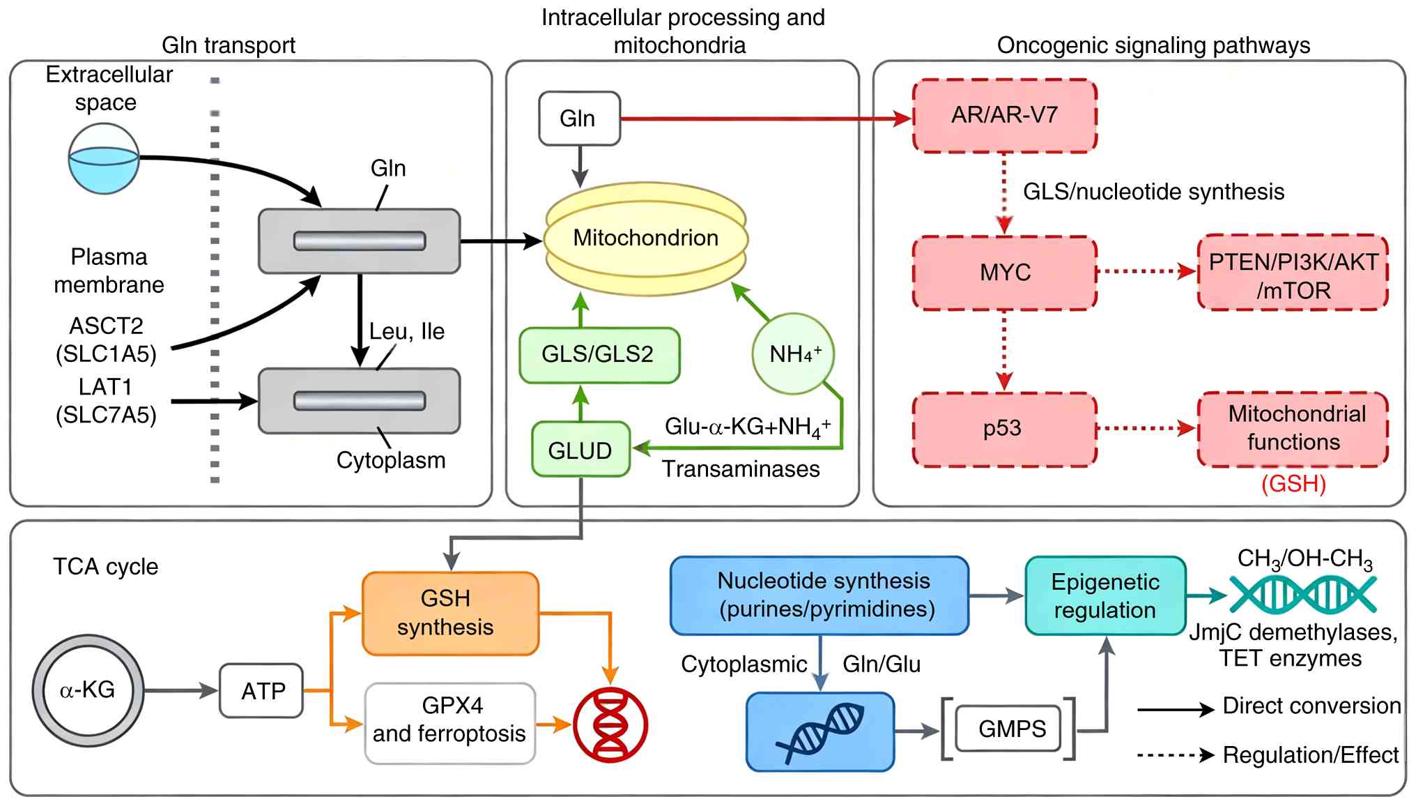 | Figure 1.Schematic overview of glutamine
metabolism in prostate cancer cells. AR-V7, androgen receptor
splice variant 7; ASCT2, alanine-serine-cysteine transporter 2;
GLS, glutaminase; GLUD, glutamate dehydrogenase; Gln, glutamine;
Glu, glutamate; GSH, glutathione; LAT1, L-type amino acid
transporter 1; α-KG, α-ketoglutarate; TCA, tricarboxylic acid. |
Glutamine dependency in PCa
PCa cells exhibit dependency on exogenous glutamine,
a phenomenon termed glutamine addiction that distinguishes
malignant metabolism from normal prostate physiology. This
metabolic vulnerability arises from the unique bioenergetic and
biosynthetic demands of proliferating tumor cells, which require
glutamine not only as a nitrogen donor for nucleotide and amino
acid synthesis but also as a carbon source for tricarboxylic acid
cycle anaplerosis. PCa cell lines PC-3, DU145 and LNCaP display
distinct bioenergetic properties, with mitochondrial adaptations
that render them sensitive to glutamine deprivation (16). Targeting alanine-serine-cysteine
transporter 2 (ASCT2)-mediated glutamine uptake effectively blocks
PCa growth and tumor development, establishing glutamine dependency
as a potential therapeutic target (17).
Plasma glutamine levels may serve as prognostic
biomarkers in localized PCa, with alterations in circulating
glutamine associated with disease outcomes (18). The degree of glutamine dependency
varies across disease states: CRPCa cells exhibit heightened
reliance on glutamine metabolism compared with hormone-naïve
(untreated by androgen deprivation therapy) counterparts,
suggesting that therapeutic pressure selects for metabolic
adaptations that amplify glutamine addiction (19). This evolutionary trajectory
positions glutamine metabolism as a key driver of lethal PCa
phenotypes, providing a rationale for developing therapeutic
strategies that exploit this dependency across the spectrum of
disease progression.
Key transporters and enzymes
The execution of glutamine-dependent metabolic
programs requires coordinated action of specialized transporters
that mediate glutamine influx and efflux. Among glutamine
transporters, ASCT2 (encoded by SLC1A5) is the dominant mediator of
glutamine uptake in PCa cells, with elevated expression
demonstrated across multiple model systems and clinical specimens
(17,20). Mechanistic studies have revealed
that ASCT2 expression is directly regulated by multiple oncogenic
signaling pathways, positioning this transporter as a nodal point
integrating proliferative signals (20,21).
The functional importance of ASCT2 is further supported by
observations that CRPCa cells exhibit increased ASCT2 expression
and glutamine uptake, linking transporter upregulation to
therapeutic resistance (21).
Beyond ASCT2, the heterodimeric amino acid transporter L-type amino
acid transporter 1 (encoded by SLC7A5) participates in glutamine
exchange by coupling glutamine efflux with essential amino acid
import, which sustains mTOR signaling and protein synthesis
(11).
Once internalized, intracellular enzymes channel
glutamine into downstream metabolic pathways. Glutamine enters
mitochondria where glutaminase (GLS) catalyzes its deamination to
glutamate, representing the first committed step of glutaminolysis
(22,23). Elevated GLS expression confers
enhanced glucose use capacity in PCa cells, linking glutamine
catabolism with broader metabolic reprogramming (22). Pharmacological inhibition of GLS
suppresses proliferation and promotes apoptosis in PCa models,
validating this enzyme as a druggable vulnerability (23,24).
The existence of two GLS isoforms, GLS and GLS2, adds regulatory
complexity; an isoform switch from GLS to GLS2 is implicated in
therapeutic resistance and disease progression, indicating that
isoform-specific targeting strategies may be required for optimal
efficacy (25). Glutamate
dehydrogenase (GLUD) converts glutamate to α-ketoglutarate, feeding
carbon into the tricarboxylic acid cycle while releasing ammonium
as a byproduct. The coordinated upregulation of these transporters
and enzymes establishes a metabolic axis that sustains PCa cell
survival and proliferation (26).
Oncogenic signaling pathways
controlling glutamine use
Glutamine metabolism in PCa integrates signals from
dominant oncogenic pathways that orchestrate metabolic
reprogramming in response to extracellular cues and therapeutic
pressures. The androgen receptor (AR) pathway, central to PCa
biology throughout disease evolution, exerts control over glutamine
metabolism. Differential regulation of metabolic pathways by
full-length AR vs. its constitutively active splice variant AR-V7
reveals that androgen signaling directs glutamine use patterns,
with AR-V7-expressing cells exhibiting distinct metabolic
dependency (27). Glutaminolysis
itself is regulated by 5α-dihydrotestosterone, establishing direct
hormonal control over this key metabolic route (28). The AR coactivator steroid receptor
coactivator-2 coordinates metabolic reprogramming that supports PCa
survival and metastasis, linking transcriptional coactivation with
glutamine-dependent phenotypes (29).
Myc and the PTEN/PI3K/AKT/mTOR pathway further
amplify glutamine use through complementary mechanisms. Myc drives
glutamine metabolism through transcriptional activation of GLS and
suppression of negative regulators; a long non-coding RNA connects
c-Myc to tumor metabolism, illustrating the layered regulatory
architecture controlling glutamine use (30). c-Myc-driven glycolysis via
thioredoxin-interacting protein suppression depends on the
GLS-MLX-interacting protein) axis, revealing crosstalk between
glucose and glutamine metabolic programs (31). PTEN loss, typically observed in PCa,
induces metabolic reprogramming characterized by enhanced glutamine
dependency, linking tumor suppressor inactivation with acquired
metabolic vulnerability (32). AKT
inhibitors elicit metabolic responses detectable by hyperpolarized
magnetic resonance spectroscopy, providing tools to monitor pathway
engagement (33). The mTOR pathway,
serving as a nutrient sensor, integrates amino acid availability
with growth signaling; dual mTOR inhibition alters tumor
heterogeneity and metabolic profiles in patient-derived xenografts
(34). p53 status modulates
glutamine metabolism through transcriptional programs that
influence mitochondrial function and redox balance. Knockdown of
the cochaperone small glutamine-rich tetratricopeptide
repeat-containing protein α suppresses both androgen and PI3K/Akt
signaling while inhibiting proliferation, illustrating the
interconnectedness of these pathways (35). The convergence of these oncogenic
signals on glutamine metabolism genes establishes a complex
regulatory network that adapts to therapeutic intervention and
disease progression (36).
Glutamine-derived metabolic fate
Once imported and processed through the
glutaminolysis pathway, glutamine-derived carbon is distributed
among multiple metabolic fates that support PCa cell survival and
proliferation. The most common fate of glutamine carbon is
anaplerotic entry into the tricarboxylic acid cycle via conversion
to α-ketoglutarate, replenishing intermediates extracted for
biosynthetic purposes (37,38). This anaplerotic function is key
under conditions of metabolic stress; metformin treatment decreases
glucose oxidation and increases the dependency of PCa cells on
reductive glutamine metabolism, demonstrating metabolic flexibility
that sustains tricarboxylic acid cycle function under
pharmacological pressure (37).
Pyruvate dehydrogenase E1 subunit α1 knockout in PCa cells results
in metabolic reprogramming toward greater glutamine dependence,
emphasizing the compensatory association between glucose and
glutamine carbon sources (38).
Beyond energy metabolism, glutamine serves as the
primary precursor for glutathione synthesis, providing the building
blocks required for this notable cellular antioxidant.
Glutamine-derived glutathione metabolism is key for survival under
chronic cycling hypoxia, where fluctuating oxygen levels generate
oxidative stress that must be neutralized to prevent cell death
(39). In enzalutamide-resistant
PCa, antioxidant programs including glutathione metabolism serve
critical roles in sustaining viability under therapeutic pressure
(24). The glutamine antagonist
JHU083 exerts antitumor effects partly through glutathione
depletion, linking pharmacological glutamine interference with
redox disruption (40). Glutamine
nitrogen supports nucleotide biosynthesis through multiple routes:
Amide nitrogen contributes directly to purine and pyrimidine ring
synthesis, while amine nitrogen provides nitrogen for non-essential
amino acids that feed into nucleotide production. Inhibition of
guanosine monophosphate synthetase, which uses glutamine amide
nitrogen, blocks glutamine metabolism and PCa growth, validating
nucleotide synthesis as a key downstream effector (41).
Glutamine-derived α-ketoglutarate serves as a
substrate for epigenetic modifying enzymes including Jumonji C
domain-containing histone demethylases and ten-eleven translocation
DNA hydroxylases. By modulating the activity of these
α-ketoglutarate-dependent dioxygenases, glutamine availability
influences histone methylation status and DNA hydroxymethylation
patterns, establishing a direct link between nutrient availability
and epigenetic regulation (13,42).
This metabolic-epigenetic axis allows fluctuations in glutamine
supply to translate into heritable changes in gene expression
programs that may contribute to phenotypic plasticity and therapy
resistance (13,42). The partitioning of glutamine among
these competing fates is dynamically regulated in response to
microenvironmental conditions and therapeutic interventions,
creating metabolic vulnerability that can be exploited for
therapeutic benefit (43).
Cross-regulation between oncogenic
signaling pathways in controlling glutamine metabolism
The regulation of glutamine metabolism in PCa is not
governed by individual oncogenic pathways acting in isolation, but
rather through a network of cross-regulatory interactions between
AR/AR-V7, Myc and PI3K/AKT/mTOR signaling. These pathways converge
on common downstream effectors while simultaneously modulating
their activity, creating a highly integrated and adaptive
regulatory system. A key distinction exists between full-length AR
and its constitutively active splice variant AR-V7 in regulating
glutamine metabolism (30,31). While AR activation increases citrate
levels, AR-V7 reduces citrate due to enhanced use rather than
impaired synthesis, mirroring the metabolic shifts observed in
patients with CRPCa (7).
Furthermore, flux assays have demonstrated that compared with AR,
AR-V7 exhibits increased dependence on glutaminolysis and reductive
carboxylation to generate tricarboxylic acid cycle intermediates,
establishing AR-V7 as a driver of distinct metabolic
vulnerabilities (7). The activity
of AR-V7 is regulated by FOXO1 in a PTEN-PI3K-AKT-dependent manner,
positioning AR-V7 as both a downstream effector and a proximal node
integrating PI3K/AKT signaling with glutamine metabolic
reprogramming (11). AR and Myc
exhibit reciprocal regulation that amplifies glutamine metabolic
reprogramming. AR directly transactivates Myc expression, and Myc
enhances AR transcriptional activity through multiple mechanisms,
establishing a positive feed-forward loop that drives GLS
expression and glutamine uptake (30,31).
This AR-Myc axis is further reinforced by long non-coding RNAs that
connect c-Myc to tumor metabolism (30). Concurrently, AR signaling intersects
with the PI3K/AKT/mTOR pathway at multiple levels. PTEN loss, which
activates PI3K/AKT signaling, enhances AR transcriptional activity
and promotes glutamine dependency (32). AKT phosphorylation of AR modulates
its stability and transcriptional output, while mTOR activation
integrates amino acid availability with AR-dependent metabolic
programs (34). Regarding the
Myc/PI3K/AKT/mTOR interaction, emerging evidence indicates that
mTORC1 positively regulates GLS and glutamine flux through
ribosomal protein S6 kinase β-1 (S6K1)-dependent control of c-Myc
translation, wherein S6K1 enhances Myc translation efficiency by
modulating eukaryotic initiation factor 4B phosphorylation
(12). The role of Myc in
regulating glutamine transporter expression is context-dependent
and influenced by PTEN/PI3K status: Myc is unable to upregulate the
glutamine transporters SLC1A4 and SLC1A5 in PTEN wild-type cells,
whereas this regulation is enabled by PTEN loss (9,16).
mTORC1, in contrast, is required for maximal AR-mediated glutamine
transporter expression and cell proliferation independent of PTEN
status (9). The convergence of
these pathways on common transcriptional targets, including GLS and
glutamine transporters such as ASCT2, creates a regulatory hub
wherein pathway activation at any node sustains glutamine
metabolism when other nodes are inhibited (20,36).
This network redundancy explains the limited efficacy of
single-pathway inhibitors and provides the rationale for
combinatorial strategies targeting multiple nodes simultaneously.
The adaptive plasticity of this regulatory network enables PCa
cells to maintain glutamine addiction under therapeutic pressure,
contributing to the evolution of CR phenotypes (19,25).
Ammonium metabolism
Ammonium has traditionally been viewed as a toxic
metabolic byproduct requiring efficient detoxification and
elimination. However, evidence positions ammonium as a key
signaling molecule and metabolic node within the prostate TME,
where its production, partitioning and accumulation profoundly
influence both cancer cell behavior and immune cell function
(12,13). The rewiring of ammonium metabolism
represents an overlooked dimension of PCa pathogenesis that extends
beyond nitrogen disposal to encompass pH regulation, stress
adaptation and immunosuppressive crosstalk (12,13).
Fig. 2 provides a schematic
overview of ammonium metabolism in PCa.
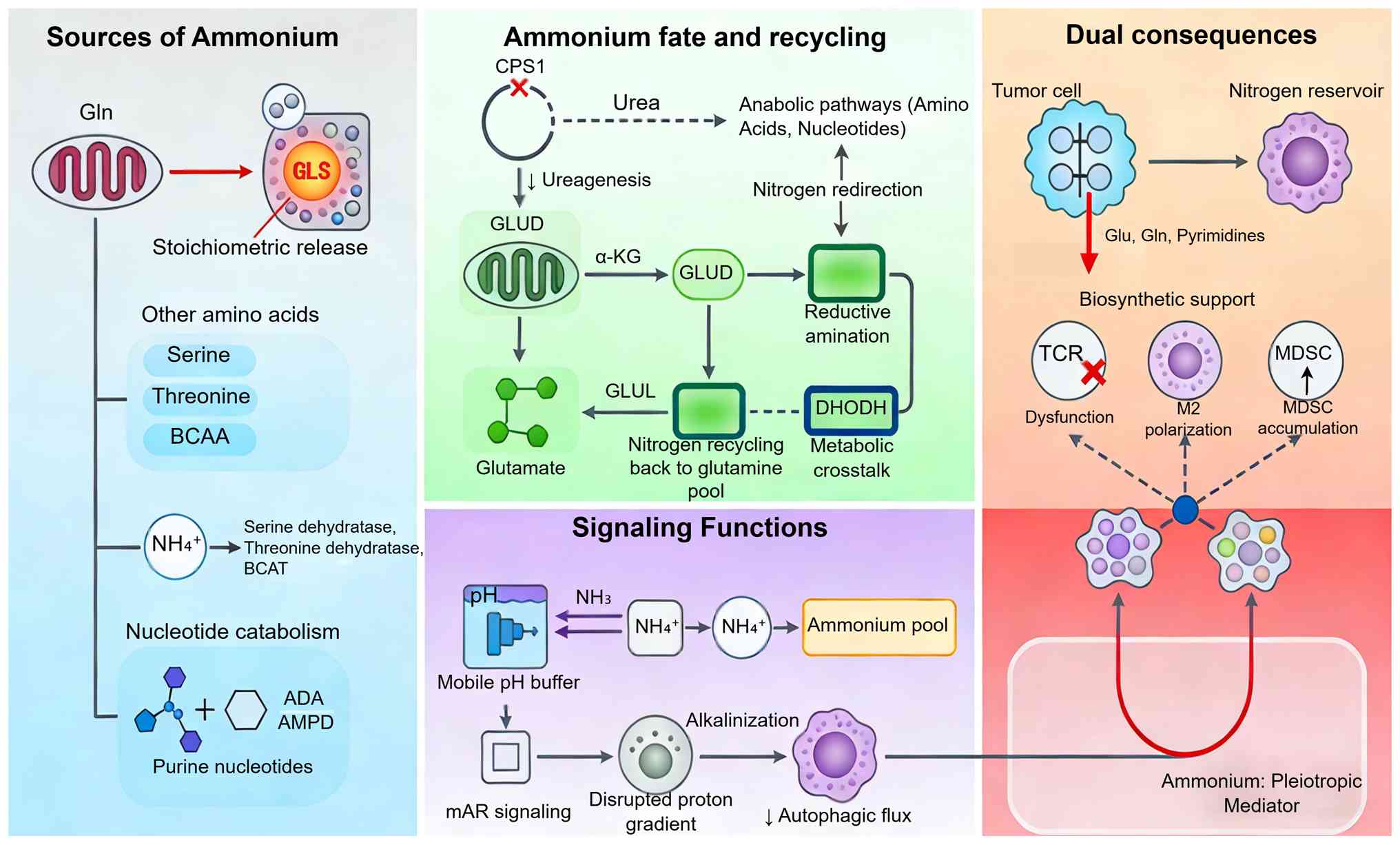 | Figure 2.Schematic overview of ammonium
metabolism in the prostate tumor microenvironment. ADA, adenosine
deaminase; AMPD, adenosine monophosphate deaminase; BCAA,
branched-chain amino acid; CPS1, carbamoyl phosphate synthetase I;
GLS, glutaminase; GLUD, glutamate dehydrogenase; GLUL, glutamine
synthetase; Gln, glutamine; Glu, glutamate; MDSC, myeloid-derived
suppressor cell; TAM, tumor-associated macrophage; α-KG,
α-ketoglutarate. |
Sources of ammonium in the prostate
TME
Ammonium within the prostate TME originates from
multiple metabolic sources that reflect the heightened catabolic
activity of malignant cells. The predominant source is
glutaminolysis, wherein GLS catalyzes the deamination of glutamine
to glutamate, releasing ammonium as a stoichiometric byproduct.
This pathway operates at elevated flux in PCa cells due to
glutamine addiction, resulting in constitutive ammonium production
that surpasses the rate at which the cells use nitrogen for amino
acid and nucleotide biosynthesis, causing ammonium to accumulate
and be released into the TME (38).
Beyond glutaminolysis, deamination reactions
involving other amino acids contribute to the ammonium pool.
Catabolism of serine, threonine and branched-chain amino acids
generates ammonium through the action of serine dehydratase,
threonine dehydratase and branched-chain amino acid transaminases
coupled with GLUD (10,11). Nucleotide catabolism represents an
additional source, with adenosine and AMP deaminase releasing
ammonium during purine nucleotide degradation (12,13).
The relative contribution of each source varies according to
metabolic context, nutrient availability and oncogenic driver
status. Ye et al (12)
reviewed ammonium metabolism rewiring in the PCa ME, emphasizing
that the convergence of these pathways creates sustained ammonium
flux that distinguishes malignant from benign prostate tissue.
Ammonium fate and recycling: Urea
cycle dysregulation and nitrogen partitioning
Once generated, ammonium faces several potential
fates determined by the expression and activity of
nitrogen-handling enzymes within PCa cells. The urea cycle
represents the canonical pathway for ammonium detoxification,
converting ammonium and bicarbonate to urea through enzymatic
reactions distributed between mitochondria and cytoplasm. However,
PCa cells exhibit notable urea cycle dysregulation characterized by
downregulation of key enzymes including carbamoyl phosphate
synthetase I and ornithine transcarbamylase. Bruzzone et al
(44) demonstrated using nuclear
magnetic resonance-based urine metabolomics that patients with PCa
display enhanced carbon and nitrogen recycling, with metabolomic
signatures indicating redirection of nitrogen away from urea
production toward anabolic pathways. This metabolic rewiring favors
nitrogen incorporation into amino acids and nucleotides at the
expense of ureagenesis, supporting biosynthetic demands of
proliferating tumor cells.
GLUD and glutamine synthetase (GLUL) represent
alternative fates for ammonium assimilation. GLUD catalyzes the
reversible reductive amination of α-ketoglutarate to glutamate,
incorporating ammonium into amino acid pools, while GLUL ligates
ammonium with glutamate to form glutamine, recycling nitrogen back
into the glutamine pool. Luo et al (45) revealed that PCa stem cells exhibit
aberrant urea cycle activity with selective upregulation of
specific urea cycle enzymes that support stemness maintenance,
suggesting that nitrogen partitioning is dynamically regulated
across tumor subpopulations. Labroy et al (46) further demonstrated metabolic
crosstalk between the urea cycle and pyrimidine synthesis,
identifying dihydroorotate dehydrogenase (DHODH) as a metabolic
vulnerability that integrates nitrogen metabolism with nucleotide
biosynthesis.
Ammonium as a signaling molecule:
Impact on pH homeostasis and autophagy
Beyond its role as a metabolic substrate, ammonium
serves as an intracellular signaling molecule that modulates key
cell processes including pH homeostasis, autophagy and stress
adaptation. Ammonium exists in equilibrium with ammonia, which
freely diffuses across membranes and accepts protons to form
ammonium, thereby serving as a mobile pH buffer. Chatterjee et
al (47) demonstrated that
membrane AR signaling influences Na+/H+ exchanger activity in PCa
cells, establishing a link between hormonal signaling and pH
regulatory mechanisms that interface with ammonium
partitioning.
Accumulation of intracellular ammonium alkalinizes
acidic compartments including lysosomes, disrupting the proton
gradient required for optimal lysosomal hydrolase activity and
autophagic flux. Yang et al (48) reported that curcumin induces both
apoptosis and protective autophagy in CRPCa cells through
mechanisms involving iron chelation, highlighting the
interconnectedness of metal homeostasis, oxidative stress and
autophagic regulation. Ammonium accumulation influences stress
adaptation pathways by modulating mTOR signaling and activating
stress-responsive transcription factors. Chen et al
(49) developed an ammonia-induced
calcium phosphate nanostructure that provides insight into how
local ammonium concentrations may influence bone metastasis,
linking nitrogen metabolism with the establishment of metastatic
niches. These signaling functions establish ammonium as a
pleiotropic mediator that integrates metabolic state with cell
stress responses.
Ammonium accumulation: Metabolic
support vs. immunosuppression
Ammonium accumulation within the prostate TME exerts
paradoxical effects, simultaneously supporting tumor metabolic
adaptation while suppressing antitumor immune responses. For cancer
cells, ammonium serves as a nitrogen reservoir that can be
assimilated into amino acids and nucleotides via GLUD and GLUL,
supporting biosynthetic capacity under nutrient-limited conditions.
Metabolomic profiling by Yu et al (50) identified distinct metabolic
signatures in plasma and urine from patients with PCa, revealing
alterations in nitrogenous compounds that reflect systemic
metabolic reprogramming.
However, the ammonium accumulation that supports
tumor metabolism imposes immunosuppressive consequences on
infiltrating immune cells. Ye et al (12) reviewed how ammonium metabolic
reprogramming drives immunosuppression within the prostate TME
through multiple mechanisms including polarization of
tumor-associated macrophages (TAMs) toward the M2 phenotype,
induction of T cell dysfunction and promotion of myeloid-derived
suppressor cell (MDSC) accumulation; direct primary evidence for
ammonium-induced T cell dysfunction has been reported in colorectal
cancer and effector T cell models (51,52).
Ammonium impairs T cell proliferation and effector function by
interfering with T cell receptor signaling and inducing metabolic
stress, while simultaneously promoting macrophage arginase activity
and anti-inflammatory cytokine production. The dual nature of
ammonium accumulation creates a therapeutic paradox: Interventions
that block ammonium production may deprive tumors of nitrogen for
biosynthesis, but interventions that promote ammonium accumulation
may reinforce immunosuppression. Understanding this duality is key
for developing therapeutic strategies that target nitrogen
metabolism without compromising antitumor immunity, positioning
ammonium metabolism as a key node connecting tumor biology with
immune evasion in PCa (51,52).
Metabolic reprogramming of immune cells in
the prostate TME
The prostate TME represents a dynamic ecosystem
wherein metabolic reprogramming extends beyond cancer cells to
encompass diverse immune populations. The competition for limited
nutrients, accumulation of metabolic waste products and
establishment of hypoxic niches reshape immune cell metabolism,
driving functional polarization toward immunosuppressive phenotypes
that facilitate tumor progression. Understanding the metabolic
adaptations of TAMs, T cells, MDSCs and dendritic cells (DCs) is
essential for appreciating how glutamine and ammonium metabolism
intersect with antitumor immunity (Table I).
 | Table I.Metabolic reprogramming of immune
cells in the prostate TME. |
Table I.
Metabolic reprogramming of immune
cells in the prostate TME.
| First author,
year | Immune cell
type | Model system | Findings | Metabolic
features | Immune
consequence | (Refs.) |
|---|
| Bharti et
al, 2019 | General TME | Primary and
metastatic PCa tissue | Hypoxia shapes
regional metabolic heterogeneity and influences therapeutic
responses | Hypoxia-driven
metabolic adaptation | Immune cell
dysfunction | (53) |
| Arocena et
al, 2019 | General TME | Coverslip hypoxia
model | Oxygen gradients
drive metabolic adaptation in tumor cells | Hypoxia gradient
sensing | Altered immune
landscape | (54) |
| Bery et al,
2020 | General TME | PCa cell lines
(including 22Rv1, PC-3, DU145) and organotypic cultures of human
prostate tissue | Hypoxia upregulates
Zeb1 and potassium channels, promoting aggressiveness | Hypoxia-induced
transcriptional reprogramming | Enhanced metastatic
potential | (55) |
| Bok et al,
2019 | General TME | Dual-agent
hyper-polarized 13C MRSI | Lactate serves as
metabolic fuel and signaling molecule in PCa | Lactate
metabolism | Metabolic fuel for
tumor | (56) |
| Comito et
al, 2019 | CD4+ T
cells | PCa cell
line-derived xenograft models in immunodeficient mice | Lactate modulates
CD4+ T cell polarization via TLR8/miR21 signaling | Lactate
signaling | Immunosuppressive
environment | (57) |
| Chetta et
al, 2023 | General TME | Review | Lactate promotes
angiogenesis and suppresses antitumor immunity | Lactate as key
metabolite | Multifaceted
immunosuppression | (58) |
| El-Kenawi et
al, 2019 | TAMs | TRAMP transgenic
model, subcutaneous grafts, and in vitro macrophage
cultures | Acidity promotes
tumor progression by altering macrophage phenotype | pH sensing | M2
polarization | (59) |
| Banerjee et
al, 2019 | TAMs | Patient-derived PCa
tumor-conditioned medium and co-culture with patient-derived cancer
cells | Context-dependent
transcriptional programs shape macrophage function | Metabolic
plasticity | Altered phagocytic
function | (60) |
| Han et al,
2020 | TAMs | THP-1
macrophages | IL-6 from prostate
epithelial cells induces M2 polarization | Cytokine-driven
metabolism | M2
polarization | (61) |
| Praharaj et
al, 2024 | TAMs | RM-1 prostate
cancer subcutaneous graft models | JHU083 reprograms
TAMs toward pro-inflammatory phenotype | Glutamine
metabolism | Enhanced
phagocytosis, decreased angiogenesis | (14) |
| Masetti et
al, 2022 | TAMs | PCa cell
line-derived xenograft models (PC-3, DU145) in immuno-deficient
mice | Lipid-loaded TAMs
sustain tumor growth and invasiveness | Lipid
metabolism | Tumor-supportive
function | (62) |
| Li et al,
2023 | TAMs | Mouse models and
PCa cell lines (RM-1, TRAMP-C1) | Dauricine inhibits
PI3K/AKT-dependent M2 polarization | PI3K/AKT
signaling | Decreased M2
polarization | (63) |
| Guan et al,
2022 | T cells | Immunocompetent
mouse models | AR activity in T
cells limits checkpoint blockade efficacy | Hormonal
signaling | T cell
dysfunction | (64) |
| Chang et al,
2024 | T cells | Prostate tumor
tissue-derived microenvironment from mouse models and human PCa
samples |
1-Pyrroline-5-carboxylate inhibits T cell
glycolysis via SHP1/PKM2/LDHB | Glycolysis
inhibition | T cell
dysfunction | (65) |
| Rastogi and McNeel,
2025 | T cells | Post-immunotherapy
PCa patient samples | Shared features
between antitumor response and immune-associated adverse
events | Metabolic
adaptation | Altered T cell
function | (66) |
| Zhou et al,
2025 | CD8+ T
cells | PCa cell lines
(LNCaP, PC-3, DU145) | EP4 upregulation
attenuates CD8+ T cell killing | PI3K/AKT
signaling | Decreased
cytotoxicity | (67) |
| Molina et
al, 2024 | T lymphocytes | Prostate tumor
tissues from radical prostatectomy patients | Regulatory and
memory T cell infiltration predicts long-term outcomes | T cell
metabolism | Prognostic
value | (68) |
| Hellsten et
al, 2019 | MDSCs | PCa cell lines
(LNCaP, PC-3, DU145) | STAT3 inhibitor
inhibits MDSC-like monocyte generation | STAT3
signaling | Decreased
immunosuppression | (69) |
| Fu et al,
2021 | MDSCs |
High-dose-irradiated TRAMP-C1 tumors | MDSCs serve as
therapeutic target and index for TME status | Radiation-induced
metabolism | Immunosuppressive
barrier | (70) |
| Koinis et
al, 2021 | MDSCs | Review | MDSCs are involved
in PCa immune evasion | MDSC
metabolism | Key role in
immunosuppression | (71) |
| Siemińska and
Baran, 2022 | MDSCs | Review | MDSCs are key
players and promising therapy targets in PCa | MDSC
metabolism | Therapeutic
targeting potential | (72) |
| Feriz et al,
2023 | DCs | Human PCa tumor
tissues | Heterogeneous
transcriptional signatures in tumor-infiltrating DCs | Transcriptional
heterogeneity | Differential
immunostimulatory capacity | (73) |
| Hawlina et
al, 2022 | DCs | DC-based vaccine
trial in patients with PCa | DC-based vaccines
prolong survival independently of vaccine cell number | DC metabolism | Clinical
immunotherapeutic efficacy | (74) |
| Hensler et
al, 2022 | DCs | Patients with PCa
(peripheral blood gene signature analysis) | Peripheral gene
signatures distinguish patient immunotypes for DC-based
vaccines | Metabolic
signatures | Biomarker for
patient selection | (75) |
| Li et al,
2022 | MDSCs | ARID1A knockout
genetically engineered mouse models | ARID1A loss induces
polymorphonuclear MDSC chemotaxis | Genetic regulation
of metabolism | Enhanced MDSC
recruitment | (76) |
Metabolic landscape of the prostate
TME: Nutrient competition, hypoxia and lactate accumulation
The prostate TME is characterized by metabolic
alterations that create a hostile landscape for infiltrating immune
cells while supporting tumor growth. Hypoxia is a dominant feature,
arising from aberrant vascularization and elevated oxygen
consumption by proliferating tumor cells. Bharti et al
(53) demonstrated distinct hypoxia
patterns in primary and metastatic PCa environments, revealing that
oxygen gradients shape regional metabolic heterogeneity and
influence therapeutic responses. Arocena et al (54) developed a variant of coverslip
hypoxia to visualize tumor cell alterations at increasing distances
from an oxygen source, providing mechanistic insight into how
oxygen tension gradients drive metabolic adaptation. Bery et
al (55) demonstrated that
hypoxia promotes PCa aggressiveness by upregulating
epithelial-mesenchymal transition activator zinc finger E-box
binding homeobox 1 and potassium channel expression, linking oxygen
deprivation with enhanced metastatic potential.
Lactate accumulation is a defining feature of the
PCa TME, resulting from elevated glycolytic flux and the Warburg
effect in cancer cells. Bok et al (56) used dual-agent hyperpolarized
carbon-13 magnetic resonance spectroscopic imaging to reveal the
role of lactate metabolism in PCa progression and metastases,
demonstrating that lactate serves not only as a waste product but
as a critical metabolic fuel and signaling molecule. Comito et
al (57) demonstrated that
lactate modulates CD4+ T cell polarization and induces
an immunosuppressive environment through the TLR8/microRNA21
pathway, sustaining prostate carcinoma progression. Chetta et
al (58) recently reviewed the
clinical implications of lactate as a key metabolite in PCa
progression, emphasizing its multifaceted roles in promoting
angiogenesis, suppressing antitumor immunity and driving therapy
resistance. The convergence of hypoxia, nutrient competition and
lactate accumulation establishes a metabolic milieu that influences
immune cell function and polarization.
TAMs: Glutamine-driven M2 polarization
and metabolic plasticity
TAMs represent the most abundant immune population
within the prostate TME and exhibit metabolic plasticity that
enables adaptation to microenvironmental cues. El-Kenawi et
al (59) demonstrated that
acidity promotes tumor progression by altering macrophage phenotype
in PCa, establishing a direct link between extracellular pH and TAM
polarization toward immunosuppressive states. Banerjee et al
(60) examined differential
expression of efferocytosis- and phagocytosis-associated genes in
TAMs exposed to patient-derived PCa ME, revealing context-dependent
transcriptional programs that shape macrophage function. Han et
al (61) demonstrated that IL-6
produced by prostate epithelial cells stimulated with
Trichomonas vaginalis promotes proliferation of PCa cells by
inducing M2 polarization of human monocytic leukemia cell line
THP-1-derived macrophages, highlighting the role of infectious
agents in shaping TAM phenotypes.
Glutamine metabolism serves a key role in driving
TAM polarization and function. Praharaj et al (14) demonstrated that metabolic
reprogramming of TAMs using the glutamine antagonist JHU083 drives
tumor immunity in myeloid-rich PCa and bladder cancer: JHU083
treatment reprogrammed immunosuppressive TAMs toward a
pro-inflammatory phenotype, increasing tumor cell phagocytosis,
diminishing pro-angiogenic capacity and promoting inflammatory
signaling. Masetti et al (62) identified lipid-loaded TAMs as key
sustainers of tumor growth and invasiveness in PCa, revealing that
metabolic substrate availability shapes macrophage effector
functions. Li et al (63)
demonstrated that dauricine regulates PCa progression by inhibiting
PI3K/AKT-dependent M2 polarization of macrophages, providing
pharmacological evidence for targeting macrophage metabolism. The
metabolic plasticity of TAMs represents both a vulnerability and a
therapeutic opportunity in PCa.
T cell metabolism: Glutamine
deprivation, effector dysfunction and exhaustion
T cells infiltrating the prostate TME face metabolic
challenges that compromise their antitumor effector function and
promote exhaustion. Nutrient competition between rapidly
proliferating cancer and T cells creates an environment of
glutamine deprivation that impairs T cell activation and
proliferation. Guan et al (64) demonstrated that AR activity in T
cells limits checkpoint blockade efficacy, revealing that hormonal
signaling directly influences T cell metabolism and function within
the prostate TME. Chang et al (65) recently demonstrated that
1-pyrroline-5-carboxylate inhibits T cell glycolysis in the PCa
microenvironment through the Src homology region 2
domain-containing phosphatase 1/pyruvate kinase M2/lactate
dehydrogenase B pathway, identifying a novel metabolite-driven
immunosuppressive mechanism.
Memory T cell differentiation and maintenance are
influenced by the metabolic landscape. Rastogi and McNeel (66) characterized prostate tumor immune ME
changes following immunotherapy, revealing shared features between
patients who developed antitumor responses and those experiencing
immune-associated adverse events. Zhou et al (67) demonstrated that upregulation of
E-prostanoid receptor 4 attenuates the killing ability of
CD8+ T cells against PCa cells via the PI3K/AKT
signaling pathway, identifying a potential target for restoring T
cell cytotoxicity. The metabolic competition between tumors and T
cells for glutamine and other nutrients shapes the outcome of
antitumor immune responses. Molina et al (68) demonstrated that regulatory and
memory T lymphocytes infiltrating prostate tumors predict long-term
clinical outcomes, emphasizing the prognostic value of T cell
subset distribution.
MDSCs and DCs: Metabolic
adaptation
MDSCs and DCs represent complementary arms of the
myeloid compartment that orchestrate immunosuppression within the
prostate TME. MDSCs expand in patients with PCa and suppress T cell
responses through multiple mechanisms including arginase-1
expression, reactive oxygen species production and nutrient
depletion. Hellsten et al (69) demonstrated the STAT3 inhibitor
galiellalactone inhibits the generation of MDSC-like monocytes by
PCa cells and decreases immunosuppressive and tumorigenic factors,
validating MDSC targeting as a therapeutic strategy. Fu et
al (70) examined the role of
MDSCs in high-dose-irradiated transgenic adenocarcinoma of the
mouse prostate cell line c1 tumors, identifying them as both a
therapeutic target and an index for assessing TME status. Koinis
et al (71) reviewed MDSCs
in PCa, emphasizing their key role in immune evasion. Siemińska and
Baran (72) further positioned
MDSCs as key players and promising therapy targets in PCa,
reviewing strategies for MDSC depletion and functional
inhibition.
DCs, key for priming antitumor T cell responses,
exhibit metabolic and functional alterations within the prostate
TME. Feriz et al (73) used
single-cell RNA sequencing to demonstrate heterogeneous
transcriptional signatures in tumor-infiltrating DCs in PCa,
revealing distinct DC subsets with differential immunostimulatory
capacities. Hawlina et al (74) demonstrated that DC-based vaccines
prolong survival and time to next therapy independently of vaccine
cell number, providing clinical evidence for DC-based
immunotherapy. Hensler et al (75) identified peripheral gene signatures
that distinguish distinct immunotypes of patients with cancer with
therapeutic implications for autologous DC-based vaccines, offering
biomarkers for patient selection. The metabolic adaptation of MDSCs
and DCs to the glutamine-depleted, ammonium-accumulating prostate
TME shapes their immunosuppressive vs. immunostimulatory functions.
Li et al (76) demonstrated
that AT-rich interactive domain-containing protein 1A loss induces
polymorphonuclear MDSC chemotaxis and promotes PCa progression,
revealing genetic determinants of myeloid cell recruitment. The
immunosuppressive effects of MDSCs and DCs, coupled with their
metabolic reprogramming, establishes a-barrier to antitumor
immunity that must be overcome for successful immunotherapy.
Bidirectional metabolic-immune crosstalk:
Glutamine and ammonium as key mediators
The association between tumor metabolism and
antitumor immunity is not unidirectional but represents a dynamic,
bidirectional association wherein metabolic reprogramming of cancer
cells shapes immune cell function, and immune-derived signals
feedback to modulate tumor metabolic pathways (9,10).
This reciprocal crosstalk establishes a self-reinforcing cycle that
drives PCa progression and immune evasion. Glutamine and ammonium
metabolism occupy central positions within this association,
serving both as substrates for tumor growth and as critical
mediators of immunosuppression. Understanding these bidirectional
interactions is key for developing therapeutic strategies that
simultaneously target tumor metabolism and enhance antitumor
immunity (12,13). Fig.
3 provides a schematic overview of the bidirectional
metabolic-immune crosstalk, illustrating the tumor-derived signals,
nutrient competition, ammonium-mediated immunosuppression, redox
interplay and immune-derived feedback loops that shape the prostate
TME.
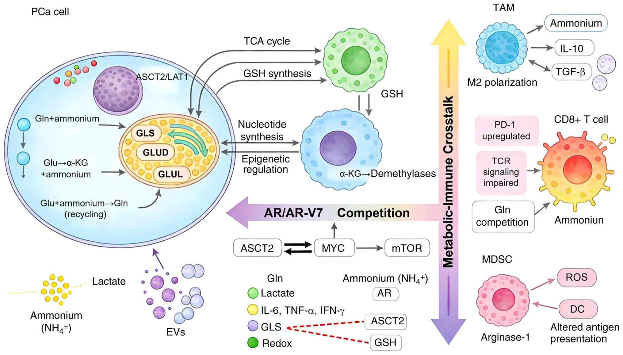 | Figure 3.Schematic illustration of the
metabolic-immune association mediated by glutamine and ammonium in
the prostate tumor microenvironment. ASCT2, alanine-serine-cysteine
transporter 2; DC, dendritic cell; EV, extracellular vesicle; GLS,
glutaminase; GLUD, glutamate dehydrogenase; GLUL, glutamine
synthetase; Gln, glutamine; Glu, glutamate; GSH: Glutathione; LAT1:
L-type amino acid transporter 1; MDSC, myeloid-derived suppressor
cell; PCa, prostate cancer; ROS, reactive oxygen species; TAM,
tumor-associated macrophage; TCR, T cell receptor; α-KG,
α-ketoglutarate. |
Tumor-derived signals shaping immune
cell metabolism: Lactate, kynurenine and extracellular
vesicles
PCa cells actively remodel the metabolic landscape
of the TME through secretion of metabolites, enzymes and
extracellular vesicles that directly modulate immune cell function.
Lactate, long considered a metabolic waste product, has emerged as
a potent signaling molecule that reprograms immune cell metabolism
and polarization (77). Stepka
et al (77) reviewed
metabolic and amino acid alterations in the TME, emphasizing that
lactate accumulation suppresses T cell proliferation and promotes
regulatory T cell differentiation while driving macrophage
polarization toward immunosuppressive M2 phenotypes.
Extracellular vesicles represent an additional
mechanism for metabolic communication between tumor cells and
immune populations. Mo et al (78) demonstrated that long non-coding RNA
nuclear-enriched abundant transcript 1 shuttled by PCa
cell-secreted exosomes initiates osteoblastic phenotypes in the
bone metastatic ME through the microRNA-205-5p/runt-related
transcription factor 2/splicing factor proline- and glutamine-rich
pathway, revealing that exosomal cargo shapes distant ME niches.
Lee et al (79) demonstrated
that extracellular vesicles derived from PCa cells induce metabolic
reprogramming toward a glycolysis phenotype in recipient cells,
suggesting vesicle-mediated transfer of metabolic regulators
contributes to the establishment of a glycolytic, immunosuppressive
ME. Ippolito et al (80)
showed that extracellular pH modulates neuroendocrine PCa cell
metabolism and susceptibility to mitochondrial inhibitors,
indicating that the acidic ME resulting from tumor metabolism
further influences immune cell function. These tumor-derived
signals establish a metabolic landscape that favors
immunosuppression while supporting tumor progression.
Glutamine competition between tumor
cells and infiltrating lymphocytes
Glutamine is a key nutrient for both proliferating
tumor cells and activated lymphocytes, creating a metabolic
competition within the prostate TME that shapes antitumor immunity.
Alhallaq and Sultan (9) positioned
glutamine and glutamate metabolic reprogramming at the center of
PCa pathogenesis, describing the dynamic competition between tumor
and immune cells for this essential amino acid. This competition
deprives infiltrating T cells of the glutamine required for
activation, proliferation and effector function, contributing to
the exhausted T cell phenotype characteristic of advanced PCa.
Matos et al (81) examined
the role of arginine and arginases in modulating metabolism, TME
and PCa progression, demonstrating that competition for amino acid
substrates extends beyond glutamine to encompass multiple
nitrogenous nutrients.
Bhowmick et al (10) reviewed strategies for targeting
glutamine metabolism in PCa, emphasizing that therapeutic
interventions should consider the differential glutamine
requirements of tumor cells vs. antitumor immune populations. This
dual benefit arises from differential sensitivity to glutamine
antagonism, with tumor-associated macrophages being susceptible to
metabolic reprogramming toward pro-inflammatory phenotypes.
Ammonium as an immunosuppressive
metabolite: Mechanisms of T cell dysfunction and macrophage
polarization toward M2 phenotype
Beyond its role as a nitrogenous waste product,
ammonium serves as an active immunosuppressive metabolite that
directly impairs T cell function and promotes macrophage
polarization toward tumor-supportive phenotypes. Ye et al
(12) reviewed ammonium metabolism
rewiring in the PCa ME, demonstrating that ammonium accumulation
drives immunosuppression through multiple mechanisms including
polarization of TAMs toward the M2 phenotype, induction of T cell
dysfunction and promotion of MDSC accumulation. Primary studies
have shown that ammonia directly impairs T cell proliferation and
effector function through lysosomal alkalization, mitochondrial
swelling and impaired autophagic flux (51,52):
Ammonium impairs T cell proliferation and effector function by
interfering with T cell receptor signaling and inducing metabolic
stress, while simultaneously promoting macrophage arginase activity
and anti-inflammatory cytokine production. Elia et al
(13) demonstrated that nitrogen
handling varies between tumor types, with PCa exhibiting unique
adaptations that favor ammonium accumulation and recycling. Yang
et al (82) reviewed
TME-driven drug resistance in urological cancers, highlighting that
ammonium-mediated immunosuppression contributes to the failure of
immunotherapeutic approaches and represents a potential therapeutic
target. The immunosuppressive consequences of ammonium accumulation
create a therapeutic paradox: Interventions that block ammonium
production may deprive tumors of nitrogen for biosynthesis, but
interventions that promote ammonium accumulation may reinforce
immunosuppression. This duality positions ammonium metabolism as a
key node connecting tumor biology with immune evasion in PCa.
Redox interplay: Glutathione
metabolism linking tumor antioxidant defense and immune
suppression
Glutathione metabolism represents a key intersection
where tumor antioxidant defense mechanisms directly influence
immune cell function within the prostate microenvironment. Wang
et al (83) reviewed the
integrated regulation of ferroptosis in PCa, demonstrating that
glutathione peroxidase 4 and glutathione metabolism protect tumor
cells from oxidative stress while simultaneously depleting the
antioxidant capacity available to infiltrating immune cells.
Linares et al (84)
demonstrated that activating transcription factor 4-induced
metabolic reprogramming represents a synthetic vulnerability of
p62-deficient tumor stroma, revealing that oxidative stress
responses in the TME are coordinately regulated and influence both
cancer cells and supporting stromal elements. Zhang et al
(15) showed that improved
antitumor activity against PCa can be achieved through combined
targeting of Myc and GFAT-1, the rate-limiting enzyme in the
hexosamine biosynthetic pathway that branches from glutamine
metabolism and influences protein glycosylation and redox balance.
The glutathione synthesis pathway directly competes with other
glutamine-using pathways for substrate, creating metabolic
trade-offs that influence both tumor survival and immune function.
Tumor cells with elevated glutathione synthesis capacity exhibit
enhanced resistance to oxidative stress induced by inflammatory
cytokines, while depleting the local cysteine and glutamate pools
required for optimal T cell activation and proliferation. This
redox interplay establishes a feedback loop wherein tumor
antioxidant defense mechanisms actively suppress the oxidative
burst required for effective antitumor immunity (15,24).
Feedback loops: Immune-derived
cytokines modulating tumor glutamine and ammonium flux
The bidirectional nature of metabolic-immune
crosstalk is evident in the feedback loops wherein immune-derived
cytokines directly modulate tumor cell glutamine and ammonium
metabolism. Hönscheid et al (11) demonstrated that PCa-associated
fibroblasts manipulate glutamine metabolism in cancer cells through
paracrine signaling, revealing that non-immune stromal elements
also participate in this metabolic interaction. Lasorsa et
al (7) demonstrated that
inflammatory cytokines including interferon-γ and tumor necrosis
factor-α produced by infiltrating lymphocytes feedback to modulate
glutamine transporter expression, GLS activity and urea cycle
enzyme expression in cancer cells. These immune-derived signals
constrain or enhance tumor metabolic capacity depending on the
context and duration of exposure. Acute exposure to inflammatory
cytokines may suppress glutamine uptake and metabolism,
contributing to the anti-proliferative effects of immune activation
(7). However, chronic exposure to
low-level inflammatory signals, characteristic of the TME, may
select for tumor cells with adaptive metabolic programs that
maintain glutamine flux despite cytokine-mediated stress. By
intervening at the metabolic level, it is possible to
simultaneously disrupt tumor metabolic adaptation and enhance the
immunostimulatory capacity of TAMs, creating a cycle of antitumor
immunity (7). Understanding these
complex feedback associations is key for developing combination
therapies that maximize therapeutic benefit while minimizing
resistance mechanisms.
Integrated regulatory network of
glutamine-ammonium mediated metabolic-immune crosstalk
The bidirectional crosstalk between tumor metabolism
and antitumor immunity in PCa operates through an integrated
regulatory network organized across three interconnected levels. At
the transcriptional level, oncogenic drivers including AR, Myc and
mutant p53 coordinate glutamine metabolic gene expression through
direct transcriptional activation and epigenetic modulation
(9,13). AR differentially regulates metabolic
pathways via full-length AR vs. AR-V7, while Myc drives GLS
expression and glutamine uptake through transcriptional programs
(64). PTEN loss enhances glutamine
dependency via PI3K/AKT/mTOR pathway activation, and p53 status
modulates mitochondrial glutamine use. At the metabolite signaling
level, glutamine-derived ammonium directly impairs T cell receptor
signaling and promotes macrophage arginase activity, establishing
metabolite-driven immunosuppression (12,65).
Simultaneously, glutamine-derived glutathione metabolism links
tumor antioxidant defense with immune suppression through depletion
of local antioxidant capacity (77). At the cell interaction level,
feedback loops complete the bidirectional circuit: Immune-derived
cytokines including interferon-γ and tumor necrosis factor-α
modulate glutamine transporter expression, GLS activity and urea
cycle enzyme expression in cancer cells, while tumor-secreted
lactate, kynurenine and extracellular vesicles reshape immune cell
metabolism and polarization (11,13,77).
This multi-level network exhibits redundancy and adaptive
plasticity, wherein pathway inhibition at one node typically
triggers compensatory activation at alternative nodes, explaining
the limited efficacy of monotherapy and providing the rationale for
combinatorial strategies targeting multiple network nodes
simultaneously.
Therapeutic strategies targeting the
metabolic-immune interface
The recognition that glutamine and ammonium
metabolism orchestrate bidirectional crosstalk between tumor cells
and immune populations has opened novel therapeutic avenues
targeting the metabolic-immune interface. Rather than pursuing
tumor cell-intrinsic metabolic disruption or immune activation as
separate strategies, emerging approaches aim to simultaneously
inhibit tumor metabolic adaptation while reprogramming the
immunosuppressive TME toward antitumor immunity (Table II).
| Table II.Therapeutic strategies targeting the
metabolic-immune interface in PCa. |
Table II.
Therapeutic strategies targeting the
metabolic-immune interface in PCa.
| First author,
year | Therapeutic
agent/strategy | Model system |
Target/mechanism | Findings | Combination
partner | (Refs.) |
|---|
| Cha et al,
2020 | CB-839
(telaglenastat) | Preclinical PCa
xenograft | Glutaminase
inhibition | Enhances
radiosensitivity via redox state, stemness and autophagy
modulation | Radiotherapy | (85) |
| Praharaj et
al, 2024 | JHU083 (glutamine
antagonist prodrug) | Myeloid-rich PCa
and bladder cancer models | Glutamine
antagonism | Reprograms TAMs to
pro-inflammatory phenotype; increases phagocytosis; decreases
angiogenesis | JHU083
(monotherapy) | (14) |
| Moon et al,
2024 | DRP-104 (glutamine
antagonist) | Human CRPCa cell
lines (LNCaP, LAPC4, C4-2/MDVR, PC-3, 22RV1, NCI-H660) and NCI-H660
neuroendocrine PCa xenograft model | Glutamine
antagonism | Inhibits
proliferation and growth of CRPCa | DRP-104
(monotherapy) | (40) |
| Ono et al,
2015 | ASCT2 transporter
blockade | CRPCa-like cell
lines (LNCaP-SF, LN-REC4) and androgen-dependent LNCaP | ASCT2
inhibition | Fluciclovine uptake
is associatedwith ASCT2 expression; ASCT2 is a potential target and
imaging biomarker | Imaging
(theranostic) | (21) |
| Ye et al,
2025 | Urea cycle enzyme
targeting; GLUD inhibition; ammonia scavengers | Review | Urea cycle
dysregulation; GLUD; nitrogen recycling | Urea cycle
dysregulation creates vulnerabilities; targeting nitrogen handling
disrupts recycling and alleviates immunosuppression | Not specified | (12) |
| Labroy et
al, 2026 | DHODH
inhibitors | AR-positive and
-negative PCa cells | Dihydroorotate
dehydrogenase | Metabolic crosstalk
between urea cycle and pyrimidine synthesis; DHODH is a metabolic
vulnerability | Not specified | (46) |
| Luo et al,
2024 | Urea cycle enzyme
targeting | Prostate cancer
stem cells | Urea cycle enzymes
(stemness maintenance) | Aberrant urea cycle
activity supports stemness; nitrogen partitioning is dynamically
regulated | Combination
strategies required | (45) |
| Sharma et
al, 2020 | Nivolumab +
ipilimumab | Patients with
mCRPCa (CheckMate 650 trial) | PD-1 + CTLA-4
blockade | Clinical activity
in mCRPCa; response is associated with tumor mutational burden and
immune infiltration | Combination
immunotherapy | (87) |
| Shenderov et
al, 2021 | Nivolumab +
ipilimumab ± enzalutamide | Patients with
AR-V7-expressing mCRPCa | PD-1 + CTLA-4
blockade | Nivolumab +
ipilimumab modestly overcame enzalutamide enzalutamide resistance
in a subset of patients with AR-V7-expressing mCRPCa | Enzalutamide | (88) |
| Powles et
al, 2022 | Atezolizumab +
enzalutamide vs. enzalutamide alone | Patients with
mCRPCa (randomized phase 3) | PD-L1 blockade | No improvement in
unselected patients; biomarker-defined subgroups may benefit | Enzalutamide | (89) |
| Hegde et al,
2021 | Evofosfamide +
ipilimumab | Advanced solid
malignancy (phase 1) | Hypoxia-activated
prodrug + CTLA-4 blockade | Proof-of-concept
for combining microenvironment-targeted metabolic agents with
immune checkpoint blockade | Ipilimumab | (90) |
| Ono et al,
2015 | [18F]Fluciclovine
PET | CRPCa-like and
androgen-dependent PCa cell lines | ASCT2 expression
imaging | Fluciclovine uptake
is associated with ASCT2 expression; validates amino acid PET for
target engagement monitoring | Target engagement
monitoring | (21) |
| Lowentritt and
Kipper, 2020 | [18F]Fluciclovine
PET/CT | Patients with
biochemical recurrence of PCa | ASCT2 and LAT1
imaging | Clinical utility
for detecting recurrent disease; guides management decisions | Imaging-guided
therapy | (91) |
| Smith et al,
2021 | Plasma glutamine
levels | Patients with
localized PCa | Circulating
glutamine | Plasma glutamine as
prognostic biomarker; alterations are associated with disease
outcome | Liquid biopsy | (18) |
Glutamine metabolism inhibitors: GLS
inhibitors and transporter blockade in preclinical and clinical
development
Pharmacological targeting of glutamine metabolism
has emerged as a promising therapeutic strategy, with multiple
agents advancing through preclinical and clinical development for
PCa. The GLS inhibitor CB-839 (telaglenastat) is the most
clinically advanced agent, having demonstrated the ability to
enhance PCa radiosensitivity by regulating redox state, stemness
and autophagy (85). Bhowmick et
al (10) reviewed strategies
for targeting glutamine metabolism in PCa, emphasizing that
therapeutic interventions should consider the differential
glutamine requirements of tumor cells vs. antitumor immune
populations. The glutamine antagonist JHU083, a prodrug that
selectively activates within the TME, has shown promise in a
preclinical study by reprogramming tumor-associated macrophages and
enhancing antitumor immunity (14).
Moon et al (40)
demonstrated that targeting glutamine dependence with DRP-104, a
novel glutamine antagonist, inhibits proliferation and tumor growth
of CRPCa, validating glutamine metabolism as a therapeutic
vulnerability in advanced disease. Beyond enzyme inhibition,
transporter blockade targeting ASCT2 has shown efficacy in a
preclinical model with Ono et al (21) demonstrating that fluciclovine uptake
is associated with ASCT2 expression in CRPCa cells, providing both
a therapeutic target and an imaging biomarker.
GLS inhibitor CB-839, glutamine antagonist prodrug
JHU083 and glutamine antagonist DRP-104, exhibit distinct
advantages and limitations that inform their therapeutic
applicability. CB-839 offers the advantage of selective GLS
inhibition with an established safety profile in clinical trials,
being well-tolerated with mostly mild adverse effects including
nausea, fatigue and photophobia (6,86).
However, its clinical efficacy as monotherapy in PCa is limited;
single-drug trials do not demonstrate notable clinical benefits,
and combination studies with talazoparib were terminated due to
challenges in demonstrating efficacy and slow recruitment (6). Furthermore, residual glutamine
metabolism via GLS2 or other pathways may circumvent
CB-839-mediated blockade (25). By
contrast, JHU083 and DRP-104, as prodrugs of the broad glutamine
antagonist 6-diazo-5-oxo-L-norleucine, provide broader metabolic
inhibition by targeting multiple glutamine-utilizing enzymes beyond
GLS, including amidotransferases involved in nucleotide
biosynthesis (10). JHU083 offers
the advantage of TME-selective activation, which reduces systemic
toxicity while achieving potent local glutamine antagonism
(14). DRP-104 exhibits broader
metabolic inhibition compared with CB-839, further suppressing
glycolysis and glutamine-dependent nucleotide biosynthesis
(3). However, the broad-spectrum
nature of these antagonists raises concerns regarding off-target
effects and potential toxicity and their clinical development
remains at earlier stages compared with CB-839 (6). The differential mechanisms of these
agents and selective GLS inhibition vs. broad glutamine antagonism
suggest that patient stratification based on tumor metabolic
dependency may be key for optimizing therapeutic benefit.
Targeting ammonium metabolism and
nitrogen balance: Urea cycle enzymes, GLUD and ammonia
scavengers
The recognition that ammonium metabolism represents
a potential therapeutic axis has spurred investigation into
strategies targeting nitrogen handling in PCa. Ye et al
(12) reviewed ammonium metabolism
rewiring in the PCa ME, highlighting that urea cycle dysregulation
creates tumor-specific vulnerabilities that can be exploited
therapeutically. Targeting urea cycle enzymes, GLUD and ammonia
scavengers represents an emerging therapeutic frontier with
potential to disrupt nitrogen recycling while alleviating
ammonium-mediated immunosuppression. Labroy et al (46) demonstrated metabolic crosstalk
between the urea cycle and pyrimidine synthesis, identifying DHODH
as a metabolic vulnerability that integrates nitrogen metabolism
with nucleotide biosynthesis in both AR-positive and -negative PCa
cells. This finding suggests that targeting nitrogen handling may
be effective across PCa subtypes. Luo et al (45) revealed that PCa stem cells exhibit
aberrant urea cycle activity with selective upregulation of
specific urea cycle enzymes that support stemness maintenance,
indicating that nitrogen partitioning is dynamically regulated
across tumor subpopulations and may require combination strategies
for effective targeting. The therapeutic potential of targeting
ammonium metabolism extends beyond direct enzyme inhibition to
include ammonia scavengers that may alleviate immunosuppression
while depriving tumors of recyclable nitrogen.
Ammonium-targeting strategies are categorized into
three principal approaches with distinct translational profiles.
Urea cycle enzyme modulation is the most mechanistically direct
strategy, aiming to restore ureagenesis and redirect nitrogen away
from anabolic pathways, however, the multi-enzyme nature of the
urea cycle creates pharmacological complexity, and, to the best of
our knowledge, no urea cycle-activating agent has entered PCa
clinical development (12,44). GLUD and GLUL inhibition offers a
more pharmacologically tractable single-target approach to block
nitrogen recycling within tumor cells, but creates a key
immunological paradox: Inhibiting these enzymes may local ammonium
concentrations, potentially exacerbating T cell dysfunction and
macrophage M2 polarization (51,52).
Ammonia scavengers, such as sodium phenylbutyrate, are the most
translationally attractive strategy due to their US Food and Drug
Administration-approved status for urea cycle disorders, yet their
efficacy in the PCa TME remains unproven and systemic ammonia
scavenging may affect nitrogen homeostasis (12).
To the best of our knowledge, none of the
aforementioned ammonium-targeting strategies has been evaluated in
combination with immune checkpoint blockade in PCa, representing a
translational gap (12). The
selection of optimal strategy will likely depend on balancing tumor
metabolic disruption against the preservation of antitumor
immunity: Urea cycle modulation and ammonia scavengers may
alleviate ammonium-mediated immunosuppression but may support tumor
nitrogen recycling, whereas GLUD/GLUL inhibition may starve tumors
of nitrogen at the cost of worsening the immunosuppressive TME.
Future research should prioritize combinatorial regimens that
simultaneously target multiple nodes of ammonium metabolism, though
such approaches remain hypothetical (12,82).
Combinatorial approaches: Metabolic
inhibitors + immune checkpoint blockade
The limited efficacy of immune checkpoint
inhibitors as monotherapy in PCa has driven investigation into
combination strategies that sensitize tumors to immunotherapy.
Sharma et al (87) reported
preliminary analysis of patients in the CheckMate 650 trial,
demonstrating that nivolumab + ipilimumab exhibits clinical
activity in metastatic CRPCa, with response rates associated with
tumor mutational burden and immune infiltration. Shenderov et
al (88) conducted a phase 2
non-randomized clinical trial of nivolumab + ipilimumab, with or
without enzalutamide, in AR-V7-expressing metastatic CRPCa,
revealing that combination immunotherapy overcomes some mechanisms
of treatment resistance. Powles et al (89) reported results from a randomized
phase 3 trial of atezolizumab with enzalutamide vs. enzalutamide
alone in metastatic CRPCa, demonstrating that while the addition of
immunotherapy did not improve outcomes in unselected patients,
biomarker-defined subgroups may derive benefit. Hegde et al
(90) conducted a phase 1
dose-escalation study evaluating the safety and tolerability of
evofosfamide, a hypoxia-activated prodrug, in combination with
ipilimumab in advanced solid malignancy, providing proof-of-concept
for combining ME-targeted metabolic agents with immune checkpoint
blockade. These studies suggest that metabolic inhibitors targeting
glutamine and ammonium metabolism may enhance the efficacy of
immune checkpoint blockade by alleviating metabolic competition and
immunosuppression within the TME.
The combination of metabolic inhibitors with immune
checkpoint blockade presents both opportunities and challenges.
Preclinical evidence demonstrates that combining glutamine
antagonism (via JHU083 or DRP-104) or GLS inhibition (via CB-839)
with PD-1/PD-L1 blockade enhances antitumor immunity in mouse
models (3,14). However, metabolic inhibition can
paradoxically impair CD8+ T cell function by upregulating PD-L1
expression on tumor cells via a reactive oxygen species-dependent
EGFR/ERK1/2/c-Jun pathway, necessitating concurrent checkpoint
blockade to restore T cell function (9). The differential effects of GLS
inhibition vs. broad glutamine antagonism on immune cell
populations remain incompletely characterized, representing a key
knowledge gap for optimizing combination strategies.
Imaging metabolic vulnerability and
biomarker development for patient stratification
The clinical translation of metabolic-immune
therapies requires parallel development of imaging biomarkers for
patient selection and response monitoring. Ono et al
(21) demonstrated that
fluciclovine uptake is associated with ASCT2 expression in CRPCa
cells, validating amino acid positron emission tomography (PET)
imaging as a non-invasive approach to assess glutamine transporter
expression and target engagement. Lowentritt and Kipper (91) provided a guide for treating patients
with biochemical recurrence of PCa using fluciclovine PET/computed
tomography, emphasizing the clinical utility of metabolic imaging
for detecting recurrent disease. Bhowmick et al (10) reviewed strategies for targeting
glutamine metabolism in PCa, highlighting the need for predictive
biomarkers to identify patients most likely to benefit from
metabolic interventions. Smith et al (18) demonstrated that plasma glutamine
levels may serve as prognostic biomarkers in localized PCa, with
alterations in circulating glutamine associated with disease
outcome, suggesting that liquid biopsy approaches may complement
imaging for patient stratification. The integration of metabolic
imaging with circulating biomarkers may enable precision medicine
approaches that match patients with specific metabolic
vulnerabilities to targeted therapy, ultimately improving outcomes
while minimizing unnecessary toxicity.
Future perspectives
The present review established the
glutamine-ammonium metabolic axis as a key hub integrating tumor
progression, immune evasion and therapeutic resistance in PCa. From
glutamine addiction driven by oncogenic signaling pathways
including AR and Myc, to the role of ammonium as an active
immunosuppressive metabolite rather than waste, the bidirectional
crosstalk between tumor metabolism and antitumor immunity affects
disease outcomes. Alhallaq and Sultan (9) reviewed the central role of glutamine
and glutamate metabolic reprogramming in promoting PCa, emphasizing
that this axis represents both a vulnerability and a therapeutic
opportunity. The convergence of glutamine-dependent bioenergetics,
redox homeostasis and epigenetic regulation with ammonium-mediated
immunosuppression creates a self-reinforcing cycle that promotes
tumor progression while inhibiting antitumor immune responses.
Understanding this integrated network is key for developing
next-generation therapeutic strategies that simultaneously target
tumor metabolism and enhance immune function.
Despite progress, numerous unanswered questions
remain regarding the spatiotemporal dynamics and heterogeneity of
metabolic-immune interactions within the prostate TME. Single-cell
omics technologies are transforming the understanding of cell
heterogeneity, with Yu et al (92) demonstrating that single-cell
approaches trace the heterogeneity of PCa cells and the TME,
revealing distinct metabolic programs across cell subpopulations.
Byrne et al (93)
demonstrated that metabolic reprogramming is spatially
heterogeneous, with distinct metabolic profiles across different
regions of the tissue that influence therapeutic response. Wang
et al (94) reviewed
integrating multi-omics proteomic approaches in the TME and
therapeutic resistance mechanisms, suggesting that comprehensive
molecular profiling is key for identifying context-dependent
metabolic vulnerabilities. The dynamic nature of metabolic
adaptation during disease progression and in response to therapy
remains poorly understood, with Mizuno and Beltran emphasizing that
future directions for precision oncology in PCa should account for
temporal evolution of metabolic phenotypes (95). These technological advances may
resolve questions about metabolic heterogeneity and its
implications for therapeutic targeting.
The TME represents a complex ecosystem wherein
multiple cell types beyond cancer cells contribute to
metabolic-immune crosstalk. Cancer-associated fibroblasts are
critical regulators of tumor metabolism and immune function, with
ChallaSivaKanaka et al (96)
and Owen et al (97)
demonstrating that CAFs exhibit marked functional heterogeneity and
actively promote tumor progression, metastatic dissemination, and
immunosuppression through paracrine signaling and extracellular
matrix remodeling. Lupsa et al (98) demonstrated PCa-associated
fibroblasts, exhibit pronounced functional heterogeneity and
actively promote therapeutic resistance through paracrine cytokine
signaling and extracellular matrix remodeling, while
organ-on-a-chip platforms offer a novel approach for modeling
CAF-tumor interactions and screening targeted therapies. Zhou et
al (99) elucidated that IL-6
and STAT3 signaling in PCa, driven by cancer-associated
fibroblasts, promotes immune evasion and represents a therapeutic
opportunity. Chen et al (100) comprehensively reviewed the PCa ME,
emphasizing multidimensional regulation of immune cells, vascular
system, stromal cells and microbiota. Li et al (101) examined TME-mediated immune evasion
and resistance in PCa, synthesizing the key mechanisms by which
tumor-stroma crosstalk drives immunosuppression and therapeutic
failure, including CAF-derived immunosuppressive cytokines and
chemokines, extracellular matrix remodeling that forms a physical
barrier to T cell infiltration, and the recruitment of MDSCs and
TAMs via metabolic competition and chemokine gradients. The
integration of these cell elements creates a highly redundant and
adaptive system that complicates therapeutic intervention.
Therapeutic frontiers extend beyond direct
targeting of glutamine and ammonium metabolism to encompass
epigenetic regulation, ferroptosis and the gut microbiome. Chen
et al (102) reviewed
advances in ferroptosis for CRPCa treatment, identifying drug
targets and combination therapy strategies that intersect with
glutamine-dependent glutathione metabolism. Rossetto et al
(103) explored the association
between purinergic signaling and oxidative stress in PCa, offering
perspectives for therapy that may complement metabolic
interventions. Epigenetic regulation of metabolism represents
another promising avenue, with Espitia-Pérez et al (104) identifying targeting PCa metabolism
through transcriptional and epigenetic modulation as a multi-target
approach to therapeutic innovation. Naik and Thakur (105) reviewed the association between
epigenetic regulation of TGF-β in cancers, highlighting reciprocal
associations between signaling pathways and chromatin
modifications. The gut microbiome is a modifiable factor
influencing PCa progression and treatment response, with Hao et
al (106) reviewing gut
microbiota as a multifaceted modulator of PCa. Qasem and El-Sayed
(107) examined the bacterial
microbiome and cancer more broadly, summarizing the association
between specific bacterial species and cancer development,
diagnosis, and treatment, while also discussing mechanisms
including genotoxin production, immune modulation, and drug
metabolism, alongside future clinical applications such as fecal
transplantation, probiotics, prebiotics, and microbiome
biomarkers.
The translation of these insights into clinical
practice requires parallel development of predictive biomarkers,
advanced imaging modalities and personalized treatment strategies.
Fidelito et al (108)
examined whether targeting metabolism is a realistic goal for
personalized medicine in PCa, identifying challenges including
tumor metabolic heterogeneity, genetic driver variability, and
tumor microenvironment complexity, while highlighting opportunities
such as the development of more physiologically relevant
preclinical models (e.g., patient-derived xenografts) that better
recapitulate in vivo tumor metabolism to inform patient
stratification. Ottini et al (109) discussed biomarker-driven
immunotherapy for precision medicine in PCa, emphasizing the need
for molecular stratification to identify patients most likely to
benefit from immune-based approaches. San-Jose Manso et al
(110) provided a guide for
immunome profiling in PCa, outlining how comprehensive immune
characterization can inform treatment decisions. Theranostic
approaches combining imaging and therapy are evolving, with Sollini
et al (111) reviewing
radiopharmaceuticals and the future of theranostics in
genitourinary cancer. Ma et al (112) discussed nanoparticle-based drug
delivery systems in urological oncology, from targeted therapy to
precision theranostics, offering strategies for improving
therapeutic index. Pati et al (113) reviewed clinical translation for
mRNA vaccines in cancer immunotherapy, representing a platform for
PCa treatment. As resistance to current therapy remains inevitable
due to cell heterogeneity and adaptive metabolic reprogramming,
combinatorial strategies targeting multiple nodes of the
glutamine-ammonium immune axis may be required for durable clinical
benefit.
At the basic research level, priority should be
given to elucidating the spatiotemporal dynamics of ammonium
accumulation using single-cell and spatial omics technologies
(92,93), defining the molecular sensors and
effectors by which ammonium impairs T cell signaling and drives
macrophage M2 polarization (12,65)
and identifying the causal feedback loops between immune-derived
cytokines and tumor ammonium flux using genetically engineered
models and organoid systems (11,13).
At the clinical translation level, predictive biomarkers, including
plasma ammonium, urea cycle enzyme signatures and fluciclovine PET
for ASCT2 expression, should be developed to enable patient
stratification (18,21). Clinical trials should prioritize
biomarker-selected populations for glutamine antagonists (CB-839,
JHU083, DRP-104) and combination strategies with immune checkpoint
blockade, with attention to sequencing and dosing (10,14,87,114).
The development of ammonia scavengers and urea cycle modulators to
alleviate ammonium-mediated immunosuppression represents an
additional opportunity for preclinical and early-phase clinical
evaluation (12). Integration of
multi-omics profiling with longitudinal monitoring of metabolic and
immune parameters in clinical trials may be key for identifying
resistance mechanisms and refining therapeutic strategies.
Conclusion
The glutamine-ammonium metabolic axis represents a
key hub integrating PCa progression with immune evasion. Glutamine
dependency fuels tumor growth while ammonium accumulation,
traditionally viewed as waste, actively suppresses antitumor
immunity through T cell dysfunction and macrophage M2 polarization.
This bidirectional crosstalk creates a self-reinforcing cycle that
drives therapeutic resistance. Disrupting this metabolic-immune
interface through glutamine antagonism or nitrogen balance
modulation offers a promising therapeutic strategy for
simultaneously inhibiting tumor progression and enhancing antitumor
immunity. However, the majority of evidence supporting this
approach is derived from preclinical models, and, to the best of
our knowledge, no glutamine-targeted agent has been evaluated in
combination with immunotherapy in PCa clinical trials to date.
Acknowledgements
Not applicable.
Funding
The present study was supported by the Gansu Provincial Science
and Technology Program (grant no. 23JRRA1628) and Cuiying Science
and Technology Innovation (grant no. CY2023-MS-A16).
Availability of data and materials
Not applicable.
Authors' contributions
PW, XL, TM and JM conceived and designed the study.
PW, XL and TM performed the literature review. PW and JM wrote and
revised the manuscript. Data authentication is not applicable. All
authors have read and approved the final manuscript.
Ethics approval and consent to
participate
Not applicable.
Patient consent for publication
Not applicable.
Competing interests
The authors declare that they have no competing
interests.
References
|
1
|
Tilki D, van den Bergh RCN, Briers E, Van
den Broeck T, Brunckhorst O, Darraugh J, Eberli D, De Meerleer G,
De Santis M, Farolfi A, et al: EAU-EANM-ESTRO-ESUR-ISUP-SIOG
guidelines on prostate cancer. Part II-2024 update: Treatment of
relapsing and metastatic prostate cancer. Eur Urol. 86:164–182.
2024. View Article : Google Scholar : PubMed/NCBI
|
|
2
|
Abida W, Cyrta J, Heller G, Prandi D,
Armenia J, Coleman I, Cieslik M, Benelli M, Robinson D, Van Allen
EM, et al: Genomic correlates of clinical outcome in advanced
prostate cancer. Proc Natl Acad Sci USA. 116:11428–11436. 2019.
View Article : Google Scholar : PubMed/NCBI
|
|
3
|
Armstrong AJ, Halabi S, Luo J, Nanus DM,
Giannakakou P, Szmulewitz RZ, Danila DC, Healy P, Anand M, Rothwell
CJ, et al: Prospective multicenter validation of androgen receptor
splice variant 7 and hormone therapy resistance in high-risk
castration-resistant prostate cancer: The PROPHECY study. J Clin
Oncol. 37:1120–1129. 2019. View Article : Google Scholar : PubMed/NCBI
|
|
4
|
Chen WS, Aggarwal R, Zhang L, Zhao SG,
Thomas GV, Beer TM, Quigley DA, Foye A, Playdle D, Huang J, et al:
Genomic drivers of poor prognosis and enzalutamide resistance in
metastatic castration-resistant prostate cancer. Eur Urol.
76:562–571. 2019. View Article : Google Scholar : PubMed/NCBI
|
|
5
|
Davies A, Conteduca V, Zoubeidi A and
Beltran H: Biological evolution of castration-resistant prostate
cancer. Eur Urol Focus. 5:147–154. 2019. View Article : Google Scholar : PubMed/NCBI
|
|
6
|
Ahmad F, Cherukuri MK and Choyke PL:
Metabolic reprogramming in prostate cancer. Br J Cancer.
125:1185–1196. 2021. View Article : Google Scholar : PubMed/NCBI
|
|
7
|
Lasorsa F, di Meo NA, Rutigliano M, Ferro
M, Terracciano D, Tataru OS, Battaglia M, Ditonno P and Lucarelli
G: Emerging hallmarks of metabolic reprogramming in prostate
cancer. Int J Mol Sci. 24:9102023. View Article : Google Scholar : PubMed/NCBI
|
|
8
|
Meng J, Liu Y, Guan S, Fan S, Zhou J,
Zhang M and Liang C: The establishment of immune infiltration based
novel recurrence predicting nomogram in prostate cancer. Cancer
Med. 8:5202–5213. 2019. View Article : Google Scholar : PubMed/NCBI
|
|
9
|
Alhallaq AS and Sultan NS: Fueling
prostate cancer: The central role of glutamine/glutamate metabolic
reprogramming. Asian Pac J Cancer Prev. 26:3157–3174. 2025.
View Article : Google Scholar : PubMed/NCBI
|
|
10
|
Bhowmick N, Posadas E, Ellis L, Freedland
SJ, Vizio DD, Freeman MR, Theodorescu D, Figlin R and Gong J:
Targeting glutamine metabolism in prostate cancer. Front Biosci
(Elite Ed). 15:22023. View Article : Google Scholar : PubMed/NCBI
|
|
11
|
Hönscheid PV, Baretton GB, Puhr M,
Siciliano T, Israel JS, Stope MB, Ebersbach C, Beier AK, Thomas C
and Erb HHH: Prostate cancer's silent partners: Fibroblasts and
their influence on glutamine metabolism manipulation. Int J Mol
Sci. 25:92752024. View Article : Google Scholar : PubMed/NCBI
|
|
12
|
Ye Z, Wu H, Li Z, Ye R, Rao Y, Liu B and
Gao B: Ammonium metabolism rewiring in the prostate cancer
microenvironment: Mechanisms and clinical prospects. Front Oncol.
15:16735132025. View Article : Google Scholar : PubMed/NCBI
|
|
13
|
Elia I, Schmieder R, Christen S and Fendt
SM: Organ-specific cancer metabolism and its potential for therapy.
Handb Exp Pharmacol. 233:321–353. 2016. View Article : Google Scholar : PubMed/NCBI
|
|
14
|
Praharaj M, Shen F, Lee AJ, Zhao L,
Nirschl TR, Theodros D, Singh AK, Wang X, Adusei KM, Lombardo KA,
et al: Metabolic reprogramming of tumor-associated macrophages
using glutamine antagonist JHU083 drives tumor immunity in
myeloid-rich prostate and bladder cancers. Cancer Immunol Res.
12:854–875. 2024. View Article : Google Scholar : PubMed/NCBI
|
|
15
|
Zhang Y, Li J, Huang Y, Chen Y, Luo Z,
Huang H, West RE III, Nolin TD, Wang Z and Li S: Improved antitumor
activity against prostate cancer via synergistic targeting of Myc
and GFAT-1. Theranostics. 13:578–595. 2023. View Article : Google Scholar : PubMed/NCBI
|
|
16
|
Panov A and Orynbayeva Z: Bioenergetic and
antiapoptotic properties of mitochondria from cultured human
prostate cancer cell lines PC-3, DU145 and LNCaP. PLoS One.
8:e720782013. View Article : Google Scholar : PubMed/NCBI
|
|
17
|
Wang Q, Hardie RA, Hoy AJ, van
Geldermalsen M, Gao D, Fazli L, Sadowski MC, Balaban S, Schreuder
M, Nagarajah R, et al: Targeting ASCT2-mediated glutamine uptake
blocks prostate cancer growth and tumour development. J Pathol.
236:278–289. 2015. View Article : Google Scholar : PubMed/NCBI
|
|
18
|
Smith B, Kim S, Tighiouart M, Posadas E,
Chung L, Figlin R, Bhowmick N and Gong J: Plasma glutamine as a
prognostic biomarker in localized prostate cancer: Comparison of
conventional variables in risk stratification. Oncology (Williston
Park). 35:528–535. 2021.PubMed/NCBI
|
|
19
|
Zhao B, Wang J, Chen L, Wang H, Liang CZ,
Huang J and Xu LF: The role of glutamine metabolism in
castration-resistant prostate cancer. Asian J Androl. 25:192–197.
2023.PubMed/NCBI
|
|
20
|
White MA, Lin C, Rajapakshe K, Dong J, Shi
Y, Tsouko E, Mukhopadhyay R, Jasso D, Dawood W, Coarfa C and Frigo
DE: Glutamine transporters are targets of multiple oncogenic
signaling pathways in prostate cancer. Mol Cancer Res.
15:1017–1028. 2017. View Article : Google Scholar : PubMed/NCBI
|
|
21
|
Ono M, Oka S, Okudaira H, Nakanishi T,
Mizokami A, Kobayashi M, Schuster DM, Goodman MM, Shirakami Y and
Kawai K: [(14)C]Fluciclovine (alias anti-[(14)C]FACBC) uptake and
ASCT2 expression in castration-resistant prostate cancer cells.
Nucl Med Biol. 42:887–892. 2015. View Article : Google Scholar : PubMed/NCBI
|
|
22
|
Pan T, Gao L, Wu G, Shen G, Xie S, Wen H,
Yang J, Zhou Y, Tu Z and Qian W: Elevated expression of glutaminase
confers glucose utilization via glutaminolysis in prostate cancer.
Biochem Biophys Res Commun. 456:452–458. 2015. View Article : Google Scholar : PubMed/NCBI
|
|
23
|
Zhang J, Mao S, Guo Y, Wu Y, Yao X and
Huang Y: Inhibition of GLS suppresses proliferation and promotes
apoptosis in prostate cancer. Biosci Rep. 39:BSR201818262019.
View Article : Google Scholar : PubMed/NCBI
|
|
24
|
Blatt EB, Parra K, Neeb A, Buroni L,
Bogdan D, Yuan W, Gao Y, Gilbreath C, Paschalis A, Carreira S, et
al: Critical role of antioxidant programs in enzalutamide-resistant
prostate cancer. Oncogene. 42:2347–2359. 2023. View Article : Google Scholar : PubMed/NCBI
|
|
25
|
Xu L, Yin Y, Li Y, Chen X, Chang Y, Zhang
H, Liu J, Beasley J, McCaw P, Zhang H, et al: A glutaminase isoform
switch drives therapeutic resistance and disease progression of
prostate cancer. Proc Natl Acad Sci USA. 118:e20127481182021.
View Article : Google Scholar : PubMed/NCBI
|
|
26
|
Dorai T, Dorai B, Pinto JT, Grasso M and
Cooper AJL: High levels of glutaminase II pathway enzymes in normal
and cancerous prostate suggest a role in ‘glutamine addiction’.
Biomolecules. 10:22019. View Article : Google Scholar : PubMed/NCBI
|
|
27
|
Shafi AA, Putluri V, Arnold JM, Tsouko E,
Maity S, Roberts JM, Coarfa C, Frigo DE, Putluri N, Sreekumar A and
Weigel NL: Differential regulation of metabolic pathways by
androgen receptor (AR) and its constitutively active splice
variant, AR-V7, in prostate cancer cells. Oncotarget.
6:31997–32012. 2015. View Article : Google Scholar : PubMed/NCBI
|
|
28
|
Cardoso HJ, Figueira MI, Vaz CV, Carvalho
TMA, Brás LA, Madureira PA, Oliveira PJ, Sardão VA and Socorro S:
Glutaminolysis is a metabolic route essential for survival and
growth of prostate cancer cells and a target of
5α-dihydrotestosterone regulation. Cell Oncol (Dordr). 44:385–403.
2021. View Article : Google Scholar : PubMed/NCBI
|
|
29
|
Dasgupta S, Putluri N, Long W, Zhang B,
Wang J, Kaushik AK, Arnold JM, Bhowmik SK, Stashi E, Brennan CA, et
al: Coactivator SRC-2-dependent metabolic reprogramming mediates
prostate cancer survival and metastasis. J Clin Invest.
125:1174–1188. 2015. View Article : Google Scholar : PubMed/NCBI
|
|
30
|
Hung CL, Wang LY, Yu YL, Chen HW,
Srivastava S, Petrovics G and Kung HJ: A long noncoding RNA
connects c-Myc to tumor metabolism. Proc Natl Acad Sci USA.
111:18697–18702. 2014. View Article : Google Scholar : PubMed/NCBI
|
|
31
|
Qu X, Sun J, Zhang Y, Li J, Hu J, Li K,
Gao L and Shen L: c-Myc-driven glycolysis via TXNIP suppression is
dependent on glutaminase-MondoA axis in prostate cancer. Biochem
Biophys Res Commun. 504:415–421. 2018. View Article : Google Scholar : PubMed/NCBI
|
|
32
|
Zhou X, Yang X, Sun X, Xu X, Li X, Guo Y,
Wang J, Li X, Yao L, Wang H and Shen L: Effect of PTEN loss on
metabolic reprogramming in prostate cancer cells. Oncol Lett.
17:2856–2866. 2019.PubMed/NCBI
|
|
33
|
Al-Saffar NMS, Troy H, Wong Te Fong AC,
Paravati R, Jackson LE, Gowan S, Boult JKR, Robinson SP, Eccles SA,
Yap TA, et al: Metabolic biomarkers of response to the AKT
inhibitor MK-2206 in pre-clinical models of human colorectal and
prostate carcinoma. Br J Cancer. 119:1118–1128. 2018. View Article : Google Scholar : PubMed/NCBI
|
|
34
|
La Manna F, De Menna M, Patel N,
Karkampouna S, De Filippo MR, Klima I, Kloen P, Beimers L, Thalmann
GN, Pelger RCM, et al: Dual-mTOR inhibitor rapalink-1 reduces
prostate cancer patient-derived xenograft growth and alters tumor
heterogeneity. Front Oncol. 10:10122020. View Article : Google Scholar : PubMed/NCBI
|
|
35
|
Trotta AP, Need EF, Selth LA, Chopra S,
Pinnock CB, Leach DA, Coetzee GA, Butler LM, Tilley WD and Buchanan
G: Knockdown of the cochaperone SGTA results in the suppression of
androgen and PI3K/Akt signaling and inhibition of prostate cancer
cell proliferation. Int J Cancer. 133:2812–2823. 2013. View Article : Google Scholar : PubMed/NCBI
|
|
36
|
Schcolnik-Cabrera A and Juárez-López D:
Dual contribution of the mTOR pathway and of the metabolism of
amino acids in prostate cancer. Cell Oncol (Dordr). 45:831–859.
2022. View Article : Google Scholar : PubMed/NCBI
|
|
37
|
Fendt SM, Bell EL, Keibler MA, Davidson
SM, Wirth GJ, Fiske B, Mayers JR, Schwab M, Bellinger G, Csibi A,
et al: Metformin decreases glucose oxidation and increases the
dependency of prostate cancer cells on reductive glutamine
metabolism. Cancer Res. 73:4429–4438. 2013. View Article : Google Scholar : PubMed/NCBI
|
|
38
|
Li Y, Li X, Li X, Zhong Y, Ji Y, Yu D,
Zhang M, Wen JG, Zhang H, Goscinski MA, et al: PDHA1 gene knockout
in prostate cancer cells results in metabolic reprogramming towards
greater glutamine dependence. Oncotarget. 7:53837–53852. 2016.
View Article : Google Scholar : PubMed/NCBI
|
|
39
|
Matschke J, Riffkin H, Klein D, Handrick
R, Lüdemann L, Metzen E, Shlomi T, Stuschke M and Jendrossek V:
targeted inhibition of glutamine-dependent glutathione metabolism
overcomes death resistance induced by chronic cycling hypoxia.
Antioxid Redox Signal. 25:89–107. 2016. View Article : Google Scholar : PubMed/NCBI
|
|
40
|
Moon D, Hauck JS, Jiang X, Quang H, Xu L,
Zhang F, Gao X, Wild R, Everitt JI, Macias E, et al: Targeting
glutamine dependence with DRP-104 inhibits proliferation and tumor
growth of castration-resistant prostate cancer. Prostate.
84:349–357. 2024. View Article : Google Scholar : PubMed/NCBI
|
|
41
|
Wang Q, Guan YF, Hancock SE, Wahi K, van
Geldermalsen M, Zhang BK, Pang A, Nagarajah R, Mak B, Freidman N,
et al: Inhibition of guanosine monophosphate synthetase (GMPS)
blocks glutamine metabolism and prostate cancer growth. J Pathol.
254:135–146. 2021. View Article : Google Scholar : PubMed/NCBI
|
|
42
|
Erb HHH, Polishchuk N, Stasyk O, Kahya U,
Weigel MM and Dubrovska A: Glutamine metabolism and prostate
cancer. Cancers (Basel). 16:28712024. View Article : Google Scholar : PubMed/NCBI
|
|
43
|
Xu L, Zhao B, Butler W, Xu H, Song N, Chen
X, Spencer Hauck J, Gao X, Zhang H, Groth J, et al: Targeting
glutamine metabolism network for the treatment of therapy-resistant
prostate cancer. Oncogene. 41:1140–1154. 2022. View Article : Google Scholar : PubMed/NCBI
|
|
44
|
Bruzzone C, Loizaga-Iriarte A,
Sánchez-Mosquera P, Gil-Redondo R, Astobiza I, Diercks T, Cortazar
AR, Ugalde-Olano A, Schäfer H, Blanco FJ, et al: 1H
NMR-based urine metabolomics reveals signs of enhanced carbon and
nitrogen recycling in prostate cancer. J Proteome Res.
19:2419–2428. 2020. View Article : Google Scholar : PubMed/NCBI
|
|
45
|
Luo Y, Yu J, Lin Z, Wang X, Zhao J, Liu X,
Qin W and Xu G: Metabolic characterization of sphere-derived
prostate cancer stem cells reveals aberrant urea cycle in stemness
maintenance. Int J Cancer. 155:742–755. 2024. View Article : Google Scholar : PubMed/NCBI
|
|
46
|
Labroy M, Paré MO, Berthiaume L, Thomas M,
Jobin C, Veilleux A, Pelletier M, Pouliot F, Masson JY and
Audet-Walsh É: Targeting DHODH reveals a metabolic vulnerability in
AR-positive and AR-negative prostate cancer cells via pyrimidine
synthesis and metabolic crosstalk with the TCA and urea cycles. Mol
Metab. 104:1023162026. View Article : Google Scholar : PubMed/NCBI
|
|
47
|
Chatterjee S, Schmidt S, Pouli S, Honisch
S, Alkahtani S, Stournaras C and Lang F: Membrane androgen receptor
sensitive Na+/H+ exchanger activity in prostate cancer cells. FEBS
Lett. 588:1571–1579. 2014. View Article : Google Scholar : PubMed/NCBI
|
|
48
|
Yang C, Ma X, Wang Z, Zeng X, Hu Z, Ye Z
and Shen G: Curcumin induces apoptosis and protective autophagy in
castration-resistant prostate cancer cells through iron chelation.
Drug Des Devel Ther. 11:431–439. 2017. View Article : Google Scholar : PubMed/NCBI
|
|
49
|
Chen S, Wang Q, Eltit F, Guo Y, Cox M and
Wang R: An ammonia-induced calcium phosphate nanostructure: A
potential assay for studying osteoporosis and bone metastasis. ACS
Appl Mater Interfaces. 13:17207–17219. 2021. View Article : Google Scholar : PubMed/NCBI
|
|
50
|
Yu C, Niu L, Li L, Li T, Duan L, He Z,
Zhao Y, Zou L, Wu X and Luo C: Identification of the metabolic
signatures of prostate cancer by mass spectrometry-based plasma and
urine metabolomics analysis. Prostate. 81:1320–1328. 2021.
View Article : Google Scholar : PubMed/NCBI
|
|
51
|
Bell HN, Huber AK, Singhal R, Korimerla N,
Rebernick RJ, Kumar R, El-Derany MO, Sajjakulnukit P, Das NK, Kerk
SA, et al: Microenvironmental ammonia enhances T cell exhaustion in
colorectal cancer. Cell Metab. 35:134–149.e6. 2023. View Article : Google Scholar : PubMed/NCBI
|
|
52
|
Zhang H, Liu J, Yuan W, Zhang Q, Luo X, Li
Y, Peng Y, Feng J, Liu X, Chen J, et al: Ammonia-induced lysosomal
and mitochondrial damage causes cell death of effector
CD8+ T cells. Nat Cell Biol. 26:1892–1902. 2024.
View Article : Google Scholar : PubMed/NCBI
|
|
53
|
Bharti SK, Kakkad S, Danhier P, Wildes F,
Penet MF, Krishnamachary B and Bhujwalla ZM: Hypoxia patterns in
primary and metastatic prostate cancer environments. Neoplasia.
21:239–246. 2019. View Article : Google Scholar : PubMed/NCBI
|
|
54
|
Arocena M, Landeira M, Di Paolo A, Silva
A, Sotelo-Silveira J, Fernández A and Alonso J: Using a variant of
coverslip hypoxia to visualize tumor cell alterations at increasing
distances from an oxygen source. J Cell Physiol. 234:16671–16678.
2019. View Article : Google Scholar : PubMed/NCBI
|
|
55
|
Bery F, Figiel S, Kouba S, Fontaine D,
Guéguinou M, Potier-Cartereau M, Vandier C, Guibon R, Bruyère F,
Fromont G and Mahéo K: Hypoxia promotes prostate cancer
aggressiveness by upregulating EMT-activator Zeb1 and SK3 channel
expression. Int J Mol Sci. 21:47862020. View Article : Google Scholar : PubMed/NCBI
|
|
56
|
Bok R, Lee J, Sriram R, Keshari K, Sukumar
S, Daneshmandi S, Korenchan DE, Flavell RR, Vigneron DB,
Kurhanewicz J and Seth P: The role of lactate metabolism in
prostate cancer progression and metastases revealed by dual-agent
hyperpolarized 13C MRSI. Cancers (Basel). 11:2572019.
View Article : Google Scholar : PubMed/NCBI
|
|
57
|
Comito G, Iscaro A, Bacci M, Morandi A,
Ippolito L, Parri M, Montagnani I, Raspollini MR, Serni S, Simeoni
L, et al: Lactate modulates CD4+ T-cell polarization and
induces an immunosuppressive environment, which sustains prostate
carcinoma progression via TLR8/miR21 axis. Oncogene. 38:3681–3695.
2019. View Article : Google Scholar : PubMed/NCBI
|
|
58
|
Chetta P, Sriram R and Zadra G: Lactate as
key metabolite in prostate cancer progression: What are the
clinical implications? Cancers (Basel). 15:34732023. View Article : Google Scholar : PubMed/NCBI
|
|
59
|
El-Kenawi A, Gatenbee C, Robertson-Tessi
M, Bravo R, Dhillon J, Balagurunathan Y, Berglund A, Vishvakarma N,
Ibrahim-Hashim A, Choi J, et al: Acidity promotes tumour
progression by altering macrophage phenotype in prostate cancer. Br
J Cancer. 121:556–566. 2019. View Article : Google Scholar : PubMed/NCBI
|
|
60
|
Banerjee H, Krauss C, Worthington M,
Banerjee N, Walker RS, Hodges S, Chen L, Rawat K, Dasgupta S, Ghosh
S and Mandal S: Differential expression of efferocytosis and
phagocytosis associated genes in tumor associated macrophages
exposed to African American patient derived prostate cancer
microenvironment. J Solid Tumors. 9:22–27. 2019. View Article : Google Scholar : PubMed/NCBI
|
|
61
|
Han IH, Song HO and Ryu JS: IL-6 produced
by prostate epithelial cells stimulated with Trichomonas
vaginalis promotes proliferation of prostate cancer cells by
inducing M2 polarization of THP-1-derived macrophages. PLoS Negl
Trop Dis. 14:e00081262020. View Article : Google Scholar : PubMed/NCBI
|
|
62
|
Masetti M, Carriero R, Portale F, Marelli
G, Morina N, Pandini M, Iovino M, Partini B, Erreni M, Ponzetta A,
et al: Lipid-loaded tumor-associated macrophages sustain tumor
growth and invasiveness in prostate cancer. J Exp Med.
219:e202105642022. View Article : Google Scholar : PubMed/NCBI
|
|
63
|
Li M, Che N, Liu X, Xuan Y and Jin Y:
Dauricine regulates prostate cancer progression by inhibiting
PI3K/AKT-dependent M2 polarization of macrophages. Biochem
Pharmacol. 217:1158382023. View Article : Google Scholar : PubMed/NCBI
|
|
64
|
Guan X, Polesso F, Wang C, Sehrawat A,
Hawkins RM, Murray SE, Thomas GV, Caruso B, Thompson RF, Wood MA,
et al: Androgen receptor activity in T cells limits checkpoint
blockade efficacy. Nature. 606:791–796. 2022. View Article : Google Scholar : PubMed/NCBI
|
|
65
|
Chang L, Li G, Jiang S, Li J, Yang J, Shah
K, Zhou L, Song H, Deng L, Luo Z, et al: 1-Pyrroline-5-carboxylate
inhibit T cell glycolysis in prostate cancer microenvironment by
SHP1/PKM2/LDHB axis. Cell Commun Signal. 22:1012024. View Article : Google Scholar : PubMed/NCBI
|
|
66
|
Rastogi I and McNeel DG: Prostate tumor
immune microenvironment changes following immunotherapy shared by
patients who developed anti-tumor response or immune-related
adverse events. Oncoimmunology. 14:25957882025. View Article : Google Scholar : PubMed/NCBI
|
|
67
|
Zhou Y, Zou L, Xu J, Zhou X and Zhao H:
The overexpression of EP4 attenuates the killing ability of CD8+ T
cells against prostate cancer cells through the PI3K/AKT signaling
pathway. Crit Rev Immunol. 45:1–13. 2025. View Article : Google Scholar
|
|
68
|
Molina OE, LaRue H, Simonyan D, Hovington
H, Vittrant B, Têtu B, Fradet V, Lacombe L, Bergeron A and Fradet
Y: Regulatory and memory T lymphocytes infiltrating prostate tumors
predict long term clinical outcomes. Front Immunol. 15:13728372024.
View Article : Google Scholar : PubMed/NCBI
|
|
69
|
Hellsten R, Lilljebjörn L, Johansson M,
Leandersson K and Bjartell A: The STAT3 inhibitor galiellalactone
inhibits the generation of MDSC-like monocytes by prostate cancer
cells and decreases immunosuppressive and tumorigenic factors.
Prostate. 79:1611–1621. 2019. View Article : Google Scholar : PubMed/NCBI
|
|
70
|
Fu SY, Chen FH, Wang CC, Yu CF, Chiang CS
and Hong JH: Role of myeloid-derived suppressor cells in
high-dose-irradiated TRAMP-C1 tumors: A therapeutic target and an
index for assessing tumor microenvironment. Int J Radiat Oncol Biol
Phys. 109:1547–1558. 2021. View Article : Google Scholar : PubMed/NCBI
|
|
71
|
Koinis F, Xagara A, Chantzara E,
Leontopoulou V, Aidarinis C and Kotsakis A: Myeloid-derived
suppressor cells in prostate cancer: Present knowledge and future
perspectives. Cells. 11:202021. View Article : Google Scholar : PubMed/NCBI
|
|
72
|
Siemińska I and Baran J: Myeloid-derived
suppressor cells as key players and promising therapy targets in
prostate cancer. Front Oncol. 12:8624162022. View Article : Google Scholar : PubMed/NCBI
|
|
73
|
Feriz AM, Khosrojerdi A, Lotfollahi M,
Shamsaki N, GhasemiGol M, HosseiniGol E, Fereidouni M, Rohban MH,
Sebzari AR, Saghafi S, et al: Single-cell RNA sequencing uncovers
heterogeneous transcriptional signatures in tumor-infiltrated
dendritic cells in prostate cancer. Heliyon. 9:e156942023.
View Article : Google Scholar : PubMed/NCBI
|
|
74
|
Hawlina S, Chowdhury HH, Smrkolj T and
Zorec R: Dendritic cell-based vaccine prolongs survival and time to
next therapy independently of the vaccine cell number. Biol Direct.
17:52022. View Article : Google Scholar : PubMed/NCBI
|
|
75
|
Hensler M, Rakova J, Kasikova L, Lanickova
T, Pasulka J, Holicek P, Hraska M, Hrnciarova T, Kadlecova P,
Schoenenberger A, et al: Peripheral gene signatures reveal distinct
cancer patient immunotypes with therapeutic implications for
autologous DC-based vaccines. Oncoimmunology. 11:21015962022.
View Article : Google Scholar : PubMed/NCBI
|
|
76
|
Li N, Liu Q, Han Y, Pei S, Cheng B, Xu J,
Miao X, Pan Q, Wang H, Guo J, et al: ARID1A loss induces
polymorphonuclear myeloid-derived suppressor cell chemotaxis and
promotes prostate cancer progression. Nat Commun. 13:72812022.
View Article : Google Scholar : PubMed/NCBI
|
|
77
|
Stepka P, Vsiansky V, Raudenska M, Gumulec
J, Adam V and Masarik M: Metabolic and amino acid alterations of
the tumor microenvironment. Curr Med Chem. 28:1270–1289. 2021.
View Article : Google Scholar : PubMed/NCBI
|
|
78
|
Mo C, Huang B, Zhuang J, Jiang S, Guo S
and Mao X: LncRNA nuclear-enriched abundant transcript 1 shuttled
by prostate cancer cells-secreted exosomes initiates osteoblastic
phenotypes in the bone metastatic microenvironment via
miR-205-5p/runt-related transcription factor 2/splicing factor
proline- and glutamine-rich/polypyrimidine tract-binding protein 2
axis. Clin Transl Med. 11:e4932021. View Article : Google Scholar : PubMed/NCBI
|
|
79
|
Lee YJ, Seo CW, Chae S, Lee CY, Kim SS,
Shin YH, Park HM, Gho YS, Ryu S, Lee SH and Choi D: Metabolic
reprogramming into a glycolysis phenotype induced by extracellular
vesicles derived from prostate cancer cells. Mol Cell Proteomics.
24:1009442025. View Article : Google Scholar : PubMed/NCBI
|
|
80
|
Ippolito JE, Brandenburg MW, Ge X, Crowley
JR, Kirmess KM, Som A, D'Avignon DA, Arbeit JM, Achilefu S,
Yarasheski KE and Milbrandt J: Extracellular pH modulates
neuroendocrine prostate cancer cell metabolism and susceptibility
to the mitochondrial inhibitor niclosamide. PLoS One.
11:e01596752016. View Article : Google Scholar : PubMed/NCBI
|
|
81
|
Matos A, Carvalho M, Bicho M and Ribeiro
R: Arginine and arginases modulate metabolism, tumor
microenvironment and prostate cancer progression. Nutrients.
13:45032021. View Article : Google Scholar : PubMed/NCBI
|
|
82
|
Yang GE, Yoon SY, Lee JS, Leem SH and Choi
YH: Tumor microenvironment-driven drug resistance in urologic
cancers: Mechanisms and therapeutic targets. Genes Genomics.
48:173–184. 2026. View Article : Google Scholar : PubMed/NCBI
|
|
83
|
Wang Z, Chen G and Liu Z: Integrated
regulation of ferroptosis in prostate cancer covering mechanisms,
resistance, and translational opportunities. J Mol Med (Berl).
104:402026. View Article : Google Scholar : PubMed/NCBI
|
|
84
|
Linares JF, Cordes T, Duran A,
Reina-Campos M, Valencia T, Ahn CS, Castilla EA, Moscat J, Metallo
CM and Diaz-Meco MT: ATF4-induced metabolic reprograming is a
synthetic vulnerability of the p62-deficient tumor stroma. Cell
Metab. 26:817–829.e6. 2017. View Article : Google Scholar : PubMed/NCBI
|
|
85
|
Cha HR, Lee JH and Ponnazhagan S:
Revisiting immunotherapy: A focus on prostate cancer. Cancer Res.
80:1615–1623. 2020. View Article : Google Scholar : PubMed/NCBI
|
|
86
|
Gouda MA, Voss MH, Tawbi H, Gordon M,
Tykodi SS, Lam ET, Vaishampayan U, Tannir NM, Chaves J, Nikolinakos
P, et al: A phase I/II study of the safety and efficacy of
telaglenastat (CB-839) in combination with nivolumab in patients
with metastatic melanoma, renal cell carcinoma, and non-small-cell
lung cancer. ESMO Open. 10:1045362025. View Article : Google Scholar : PubMed/NCBI
|
|
87
|
Sharma P, Pachynski RK, Narayan V, Fléchon
A, Gravis G, Galsky MD, Mahammedi H, Patnaik A, Subudhi SK,
Ciprotti M, et al: Nivolumab plus ipilimumab for metastatic
castration-resistant prostate cancer: Preliminary analysis of
patients in the CheckMate 650 trial. Cancer Cell. 38:489–499.e3.
2020. View Article : Google Scholar : PubMed/NCBI
|
|
88
|
Shenderov E, Boudadi K, Fu W, Wang H,
Sullivan R, Jordan A, Dowling D, Harb R, Schonhoft J, Jendrisak A,
et al: Nivolumab plus ipilimumab, with or without enzalutamide, in
AR-V7-expressing metastatic castration-resistant prostate cancer: A
phase-2 nonrandomized clinical trial. Prostate. 81:326–338. 2021.
View Article : Google Scholar : PubMed/NCBI
|
|
89
|
Powles T, Yuen KC, Gillessen S, Kadel EE
III, Rathkopf D, Matsubara N, Drake CG, Fizazi K, Piulats JM,
Wysocki PJ, et al: Atezolizumab with enzalutamide versus
enzalutamide alone in metastatic castration-resistant prostate
cancer: A randomized phase 3 trial. Nat Med. 28:144–153. 2022.
View Article : Google Scholar : PubMed/NCBI
|
|
90
|
Hegde A, Jayaprakash P, Couillault CA,
Piha-Paul S, Karp D, Rodon J, Pant S, Fu S, Dumbrava EE, Yap TA, et
al: A phase I dose-escalation study to evaluate the safety and
tolerability of evofosfamide in combination with ipilimumab in
advanced solid malignancies. Clin Cancer Res. 27:3050–3060. 2021.
View Article : Google Scholar : PubMed/NCBI
|
|
91
|
Lowentritt BH and Kipper MS: Understanding
and improving 18F-fluciclovine PET/CT reports: A guide
for physicians treating patients with biochemical recurrence of
prostate cancer. Prostate Cancer. 2020:19295652020. View Article : Google Scholar : PubMed/NCBI
|
|
92
|
Yu X, Liu R, Gao W, Wang X and Zhang Y:
Single-cell omics traces the heterogeneity of prostate cancer cells
and the tumor microenvironment. Cell Mol Biol Lett. 28:382023.
View Article : Google Scholar : PubMed/NCBI
|
|
93
|
Byrne MHV, Anbarasan T, Browning L and
Woodcock DJ: What spatial omics is teaching us about field
cancerisation in prostate and bladder cancer. BJU Int. 136:578–589.
2025. View Article : Google Scholar : PubMed/NCBI
|
|
94
|
Wang Y, Wang F, Pan Q and Wang J:
Integrating multi-omics proteomic approaches in deciphering the
tumor microenvironment and therapeutic resistance mechanisms in
prostate cancer. Biochim Biophys Acta Rev Cancer. 1880:1894262025.
View Article : Google Scholar : PubMed/NCBI
|
|
95
|
Mizuno K and Beltran H: Future directions
for precision oncology in prostate cancer. Prostate. 82 (Suppl
1):S86–S96. 2022. View Article : Google Scholar : PubMed/NCBI
|
|
96
|
ChallaSivaKanaka S, Vickman RE, Kakarla M,
Hayward SW and Franco OE: Fibroblast heterogeneity in prostate
carcinogenesis. Cancer Lett. 525:76–83. 2022. View Article : Google Scholar : PubMed/NCBI
|
|
97
|
Owen JS, Clayton A and Pearson HB:
Cancer-associated fibroblast heterogeneity, activation and
function: Implications for prostate cancer. Biomolecules.
13:672022. View Article : Google Scholar : PubMed/NCBI
|
|
98
|
Lupsa N, Heninger E, Ding AB, Sanchez De
Diego C, Vietor K, Reese SR, LeBeau AM, Kosoff D, Beebe DJ, Kerr SC
and Lang JM: Prostate cancer-associated fibroblasts: A review on
CAF functions, heterogeneity, resistance mechanisms, and future in
a chip. Int J Mol Sci. 27:15852026. View Article : Google Scholar : PubMed/NCBI
|
|
99
|
Zhou T, Li Y, Liu Z, Zeng Z, Li T, Chen L,
Zhao H, Wu X, Shen Y, Fan H, et al: IL-6/STAT3 signaling in
prostate cancer: CAF-driven immune evasion and therapeutic
opportunities. Front Immunol. 16:17366062026. View Article : Google Scholar : PubMed/NCBI
|
|
100
|
Chen L, Xu YX, Wang YS, Ren YY, Dong XM,
Wu P, Xie T, Zhang Q and Zhou JL: Prostate cancer microenvironment:
multidimensional regulation of immune cells, vascular system,
stromal cells, and microbiota. Mol Cancer. 23:2292024. View Article : Google Scholar : PubMed/NCBI
|
|
101
|
Li M, Kwantwi LB, Wang C and Xiao Q: Tumor
microenvironment-mediated immune evasion and resistance in prostate
cancer: Mechanisms, cross-talk, and therapeutic opportunities. Clin
Exp Med. 26:602025. View Article : Google Scholar : PubMed/NCBI
|
|
102
|
Chen H, Lyu F and Gao X: Advances in
ferroptosis for castration-resistant prostate cancer treatment:
Novel drug targets and combination therapy strategies. Prostate
Cancer Prostatic Dis. 29:36–46. 2026. View Article : Google Scholar : PubMed/NCBI
|
|
103
|
Rossetto RZ, Maciel SFVO and Cardoso AM:
Relationship between purinergic signalling and oxidative stress in
prostate cancer: Perspectives for future therapy. Crit Rev Oncol
Hematol. 209:1046752025. View Article : Google Scholar : PubMed/NCBI
|
|
104
|
Espitia-Pérez PJ, Espitia-Perez LM and
Negrette-Guzmán M: Targeting prostate cancer metabolism through
transcriptional and epigenetic modulation: A multi-target approach
to therapeutic innovation. Int J Mol Sci. 26:60132025. View Article : Google Scholar : PubMed/NCBI
|
|
105
|
Naik A and Thakur N: Epigenetic regulation
of TGF-β and vice versa in cancers-A review on recent developments.
Biochim Biophys Acta Rev Cancer. 1879:1892192024. View Article : Google Scholar : PubMed/NCBI
|
|
106
|
Hao Z, Xie Y, Zhang R, Sang H, Li L, Liu
Y, Hu J, Wang J, Chai K and Zhao Q: Gut microbiota as a
multifaceted modulator of prostate cancer: Mechanistic insights,
therapeutic opportunities and clinical challenges (review). Mol Med
Rep. 33:652026.PubMed/NCBI
|
|
107
|
Qasem HH and El-Sayed WM: The bacterial
microbiome and cancer: Development, diagnosis, treatment, and
future directions. Clin Exp Med. 25:122024. View Article : Google Scholar : PubMed/NCBI
|
|
108
|
Fidelito G, Watt MJ and Taylor RA:
Personalized medicine for prostate cancer: Is targeting metabolism
a reality? Front Oncol. 11:7787612022. View Article : Google Scholar : PubMed/NCBI
|
|
109
|
Ottini A, Sepe P, Beninato T, Claps M,
Guadalupi V, Verzoni E, Giannatempo P, Baciarello G, de Braud F and
Procopio G: Biomarker-driven immunotherapy for precision medicine
in prostate cancer. Per Med. 19:51–66. 2022. View Article : Google Scholar : PubMed/NCBI
|
|
110
|
San-Jose Manso L, Alfranca A, Moreno-Pérez
I, Ruiz-Vico M, Velasco C, Toquero P, Pacheco M, Zapatero A, Aldave
D, Celada G, et al: Immunome profiling in prostate cancer: A guide
for clinicians. Front Immunol. 15:13981092024. View Article : Google Scholar : PubMed/NCBI
|
|
111
|
Sollini M, Calais J, Chiti A, Emmett L,
Fanti S, Fendler W, Herrmann K, Hope TA, Sartor O, Shuch B, et al:
Novel Radiopharmaceuticals and future of theranostics in
genitourinary cancers. Eur Urol. 87:125–139. 2025. View Article : Google Scholar : PubMed/NCBI
|
|
112
|
Ma J, Hai Y, Zheng K, Hu X and Ni K:
Nanoparticle-based drug delivery systems in urologic oncology: From
targeted therapy to precision theranostics. Mater Today Bio.
35:1025852025. View Article : Google Scholar : PubMed/NCBI
|
|
113
|
Pati SS, Dhal S, Pattnaik R, Tripathy S
and Kumar S: mRNA vaccines in cancer immunotherapy: Recent
advances, clinical translation, and future perspectives. Curr Med
Sci. 45:985–1002. 2025. View Article : Google Scholar : PubMed/NCBI
|
|
114
|
Olino K, Park T and Ahuja N: Exposing
hidden targets: Combining epigenetic and immunotherapy to overcome
cancer resistance. Semin Cancer Biol. 65:114–122. 2020. View Article : Google Scholar : PubMed/NCBI
|

















