Introduction
The anatomical extent of disease and surgical
feasibility usually define the management of resectable non-small
cell lung cancer (NSCLC), yet postoperative recurrence is often
driven by biological events that precede surgery. Despite advances
in neoadjuvant, adjuvant, and perioperative systemic therapies,
postoperative relapse remains a major cause of treatment failure
following curative-intent therapy (1–5). A
tumor that has been completely resected macroscopically may harbour
genetically diverse subclones with the capacity for invasion,
dissemination, immune escape, and survival under therapeutic
pressure. This distinction is important because postoperative
recurrence is not simply a failure of local control. In many
patients, it may represent the delayed clinical manifestation of
tumor populations that have disseminated and evolved before
resection.
Evolutionary studies have provided a biological
explanation for the disconnect between anatomical resectability and
postoperative risk of recurrence. The TRACERx program has shown
that intratumoral heterogeneity, subclonal architecture, copy
number instability, and whole-genome doubling are associated with
patterns of tumor evolution, progression, and clinical outcomes in
lung cancer (6,7). Further analyses of metastatic
progression have suggested that metastases may arise from
pre-existing or evolving tumor clones subjected to selective
pressures, rather than through a simple linear process (8). In parallel, ultrasensitive
preoperative circulating tumor DNA (ctDNA) analysis of early-stage
lung adenocarcinoma has shown that molecularly detectable disease
before surgery can identify patients at increased risk of adverse
outcomes, providing evidence that clinically occult tumor burden
may persist despite apparently localized disease (9).
Genomic instability provides a conceptual framework
that links these observations. Rather than representing a single
molecular abnormality, genomic instability encompasses diverse
processes and genomic features, including chromosomal instability
(CIN), copy number alterations (CNAs), structural variants,
aneuploidy, DNA repair defects, replication stress, and genome-wide
mutational patterns (10). These
processes can increase the genetic diversity available for
selection, promote the survival of clinically important subclones,
and in some contexts, create candidate therapeutic vulnerabilities.
Therefore, genomic instability may contribute to tumor initiation
and progression, metastatic dissemination, and persistence of
residual disease.
The present review evaluated the genomic instability
as a biological framework for interpreting recurrence after NSCLC
resection. Rather than treating it as a single, interchangeable
construct, each instability process was assessed across three
translational dimensions: i) Its principal biological consequence;
ii) its potential clinical role as a prognostic marker,
treatment-predictive biomarker, or therapeutic vulnerability; and
iii) the maturity, directness, and clinical relevance of the
supporting evidence in resectable NSCLC. This approach
distinguished features with relatively direct evolutionary or
prognostic relevance from those supported mainly by mechanistic
studies or evidence derived from advanced disease. Notably, the
relationship between genomic instability and molecular residual
disease (MRD) was treated as a testable evolutionary hypothesis
rather than an established biological or causal relationship. Key
unresolved controversies were also highlighted and strategies for
integrating tumor profiling with longitudinal MRD assessment were
outlined. The aim was to define the boundaries of current evidence,
identify research priorities, and clarify where genomic instability
may complement, but should not replace, established clinical,
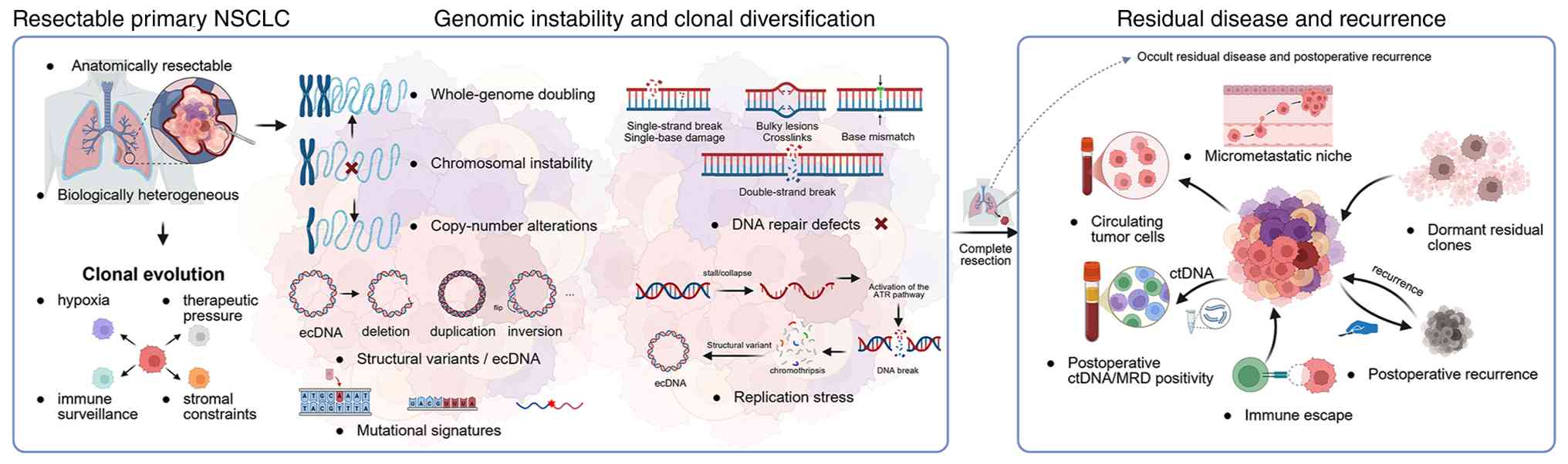
pathological, or molecular decision-making. The conceptual
relationships between genomic instability, clonal evolution, occult
residual disease, and postoperative recurrence are summarized in
Fig. 1.
Forms and sources of genomic instability in
NSCLC
Genomic instability in NSCLC is not a
single-molecule phenomenon. It arises from several related but
biologically distinct processes and genomic alterations. For
example, CIN results from ongoing errors in chromosome segregation
and maintenance, leading to aneuploidy, chromosome-arm gains or
losses, and highly complex karyotypes in some tumors (11). CNAs represent another important
component of genomic complexity. These changes may affect
oncogenes, tumor suppressor genes, or immune-related loci, and
their distribution may vary substantially across different regions
of the same tumor. Notably, a recent lung cancer study linked
clone-level copy-number diversity to survival, suggesting that
copy-number complexity is not simply a descriptive feature but may
reflect clinically relevant evolutionary behavior (12).
Whole-genome doubling increases the complexity. Once
a tumor cell duplicates its entire chromosomal complement, it may
tolerate and accumulate additional chromosomal alterations.
TRACERx-based analyses have shown that FAT atypical cadherin 1
(FAT1) loss, Hippo pathway dysregulation, CIN, and whole-genome
doubling are associated with NSCLC. Similarly, studies across
cancer types have shown that genome-doubled tumors can develop
complicated chromosomal histories that may be difficult to
reconstruct from a single sampled region (13,14).
Structural variants contribute in different ways; they can generate
actionable fusions, disrupt tumor suppressor loci, or alter
regulatory elements. Genomic changes do not occur in isolation
either; during NSCLC evolution, they may interact with epigenetic
remodeling, underscoring the close relationship between genome
structure and transcriptional state (15). Extrachromosomal DNA (ecDNA)
represents a dynamic, non-chromosomal form of amplified DNA that
can undergo rapid structural and copy-number evolution.
Experimental research suggests that ecDNA-positive cancers may
develop dependencies related to transcription-replication conflicts
and could be vulnerable to checkpoint kinase 1 (CHK1) inhibition
(16).
Instability can also arise from defective DNA repair
pathways and persistent replication stress. Deficiencies in
homologous recombination, mismatch repair, nucleotide excision
repair, base excision repair, or non-homologous end joining can
increase the accumulation of mutations, replication-associated
lesions, and structural genomic abnormalities (17). When one repair pathway is impaired,
tumor cells may become more dependent on alternative repair
pathways or cell-cycle checkpoints for survival. This provides a
biological rationale for DNA-damaging therapies and DNA damage
response (DDR)-targeted strategies in selected settings, although
the clinical relevance is likely to vary according to specific
molecular defect and cellular context (18). Mutational signatures provide a
complementary view of genomic instability and tumor evolution.
Rather than identifying a single alteration, they capture patterns
of mutations that reflect the processes shaping the tumor genome
over time, including carcinogen exposure, enzymatic editing, and
repair deficiency. In lung cancer, apolipoprotein B mRNA editing
enzyme catalytic polypeptide-like activity has been linked to tumor
evolution and treatment resistance, and whole-genome studies have
identified distinct mutagenic processes in smoking-associated and
never-smoker lung cancers (19–21).
These differences may have distinct clinical
implications. Tumors with extensive CIN may have marked clonal
diversity and greater capacity for ongoing genomic evolution;
however, CIN itself does not currently define a clinically
actionable repair defect. Conversely, a tumor with a specific
homologous recombination defect may provide a rationale for a
therapeutic hypothesis, even when broad aneuploidy is not
prominent. Collapsing these different processes into a single label
such as ‘high genomic instability’ would therefore risk obscuring
the biological and therapeutic differences that may eventually make
them useful.
Measurement is equally important. Targeted DNA or
RNA sequencing can identify established driver alterations and
selected copy-number events but is less suited to comprehensively
characterizing global chromosomal complexity, whole-genome
doubling, complex structural variation, or mutational signatures
(22). Broader genomic profiling is
often required to assess these features, while characterization of
clonal diversity may additionally require multi-region sampling or
phylogeny-informed analysis (23).
Repair deficiency presents a particular challenge because
alterations in homologous recombination repair genes and
genomic-scarring or mutational-signature measures show incomplete
concordance in NSCLC and should not be treated as interchangeable
readouts (24). This situation is
even less mature for ecDNA, which remains a research-stage
structural feature for which standardized perioperative assays are
not yet available (25). Surgical
specimens obtained from patients with resectable NSCLC offer
important opportunities for exploratory profiling. However,
clinical application requires pre-specified analytical methods,
reproducible thresholds, and prospective evidence that these
measurements add useful information beyond standard pathology,
driver status, and longitudinal MRD assessment. The potential
measurement strategies and their current translational relevance
for major genomic instability processes in resectable NSCLC are
summarized in Table I.
 | Table I.Genomic-instability processes,
potential measurement approaches and current translational
relevance in resectable NSCLC. |
Table I.
Genomic-instability processes,
potential measurement approaches and current translational
relevance in resectable NSCLC.
| Process or
consequence | Biological
meaning | Potential
measurement approach | Implications for
resectable NSCLC and recurrence | Evidence status and
current interpretation | Representative
references |
|---|
| Chromosomal
instability | Ongoing chromosome
mis-segregation causing aneuploidy and large-scale genomic
imbalance | Copy-number burden,
aneuploidy metrics or multiregion assessment; no standardized
routine clinical assay | May promote
subclonal diversification, adaptation and metastatic
progression | Emerging evidence;
investigational evolutionary and risk marker with context-dependent
immune implications | (11,13,14,71) |
| Copy-number
alterations | Gains or losses
affecting onco-genes, tumor-suppressor genes or immune-related
loci | DNA-based
copy-number profiling; clone-level diversity generally requires
multiregion or phylogeny-informed analysis | Subclonal
copy-number diversity may contribute to heterogeneity and adverse
outcome | Investigational for
risk modeling and trial stratification; no established
perioperative role | (12) |
| Whole-genome
doubling | Duplication of the
chromosomal complement, permitting further genomic complexity | Allele-specific
copy-number and ploidy inference from wholeexome or whole-genome
sequencing | May increase
evolutionary capacity and tolerance of subsequent chromosomal
alterations | Investigational
marker of evolutionary potential; no validated treatment-selection
role | (13,14) |
| Structural
variants | Rearrangements that
alter genomic architecture | Targeted DNA/RNA
sequencing for actionable rearrangements; broader complexity
requires comprehensive profiling | Actionable fusions
may guide established therapies, whereas global complexity may
reflect tumor evolution | Clinically
established for selected actionable rearrangements; broader
structural-variant complexity remains investigational | (22,92,93) |
| Extrachromosomal
DNA | Circular DNA
elements associated with increased gene dosage and rapid
adaptation | Research-level
genomic or cytogenetic assessment; no routine perioperative
assay | May support rapid
evolutionary adaptation | Mechanistic
evidence; candidate CHK1-related or
transcription-replication-conflict vulnerability | (16,25) |
| DNA repair
defects | Impaired repair of
DNA lesions or double-strand breaks | Repair-gene
profiling and genomic scar/signature assessment; these readouts are
non-equivalent | May increase
mutation accumulation and create DDR-related vulnerabilities | Candidate biomarker
for PARP- or DDR-directed trials; not validated for perioperative
selection | (17,18,24,78,79) |
| Replication
stress | Stalled or
collapsed replication forks under oncogenic or cellular stress | No validated
routine clinical assay; candidate surrogate markers require
prospective evaluation | May support
stress-tolerant clones and checkpoint dependence | Investigational
ATR/CHK1/WEE1-directed trial concept | (18,78,80–82,84) |
| Mutational
signatures | Genomic footprints
of carcinogen exposure, APOBEC activity or repair deficiency | Whole-exome or
whole-genome sequencing, depending on mutation burden and assay
resolution | May reconstruct
evolutionary history and identify processes associated with
adaptation or resistance | Potentially useful
for trial stratification, but not validated for routine
perioperative selection | (19–22) |
| Evolutionary
consequence: Intratumoral heterogeneity | Coexisting
subclones with distinct genomic or biological features | Multiregion
sequencing, spatial profiling or single-cell approaches | Minor subclones may
be missed yet later contribute to dissemination, immune escape or
recurrence | May inform sampling
and risk assessment, but is not a stand-alone treatment marker | (26–30,35) |
Clonal evolution and tumor
heterogeneity
Genomic instability can lead to carcinogenesis and
tumor progression by continuously generating genetic diversity
within the primary tumor. Resectable NSCLC should therefore not be
viewed as a single uniform lesion, but as a spatially structured
population of genetically related subclones. Single-cell and
spatial transcriptomic studies have shown that malignant cells,
immune populations, and stromal components in NSCLC form organized
spatial communities rather than random cellular mixtures (26,27).
This complexity is clinically important because the subclone that
ultimately drives recurrence may not be the dominant clone
represented in a single resected specimen.
However, sampling limitations further complicate
this interpretation. A focal biopsy or a single tissue block may
capture only a fraction of the genomic diversity within a lung
tumor (28). Longitudinal
evolutionary analyses have shown that multiple subclones from a
primary tumor may disseminate and contribute to subsequent
metastatic disease (29).
Therefore, a minor subclone may become clinically important if it
acquires or possesses selective advantages in invasion, survival,
immune escape, or adaptation to distant microenvironments.
The tumor microenvironment helps determine which
subclones persist and expand. Hypoxia, inflammation, stromal
pressure, nutrient limitation, vascular invasion, and immune
surveillance can act as selective forces during tumor evolution
before surgery. Spatial Tumor Atlas studies have shown that
malignant cell evolution is closely linked to the surrounding
microenvironment, and the spatial organization of immune cells in
lung cancer is associated with immune evasion and clinical outcomes
(30,31). In this setting, genomic instability
should not be viewed as a direct or sufficient cause of recurrence.
Rather, it expands the pool of variants, from which
microenvironment and environmental and therapeutic selection may
favor subclones with greater metastatic potential, immune-evasive
capacity, and treatment resistance. This provides a biological link
between genomic instability and metastatic progression. Tumor cells
capable of leaving the primary lesion before surgery must survive
in circulation, adapt to distant tissue environments, and either
establish immediate growth or enter a state of disseminated tumor
cell dormancy. Dormant disseminated tumor cells may remain
clinically invisible for prolonged periods while evading immune
surveillance, and their later outgrowth can be supported by
stromal, immune, and tissue-niche interactions (32–34). A
recent functional study on NSCLC showed that immune-evasive and
non-immune-evasive subclones can coexist within the same tumor,
suggesting that immune escape may emerge during subclonal evolution
(35). The clinical relevance of
genomic instability therefore lies not in a direct deterministic
effect on recurrence, but in its potential to generate diversity
that enables adaptation, dissemination, immune evasion, and
persistence.
Postoperative recurrence biology
Postoperative recurrence is the clinical endpoint of
a biological process that may begin well before recurrence becomes
clinically detectable. Although recurrence is typically recognized
by imaging, pathology, or clinical symptoms, the underlying events
may include preoperative clonal diversification, occult
dissemination, persistence of residual tumor cells, and subsequent
metastatic outgrowth. Multiomics profiling of recurrent stage I
NSCLC has shown that tumors that subsequently relapse may differ in
their genomic, epigenomic, transcriptomic, and microenvironmental
features (36). This biological
heterogeneity may help explain why patients with similar anatomical
stages and apparently complete resection can experience markedly
different postoperative outcomes.
Genomic instability may contribute to this process
on several levels. First, it increases the diversity from which
invasive or stress-tolerant subclones emerge within the primary
tumor. Second, it may support metastatic competence by generating
alterations that favor cellular motility, vascular invasion, immune
escape, or adaptation to distant microenvironments. Third, it may
facilitate the persistence of residual tumor cells under
postoperative and therapeutic stress. Pulmonary venous circulating
tumor cell research has shown that tumor cells may enter the
circulation before resection, and that such early dissemination is
associated with subsequent relapse and metastatic disease (37). Tumor dormancy research further
suggests that disseminated residual tumor cells can persist for
prolonged periods before undergoing clinical outgrowth (38).
This provides an important context for understanding
the role of MRD. Presurgical ctDNA detection and postoperative
ctDNA-defined MRD both have demonstrated prognostic value in
early-stage NSCLC (39,40). Imaging-based features, including
radiological tumor volume and positron emission tomography/computed
tomography-derived parameters, may add another layer of relapse
risk assessment when interpreted in conjunction with ctDNA
(41,42). In resected epidermal growth factor
receptor-mutated NSCLC, exploratory analyses from the ADAURA trial
further suggested that MRD signals can remain informative, even in
patients receiving effective adjuvant epidermal growth factor
receptor (EGFR)-targeted therapy (43).
However, MRD should not be interpreted as a complete
or infallible measure of residual disease. The recurrence risk
remains heterogeneous, and MRD assays require further analytical
and clinical standardization before they can routinely guide
treatment decisions (44,45). A positive postoperative ctDNA result
may indicate that one or more tumor populations have survived
resection and remain detectable in the circulation. Conversely, a
negative result is reassuring but does not exclude residual
disease, particularly in tumors with low shedding, limited
representation of the primary tumor in the sampled tissue, or
marked spatial heterogeneity.
Complete resection remains essential, but it cannot
reverse evolutionary events that have already occurred. Resected
tumors with extensive clonal diversity may harbor subclones that
have disseminated, entered dormancy, or acquired immune-evasive
properties before surgery. Another tumor at the same stage may not
have undergone the same extent of evolutionary diversification.
What later appears as postoperative recurrence may, therefore, be
the delayed manifestation of carcinogenic and metastatic processes
that were established before surgery in some patients.
Genomic instability and MRD
Following surgery, ctDNA-defined MRD may provide one
of the earliest indications that tumor-derived molecular material
remains detectable after resection of the primary lesion. A
positive result is consistent with the presence of residual
tumor-derived DNA and may reflect clones that disseminated before
surgery and persist during the early postoperative period. However,
ctDNA-defined MRD is an assay-dependent molecular signal and is not
a direct measure of viable residual tumor cells, residual tumor
burden, or genomic instability. Therefore, it is best understood as
an imperfect but potentially informative biomarker of molecular
residual disease and early recurrence risk, rather than a direct
measure of residual clonal burden or evolution (46,47).
The relationship between genomic instability and MRD
should be regarded as a biologically plausible, testable hypothesis
rather than an established causal pathway. Experimental and
evolutionary studies have suggested that genomic instability
expands clonal diversity and increases the probability that
invasive, stress-tolerant, or immune-evasive populations emerge and
persist (48–50). However, direct clinical evidence
linking specific instability features, such as CIN, copy number
complexity, whole-genome doubling, or repair deficiency, to the
presence or longitudinal dynamics of postoperative MRD in
resectable NSCLC remains limited. Studies characterizing genomic
instability patterns and their clinical associations (51) and those linking postoperative MRD
with subsequent relapse in resectable NSCLC (52) currently represent related but
largely distinct evidence streams.
An MRD result reflects both the biological presence
of residual disease and its detectability in the sampled blood
compartment. Detectability is influenced by the residual tumor
burden, biological shedding, access to the circulation, blood
collection timing, and assay sensitivity (53,54).
Spatial and temporal intratumoral heterogeneity introduce
additional uncertainty, particularly in tissue-informed assays. A
tissue block selected for sequencing may represent the dominant
primary tumor clone while failing to capture a minor clone that
subsequently persists or disseminates. Tumor-derived sequence
variants can be used as tracking markers in tumor-informed or
targeted sequencing assays (55–58),
whereas fragmentomic (59–61) and methylation-based approaches
(62) may capture broader signals;
however, none eliminate the effects of low shedding, limited
vascular access, or very small residual burden. Consequently, a
negative result does not establish the biological absence of
residual disease (63). Conversely,
an association between a genomic instability feature and MRD
positivity could partly reflect differences in tumor burden,
proliferation, necrosis, or ctDNA shedding rather than a direct
effect of genomic instability on the persistence of residual tumor
cells.
Therefore, three levels of evidence should be
distinguished. First, mechanistic evidence may explain how an
unstable process facilitates genomic diversification,
dissemination, and survival under therapeutic pressure. Second,
prognostic evidence may indicate that a genomic instability feature
or postoperative MRD status is associated with recurrence or
survival. Third, predictive evidence must demonstrate that a
biomarker identifies differential treatment benefit, ideally
through a biomarker-by-treatment interaction. Mechanistic
plausibility and prognostic associations do not establish
predictive utility. At present, no genomic instability feature has
demonstrated treatment-predictive value in resectable NSCLC, and no
validated perioperative model has established that genomic
instability profiling provides prognostic or predictive information
beyond standard clinicopathological assessment and longitudinal
MRD.
Tissue profiling and serial blood monitoring can
provide complementary but distinct information. Analysis of
resected tumors can characterize their evolutionary architecture,
including chromosomal complexity, repair defects, and other
instability-related features. Serial ctDNA testing can determine
whether tumor-derived molecular signals persist, clear, or
re-emerge after surgery or perioperative treatment (64). Persistent MRD after the resection of
a genomically complex tumor may represent a different biological
context from a transient low-level signal arising from a tumor with
limited pathological risk; however, whether such combined profiling
provides clinically actionable information remains unproven and
should not yet be considered a validated basis for treatment
intensification or surveillance planning.
Prospective studies should pair prespecified,
analytically validated tissue-based measures of genomic instability
with standardized postoperative and longitudinal blood sampling.
Such studies should account for residual tumor burden, DNA
shedding, vascular accessibility, spatial and temporal
heterogeneity, sampling time, and assay sensitivity while using
clinically relevant endpoints such as recurrence-free survival and
MRD clearance or conversion. They should also assess whether
integrating instability profiling with MRD improves risk
stratification beyond established clinicopathological factors and
driver status, and whether any instability phenotype identifies
differential benefit from a matched intervention through a
biomarker-by-treatment interaction. Until these questions are
resolved, the genomic instability-MRD relationship should remain a
research framework rather than a basis for routine perioperative
treatment decisions.
Immune microenvironment and perioperative
immunotherapy
The immune consequences of genomic instability
cannot easily be reduced to simple rules. An unstable genome may
generate mutations, structural changes, and novel peptides that can
serve as potential tumor antigens. However, a greater number of
mutations does not automatically make a tumor more susceptible to
immune recognition or control. What matters is whether the relevant
neoantigens are expressed and clonally represented, whether T cells
can recognize and reach the tumor, and whether the surrounding
microenvironment permits an immune response (65,66).
This explains why tumors with a high mutational burden may still
evade immunity when antigen presentation is impaired, effector T
cells are excluded, or immunosuppressive programs predominate. In
NSCLC, acquired resistance to immunotherapy has been linked to
defects in antigen processing, interferon signaling, and immune
cell function (67). The genomic
background also matters: Alterations in serine/threonine kinase 11
and Kelch-like ECH-associated protein 1 have been associated with
distinct immune profiles and poorer outcomes after programmed cell
death protein 1/programmed death-ligand 1 (PD-L1) blockade,
particularly in KRAS-mutated lung adenocarcinoma (68,69).
CIN introduces an additional layer of complexity.
Chromosome missegregation can generate micronuclei and activate the
cyclic GMP-AMP synthase (cGAS)-stimulator of interferon genes
(STING) pathway. In experimental settings, this response may induce
type I interferon signaling and enhance antitumor immune responses
(70). In other contexts,
particularly when signaling becomes chronic or dysregulated, it may
promote a pro-metastatic and immunosuppressive environment
(71). CIN is therefore not simply
‘beneficial’ or ‘detrimental’ for antitumor immunity. Its effects
are likely to depend on the magnitude and duration of cGAS-STING
signaling, as well as on the local cellular and molecular context
of the tumor.
This uncertainty is clinically relevant now that
perioperative immunotherapy has become an established component of
treatment for selected patients with resectable NSCLC. Phase III
studies evaluating perioperative durvalumab and nivolumab-based
strategies have demonstrated improvements in event-free survival,
supporting the efficacy of perioperative chemoimmunotherapy in
appropriately selected patients (72,73).
Meta-analytic evidence similarly supports neoadjuvant
chemoimmunotherapy across pathological response, surgical, and
efficacy outcomes, although the magnitude and interpretation vary
according to disease stage, PD-L1 expression, treatment regimen,
and clinical setting (74). Genomic
instability features may help explain some of this heterogeneity;
however, they are not yet sufficiently validated to serve as
independent biomarkers for perioperative immunotherapy selection.
Their potential relevance should instead be interpreted alongside
established clinical, molecular, and immune biomarkers (75). Postoperative ctDNA-defined MRD may
provide a complementary, dynamic measure of residual disease risk
within this broader assessment (76).
Candidate therapeutic vulnerabilities of
unstable genomes
Genomic instability can accelerate tumor evolution.
In some biological settings, it also creates dependencies on
pathways that enable tumor cells to tolerate ongoing DNA damage,
including residual repair capacity, cell-cycle checkpoints, and
stress-response mechanisms. These dependencies provide a rationale
for investigating DDR-directed strategies and agents targeting
replication stress or checkpoint pathways. However, in resectable
NSCLC, this remains a research hypothesis rather than an
established treatment-selection strategy. Platinum-based
chemotherapy is an established perioperative systemic treatment,
and radiotherapy is used in selected locoregional settings. Neither
approach is currently routinely selected on the basis of CIN,
whole-genome doubling, homologous recombination deficiency,
replication stress, or other genomic instability biomarkers
(77,78).
Homologous recombination deficiency provides one
potential example. In early-stage resectable NSCLC, a genomic
scarring score has been explored as a candidate marker of
poly(ADP-ribose) polymerase (PARP) inhibitor sensitivity, although
the supporting therapeutic evidence remains largely preclinical,
and PARP inhibitors are not part of standard perioperative
treatment (79). Replication stress
raises additional therapeutic possibilities. Tumors with oncogenic
activation, tumor protein p53 dysfunction, or impaired checkpoint
control may become increasingly reliant on ataxia telangiectasia
and Rad3-related (ATR), CHK1, or WEE1 G2 checkpoint kinase (WEE1)
to tolerate persistent DNA damage. ATR inhibitors have shown early
clinical activity in biomarker-selected advanced solid tumors
(80,81). WEE1 inhibition has preclinical
support for KRAS/tumor protein p53-mutant NSCLC (82), whereas DNA-dependent protein kinase
(DNA-PK) inhibition has entered early-phase clinical evaluation in
advanced cancers (83). These
findings provide a rationale for developing and prospectively
validating predictive biomarkers, but they do not support treatment
selection for resectable NSCLC.
The broader experience with targeting genomic
instability-related dependencies also underscores the need for
caution. Therapeutic strategies directed at replication stress and
DDR dependencies are biologically plausible (84), but most clinical evidence comes from
advanced solid tumors or metastatic NSCLC rather than from patients
treated with curative intent. In the HUDSON study,
biomarker-directed treatment incorporating ATR inhibition and PD-L1
blockade showed clinical activity in advanced NSCLC. By contrast,
phase III trials evaluating PARP inhibitor-immunotherapy
maintenance approaches have not established a broadly applicable
treatment strategy for metastatic disease (85–87).
Therefore, it is premature to extend these approaches to routine
perioperative care. Homologous recombination deficiency,
replication stress phenotypes, and checkpoint dependence are better
viewed as candidate biomarkers for prospective, biomarker-defined
clinical trials (88). ecDNA
represents a distinct candidate biomarker, and the bromodomain and
extraterminal domain-containing protein 4-dependent organization of
ecDNA hubs may represent a therapeutic vulnerability that requires
prospective validation (89,90).
Notably, these candidate vulnerabilities are
unlikely to be interchangeable. Repair-deficient tumors may be
suitable for prospective evaluation of DDR-directed therapies,
whereas tumors with prominent replication stress may be more
relevant for studies on ATR, CHK1, or WEE1 dependence.
ecDNA-positive cancers may represent another biological setting in
which transcription-replication conflicts or CHK1-related
dependencies warrant investigation. CIN is less directly
actionable, but it may influence the tumor immune microenvironment
through context-dependent inflammatory signaling. Its therapeutic
implications remain uncertain and should not be used to guide
treatment selection without prospective validation. The practical
aim, then, is not to assign existing perioperative therapies based
on genomic instability, but to identify well-defined instability
phenotypes that can be prospectively and rigorously evaluated in
future clinical trials.
Translational prioritization and
perioperative interpretation
The perioperative management of resectable NSCLC
remains grounded in anatomical stage, pathological risk features,
actionable driver alterations, and treatment strategies supported
by prospective clinical trials. The ninth edition of the
tumor-node-metastasis classification provides the current
anatomical framework (91), whereas
established molecular alterations identify patients eligible for
specific targeted therapies (92–94).
Postoperative ctDNA-defined MRD is emerging as a longitudinal
indicator of residual disease risk, although its utility for
treatment selection remains under investigation. Genomic
instability features are less clinically mature and should be
evaluated on a case-by-case basis, according to the specific
clinical claim under consideration, rather than treated as a
single, interchangeable biomarker category.
Therefore, a three-axis translational framework is
proposed. The first axis defines the principal biological
consequence of each feature: The generation of clonal diversity,
tolerance to genomic disruption, defective DNA repair, dependence
on replication-stress pathways, or dynamic adaptation mediated by
ecDNA. The second axis defines its potential clinical role as a
prognostic marker, treatment-predictive biomarker, or therapeutic
vulnerability. The third axis considers evidence maturity, ranging
from mechanistic and preclinical observations to retrospective
clinical associations, prospective prognostic validation, and
prospective treatment-specific predictive evidence. These levels
are not interchangeable. A biologically plausible dependency is not
necessarily a predictive biomarker, and an association with
recurrence does not establish that treatment decisions based on the
feature will improve clinical outcomes.
Among the current candidates, clonal diversity and
copy-number complexity have the clearest near-term prognostic
rationale. By contrast, CIN and whole-genome doubling have stronger
evolutionary and retrospective clinical support than prospective
clinical validation. Pan-cancer copy-number signature analyses have
linked distinct CNA patterns to whole-genome doubling, aneuploidy,
CIN-associated processes, and disease-specific survival (95). In lung adenocarcinoma, multi-region
TRACERx validation of ORACLE showed that clonal expression patterns
capture prognostic information associated with genetic evolutionary
features, including CIN, and are associated with survival (96). However, ORACLE is a clonal
expression-based biomarker, rather than a genomic instability
phenotype. For genomic instability features, assay definitions,
analytical thresholds, and incremental prognostic value beyond
established clinicopathological factors remain insufficiently
validated, and none have demonstrated treatment-predictive utility
in resectable NSCLC. Accordingly, their appropriate near-term role
is prospective validation within risk-stratification models, rather
than routine treatment assignment.
Repair deficiency, replication stress, and ecDNA
represent different translational categories. Their principal
promise lies in identifying candidate therapeutic dependencies
rather than established prognostic markers of recurrence.
Repair-deficient tumors may warrant evaluation in DDR-directed
trials, whereas tumors with prominent replication stress may be
candidates for ATR-, CHK1-, or WEE1-directed strategies (97). ecDNA-positive cancers may represent
another setting in which transcription-replication conflict or
checkpoint dependencies can be therapeutically explored (98). However, alterations in repair genes,
genomic scars, and functional measures of repair deficiency are not
interchangeable, and most therapeutic evidence has been derived
from preclinical studies, early phase trials, or advanced cancers.
Therefore, these features should be regarded as priorities for
biomarker development and prospective clinical testing, rather than
validated predictive biomarkers for resectable NSCLC.
The immune effects of genomic instability are highly
context-dependent. Mutational or structural complexity may increase
the pool of potential tumor antigens; however, immune recognition
also depends on antigen clonality and presentation, T-cell access
and function, and the surrounding tumor microenvironment.
CIN-associated inflammatory signaling may enhance immune
recognition in some settings, while promoting immune suppression or
metastatic behavior in others (99). Therefore, genomic instability
features should not currently supersede established clinical,
pathological, molecular, or immune factors in the selection of
perioperative immunotherapy. Their immediate role is to generate
testable hypotheses regarding heterogeneous treatment responses
that can be evaluated prospectively.
Tissue profiling, longitudinal MRD assessment, and
immune characterization may provide complementary but
non-equivalent information. Tissue analysis describes the
evolutionary architecture and candidate therapeutic dependencies of
the resected tumors. Postoperative ctDNA-defined MRD provides a
dynamic, assay-dependent signal of residual disease rather than a
direct measurement of genomic instability. Immune biomarkers
characterize the tumor-immune context in which residual clones may
persist or respond to treatment. Among these layers, ctDNA-defined
MRD currently has the most direct prognostic evidence for
postoperative recurrence, although it is not itself a genomic
instability phenotype and should not be used as a standalone basis
for treatment escalation or de-escalation (100).
This translational prioritization must also be
interpreted in the context of histological subtype, driver status,
and treatment setting. Evidence derived from lung adenocarcinoma or
molecularly selected cohorts should not be assumed to apply
uniformly to all patients with resectable NSCLC. EGFR-mutated,
anaplastic lymphoma kinase-positive, driver-negative non-squamous
and squamous tumors may differ in their patterns of genomic
instability, immune context, and therapeutic dependencies.
Therefore, the proposed framework is neither a composite score nor
a treatment algorithm. Its purpose is to identify which clinical
claims are supported for each feature, where the evidence remains
indirect or immature, and which biomarker-defined questions should
be prioritized for prospective evaluations. This evidence-based
prioritization is summarized in Table
II, and Fig. 2 places the
framework within the broader perioperative treatment pathway.
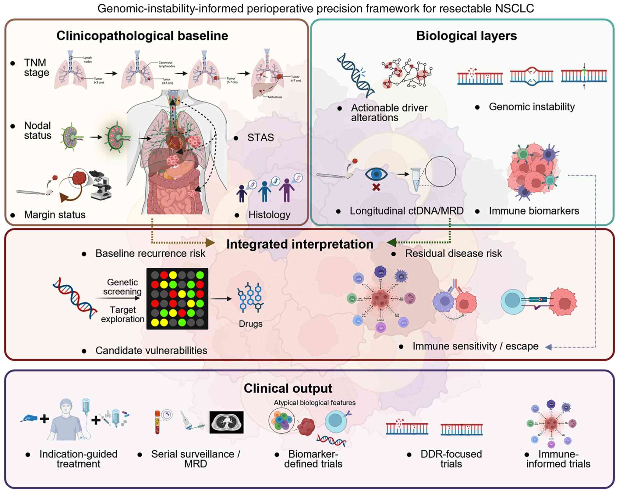 | Figure 2.Genomic-instability-informed
perioperative precision framework for resectable NSCLC. Established
clinicopathological factors and validated molecular tests remain
the basis for perioperative management. Tissue-based genomic
instability profiling, longitudinal ctDNA-defined MRD assessment,
and immune characterization may provide complementary but
non-equivalent information on tumor evolution, residual disease
risk, and candidate vulnerabilities. These investigational layers
may support risk model development and biomarker-defined clinical
trials but should not replace validated treatment indications.
NSCLC, non-small cell lung cancer; ctDNA, circulating tumor DNA;
MRD, molecular residual disease; DDR, DNA damage response; STAS,
spread through air spaces; TNM, tumor-node-metastasis. |
| Table II.Translational prioritization of
genomic-instability-related features for perioperative
interpretation in resectable NSCLC. |
Table II.
Translational prioritization of
genomic-instability-related features for perioperative
interpretation in resectable NSCLC.
| Component | Evidence relevant
to resectable NSCLC | Principal potential
clinical role | Evidence maturity
and translational priority | Current
interpretation | Representative
references |
|---|
| Intratumoral
heterogeneity and clonal evolution | Multiregion and
longitudinal studies link subclonal architecture and selection to
lung cancer progression and metastasis | Prognostic and
biological- context marker | Relatively mature
translational evidence; near-term risk-stratification priority | May help explain
recurrence after complete resection, but is not a
treatment-selection marker | (6–8,26–31,35) |
| Copy-number
complexity, CIN and whole-genome doubling | Copy-number
diversity, CIN and whole-genome doubling are associated with
evolutionary capacity and adverse outcomes | Candidate
prognostic or evolutionary-context marker | Mixed evidence;
strongest prognostic rationale for copy-number complexity, with
limited prospective validation for CIN and whole-genome
doubling | Promising for risk
stratification, but assays, thresholds and incremental value remain
unvalidated | (6,7,11–14) |
| Occult
dissemination before surgery | Circulating
tumor-cell and ctDNA studies support dissemination before
clinically detectable relapse | Mechanistic and
prognostic context | Direct evidence of
pre-relapse dissemination; contextual priority | Supports the
premise that surgery may not eliminate previously disseminated
clones | (37,64) |
| Postoperative
ctDNA-defined MRD | Serial ctDNA
studies after curative-intent treatment show strong associations
with relapse | Dynamic prognostic
marker; potential treatment-guiding biomarker | Most mature
postoperative risk signal, but treatment-guiding utility remains
unproven | Not a
genomic-instability phenotype or a stand-alone basis for treatment
escalation or de-escalation | (40,43,55,58,63) |
| Integration of
genomic instability with MRD | No validated
perioperative model has integrated instability profiling with
postoperative MRD | Composite
prognostic and treatment-selection hypothesis | Conceptual
framework; high prospective validation priority | Requires paired
tissue and serial blood studies accounting for burden, shedding,
heterogeneity, timing and assay sensitivity | (6,12,40,51,52,55) |
| Repair deficiency
and genomic scarring | Genomic scars and
repair-gene alterations have been explored as markers of DDR
sensitivity, without perioperative validation | Therapeutic
vulnerability; treatment-predictive biomarker development
priority | Preliminary
evidence; biomarker-development priority | These readouts are
non-equivalent, and neither is validated for perioperative
treatment selection | (17,18,24,78,79) |
| Replication stress
and checkpoint dependence | ATR-, CHK1-, WEE1-
and DNA-PK-directed strategies are supported mainly by preclinical,
advanced-disease or early-phase evidence | Therapeutic
vulnerability; exploratory biomarker-development priority | Indirect evidence
for resectable NSCLC; early-phase clinical trial priority | Appropriate for
biomarker-selected trials, but not routine perioperative use | (78,80–85,88) |
| Extrachromosomal
DNA | Experimental
studies identify transcription-replication conflict and
CHK1-related dependence in ecDNA-positive cancers | Candidate
therapeutic vulnerability | Mechanistic
evidence; exploratory priority | Biologically
compelling, but lacks a validated assay and direct perioperative
evidence | (16,25,98) |
| CIN-related immune
context | CIN may promote
inflammatory or immunosuppressive microen-vironmental states, but
predictive evidence is lacking | Potential
immunotherapy-response modifier | Mechanistic and
translational evidence; exploratory priority | Cannot currently
guide perioperative immunotherapy selection | (11,71,99) |
Challenges and future directions
Clinical translation requires standardized,
phenotype-specific, and analytically validated assays rather than a
generic designation of ‘genomic instability’. Future studies should
prospectively define the instability phenotype, assay, analytical
thresholds, tissue and blood sampling strategy, and clinically
relevant endpoints (101,102). The key questions are whether
genomic instability features add prognostic information beyond
stage, pathology, driver status, and longitudinal MRD, and whether
they identify differential benefit from matched therapies.
Prospective biomarker-defined trials will be required to establish
clinical utility (103).
Conclusions
Genomic instability provides a useful evolutionary
framework for understanding postoperative recurrence in resectable
NSCLC, but its individual components have distinct and unequally
mature clinical implications. Clonal diversity and copy-number
complexity have the strongest near-term prognostic rationale,
whereas CIN and whole-genome doubling remain primarily evolutionary
and prognostic candidates. Repair defects, replication stress, and
ecDNA represent candidate therapeutic vulnerabilities rather than
validated predictive biomarkers. Postoperative ctDNA-defined MRD
provides more direct prognostic information, but its relationship
with specific genomic instability features remains unproven. At
present, genomic instability profiling should support biomarker
development and prospective clinical trials rather than routine
perioperative treatment decisions.
Acknowledgements
Not applicable.
Funding
Funding: No funding was received.
Availability of data and materials
Not applicable.
Authors' contributions
LW and YF wrote the original draft. LW and QL
contributed to conceptualization, literature search and selection,
interpretation of the literature, and critical revision of the
manuscript. LW provided project administration. Data authentication
is not applicable. All authors have read and approved the final
manuscript.
Ethics approval and consent to
participate
Not applicable.
Patient consent for publication
Not applicable.
Competing interests
The authors declare that they have no competing
interests.
References
|
1
|
Forde PM, Spicer J, Lu S, Provencio M,
Mitsudomi T, Awad MM, Felip E, Broderick SR, Brahmer JR, Swanson
SJ, et al: Neoadjuvant nivolumab plus chemotherapy in resectable
lung cancer. N Engl J Med. 386:1973–1985. 2022. View Article : Google Scholar : PubMed/NCBI
|
|
2
|
Wakelee H, Liberman M, Kato T, Tsuboi M,
Lee SH, Gao S, Chen KN, Dooms C, Majem M, Eigendorff E, et al:
Perioperative pembrolizumab for Early-stage non-small-cell lung
cancer. N Engl J Med. 389:491–503. 2023. View Article : Google Scholar : PubMed/NCBI
|
|
3
|
Tsuboi M, Herbst RS, John T, Kato T, Majem
M, Grohé C, Wang J, Goldman JW, Lu S, Su WC, et al: Overall
survival with osimertinib in resected EGFR-Mutated NSCLC. N Engl J
Med. 389:137–147. 2023. View Article : Google Scholar : PubMed/NCBI
|
|
4
|
Felip E, Altorki N, Zhou C, Csőszi T,
Vynnychenko I, Goloborodko O, Luft A, Akopov A, Martinez-Marti A,
Kenmotsu H, et al: Adjuvant atezolizumab after adjuvant
chemotherapy in resected stage IB-IIIA non-small-cell lung cancer
(IMpower010): A randomised, multicentre, open-label, phase 3 trial.
Lancet. 398:1344–1357. 2021. View Article : Google Scholar : PubMed/NCBI
|
|
5
|
Mountzios G, Remon J, Hendriks LEL,
Garcia-Campelo R, Rolfo C, Van Schil P, Forde PM, Besse B, Subbiah
V, Reck M, et al: Immune-checkpoint inhibition for resectable
non-small-cell lung cancer-opportunities and challenges. Nat Rev
Clin Oncol. 20:664–677. 2023. View Article : Google Scholar : PubMed/NCBI
|
|
6
|
Frankell AM, Dietzen M, Al Bakir M, Lim
EL, Karasaki T, Ward S, Veeriah S, Colliver E, Huebner A, Bunkum A,
et al: The evolution of lung cancer and impact of subclonal
selection in TRACERx. Nature. 616:525–533. 2023. View Article : Google Scholar : PubMed/NCBI
|
|
7
|
Martínez-Ruiz C, Black JRM, Puttick C,
Hill MS, Demeulemeester J, Larose Cadieux E, Thol K, Jones TP,
Veeriah S, Naceur-Lombardelli C, et al: Genomic-transcriptomic
evolution in lung cancer and metastasis. Nature. 616:543–552. 2023.
View Article : Google Scholar : PubMed/NCBI
|
|
8
|
Al Bakir M, Huebner A, Martínez-Ruiz C,
Grigoriadis K, Watkins TBK, Pich O, Moore DA, Veeriah S, Ward S,
Laycock J, et al: The evolution of non-small cell lung cancer
metastases in TRACERx. Nature. 616:534–542. 2023. View Article : Google Scholar : PubMed/NCBI
|
|
9
|
Black JRM, Bartha G, Abbott CW, Boyle SM,
Karasaki T, Li B, Chen R, Harris J, Veeriah S, Colopi M, et al:
Ultrasensitive ctDNA detection for preoperative disease
stratification in early-stage lung adenocarcinoma. Nat Med.
31:70–76. 2025. View Article : Google Scholar : PubMed/NCBI
|
|
10
|
Yap TA, Manning HC, Sapra P, Mills GB and
O'Connor MJ: Targeting genomic instability in cancer. Cell.
189:2278–2306. 2026. View Article : Google Scholar : PubMed/NCBI
|
|
11
|
Chen X, Agustinus AS, Li J, DiBona M and
Bakhoum SF: Chromosomal instability as a driver of cancer
progression. Nat Rev Genet. 26:31–46. 2025. View Article : Google Scholar : PubMed/NCBI
|
|
12
|
Pawlik P, Grigoriadis K, Bunkum A, Coggan
H, Frankell AM, Martínez-Ruiz C, Karasaki T, Huebner A, Rowan A,
Fisher J, et al: Clone copy number diversity is linked to survival
in lung cancer. Nature. 646:190–197. 2025. View Article : Google Scholar : PubMed/NCBI
|
|
13
|
Lu WT, Zalmas LP, Bailey C, Black JRM,
Martinez-Ruiz C, Pich O, Gimeno-Valiente F, Usaite I, Magness A,
Thol K, et al: TRACERx analysis identifies a role for FAT1 in
regulating chromosomal instability and whole-genome doubling via
Hippo signalling. Nat Cell Biol. 27:154–168. 2025. View Article : Google Scholar : PubMed/NCBI
|
|
14
|
Baker TM, Lai S, Lynch AR, Lesluyes T, Yan
H, Ogilvie HA, Verfaillie A, Dentro S, Bowes AL, Pillay N, et al:
The history of chromosomal instability in Genome-Doubled tumors.
Cancer Discov. 14:1810–1822. 2024. View Article : Google Scholar : PubMed/NCBI
|
|
15
|
Gimeno-Valiente F, Castignani C, Larose
Cadieux E, Mensah NE, Liu X, Chen K, Chervova O, Karasaki T, Weeden
CE, Richard C, et al: DNA methylation cooperates with genomic
alterations during non-small cell lung cancer evolution. Nat Genet.
57:2226–2237. 2025. View Article : Google Scholar : PubMed/NCBI
|
|
16
|
Tang J, Weiser NE, Wang G, Chowdhry S,
Curtis EJ, Zhao Y, Wong ITL, Marinov GK, Li R, Hanoian P, et al:
Enhancing transcription-replication conflict targets ecDNA-positive
cancers. Nature. 635:210–218. 2024. View Article : Google Scholar : PubMed/NCBI
|
|
17
|
Hopkins JL, Lan L and Zou L: DNA repair
defects in cancer and therapeutic opportunities. Genes Dev.
36:278–293. 2022. View Article : Google Scholar : PubMed/NCBI
|
|
18
|
Li Q, Qian W, Zhang Y, Hu L, Chen S and
Xia Y: A new wave of innovations within the DNA damage response.
Signal Transduct Target Ther. 8:3382023. View Article : Google Scholar : PubMed/NCBI
|
|
19
|
Caswell DR, Gui P, Mayekar MK, Law EK,
Pich O, Bailey C, Boumelha J, Kerr DL, Blakely CM, Manabe T, et al:
The role of APOBEC3B in lung tumor evolution and targeted cancer
therapy resistance. Nat Genet. 56:60–73. 2024. View Article : Google Scholar : PubMed/NCBI
|
|
20
|
Díaz-Gay M, Zhang T, Hoang PH, Leduc C,
Baine MK, Travis WD, Sholl LM, Joubert P, Khandekar A, Zhao W, et
al: The mutagenic forces shaping the genomes of lung cancer in
never smokers. Nature. 644:133–144. 2025. View Article : Google Scholar : PubMed/NCBI
|
|
21
|
Zhang T, Sang J, Hoang PH, Zhao W,
Rosenbaum J, Johnson KE, Klimczak LJ, McElderry J, Klein A, Wirth
C, et al: APOBEC affects tumor evolution and age at onset of lung
cancer in smokers. Nat Commun. 16:47112025. View Article : Google Scholar : PubMed/NCBI
|
|
22
|
Everall A, Tapinos A, Hawari A, Cornish
AJ, Sud A, Chubb D, Kinnersley B, Frangou A, Barquin M, Jung J, et
al: Comprehensive repertoire of the chromosomal alteration and
mutational signatures across 16 cancer types. Nat Genet.
58:570–581. 2026. View Article : Google Scholar : PubMed/NCBI
|
|
23
|
Jamal-Hanjani M, Wilson GA, McGranahan N,
Birkbak NJ, Watkins TBK, Veeriah S, Shafi S, Johnson DH, Mitter R,
Rosenthal R, et al: Tracking the evolution of non-small-cell lung
cancer. N Engl J Med. 376:2109–2121. 2017. View Article : Google Scholar : PubMed/NCBI
|
|
24
|
Diossy M, Sztupinszki Z, Borcsok J,
Krzystanek M, Tisza V, Spisak S, Rusz O, Timar J, Csabai I,
Fillinger J, et al: A subset of lung cancer cases shows robust
signs of homologous recombination deficiency associated genomic
mutational signatures. NPJ Precis Oncol. 5:552021. View Article : Google Scholar : PubMed/NCBI
|
|
25
|
Yan X, Mischel P and Chang H:
Extrachromosomal DNA in cancer. Nat Rev Cancer. 24:261–273. 2024.
View Article : Google Scholar : PubMed/NCBI
|
|
26
|
De Zuani M, Xue H, Park JS, Dentro SC,
Seferbekova Z, Tessier J, Curras-Alonso S, Hadjipanayis A,
Athanasiadis EI, Gerstung M, et al: Single-cell and spatial
transcriptomics analysis of non-small cell lung cancer. Nat Commun.
15:43882024. View Article : Google Scholar : PubMed/NCBI
|
|
27
|
Lomakin A, Svedlund J, Strell C, Gataric
M, Shmatko A, Rukhovich G, Park JS, Ju YS, Dentro S, Kleshchevnikov
V, et al: Spatial genomics maps the structure, nature and evolution
of cancer clones. Nature. 611:594–602. 2022. View Article : Google Scholar : PubMed/NCBI
|
|
28
|
Hertel A, Streuer A, Diehl S, Boch T,
Nörenberg D, Strittmatter A, Zöllner FG, Schoenberg SO, Hofmann WK,
Loges S, et al: Targeting tumoral heterogeneity in lung cancer: A
novel, CT-texture-guided targeted biopsy approach with exome
sequencing. NPJ Precis Oncol. 9:3422025. View Article : Google Scholar : PubMed/NCBI
|
|
29
|
Hessey S, Bunkum A, Huebner A, Haase K,
Grigoriadis K, Naceur-Lombardelli C, Liu WK, Harrigan CF, Grieco C,
Marinelli D, et al: Evolutionary characterization of lung cancer
metastasis. Nature. 653:911–922. 2026. View Article : Google Scholar : PubMed/NCBI
|
|
30
|
Enfield KSS, Colliver E, Lee C, Magness A,
Moore DA, Sivakumar M, Grigoriadis K, Pich O, Karasaki T, Hobson
PS, et al: Spatial architecture of myeloid and T cells orchestrates
immune evasion and clinical outcome in lung cancer. Cancer Discov.
14:1018–1047. 2024. View Article : Google Scholar : PubMed/NCBI
|
|
31
|
Mo CK, Liu J, Chen S, Storrs E, Targino da
Costa ALN, Houston A, Wendl MC, Jayasinghe RG, Iglesia MD, Ma C, et
al: Tumour evolution and microenvironment interactions in 2D and 3D
space. Nature. 634:1178–1186. 2024. View Article : Google Scholar : PubMed/NCBI
|
|
32
|
Gerstberger S, Jiang Q and Ganesh K:
Metastasis. Cell. 186:1564–1579. 2023. View Article : Google Scholar : PubMed/NCBI
|
|
33
|
Goddard ET, Linde MH, Srivastava S, Klug
G, Shabaneh TB, Iannone S, Grzelak CA, Marsh S, Riggio AI, Shor RE,
et al: Immune evasion of dormant disseminated tumor cells is due to
their scarcity and can be overcome by T cell immunotherapies.
Cancer Cell. 42:119–134.e12. 2024. View Article : Google Scholar : PubMed/NCBI
|
|
34
|
Li Y, Liu F, Cai Q, Deng L, Ouyang Q,
Zhang XHF and Zheng J: Invasion and metastasis in cancer: Molecular
insights and therapeutic targets. Signal Transduct Target Ther.
10:572025. View Article : Google Scholar : PubMed/NCBI
|
|
35
|
Dijkstra KK, Vendramin R, Karagianni D,
Witsen M, Gálvez-Cancino F, Hill MS, Foster KA, Barbè V, Angelova
M, Hynds RE, et al: Subclonal immune evasion in non-small cell lung
cancer. Cancer Cell. 43:1833–1849.e10. 2025. View Article : Google Scholar : PubMed/NCBI
|
|
36
|
Wang C, Li J, Chen J, Wang Z, Zhu G, Song
L, Wu J, Li C, Qiu R, Chen X, et al: Multi-omics analyses reveal
biological and clinical insights in recurrent stage I non-small
cell lung cancer. Nat Commun. 16:14772025. View Article : Google Scholar : PubMed/NCBI
|
|
37
|
Chemi F, Rothwell DG, McGranahan N, Gulati
S, Abbosh C, Pearce SP, Zhou C, Wilson GA, Jamal-Hanjani M, Birkbak
NJ, et al: Pulmonary venous circulating tumor cell dissemination
before tumor resection and disease relapse. Nat Med. 25:1534–1539.
2019. View Article : Google Scholar : PubMed/NCBI
|
|
38
|
Liang Y, Chen WM, Zhang Y and Li L:
Remodeling the tumor dormancy ecosystem to prevent recurrence and
metastasis. Signal Transduct Target Ther. 11:12026. View Article : Google Scholar : PubMed/NCBI
|
|
39
|
Hong TH, Hwang S, Dasgupta A, Abbosh C,
Hung T, Bredno J, Walker J, Shi X, Milenkova T, Horn L, et al:
Clinical utility of Tumor-Naïve presurgical circulating tumor DNA
detection in Early-stage NSCLC. J Thorac Oncol. 19:1512–1524. 2024.
View Article : Google Scholar : PubMed/NCBI
|
|
40
|
Gale D, Heider K, Ruiz-Valdepenas A,
Hackinger S, Perry M, Marsico G, Rundell V, Wulff J, Sharma G,
Knock H, et al: Residual ctDNA after treatment predicts early
relapse in patients with early-stage non-small cell lung cancer.
Ann Oncol. 33:500–510. 2022. View Article : Google Scholar : PubMed/NCBI
|
|
41
|
Tran HT, Heeke S, Sujit SJ, Vokes N, Zhang
J, Aminu M, Lam VK, Vaporciyan A, Swisher SG, Godoy MCB, et al:
Circulating tumor DNA and radiological tumor volume identify
patients at risk for relapse with resected, early-stage
non-small-cell lung cancer. Ann Oncol. 35:183–189. 2024. View Article : Google Scholar : PubMed/NCBI
|
|
42
|
Sujit SJ, Aminu M, Karpinets TV, Chen P,
Saad MB, Salehjahromi M, Boom JD, Qayati M, George JM, Allen H, et
al: Enhancing NSCLC recurrence prediction with PET/CT habitat
imaging, ctDNA, and integrative radiogenomics-blood insights. Nat
Commun. 15:31522024. View Article : Google Scholar : PubMed/NCBI
|
|
43
|
Herbst RS, John T, Grohé C, Goldman JW,
Kato T, Laktionov K, Bonanno L, Tiseo M, Majem M, Dómine M, et al:
Molecular residual disease analysis of adjuvant osimertinib in
resected EGFR-mutated stage IB-IIIA non-small-cell lung cancer. Nat
Med. 31:1958–1968. 2025. View Article : Google Scholar : PubMed/NCBI
|
|
44
|
Stetson D, Labrousse P, Russell H, Shera
D, Abbosh C, Dougherty B, Barrett JC, Hodgson D and Hadfield J:
Next-Generation molecular residual disease assays: Do we have the
tools to evaluate them properly? J Clin Oncol. 42:2736–2740. 2024.
View Article : Google Scholar : PubMed/NCBI
|
|
45
|
Boukouris AE, Michaelidou K, Joosse SA,
Charpidou A, Mavroudis D, Syrigos KN and Agelaki S: A comprehensive
overview of minimal residual disease in the management of
early-stage and locally advanced non-small cell lung cancer. NPJ
Precis Oncol. 9:1782025. View Article : Google Scholar : PubMed/NCBI
|
|
46
|
Moding EJ, Nabet BY, Alizadeh AA and Diehn
M: Detecting liquid remnants of solid tumors: Circulating tumor DNA
minimal residual disease. Cancer Discov. 11:2968–2986. 2021.
View Article : Google Scholar : PubMed/NCBI
|
|
47
|
Zheng J, Qin C, Wang Q, Tian D and Chen Z:
Circulating tumour DNA-Based molecular residual disease detection
in resectable cancers: A systematic review and meta-analysis.
EBioMedicine. 103:1051092024. View Article : Google Scholar : PubMed/NCBI
|
|
48
|
Venkatesan S, Angelova M, Puttick C, Zhai
H, Caswell DR, Lu WT, Dietzen M, Galanos P, Evangelou K, Bellelli
R, et al: Induction of APOBEC3 exacerbates DNA replication stress
and chromosomal instability in early breast and lung cancer
evolution. Cancer Discov. 11:2456–2473. 2021. View Article : Google Scholar : PubMed/NCBI
|
|
49
|
Isozaki H, Sakhtemani R, Abbasi A, Nikpour
N, Stanzione M, Oh S, Langenbucher A, Monroe S, Su W, Cabanos HF,
et al: Therapy-induced APOBEC3A drives evolution of persistent
cancer cells. Nature. 620:393–401. 2023. View Article : Google Scholar : PubMed/NCBI
|
|
50
|
Wang X, Bai H, Zhang J, Wang Z, Duan J,
Cai H, Cao Z, Lin Q, Ding X, Sun Y, et al: Genetic intratumor
heterogeneity remodels the immune microenvironment and induces
immune evasion in brain metastasis of lung cancer. J Thorac Oncol.
19:252–272. 2024. View Article : Google Scholar : PubMed/NCBI
|
|
51
|
Drews RM, Hernando B, Tarabichi M, Haase
K, Lesluyes T, Smith PS, Morrill Gavarró L, Couturier DL, Liu L,
Schneider M, et al: A pan-cancer compendium of chromosomal
instability. Nature. 606:976–983. 2022. View Article : Google Scholar : PubMed/NCBI
|
|
52
|
Chen K, Yang F, Shen H, Wang C, Li X,
Chervova O, Wu S, Qiu F, Peng D, Zhu X, et al: Individualized
tumor-informed circulating tumor DNA analysis for postoperative
monitoring of non-small cell lung cancer. Cancer Cell.
41:1749–1762.e6. 2023. View Article : Google Scholar : PubMed/NCBI
|
|
53
|
Stejskal P, Goodarzi H, Srovnal J, Hajdúch
M, van 't Veer LJ and Magbanua MJM: Circulating tumor nucleic
acids: Biology, release mechanisms, and clinical relevance. Mol
Cancer. 22:152023. View Article : Google Scholar : PubMed/NCBI
|
|
54
|
Normanno N, Morabito A, Rachiglio AM,
Sforza V, Landi L, Bria E, Delmonte A, Cappuzzo F and De Luca A:
Circulating tumour DNA in early stage and locally advanced NSCLC:
Ready for clinical implementation? Nat Rev Clin Oncol. 22:215–231.
2025. View Article : Google Scholar : PubMed/NCBI
|
|
55
|
Black JRM, Karasaki T, Abbott CW, Li B,
Veeriah S, Al Bakir M, Liu WK, Huebner A, Martínez-Ruiz C, Pawlik
P, et al: Longitudinal ultrasensitive ctDNA monitoring for
high-resolution lung cancer risk prediction. Cell.
188:7083–7098.e18. 2025. View Article : Google Scholar : PubMed/NCBI
|
|
56
|
Schuurbiers MMF, Smith CG, Hartemink KJ,
Rintoul RC, Gale D, Monkhorst K, Mandos BLR, Paterson AL, van den
Broek D, Rosenfeld N, et al: Recurrence prediction using
circulating tumor DNA in patients with early-stage non-small cell
lung cancer after treatment with curative intent: A retrospective
validation study. PLoS Med. 22:e10045742025. View Article : Google Scholar : PubMed/NCBI
|
|
57
|
Qiu B, Guo W, Zhang F, Lv F, Ji Y, Peng Y,
Chen X, Bao H, Xu Y, Shao Y, et al: Dynamic recurrence risk and
adjuvant chemotherapy benefit prediction by ctDNA in resected
NSCLC. Nat Commun. 12:67702021. View Article : Google Scholar : PubMed/NCBI
|
|
58
|
Zhang JT, Liu SY, Gao W, Liu SYM, Yan HH,
Ji L, Chen Y, Gong Y, Lu HL, Lin JT, et al: Longitudinal
undetectable molecular residual disease defines potentially cured
population in localized Non-Small cell lung cancer. Cancer Discov.
12:1690–1701. 2022. View Article : Google Scholar : PubMed/NCBI
|
|
59
|
Tsui WHA, Jiang P and Lo YMD: Cell-free
DNA fragmentomics in cancer. Cancer Cell. 43:1792–1814. 2025.
View Article : Google Scholar : PubMed/NCBI
|
|
60
|
Mathios D, Johansen JS, Cristiano S,
Medina JE, Phallen J, Larsen KR, Bruhm DC, Niknafs N, Ferreira L,
Adleff V, et al: Detection and characterization of lung cancer
using cell-free DNA fragmentomes. Nat Commun. 12:50602021.
View Article : Google Scholar : PubMed/NCBI
|
|
61
|
van 't Erve I, Alipanahi B, Lumbard K,
Skidmore ZL, Rinaldi L, Millberg LK, Carey J, Chesnick B, Cristiano
S, Portwood C, et al: Cancer treatment monitoring using cell-free
DNA fragmentomes. Nat Commun. 15:88012024. View Article : Google Scholar : PubMed/NCBI
|
|
62
|
Lo YMD, Han DSC, Jiang P and Chiu RWK:
Epigenetics, fragmentomics, and topology of cell-free DNA in liquid
biopsies. Science. 372:eaaw36162021. View Article : Google Scholar : PubMed/NCBI
|
|
63
|
Pascual J, Attard G, Bidard FC, Curigliano
G, De Mattos-Arruda L, Diehn M, Italiano A, Lindberg J, Merker JD,
Montagut C, et al: ESMO recommendations on the use of circulating
tumour DNA assays for patients with cancer: A report from the ESMO
Precision Medicine Working Group. Ann Oncol. 33:750–768. 2022.
View Article : Google Scholar : PubMed/NCBI
|
|
64
|
Abbosh C, Frankell AM, Harrison T,
Kisistok J, Garnett A, Johnson L, Veeriah S, Colliver E, Moreau M,
Ward S, et al: Tracking early lung cancer metastatic dissemination
in TRACERx using ctDNA. Nature. 616:553–562. 2023. View Article : Google Scholar : PubMed/NCBI
|
|
65
|
Peri A, Salomon N, Wolf Y, Kreiter S,
Diken M and Samuels Y: The landscape of T cell antigens for cancer
immunotherapy. Nat Cancer. 4:937–954. 2023. View Article : Google Scholar : PubMed/NCBI
|
|
66
|
Litchfield K, Reading JL, Puttick C,
Thakkar K, Abbosh C, Bentham R, Watkins TBK, Rosenthal R, Biswas D,
Rowan A, et al: Meta-analysis of tumor- and T cell-intrinsic
mechanisms of sensitization to checkpoint inhibition. Cell.
184:596–614.e14. 2021. View Article : Google Scholar : PubMed/NCBI
|
|
67
|
Memon D, Schoenfeld AJ, Ye D, Fromm G,
Rizvi H, Zhang X, Keddar MR, Mathew D, Yoo KJ, Qiu J, et al:
Clinical and molecular features of acquired resistance to
immunotherapy in non-small cell lung cancer. Cancer Cell.
42:209–224.e9. 2024. View Article : Google Scholar : PubMed/NCBI
|
|
68
|
Ricciuti B, Arbour KC, Lin JJ, Vajdi A,
Vokes N, Hong L, Zhang J, Tolstorukov MY, Li YY, Spurr LF, et al:
Diminished efficacy of programmed Death-(Ligand)1 inhibition in
STK11- and KEAP1-Mutant lung adenocarcinoma is affected by KRAS
mutation status. J Thorac Oncol. 17:399–410. 2022. View Article : Google Scholar : PubMed/NCBI
|
|
69
|
Ricciuti B and Garassino MC: Precision
immunotherapy for STK11/KEAP1-mutant NSCLC. J Thorac Oncol.
19:877–882. 2024. View Article : Google Scholar : PubMed/NCBI
|
|
70
|
Huang Y, Lu C, Wang H, Gu L, Fu YX and Li
GM: DNAJA2 deficiency activates cGAS-STING pathway via the
induction of aberrant mitosis and chromosome instability. Nat
Commun. 14:52462023. View Article : Google Scholar : PubMed/NCBI
|
|
71
|
Li J, Hubisz MJ, Earlie EM, Duran MA, Hong
C, Varela AA, Tsao JL, Petersson EJ, Lok BH, Modrek AS, et al:
Non-cell-autonomous cancer progression from chromosomal
instability. Nature. 620:1080–1088. 2023. View Article : Google Scholar : PubMed/NCBI
|
|
72
|
Heymach JV, Harpole D, Mitsudomi T, Taube
JM, Galffy G, Hochmair M, Winder T, Zukov R, Garbaos G, Gao S, et
al: Perioperative durvalumab for resectable Non-Small-Cell lung
cancer. N Engl J Med. 389:1672–1684. 2023. View Article : Google Scholar : PubMed/NCBI
|
|
73
|
Cascone T, Awad MM, Spicer JD, He J, Lu S,
Sepesi B, Tanaka F, Taube JM, Cornelissen R, Havel L, et al:
Perioperative nivolumab in resectable lung cancer. N Engl J Med.
390:1756–1769. 2024. View Article : Google Scholar : PubMed/NCBI
|
|
74
|
Sorin M, Prosty C, Ghaleb L, Nie K,
Katergi K, Shahzad MH, Dubé LR, Atallah A, Swaby A, Dankner M, et
al: Neoadjuvant Chemoimmunotherapy for NSCLC: A systematic review
and meta-analysis. JAMA Oncol. 10:621–633. 2024. View Article : Google Scholar : PubMed/NCBI
|
|
75
|
Vaccaro A, Rahal Z, Kadara H and Cascone
T: A roadmap to precision immunotherapy for Early-stage non-small
cell lung cancer. Cancer Discov. 15:884–889. 2025. View Article : Google Scholar : PubMed/NCBI
|
|
76
|
Xia L, Mei J, Kang R, Deng S, Chen Y, Yang
Y, Feng G, Deng Y, Gan F, Lin Y, et al: Perioperative ctDNA-Based
molecular residual disease detection for Non-Small cell lung
cancer: A prospective multicenter cohort study (LUNGCA-1). Clin
Cancer Res. 28:3308–3317. 2022. View Article : Google Scholar : PubMed/NCBI
|
|
77
|
Zer A, Ahn MJ, Barlesi F, Bubendorf L, De
Ruysscher D, Garrido P, Gautschi O, Hendriks LE, Jänne PA, Kerr KM,
et al: Early and locally advanced non-small-cell lung cancer: ESMO
Clinical Practice Guideline for diagnosis, treatment and follow-up.
Ann Oncol. 36:1245–1262. 2025. View Article : Google Scholar : PubMed/NCBI
|
|
78
|
Drew Y, Zenke FT and Curtin NJ: DNA damage
response inhibitors in cancer therapy: Lessons from the past,
current status and future implications. Nat Rev Drug Discov.
24:19–39. 2025. View Article : Google Scholar : PubMed/NCBI
|
|
79
|
Tsilingiri K, Chalari A, Christopoulou G,
Voutsina A, Constantoulakis P, Potaris K, Vamvakaris I, Hatzidaki
D, Zachou G, Vatsellas G, et al: Genomic scarring score predicts
the response to PARP inhibitors in non-small cell lung cancer. NPJ
Precis Oncol. 8:2912024. View Article : Google Scholar : PubMed/NCBI
|
|
80
|
Yap TA, Fontana E, Lee EK, Spigel DR,
Højgaard M, Lheureux S, Mettu NB, Carneiro BA, Carter L, Plummer R,
et al: Camonsertib in DNA damage response-deficient advanced solid
tumors: Phase 1 trial results. Nat Med. 29:1400–1411. 2023.
View Article : Google Scholar : PubMed/NCBI
|
|
81
|
Yap TA, Tan DSP, Terbuch A, Caldwell R,
Guo C, Goh BC, Heong V, Noor NR, Bashir S, Drew Y, et al:
First-in-Human trial of the oral ataxia telangiectasia and
RAD3-Related (ATR) inhibitor BAY 1895344 in patients with advanced
solid tumors. Cancer Discov. 11:80–91. 2021. View Article : Google Scholar : PubMed/NCBI
|
|
82
|
Fukuda K, Takeuchi S, Arai S, Nanjo S,
Sato S, Kotani H, Kita K, Nishiyama A, Sakaguchi H, Ohtsubo K, et
al: Targeting WEE1 enhances the antitumor effect of KRAS-mutated
non-small cell lung cancer harboring TP53 mutations. Cell Rep Med.
5:1015782024. View Article : Google Scholar : PubMed/NCBI
|
|
83
|
Yap TA, LoRusso P, Miller RE, Kristeleit
R, Paulovich AG, McMorn S, Oplustil O'Connor L, Lombardi B,
Marco-Casanova P, Gangl ET, et al: The DNA-PK inhibitor AZD7648
alone or combined with pegylated liposomal doxorubicin in patients
with advanced cancer: Results of a first-in-human Phase I/IIa
study. Br J Cancer. 133:168–177. 2025. View Article : Google Scholar : PubMed/NCBI
|
|
84
|
da Costa AABA, Chowdhury D, Shapiro GI,
D'Andrea AD and Konstantinopoulos PA: Targeting replication stress
in cancer therapy. Nat Rev Drug Discov. 22:38–58. 2023. View Article : Google Scholar : PubMed/NCBI
|
|
85
|
Besse B, Pons-Tostivint E, Park K, Hartl
S, Forde PM, Hochmair MJ, Awad MM, Thomas M, Goss G, Wheatley-Price
P, et al: Biomarker-directed targeted therapy plus durvalumab in
advanced non-small-cell lung cancer: A phase 2 umbrella trial. Nat
Med. 30:716–729. 2024. View Article : Google Scholar : PubMed/NCBI
|
|
86
|
Gray JE, Schenker M, Şendur MAN, Leonova
V, Kowalski D, Kato T, Orlova R, Yang JC, Langleben A, Pilz A, et
al: The Phase 3 KEYLYNK-006 study of pembrolizumab plus olaparib
versus pembrolizumab plus pemetrexed as maintenance therapy for
metastatic nonsquamous NSCLC. J Thorac Oncol. 20:219–232. 2025.
View Article : Google Scholar : PubMed/NCBI
|
|
87
|
Hochmair M, Schenker M, Cobo Dols M, Kim
TM, Ozyilkan O, Smagina M, Leonova V, Kato T, Fedenko A, De Angelis
F, et al: Pembrolizumab with or without maintenance olaparib for
metastatic squamous NSCLC that responded to First-Line
pembrolizumab plus chemotherapy. J Thorac Oncol. 20:203–218. 2025.
View Article : Google Scholar : PubMed/NCBI
|
|
88
|
Cleary JM, Aguirre AJ, Shapiro GI and
D'Andrea AD: Biomarker-guided development of DNA repair inhibitors.
Mol Cell. 78:1070–1085. 2020. View Article : Google Scholar : PubMed/NCBI
|
|
89
|
Bailey C, Pich O, Thol K, Watkins TBK,
Luebeck J, Rowan A, Stavrou G, Weiser NE, Dameracharla B, Bentham
R, et al: Origins and impact of extrachromosomal DNA. Nature.
635:193–200. 2024. View Article : Google Scholar : PubMed/NCBI
|
|
90
|
Hung KL, Yost KE, Xie L, Shi Q, Helmsauer
K, Luebeck J, Schöpflin R, Lange JT, Chamorro González R, Weiser
NE, et al: ecDNA hubs drive cooperative intermolecular oncogene
expression. Nature. 600:731–736. 2021. View Article : Google Scholar : PubMed/NCBI
|
|
91
|
Rami-Porta R, Nishimura KK, Giroux DJ,
Detterbeck FC, Asamura H, Goldstraw P, Crowley J, Chansky K,
Edwards J, Nicholson AG, et al: The international association for
the study of lung cancer lung cancer staging project: Proposals for
revision of the TNM stage groups in the forthcoming (Ninth) edition
of the TNM classification for lung cancer. J Thorac Oncol.
19:1007–1027. 2024. View Article : Google Scholar : PubMed/NCBI
|
|
92
|
Spicer JD, Cascone T, Wynes MW, Ahn MJ,
Dacic S, Felip E, Forde PM, Higgins KA, Kris MG, Mitsudomi T, et
al: Neoadjuvant and adjuvant treatments for early stage resectable
NSCLC: Consensus recommendations from the international association
for the study of lung cancer. J Thorac Oncol. 19:1373–1414. 2024.
View Article : Google Scholar : PubMed/NCBI
|
|
93
|
Wu YL, Dziadziuszko R, Ahn JS, Barlesi F,
Nishio M, Lee DH, Lee JS, Zhong W, Horinouchi H, Mao W, et al:
Alectinib in resected ALK-Positive Non-Small-Cell lung cancer. N
Engl J Med. 390:1265–1276. 2024. View Article : Google Scholar : PubMed/NCBI
|
|
94
|
Passaro A, Al Bakir M, Hamilton EG, Diehn
M, André F, Roy-Chowdhuri S, Mountzios G, Wistuba II, Swanton C and
Peters S: Cancer biomarkers: Emerging trends and clinical
implications for personalized treatment. Cell. 187:1617–1635. 2024.
View Article : Google Scholar : PubMed/NCBI
|
|
95
|
Steele CD, Abbasi A, Islam SMA, Bowes AL,
Khandekar A, Haase K, Hames-Fathi S, Ajayi D, Verfaillie A, Dhami
P, et al: Signatures of copy number alterations in human cancer.
Nature. 606:984–991. 2022. View Article : Google Scholar : PubMed/NCBI
|
|
96
|
Biswas D, Liu YH, Herrero J, Wu Y, Moore
DA, Karasaki T, Grigoriadis K, Lu WT, Veeriah S, Naceur-Lombardelli
C, et al: Prospective validation of ORACLE, a clonal expression
biomarker associated with survival of patients with lung
adenocarcinoma. Nat Cancer. 6:86–101. 2025. View Article : Google Scholar : PubMed/NCBI
|
|
97
|
Groelly FJ, Fawkes M, Dagg RA, Blackford
AN and Tarsounas M: Targeting DNA damage response pathways in
cancer. Nat Rev Cancer. 23:78–94. 2023. View Article : Google Scholar : PubMed/NCBI
|
|
98
|
Wong IT, Yi H, Melillo B, Cravatt BF,
Chang HY and Mischel PS: Targeting extrachromosomal DNA in human
cancers. Nat Rev Drug Discov. 25:374–389. 2026. View Article : Google Scholar : PubMed/NCBI
|
|
99
|
Al-Rawi DH, Lettera E, Li J, DiBona M and
Bakhoum SF: Targeting chromosomal instability in patients with
cancer. Nat Rev Clin Oncol. 21:645–659. 2024. View Article : Google Scholar : PubMed/NCBI
|
|
100
|
Tan AC, Liao BC, Li M, Lee D, Uehara Y,
Thamlikitkul L, Zhang JT, Zheng M, Lee CK, Pavlakis N, et al:
Consensus statement on ctDNA minimal residual disease testing in
early stage NSCLC: A Delphi study by the Asian thoracic oncology
research group. J Thorac Oncol. 21:1036962026. View Article : Google Scholar : PubMed/NCBI
|
|
101
|
Karasaki T, Moore DA, Veeriah S,
Naceur-Lombardelli C, Toncheva A, Magno N, Ward S, Bakir MA,
Watkins TBK, Grigoriadis K, et al: Evolutionary characterization of
lung adenocarcinoma morphology in TRACERx. Nat Med. 29:833–845.
2023. View Article : Google Scholar : PubMed/NCBI
|
|
102
|
Malki Y, Zhou Q, Jiang P and Lo YMD: The
comings and goings of cell-free DNA: Biological and clinical
implications. Med. 7:1009262026. View Article : Google Scholar : PubMed/NCBI
|
|
103
|
Hayes DF: Defining clinical utility of
tumor biomarker tests: A Clinician's Viewpoint. J Clin Oncol.
39:238–248. 2021. View Article : Google Scholar : PubMed/NCBI
|