Introduction
Lung cancer remains the leading cause of
cancer-related mortality worldwide. In 2022, ~2.5 million new cases
were diagnosed, accounting for 12.4% of all cancer diagnoses, with
an estimated 1.8 million deaths representing 18.7% of global cancer
mortality (1). Non-small cell lung
cancer (NSCLC) constitutes ~85% of all lung cancer cases. Over the
past decade, treatment paradigms for NSCLC have transitioned from
conventional cytotoxic chemotherapy toward biomarker-driven
precision medicine, substantially improving patient outcomes and
quality of life (2,3). Following the landmark discovery
linking epidermal growth factor receptor (EGFR) mutations to
gefitinib efficacy, inhibitors targeting actionable drivers,
including EGFR, anaplastic lymphoma kinase, ROS proto-oncogene 1,
B-Raf proto-oncogene, rearranged during transfection, neurotrophic
tyrosine receptor kinase, mesenchymal-epithelial transition factor
and kirsten rat sarcoma viral oncogene, have been integrated into
clinical practice, conferring marked survival advantages (3–5).
Nevertheless, precision therapy in NSCLC confronts
persistent challenges. First, only ~25% of patients harbor
currently targetable driver mutations (6); Second, median progression-free
survival on targeted agents frequently remains below 20 months,
constrained by acquired resistance mechanisms such as secondary
mutations, bypass pathway activation, and histologic transformation
(7); Third, immune checkpoint
inhibitors targeting programmed cell death protein-1/programmed
death-ligand 1(PD-L1) demonstrate efficacy predominantly in
patients with high PD-L1 expression, yet primary resistance occurs
in over 40% of these cases (8).
These limitations underscore the urgent need to explore novel
therapeutic strategies targeting core oncogenic signaling
pathways.
The JAK2/STAT3 signaling pathway plays a pivotal
role in tumorigenesis and cancer progression across multiple
malignancies, regulating crucial processes including cell
proliferation, apoptosis evasion, and immune modulation (9,10).
Constitutive JAK2/STAT3 activation upregulates anti-apoptotic
proteins and immunosuppressive factors, thereby facilitating tumor
survival and immune escape (11–13).
Enhancer of Zeste Homolog 2 (EZH2), the catalytic
subunit of Polycomb Repressive Complex 2 (PRC2), is a key
epigenetic regulator frequently overexpressed in various cancers.
In NSCLC, EZH2 expression is markedly elevated (~3.2-fold higher
than in adjacent normal tissues (14,15)
and contributes to tumor growth, invasion and metastasis through
diverse mechanisms (16). Notably,
emerging evidence implicates crosstalk between EZH2 and JAK2/STAT3
signaling in several cancer types (17). In bladder cancer, pharmacological
EZH2 inhibition suppresses tumor growth and metastasis via
downregulation of the JAK2/STAT3 pathway (17). In multiple myeloma, EZH2 promotes
disease progression by activating STAT3 signaling (18). In melanoma, EZH2 co-operates with
STAT3 to regulate the expression of the autophagy-related gene
Atg7, shaping the tumor immune microenvironment (19). In glioma, EZH2 modulates STAT3
expression and activity, influencing downstream inflammasome
activation (20).
Despite these advances, the functional relationship
between EZH2 and JAK2/STAT3 signaling in NSCLC remains incompletely
understood. Given the established oncogenic roles of both pathways
and their documented interplay in other malignancies, it was
hypothesized that EZH2 promotes NSCLC progression through
activation of the JAK2/STAT3 cascade. Targeting this axis may
therefore represent a promising strategy to overcome treatment
resistance and mitigate immune suppression in NSCLC.
Materials and methods
Reagents and antibodies
AG490, a JAK2 inhibitor, was obtained from
MedChemExpress and coumermycin A1, a JAK2 agonist, was purchased
from Promega Corporation. The following cell lines were obtained
from Shanghai Fuheng Biotechnology: BEAS-2B (FH0319), HCC827
(FH0048), NCI-H1650 (FH0047), NCI-H1299 (FH0908) and A549 (FH0045).
Cells were maintained in RPMI 1640 (BasalMedia; cat. no. L110KJ)
supplemented with 10% fetal bovine serum (FBS; Vazyme Biotech Co.,
Ltd.; cat. no. 7E830L4) and 1% penicillin/streptomycin (P/S,
Biosharp Life Sciences; cat. no. BL505A). Antibodies against
β-actin (cat. no. LF212S), JAK2 (cat. no. R013499), STAT3 (cat. no.
P012881), phosphorylated (p)-STAT3 (cat. no. R013862) and all
secondary antibodies were purchased from Epizyme Biotech.
Anti-p-JAK2 (cat. no. AF3024) was obtained from Affinity
Biosciences and anti-EZH2 (cat. no. 21800-1-AP) was from
Proteintech Group, Inc. Radio- immunoprecipitation assay (RIPA)
lysis buffer and bicinchoninic acid (BCA) protein assay kit were
procured from Beyotime Biotechnology. Reverse
transcription-quantitative (RT-q) PCR reagents were sourced from
Takara Bio, Inc. The FITC Annexin V Apoptosis Detection Kit was
acquired from BD Biosciences. The experimental workflow is
summarized in Fig. S1.
Dry lab section
Bioinformatics analyses (in silico procedures in
dry lab section)
EZH2 expression in lung adenocarcinoma (LUAD)
tissues and adjacent normal tissues was analyzed using the GEPIA
database (http://gepia.cancer-pku.cn/index.html) (21).
Wet lab Section
Cell culture
A total of four NSCLC cell lines, HCC827, NCI-H1650,
NCI-H1299 and A549, were initially screened for EZH2 expression.
Based on the results, NCI-H1299 and A549 cells were selected for
subsequent EZH2 knockdown experiments.
To validate the role of EZH2 in JAK2/STAT3
signaling, cells were divided into the following groups: Control,
sh-EZH2, JAK2 inhibitor and sh-EZH2+JAK2 agonist. Subsequent
pathway analyses were conducted across these groups. For the JAK2
inhibitor group, cells were treated with 25 µmol/l AG490. For the
sh-EZH2+JAK2 agonist group, EZH2-knockdown cells were treated with
10 µmol/l coumermycin A1 (CA1) (22).
Transfection
shRNA sequences targeting EZH2 and a non-targeting
control were designed and synthesized by GeneChem Co., Ltd.
(Shanghai, China). The sequences were as follows: shNC:
5′-TTCTCCGAACGTGTCACGT-3′; sh-EZH2 #1: 5′-GCTAGGTTAATTGGGACCA-3′;
sh-EZH2 #2: 5′-CCAACACAAGTCATCCCATTA-3′. Using the
second-generation lentiviral packaging system, lentiviral packaging
was performed by GeneChem Co., Ltd., which used 293T cells as the
packaging cell line. The shRNA-containing pLKO.1-TRC vector was
co-transfected with the packaging plasmid psPAX2 and envelope
plasmid pMD2.G at a ratio of 4:3:1, with a total of 10 µg of
lentiviral plasmid per transfection, using
Lipofectamine® 2000 (Thermo Fisher Scientific, Inc.).
Cells were incubated at 37°C for 48–72 h. Viral supernatant was
harvested, filtered through 0.22 µm pore-size filters (Millipore),
and used for transduction.
For transduction, NCI-H1299 and A549 cells were
seeded into 6-well plates (2×10⁵ cells/well) at 70–90%
confluence. The harvested lentivirus was diluted in complete medium
to a titer of 1×10⁸ TU/ml, and 60 µl/well (MOI=30) was
added to the cells. Polybrene (5 µg/ml; MilliporeSigma) was added
to enhance transduction efficiency. Cells were incubated at 37°C
for 24 h, after which the medium was replaced with fresh complete
medium. After an additional 48 h of culture, stable transfectants
were selected using 2.5 µg/ml puromycin for 7 days.
Cell derived xenograft
Female BALB/c nude mice (4–6 weeks old, n=6 per
group) were used for the experiments (23,24),
purchased from Jinan Pengyue Laboratory Animal Breeding Co., Ltd.
All mice were housed under specific pathogen-free (SPF) conditions
with free access to food and water and were monitored daily for
general health and behavior. The housing environment was maintained
at a temperature of 22±2°C with a relative humidity of 50±10%, and
a 12-h light/dark cycle. All efforts were made to minimize
suffering and distress throughout the experiment. A total of 12
mice were used in the present study. Female mice were selected
following the practice of previous xenograft studies (23,24).
The skin on the lumbar and abdominal regions was disinfected using
povidone-iodine. Mice were divided into two groups: The negative
control group (shNC) received a subcutaneous injection of 0.1 ml of
control A549 cell suspension (5×10⁶ cells), and the
experimental (shEZH2) group received the same volume of
EZH2-knockdown A549 cell suspension (25). Humane endpoints were defined as
tumor volume exceeding 1,500 mm3, or tumor ulceration,
necrosis, or infection accompanied by severe clinical signs of
distress (such as ruffled fur, reduced appetite, or decreased
activity). Any mouse meeting these criteria was immediately
sacrificed prior to the scheduled endpoint. The experiment was
terminated on day 28 post-inoculation. No mice were found dead or
required early sacrifice prior to the scheduled endpoint. On day 28
post-inoculation, all mice were sacrificed by CO2
inhalation at a flow rate of 30% chamber volume per min until
respiration ceased followed by cervical dislocation. Mortality was
confirmed by the absence of spontaneous respiration, loss of
corneal reflex, and lack of response to toe pinch. Tumor tissues
were then excised for subsequent experiments.
Cell Counting Kit-8 (CCK-8)
Cell viability was assessed using the CCK-8 kit
(Dojindo Laboratories, Inc.). Cells (3,000–5,000 per well) were
seeded in 96-well plates. Subsequently, 10 µl of CCK-8 solution was
added to each well and incubated at 37°C for 3 h. Absorbance was
measured at 450 nm.
RT-qPCR
Total RNA was extracted from NCI-H1299 and A549
cells using RNAiso Plus (Takara Bio, Inc.) and reverse-transcribed
into cDNA using PrimeScript™ RT Master Mix (Takara Bio, Inc.), both
according to the manufacturers' protocols. qPCR was performed on a
CFX-96 system (Bio-Rad Laboratories, Inc.) using TB Green Premix Ex
Taq (Takara Bio, Inc.) under the following conditions: 95°C for 10
sec, 55°C for 15 sec and 72°C for 20 sec. Cycle threshold values
were normalized to β-actin. Primer sequences were: EZH2 (F)
GTACACGGGGATAGAGAATGTGG, (R) GGTGGGCGGCTTTCTTTATCA; β-actin (F)
AACACCCCAGCCATGTACGTT, (R) CCATCTCTTGCTCGAAGTCCA. Relative
expression was calculated using the 2−ΔΔCq method
(26).
Colony formation assay
Cells were seeded at 500 cells per well in a 6-well
plate and culture continued for 14 days (27). The medium was changed every three
days and the cells observed. Once colonies (defined as a cluster of
>50 cells) had formed, they were washed once with PBS. Then 4%
paraformaldehyde (1 ml) was added to each well for 30 min at room
temperature. The cells were washed once with PBS. Crystal violet
staining solution (1 ml) was added to each well and the cells
stained for 10–20 min at room temperature. After washing, the cells
were air-dried and images captured using a digital camera.
Cell migration and invasion
assays
Migration and invasion were evaluated using
Transwell chambers without or with Matrigel coating, respectively.
For the invasion assay, Transwell chambers were precoated with
Matrigel at 37°C for 1 h. Following sh-EZH2 transfection, cells
were serum-starved for 12 h. Complete medium (600 µl) containing
10% FBS was added to the lower chamber, and 200 µl of cell
suspension (5×10⁵ cells/ml in serum-free medium) was
seeded into the upper chamber. After 24 h, cells on the lower
surface were fixed with 4% paraformaldehyde at room temperature for
20 min, stained with crystal violet at room temperature for 15 min,
and images captured under a light microscope at ×200 magnification.
Images of five randomly selected fields per chamber were captured
(28).
Apoptosis assay
Cells were suspended in 1X binding buffer at
1×10⁵ cells/ml, stained with Annexin V-FITC and PI at
room temperature in the dark for 20 min, and analyzed by flow
cytometry (Accuri™ C6; BD Biosciences) using FlowJo software
(v10.8.1; BD Biosciences). The apoptotic rate was calculated as the
percentage of early apoptotic cells (Annexin
V-FITC+/PI−) plus late apoptotic cells
(Annexin V-FITC+/PI+). TUNEL staining was
performed on tumor tissue using an TMR (red) tunel cell apoptosis
detection kit (Wuhan Servicebio Technology Co., Ltd.) according to
the manufacturer's protocol. Following dewaxing and rehydrating,
4-µm thick paraffin sections were incubated with proteinase K at
37°C for 20 min. Following completion of the reaction, the sections
were washed with PBS and excess liquid was gently flicked off.
Membrane-breaching solution was added and the sections were
incubated at room temperature for 20 min. After washing with PBS,
the sections were incubated with buffer for 10 min. Subsequently,
TDT enzyme, dUTP, and buffer in a 2:5:50 ratio were mixed and the
sections were incubated at 37°C for 1 h. Nuclei were counterstained
with DAPI at room temperature for 10 min, and sections were
visualized under a fluorescence microscope (Leica Microsystems
GmbH).
Western blotting
Total proteins were extracted from NCI-H1299 and
A549 cells using RIPA lysate (Beyotime Biotechnology). After
measuring the protein concentration by BCA (Beyotime
Biotechnology), the protein samples (20 µg per lane) were separated
on 10% gels using sodium dodecyl sulfate-polyacrylamide gel
electrophoresis (SDS-PAGE) and transferred onto the polyvinylidene
difluoride (PVDF) membranes blocked with 5% non-fat milk at room
temperature for 1 h and incubated with the primary antibodies [EZH2
(1:5,000; cat. no. 21800-1-AP; Proteintech Group, Inc.), JAK2
(1:500; cat. no. R013499; Epizyme Biotech), STAT3 (1:1,000; cat.
no. P012881; Epizyme Biotech), p-JAK2 (1:1,000; cat. no. AF3024;
Proteintech Group, Inc.), p-STAT3 (1:500; cat. no. R013862; Epizyme
Biotech) and β-actin (1:5,000; cat. no. LF212S; Epizyme Biotech)]
at 4°C overnight. Membranes were then incubated with HRP-conjugated
secondary antibodies (1:10,000; cat. no. LF102; Epizyme Biotech)
for 2 h at room temperature. Signals were detected using enhanced
chemiluminescence (ECL) substrate kit (Vazyme Biotech Co., Ltd.),
and band intensities were quantified with ImageJ (version 1.54p;
National Institutes of Health) (29).
Hematoxylin and eosin (H&E)
staining
A tissue sample ~1 cm in length was excised from the
tumor tissue, fixed in 4% formaldehyde solution at room temperature
for 24 h, followed by dehydration (70, 80, 90, 95 and 100%
ethanol), clearing in xylene and embedding in paraffin wax. The
paraffin-embedded tissues were cut into 4-µm-thick sections,
dewaxed and rehydrated. Sections were stained with hematoxylin at
room temperature for 5 min, followed by eosin for 2 min, and then
dehydrated, cleared and mounted. Images were captured under a light
microscope (Zeiss AG).
Immunohistochemistry (IHC)
staining
After dewaxing and rehydrating, 4-µm thick paraffin
sections were subjected to heat-induced antigen retrieval in
citrate buffer at 95°C for 15 min. Sections were incubated with 3%
H2O2 at room temperature for 30 min to block
endogenous peroxidase activity. After blocking with 3% bovine serum
albumin (BSA; Wuhan Servicebio Technology Co., Ltd.) at room
temperature for 30 min, sections were incubated with Ki-67 primary
antibody (1:800; cat. no. GB111499; Wuhan Servicebio Technology
Co., Ltd.) overnight at 4°C, followed by HRP-conjugated goat
anti-rabbit secondary antibody (1:500; cat. no. GB23303; Wuhan
Servicebio Technology Co., Ltd.) for 60 min at 37°C. Staining was
developed with DAB (cat. no. G1212-200T; Wuhan Servicebio
Technology Co., Ltd.), counterstained with hematoxylin for 3 min at
room temperature, dehydrated, cleared, and mounted. Images were
captured using a light microscope (Zeiss AG).
Statistical analysis
Data were presented as mean ± standard deviation
(SD). Statistical analyses were performed using GraphPad Prism (v
8; Dotmatics). Differences between groups were assessed using
Student's t-test or one-way ANOVA with Tukey's post hoc test.
P<0.05 was considered to indicate a statistically significant
difference.
Results
EZH2 expression is upregulated in
LUAD
To investigate the potential oncogenic role of EZH2,
the present study analyzed its expression pattern using the GEPIA
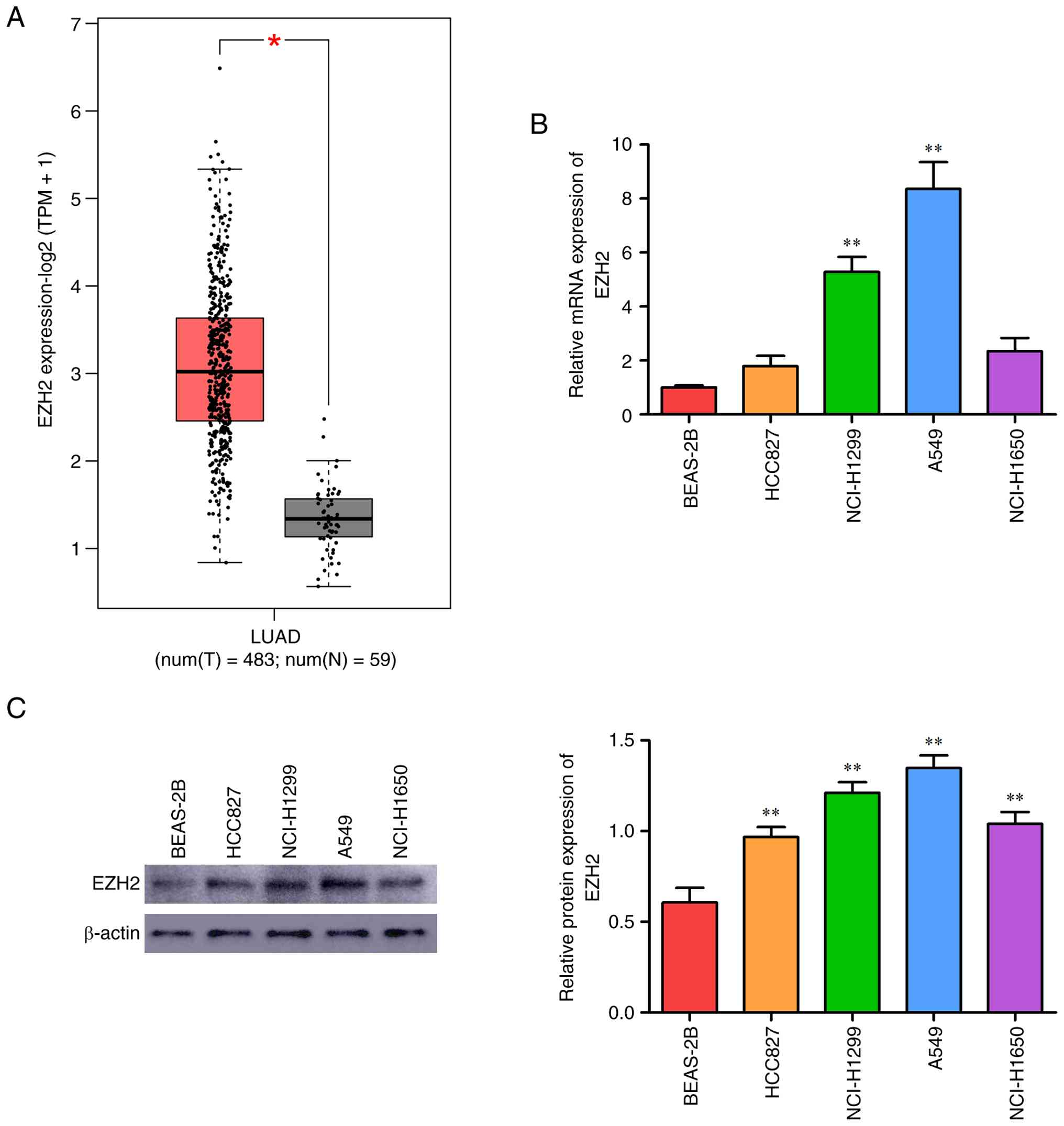
database. EZH2 was markedly upregulated in LUAD tissues compared to
adjacent normal tissues (Fig.
1A).
EZH2 is highly expressed in lung
cancer cell lines
EZH2 protein and mRNA levels were assessed in the
normal bronchial epithelial cell line BEAS-2B and four NSCLC cell
lines (HCC827, NCI-H1650, NCI-H1299, and A549) by RT-qPCR (Fig. 1B) and western blotting (Fig. 1C). EZH2 expression was markedly
higher in all cancer cell lines relative to BEAS-2B. Notably,
NCI-H1299 and A549 cells exhibited the highest EZH2 levels and were
therefore chosen for functional studies.
EZH2 knockdown suppresses
proliferation and induces apoptosis via JAK2/STAT3 pathway
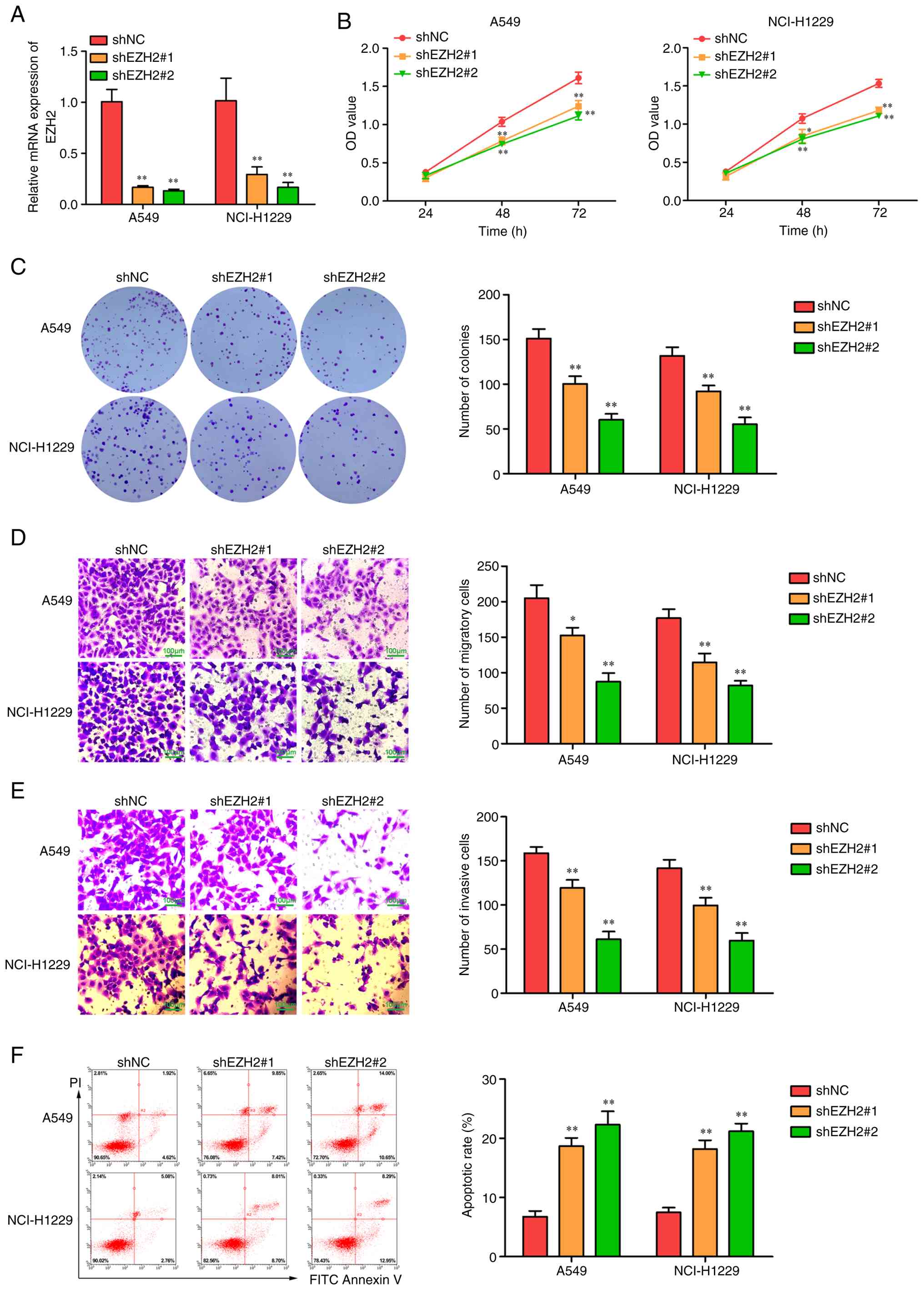
Stable EZH2-knockdown models were established in
NCI-H1299 and A549 cells using two independent shRNAs, with
knockdown efficiency confirmed by RT-qPCR (Fig. 2A). CCK-8 assays demonstrated that
EZH2 silencing markedly reduced cell viability (Fig. 2B). Moreover, migration, invasion,
and colony formation were markedly impaired (Fig. 2C-E). Flow cytometry revealed a
significant increase in apoptosis upon EZH2 knockdown (Fig. 2F).
To explore the underlying molecular mechanism, the
present study examined the effect of EZH2 knockdown on the
JAK2/STAT3 signaling pathway. Mechanistically, western blot
analysis showed that EZH2 knockdown substantially reduced the
phosphorylation levels of JAK2 and STAT3 (Fig. 3), suggesting that EZH2 promotes
tumor survival and proliferation primarily through activation of
this signaling cascade.
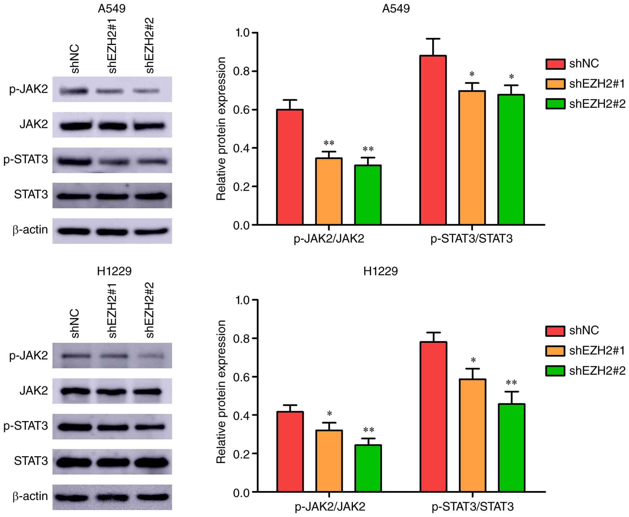 | Figure 3.Knockdown of EZH2 reduced JAK2/STAT3
phosphorylation in A549 and NCI-H1299 cells. Representative western
blots and quantitative analysis of EZH2, p-JAK2, JAK2, p-STAT3,
STAT3, and β-actin in EZH2-knockdown A549 and NCI-H1299 cells.
*P<0.05, **P<0.01 vs. shNC. EZH2, enhancer of zeste homolog
2; p-, phosphorylated; sh, short hairpin; NC, negative control. |
Rescue experiments confirm EZH2 acts
upstream of JAK2/STAT3
To define the functional hierarchy between EZH2 and
JAK2/STAT3, rescue experiments were performed using CA1 (10
µmol/l), a JAK2 agonist, and AG490 (25 µmol/l), a JAK2 inhibitor
(22). JAK2 activation restored
cell proliferation in EZH2-knockdown A549 and NCI-H1299 cells
(Fig. 4A). Likewise, JAK2
activation effectively rescued the impaired migration, invasion,
and colony formation, and attenuated the apoptosis induced by EZH2
silencing (Fig. 4B-E). Western
blotting confirmed that JAK2 inhibition reduced p-JAK2 and p-STAT3
without affecting EZH2 expression (Fig.
5), whereas JAK2 activation in EZH2-knockdown cells restored
both p-JAK2 and p-STAT3 levels.
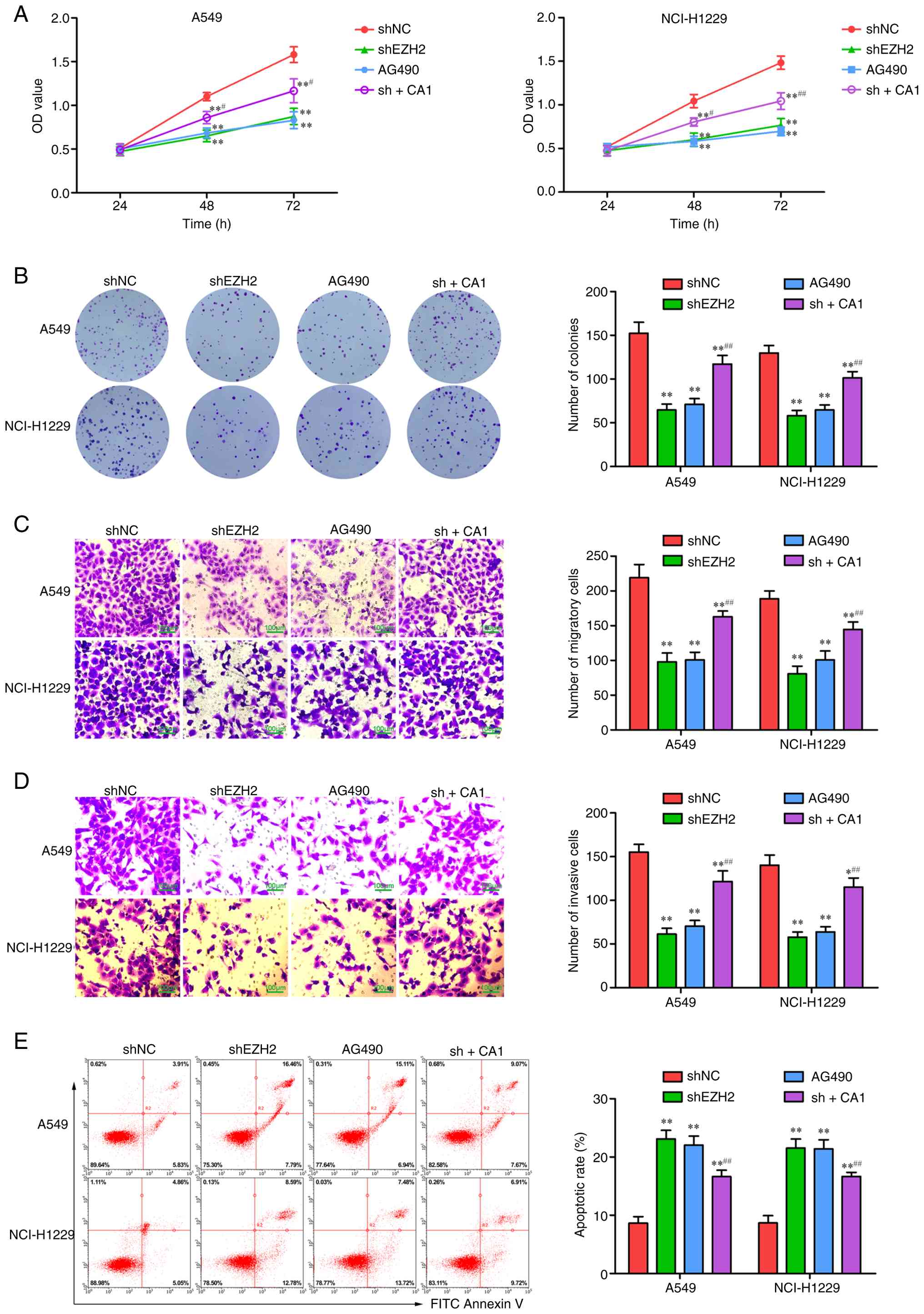 | Figure 4.Reversal by a JAK2 agonist of the
phenotypic effects of EZH2 knockdown, including inhibited
proliferation/migration and promoted apoptosis. (A) CCK-8 assay of
cell viability after transfection. (B) Colony formation assay of
clonogenic proliferation. (C) Transwell assay of cell migration.
Scale bar, 100 µm; magnification, ×200. (D) Transwell assay of cell
invasion. Scale bar, 100 µm; magnification, ×200. (E) Flow
cytometric analysis of apoptosis. *P<0.05, **P<0.01 vs. shNC;
#P<0.05, ##P<0.01 vs. shEZH2. EZH2,
enhancer of zeste homolog 2; p-, phosphorylated; sh, short hairpin;
NC, negative control; CA1, JAK2 agonist. |
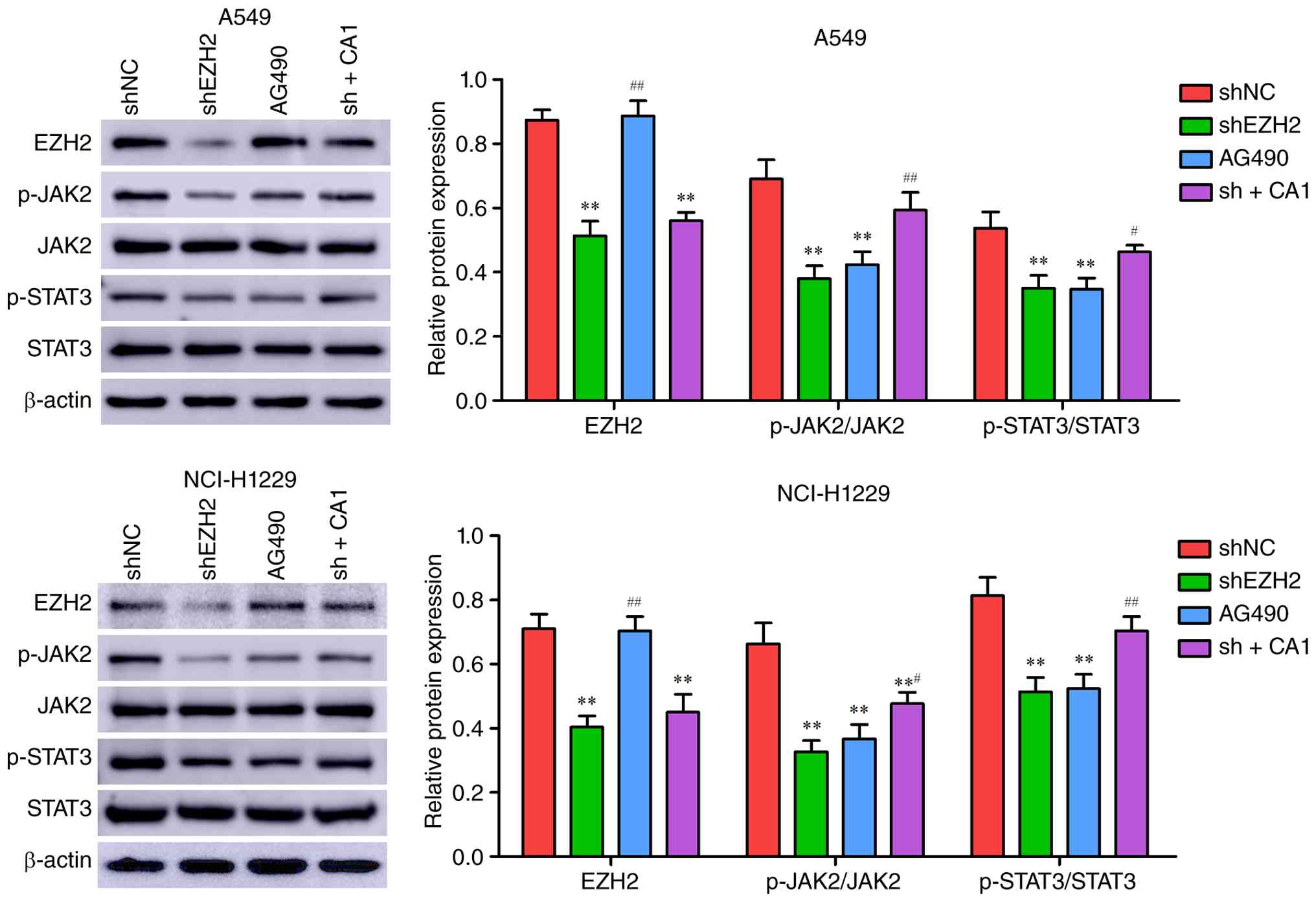 | Figure 5.The inhibition of the JAK2/STAT3
pathway resulting from EZH2 knockdown was reversed by a JAK2
agonist. Representative western blots and quantitative analyses of
p-JAK2, JAK2, p-STAT3, STAT3, and β-actin were performed in A549
and NCI-H1299 cells treated with shNC, shEZH2, AG490, and shEZH2 +
CA1. **P<0.01 vs. shNC; #P<0.05,
##P<0.01 vs. shEZH2. EZH2, enhancer of zeste homolog
2; AG490, JAK2 inhibitor; CA1, JAK2 agonist; p-, phosphorylated;
sh, short hairpin; NC, negative control. |
EZH2 silencing suppresses tumor growth
in vivo via inhibiting JAK2/STAT3
Since A549 cells exhibited more pronounced
phenotypic changes upon EZH2 knockdown compared to NCI-H1299 cells
in vitro, A549 cells were selected for the xenograft model.
Control or EZH2-knockdown A549 cells were inoculated into nude
mice. Tumors in the EZH2-knockdown group (shEZH2) exhibited
markedly slower growth, reduced volume, and decreased weight
compared to controls (Fig. 6A-C).
H&E staining revealed a looser histological architecture in
shEZH2 group tumors (Fig. 6D).
Immunohistochemical staining for Ki-67 demonstrated markedly
reduced proliferation in the shEZH2 group (Fig. 6E), while TUNEL staining indicated a
significant increase in apoptosis (Fig.
6F). Western blot analysis of tumor confirmed reduced JAK2 and
STAT3 phosphorylation, consistent with in vitro observations
(Fig. 6G). These in vivo
results corroborate that EZH2 facilitates tumor growth and survival
primarily through JAK2/STAT3 pathway activation.
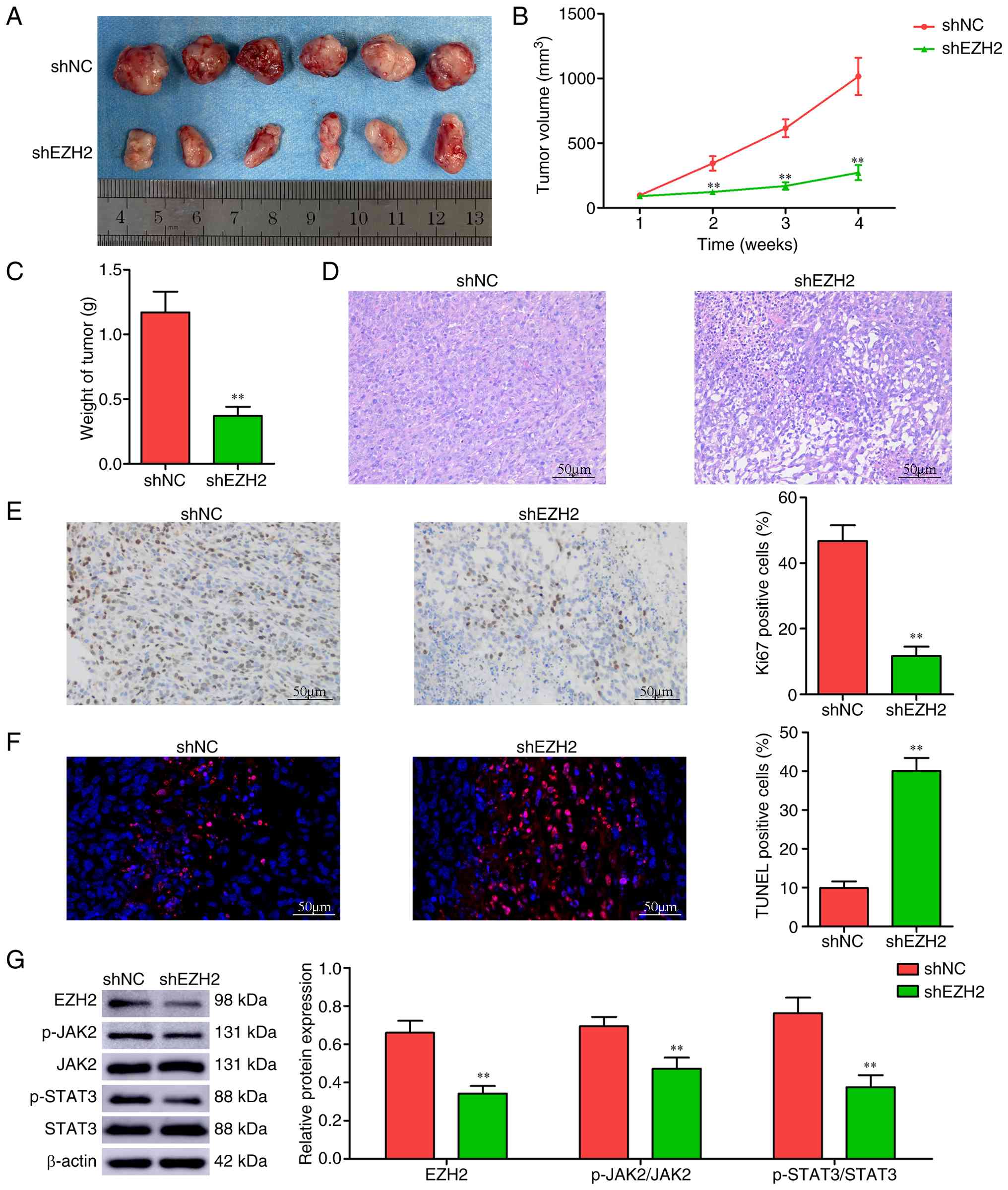 | Figure 6.EZH2 silencing suppresses tumor
growth in vivo by inhibiting JAK2/STAT3. (A) The tumor image
at day 28. (B) Tumor volumes at day 7, 14, 21 and 28 were
calculated. (C) Tumor weight on day 28. (D) Hematoxylin and eosin
staining, (E) Ki-67 immunohistochemistry and (F) TUNEL staining of
tumor tissue. (G) Representative western blot and quantitative
analysis for EZH2, p-JAK2, JAK2, p-STAT3, STAT3 and β-actin.
**P<0.01 vs. shNC. EZH2, enhancer of zeste homolog 2; p-,
phosphorylated; sh, short hairpin; NC, negative control. |
Discussion
The present study systematically investigated the
expression, function and molecular mechanism of EZH2 in NSCLC. The
key findings demonstrated that EZH2 is markedly overexpressed in
LUAD tissues and multiple NSCLC cell lines. Functional experiments,
both in vitro and in vivo, confirmed that EZH2
knockdown effectively suppresses proliferation and migration while
inducing apoptosis. All quantitative data were presented as mean ±
SD, and the differences were statistically significant, supporting
the reliability of our findings. Mechanistically, EZH2 functions as
an upstream regulator of the JAK2/STAT3 signaling pathway and
exerts its oncogenic effects through positive regulation of this
cascade, as consistently demonstrated in both in vitro and
in vivo models.
EZH2, an epigenetic regulator, is highly expressed
in various malignancies, including NSCLC. Prior studies indicate
that EZH2 expression is markedly higher in NSCLC tissues than in
normal counterparts and its overexpression correlates with tumor
invasiveness, metastasis and poor prognosis (30,31).
The findings of the present study aligned with extensive existing
research, further supporting the critical role of EZH2 in lung
cancer progression. In addition to validating the prognostic
implications, the present study demonstrated through in
vitro models and in vivo xenografts that EZH2 directly
governs malignant behaviors-proliferation and apoptosis-in
NCI-H1299 and A549 cells, providing a strong rationale for
targeting EZH2 therapeutically.
The most significant contribution of the present
study was the elucidation of a functional linkage between EZH2 and
JAK2/STAT3 signaling in NSCLC. Rescue experiments provided
compelling evidence that the oncogenic functions of EZH2 are
mediated through this pathway: JAK2 agonism effectively reversed
the proliferation inhibition, apoptosis induction and reduced
JAK2/STAT3 phosphorylation elicited by EZH2 knockdown. This finding
unequivocally positioned EZH2 upstream of the JAK2/STAT3 pathway.
Previous studies have reported similar interactions between EZH2
and JAK2/STAT3 signaling in other cancer types. In bladder cancer,
pharmacological inhibition of EZH2 was shown to suppress tumor
growth through downregulation of the JAK2/STAT3 pathway (32). Notably, our recent study
demonstrated that norcantharidin exerts anti-tumor effects in NSCLC
through downregulation of EZH2/JAK2/STAT3 signaling pathway
(17). However, these studies
primarily relied on pharmacological inhibitors and did not
establish a clear functional hierarchy between EZH2 and the
JAK2/STAT3 pathway. By contrast, the present study provided the
first characterization of this regulatory relationship in NSCLC
using genetic EZH2 knockdown combined with rescue experiments.
Mechanistically, previous studies have demonstrated that EZH2
regulates JAK2 expression by modifying the H3K27me3 histone mark in
its promoter region (33,34). However, the present study revealed
that EZH2 knockdown reduced JAK2 and STAT3 phosphorylation without
markedly altering their total protein levels. The precise mechanism
underlying this regulation requires further investigation.
Persistent JAK2/STAT3 activation is intimately
linked to tumor cell proliferation, apoptosis resistance and the
establishment of an immunosuppressive microenvironment (35–37).
Notably, Toll-like receptors (TLRs) play a role in modulating
immune responses within the tumor microenvironment and TLR-guided
therapeutic strategies have been explored as promising approaches
for cancer immunotherapy (38,39).
Given that EZH2 acts upstream of JAK2/STAT3 signaling pathway and
has been implicated in epigenetic regulation of immune-related
genes (40), targeting EZH2 may
represent a more promising strategy to suppress the abnormal
activation of this pathway and reshape the immunosuppressive
microenvironment at its source. Currently, several EZH2 inhibitors
are under clinical investigation (41–43).
Given its established role in resistance mechanisms, EZH2
inhibitors, whether used alone or in combination with JAK2 or
immune checkpoint inhibitors, merit investigation as a therapeutic
strategy for patients who have progressed on existing targeted or
immunotherapies.
The present study has several strengths. First, the
present study employed two independent shRNA sequences to knock
down EZH2, minimizing off-target effects and ensuring the
specificity of the observations. Second, the combination of genetic
knockdown with pharmacological rescue experiments using both a JAK2
agonist and inhibitor allowed the establishment of a functional
hierarchy between EZH2 and the JAK2/STAT3 pathway. Third, the
consistency of the findings across in vitro cell models and
an in vivo xenograft model provided robust evidence for the
role of the EZH2-JAK2/STAT3 axis in NSCLC progression.
Of course, the present study had certain
limitations. First, while the present study demonstrated that EZH2
acts upstream of the JAK2/STAT3 pathway, the precise molecular
mechanism by which EZH2 regulates JAK2/STAT3 phosphorylation
remains unclear. Additional mechanistic studies, such as ChIP-qPCR,
promoter analyses, or protein interaction assays, are needed to
determine whether EZH2 directly regulates JAK2 expression or
affects pathway activation indirectly. Furthermore, given the
classical role of EZH2 as the PRC2 catalytic subunit mediating
H3K27 trimethylation, future studies using H3K27me3 analysis or
pharmacological EZH2 inhibitors are required to clarify whether the
observed effects rely on EZH2 methyltransferase activity,
especially given recent evidence of methyltransferase-independent
EZH2 functions in other types of cancer (44). Second, EZH2 overexpression rescue
experiments would further strengthen the causal relationship
between EZH2 and the JAK2/STAT3 pathway, which will be pursued in
future studies. Third, the present study primarily focused on the
NCI-H1299 and A549 cell lines; the generalizability of these
findings to other NSCLC subtypes requires validation in additional
models. Additionally, the use of only female mice in the present
study may introduce sex-related bias. Future studies will include
both male and female animals to further validate the
generalizability of our findings. Finally, in vivo studies
could be expanded, such as using conditional knockdown mouse
models, to more precisely elucidate the role of EZH2 in lung cancer
development.
In summary, the present study established that EZH2
drives malignant progression in NSCLC through activation of the
JAK2/STAT3 signaling pathway. These findings deepen our
understanding of EZH2-mediated oncogenesis and, more importantly,
highlight the therapeutic potential of targeting the
EZH2-JAK2/STAT3 axis to overcome drug resistance and immune
suppression in NSCLC, offering new insights and experimental
rationale for the development of novel combination therapeutic
strategies.
Supplementary Material
Supporting Data
Acknowledgements
Not applicable.
Funding
Funding: No funding was received.
Availability of data and materials
The data generated in the present study may be
requested from the corresponding author.
Authors' contributions
SW and WZ confirm the authenticity of all the raw
data. SW and WZ contributed to the study conception and design.
Writing, reviewing and editing, resources, supervision and
conceptualization were performed by WZ. The writing of the original
draft, formal analysis, software and figure preparation
(visualization) were carried out by SW, and both authors commented
on previous versions of the manuscript. Both authors have read and
approved the final manuscript.
Ethics approval and consent to
participate
The animal experiments were performed in accordance
with ARRIVE guidelines and were approved by the Ethics Committee of
Yuhuangding Hospital (Shandong, China; approval no. K2025-381).
Patient consent for publication
Not applicable.
Competing interests
The authors declare that they have no competing
interests.
References
|
1
|
Bray F, Laversanne M, Sung H, Ferlay J,
Siegel RL, Soerjomataram I and Jemal A: Global cancer statistics
2022: GLOBOCAN estimates of incidence and mortality worldwide for
36 cancers in 185 countries. CA Cancer J Clin. 74:229–263.
2024.PubMed/NCBI
|
|
2
|
Goldstraw P, Ball D, Jett JR, Le Chevalier
T, Lim E, Nicholson AG and Shepherd FA: Non-small-cell lung cancer.
Lancet. 378:1727–1740. 2011. View Article : Google Scholar : PubMed/NCBI
|
|
3
|
Wu YL, Tsuboi M, He J, John T, Grohe C,
Majem M, Goldman JW, Laktionov K, Kim SW, Kato T, et al:
Osimertinib in resected EGFR-Mutated non-small-cell lung cancer. N
Engl J Med. 383:1711–1723. 2020. View Article : Google Scholar : PubMed/NCBI
|
|
4
|
Meyer ML, Fitzgerald BG, Paz-Ares L,
Cappuzzo F, Jänne PA, Peters S and Hirsch FR: New promises and
challenges in the treatment of advanced non-small-cell lung cancer.
Lancet. 404:803–822. 2024. View Article : Google Scholar : PubMed/NCBI
|
|
5
|
Parvaresh H, Roozitalab G, Golandam F,
Behzadi P and Jabbarzadeh Kaboli P: Unraveling the potential of
ALK-Targeted therapies in non-small cell lung cancer: Comprehensive
insights and future directions. Biomedicines. 12:2972024.
View Article : Google Scholar : PubMed/NCBI
|
|
6
|
Gao W, Wang L, Zhao Y and Zhu L: The role
of PD-L1 in EGFR-mutant non-small cell lung cancer. Discov Oncol.
16:3072025. View Article : Google Scholar : PubMed/NCBI
|
|
7
|
Wiest N, Majeed U, Seegobin K, Zhao Y, Lou
Y and Manochakian R: Role of immune checkpoint inhibitor therapy in
advanced EGFR-Mutant non-small cell lung cancer. Front Oncol.
11:7512092021. View Article : Google Scholar : PubMed/NCBI
|
|
8
|
Aguilar EJ, Ricciuti B, Gainor JF, Kehl
KL, Kravets S, Dahlberg S, Nishino M, Sholl LM, Adeni A, Subegdjo
S, et al: Outcomes to first-line pembrolizumab in patients with
non-small-cell lung cancer and very high PD-L1 expression. Ann
Oncol. 30:1653–1659. 2019. View Article : Google Scholar : PubMed/NCBI
|
|
9
|
Rah B, Rather RA, Bhat GR, Baba AB,
Mushtaq I, Farooq M, Yousuf T, Dar SB, Parveen S, Hassan R, et al:
JAK/STAT signaling: Molecular targets, therapeutic opportunities,
and limitations of targeted inhibitions in solid malignancies.
Front Pharmacol. 13:8213442022. View Article : Google Scholar : PubMed/NCBI
|
|
10
|
Thomas SJ, Snowden JA, Zeidler MP and
Danson SJ: The role of JAK/STAT signalling in the pathogenesis,
prognosis and treatment of solid tumours. Br J Cancer. 113:365–371.
2015. View Article : Google Scholar : PubMed/NCBI
|
|
11
|
Chen L, Sun R and Fang K: Erianin inhibits
tumor growth by promoting ferroptosis and inhibiting invasion in
hepatocellular carcinoma through the JAK2/STAT3/SLC7A11 pathway.
Pathol Int. 74:119–128. 2024. View Article : Google Scholar : PubMed/NCBI
|
|
12
|
Li Y, Wang M, Bai J, Li X, Xiao S and Song
L: Anthocyanins in black soybean coats promote apoptosis in
hepatocellular carcinoma cells by regulating the JAK2/STAT3
pathway. Int J Mol Sci. 26:10702025. View Article : Google Scholar : PubMed/NCBI
|
|
13
|
Wu L, Li J, Liu T, Li S, Feng J, Yu Q,
Zhang J, Chen J, Zhou Y, Ji J, et al: Quercetin shows anti-tumor
effect in hepatocellular carcinoma LM3 cells by abrogating
JAK2/STAT3 signaling pathway. Cancer Med. 8:4806–4820. 2019.
View Article : Google Scholar : PubMed/NCBI
|
|
14
|
Cheng Y, Song Z, Fang X and Tang Z:
Polycomb repressive complex 2 and its core component EZH2:
Potential targeted therapeutic strategies for head and neck
squamous cell carcinoma. Clin Epigenetics. 16:542024. View Article : Google Scholar : PubMed/NCBI
|
|
15
|
Cao W, Ribeiro Rde O, Liu D, Saintigny P,
Xia R, Xue Y, Lin R, Mao L and Ren H: EZH2 promotes malignant
behaviors via cell cycle dysregulation and its mRNA level
associates with prognosis of patient with non-small cell lung
cancer. PLoS One. 7:e529842012. View Article : Google Scholar : PubMed/NCBI
|
|
16
|
Pan YM, Wang CG, Zhu M, Xing R, Cui JT, Li
WM, Yu DD, Wang SB, Zhu W, Ye YJ, et al: STAT3 signaling drives
EZH2 transcriptional activation and mediates poor prognosis in
gastric cancer. Mol Cancer. 15:792016. View Article : Google Scholar : PubMed/NCBI
|
|
17
|
Wu S and Zhang W: Norcantharidin inhibits
the EZH2-mediated JAK2/STAT3 signaling pathway to inhibit the
proliferation of non-small cell lung cancer. Toxicol Appl
Pharmacol. 507:1177142026. View Article : Google Scholar : PubMed/NCBI
|
|
18
|
Wang Y, Tian J, Huang D, Gao Y, Lin H, Liu
L and Wang A: EZH2 promotes multiple myeloma progression via STAT3
pathway activation. Discov Med. 36:721–729. 2024. View Article : Google Scholar : PubMed/NCBI
|
|
19
|
Zimmerman SM, Suh E, Smith SR and
Souroullas GP: Stat3-mediated Atg7 expression regulates anti-tumor
immunity in mouse melanoma. Cancer Immunol Immunother. 73:2182024.
View Article : Google Scholar : PubMed/NCBI
|
|
20
|
Yu D, Wang S, Wang J, Zhang K, Niu Z and
Lin N: EZH2-STAT3 signaling pathway regulates GSDMD-mediated
pyroptosis in glioblastoma. Cell Death Discov. 10:3412024.
View Article : Google Scholar : PubMed/NCBI
|
|
21
|
Ranjbar R, Behzadi P and Mammina C:
Respiratory tularemia: Francisella tularensis and microarray probe
designing. Open Microbiol J. 10:176–182. 2016. View Article : Google Scholar : PubMed/NCBI
|
|
22
|
Xiang L, Wang Y, Liu S, Ying L, Zhang K,
Liang N, Li H, Luo G and Xiao L: Quercetin attenuates KLF4-Mediated
phenotypic switch of VSMCs to macrophage-like cells in
atherosclerosis: A critical role for the JAK2/STAT3 pathway. Int J
Mol Sci. 25:77552024. View Article : Google Scholar : PubMed/NCBI
|
|
23
|
Jin S, Zhang D, Liao Z, Yu L, Wang Y, Jia
P, Pan M, Li Y and Zhang J: TRIM46 deficiency-induced DNA damage
enhances the sensitivity of cisplatin in non-small cell lung cancer
by regulating the Akt signaling pathway. Oncol Rep. 55:582026.
View Article : Google Scholar : PubMed/NCBI
|
|
24
|
Zheng WW, Ding JX, Liang YX and Ye L:
Hydroxypropyl-β-cyclodextrin/thymoquinone inclusion complex
inhibits non-small cell lung cancer progression through
NF-κB-mediated ferroptosis. Oncol Rep. 54:1572025. View Article : Google Scholar : PubMed/NCBI
|
|
25
|
Chen H, Wu X, Zhu M, Cheng Y and Feng Q:
Glutamine starvation induces ferroptosis in NSCLC via
AMPK/PDZD8-Mediated ferritinophagy. Nutrients. 18:15962026.
View Article : Google Scholar : PubMed/NCBI
|
|
26
|
Livak KJ and Schmittgen TD: Analysis of
relative gene expression data using real-time quantitative PCR and
the 2(−Delta Delta C(T)) method. Methods. 25:402–408. 2001.
View Article : Google Scholar : PubMed/NCBI
|
|
27
|
Li JX, Tan SY, Li LQ, Zheng YH, Zhao L,
Zhu HR, He HL, Zhang YY, Li RZ, Bao TY, et al: Tricin selectively
combats KRAS-mutant non-small cell lung cancer by inhibiting the
PDGF-BB-induced SRC/MAPK/AP-1/PD-L1 signaling pathway and
potentiating the antitumor effect of an anti-PD-1 antibody. Front
Pharmacol. 16:15942132025. View Article : Google Scholar : PubMed/NCBI
|
|
28
|
Ma G, Feng J, Zeng J, Liao R, Zhong X, Lv
Z and Chen F: β-sitosterol suppresses malignant biological
behaviors and glycolysis via modulating YBX1-hnRNP K interaction in
non-small cell lung cancer. Sci Rep. 16:191052026. View Article : Google Scholar : PubMed/NCBI
|
|
29
|
Jiang P, Chen Z, Cui L, Zhang N and Gao K:
ID1 in TAMs promoted the progression of non-small-cell carcinoma
via increasing NF-κB/NPM1/SHP1/SHP2 signaling induced M2
polarization. Sci Rep. 16:142222026. View Article : Google Scholar : PubMed/NCBI
|
|
30
|
Kim NY and Pyo JS: Clinicopathological
significance and prognostic role of EZH2 expression in non-small
cell lung cancer. Pathol Res Pract. 213:778–782. 2017. View Article : Google Scholar : PubMed/NCBI
|
|
31
|
Shi B, Behrens C, Vaghani V, Riquelme EM,
Rodriguez-Canales J, Kadara H, Lin H, Lee J, Liu H, Wistuba I and
Simon G: Oncogenic enhancer of zeste homolog 2 is an actionable
target in patients with non-small cell lung cancer. Cancer Med.
8:6383–6392. 2019. View Article : Google Scholar : PubMed/NCBI
|
|
32
|
Chen Z, Du Y, Liu X, Chen H, Weng X, Guo
J, Wang M, Wang X and Wang L: EZH2 inhibition suppresses bladder
cancer cell growth and metastasis via the JAK2/STAT3 signaling
pathway. Oncol Lett. 18:907–915. 2019.PubMed/NCBI
|
|
33
|
Chandnani N, Gupta I, Thakkar V and Sarkar
K: Epigenetic regulation of enhancer of zeste homolog 2 (EZH2) -Yin
Yang 1 (YY1) axis in cancer. Pathol Res Pract. 251:1548852023.
View Article : Google Scholar : PubMed/NCBI
|
|
34
|
Dreger H, Ludwig A, Weller A, Stangl V,
Baumann G, Meiners S and Stangl K: Epigenetic regulation of cell
adhesion and communication by enhancer of zeste homolog 2 in human
endothelial cells. Hypertension. 60:1176–1183. 2012. View Article : Google Scholar : PubMed/NCBI
|
|
35
|
Johnson DE, O'Keefe RA and Grandis JR:
Targeting the IL-6/JAK/STAT3 signalling axis in cancer. Nat Rev
Clin Oncol. 15:234–248. 2018. View Article : Google Scholar : PubMed/NCBI
|
|
36
|
Li C, Song J, Guo Z, Gong Y, Zhang T,
Huang J, Cheng R, Yu X, Li Y, Chen L, et al: EZH2 inhibitors
suppress colorectal cancer by regulating macrophage polarization in
the tumor microenvironment. Front Immunol. 13:8578082022.
View Article : Google Scholar : PubMed/NCBI
|
|
37
|
Xue C, Yao Q, Gu X, Shi Q, Yuan X, Chu Q,
Bao Z, Lu J and Li L: Evolving cognition of the JAK-STAT signaling
pathway: autoimmune disorders and cancer. Signal Transduct Target
Ther. 8:2042023. View Article : Google Scholar : PubMed/NCBI
|
|
38
|
Mukherjee S, Patra R, Behzadi P, Masotti
A, Paolini A and Sarshar M: Toll-like receptor-guided therapeutic
intervention of human cancers: Molecular and immunological
perspectives. Front Immunol. 14:12443452023. View Article : Google Scholar : PubMed/NCBI
|
|
39
|
Behzadi P, García-Perdomo HA and Karpiński
TM: Toll-Like receptors: General molecular and structural biology.
J Immunol Res. 2021:99148542021. View Article : Google Scholar : PubMed/NCBI
|
|
40
|
Hamad J, Tarabain NE, Soufan F,
Fayyad-Kazan M, Issa N and Fayyad-Kazan H: Beyond oncogenesis: The
emerging role of EZH2 in tumor microenvironment. Crit Rev Oncol
Hematol. 226:1054542026. View Article : Google Scholar : PubMed/NCBI
|
|
41
|
Italiano A, Soria JC, Toulmonde M, Michot
JM, Lucchesi C, Varga A, Coindre JM, Blakemore SJ, Clawson A,
Suttle B, et al: Tazemetostat, an EZH2 inhibitor, in relapsed or
refractory B-cell non-Hodgkin lymphoma and advanced solid tumours:
A first-in-human, open-label, phase 1 study. Lancet Oncol.
19:649–659. 2018. View Article : Google Scholar : PubMed/NCBI
|
|
42
|
Song Y, Liu Y, Li ZM, Li L, Su H, Jin Z,
Zuo X, Wu J, Zhou H, Li K, et al: SHR2554, an EZH2 inhibitor, in
relapsed or refractory mature lymphoid neoplasms: A first-in-human,
dose-escalation, dose-expansion, and clinical expansion phase 1
trial. Lancet Haematol. 9:e493–e503. 2022. View Article : Google Scholar : PubMed/NCBI
|
|
43
|
Zeng D, Liu M and Pan J: Blocking EZH2
methylation transferase activity by GSK126 decreases stem cell-like
myeloma cells. Oncotarget. 8:3396–3411. 2017. View Article : Google Scholar : PubMed/NCBI
|
|
44
|
Kuser-Abali G, Noguchi F, Zhao P, Szeto P,
Zhang Y, Huang C, Barlow CK, Pomilio G, Boudes M, Chew C, et al:
Cytosolic EZH2-IMPDH2 complexes regulate melanoma progression and
metastasis via GTP. Mol Cell. 86:2521–2538.e10. 2026. View Article : Google Scholar : PubMed/NCBI
|