Introduction
Lung cancer is one of the most common malignancies
and a leading cause of cancer-related death worldwide, with
non-small cell lung cancer (NSCLC) accounting for ~79% of all lung
cancer cases (1). The advent of
immune checkpoint inhibitors (ICIs), particularly agents targeting
the programmed cell death protein 1/programmed death-ligand 1
(PD-1/PD-L1) axis, has markedly transformed the treatment landscape
for advanced NSCLC. Nevertheless, the clinical benefits of ICIs
remain highly variable. Although some patients achieve durable
tumor control, many experience only transient benefit or primary
resistance, depending on the treatment setting, biomarker status,
combination regimen and tumor microenvironmental (TME) context
(2). These variable outcomes
suggest that the limited efficacy of ICIs in NSCLC cannot be
attributed to a single immunosuppressive pathway. Rather, it likely
reflects the interplay among tumor-intrinsic characteristics,
immune-cell functional states, and spatial constraints within the
tumor immune microenvironment (TIME) (3,4).
Accumulating evidence indicates that structural
constraints arising from interactions between tumors and the host
immune system within local tissue niches may contribute to the
failure of ICI therapy. ICI efficacy is associated not only with
established biomarkers, such as PD-L1 expression and tumor
mutational burden (TMB), but also with the spatial organization of
the TIME (5). The TME comprises a
complex network of cellular and structural components, including
immune cells, fibroblasts, blood vessels, signaling molecules and
the extracellular matrix (ECM) (6,7). Its
spatial architecture influences whether immune cells can reach the
tumor, penetrate tumor-cell nests, and sustain effector function.
It also shapes immune-cell states through nutrient availability,
oxygen tension and metabolic by-products (6). In this context, NSCLC exhibits
pronounced spatial immune heterogeneity. Some tumors contain
abundant intratumoral T cells and exhibit interferon (IFN)-related
transcriptional programs, whereas others are nearly devoid of T
cells or harbor T cells that remain largely confined to tumor
margins or stromal compartments (5).
Based on these spatial patterns, tumor immune states
are commonly described in terms of three operational phenotypes:
Inflamed, immune-desert and immune-excluded (8). The inflamed phenotype is characterized
by immune-cell infiltration into tumor nests; the immune-desert
phenotype by globally limited immune infiltration; and the
immune-excluded phenotype by the accumulation of immune cells in
stromal or marginal regions with limited penetration into
tumor-cell nests. However, these phenotypes should be viewed as
context-dependent, non-mutually exclusive spatial states rather
than categories defined by universal thresholds. They may vary
according to the sampled region, disease stage, treatment exposure
and analytical platform. Elucidating the mechanisms that give rise
to these spatial states may therefore help explain why some NSCLC
tumors respond poorly to ICIs despite the presence of targetable
immune checkpoints.
Mitochondrial metabolism, which intersects with
multiple aspects of cancer biology, offers a mechanistic
perspective for understanding these spatial immune constraints.
Although the classical Warburg effect emphasizes the role of
aerobic glycolysis in tumor-cell energy metabolism, mitochondria
are also involved in tumor progression, stress adaptation,
therapeutic responses and immune regulation. Mitochondrial reactive
oxygen species (mtROS) may function as signaling mediators or
amplify cellular stress, depending on their intensity, duration and
cellular context. Tricarboxylic acid (TCA) cycle metabolites may
link mitochondrial metabolism to epigenetic regulation and
hypoxia-responsive programs. The release of mitochondrial DNA
(mtDNA) may engage innate immune sensing through the cyclic GMP-AMP
synthase-stimulator of IFN genes (cGAS-STING) pathway. In addition,
mitochondrial dynamics, mitophagy and intercellular mitochondrial
transfer may influence the functional states of tumor cells and
tumor-infiltrating lymphocytes (TILs). These mechanisms may
therefore connect mitochondrial biology to immune priming,
chemokine-driven immune recruitment, stromal barrier formation, and
the maintenance of T-cell effector function (9–11).
However, evidence linking mitochondrial processes to
spatial immune phenotypes in human NSCLC remains predominantly
correlative. Standardized, spatially resolved and cell
type-specific measures of mitochondrial function remain limited,
complicating causal inference and efforts to determine whether
mitochondrial alterations act as upstream drivers, phenotype
amplifiers, or concomitant biological states (3,5,12).
Accordingly, the present review synthesizes current evidence
linking mitochondrial biology to spatial immune phenotypes in NSCLC
within an evidence-weighted mechanistic framework. It was examined
how mitochondrial processes in tumor, immune, stromal and vascular
cells may contribute to immune-desert, immune-excluded and inflamed
states. Well-supported clinical associations from preclinical
evidence and hypothesis-generating models were also relatively
distinguished and the key experimental and translational steps
required to validate mitochondria-informed strategies for
cold-to-hot tumor conversion were outlined.
Review methodology and scope
The present review presents a focused mechanistic
narrative review. It aims to synthesize current evidence linking
mitochondrial biology to spatial immune phenotypes and responses to
ICIs in NSCLC, with particular emphasis on immune-desert,
immune-excluded and inflamed tumor states.
Relevant literature published through March 2026 was
identified through iterative searches of PubMed (https://pubmed.ncbi.nlm.nih.gov/), Web of Science
(https://clarivate.com/academia-government/scientific-and-academic-research/research-discovery-and-referencing/web-of-science/)
and Google Scholar (https://scholar.google.com/). The searches were
structured around predefined conceptual modules, including NSCLC,
spatial immune phenotypes, the TIME, ICIs, mitochondrial
metabolism, oxidative phosphorylation (OXPHOS), mtROS, mtDNA,
cGAS-STING signaling, TCA cycle metabolites, epigenetic regulation,
mitochondrial dynamics, mitophagy, mitochondrial transfer,
cancer-associated fibroblasts (CAFs), hypoxia, and cold-to-hot
tumor conversion. Additional studies were identified by screening
the reference lists of relevant original articles and reviews.
Because the available literature encompasses
heterogeneous evidence from human NSCLC cohorts, spatial profiling
studies, animal models, in vitro systems, and other tumor
types, evidence was prioritized in the following order: Human NSCLC
studies with clinical or spatial information; NSCLC studies
involving ICI response or resistance; mechanistic studies using
NSCLC models; and, when direct evidence from NSCLC was unavailable,
mechanistic evidence from other tumor types or general cancer
biology. Evidence derived from non-NSCLC models or non-spatial
experimental systems was interpreted as supportive or
hypothesis-generating rather than definitive evidence for spatial
immune phenotypes in human NSCLC.
Accordingly, the present review does not provide a
quantitative evidence synthesis. Instead, it adopts an
evidence-weighted narrative approach to distinguish relatively
well-established clinical associations from preclinical mechanistic
findings and context-dependent hypotheses requiring further
validation in spatially resolved human NSCLC samples.
Operational and clinical definitions of
spatial immune phenotypes in NSCLC
The spatial immune architecture of NSCLC is commonly
described in terms of three major patterns: Inflamed, immune-desert
and immune-excluded phenotypes. These terms provide a practical
framework for characterizing the localization of immune cells
relative to tumor-cell nests and interpreting potential differences
in ICI responses (6,8,13,14)
(Fig. 1; Table SI). However, they should be
regarded as operational, context-dependent spatial states rather
than fixed, mutually exclusive biological categories defined by
universal thresholds. Different phenotypes may coexist within the
same tumor, and their classification may vary according to the
sampled region, disease stage, prior treatment, tissue processing,
marker selection, analytical platform, and thresholding method
(8,14,15)
(Fig. 2).
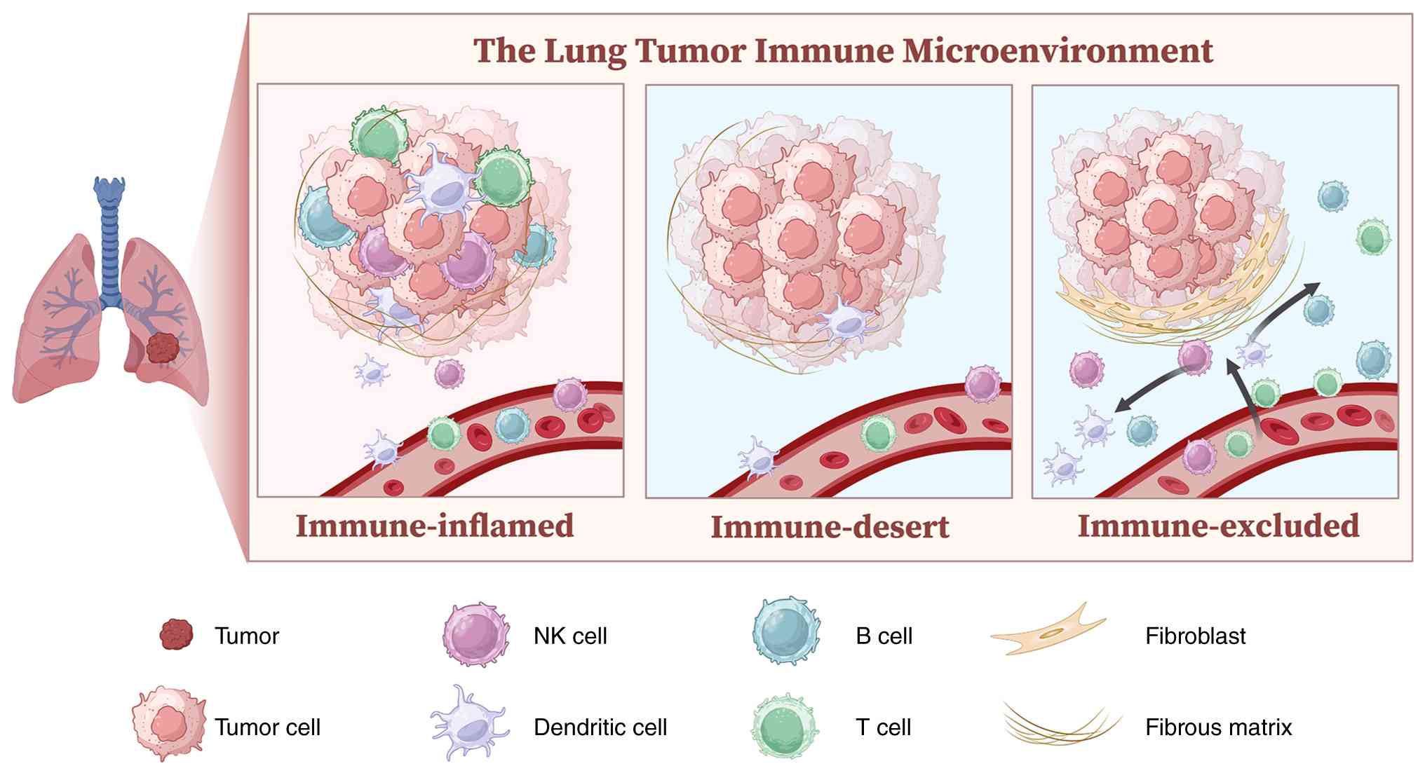 | Figure 1.Spatial immune phenotypes of the
NSCLC TME. Schematic illustration of the three major spatial immune
phenotypes in the TIME of NSCLC, conceptually categorized according
to the spatial distribution of immune cells relative to tumor
nests. Immune-inflamed tumors are characterized by abundant
immune-cell infiltration within tumor parenchyma, with immune cells
frequently located in close proximity to tumor cells. Immune-desert
tumors exhibit sparse immune-cell infiltration within both tumor
nests and surrounding stroma, suggesting limited immune recruitment
or impaired immune priming within the TME. Immune-excluded tumors
display immune cells enriched in the peritumoral stroma or invasive
margin but with limited penetration into tumor nests, often
associated with stromal remodeling or physical barriers restricting
immune-cell infiltration. The schematic also depicts representative
cellular components involved in NSCLC immune architecture,
including tumor cells, T cells, B cells, NK cells, dendritic cells,
fibroblasts, and fibrous ECM. NSCLC, non-small cell lung cancer;
TME, tumor microenvironment; NK, natural killer. |
 | Figure 2.Metabolic geography of NSCLC: Spatial
heterogeneity of tumor metabolism and potential immune
consequences. Tumor regions within NSCLC exhibit substantial
metabolic heterogeneity, with distinct bioenergetic states
potentially influencing the local immune microenvironment. This
schematic illustrates a conceptual spatial map integrating
tumor-cell mitochondrial activity, metabolic adaptation and
immune-cell responses across different tumor niches. Highly
proliferative tumor regions may exhibit increased glucose and
glutamine utilization, enhanced ETC activity, and elevated OXPHOS,
supporting biosynthetic demands and stress adaptation. By contrast,
poorly perfused or hypoxic regions are characterized by increased
glycolytic activity, lactate accumulation, extracellular
acidification and altered immune-cell function. The upper right
panel illustrates potential effects of tumor-associated metabolic
conditions on CD8⁺ T-cell mitochondrial fitness. Although oxidative
metabolism is essential for effector T-cell persistence and
function, nutrient limitation and altered metabolite availability
within tumor regions may impose metabolic constraints on
infiltrating immune cells. Hypoxia-associated metabolic remodeling
may further influence immune-cell behavior through lactate
accumulation, reduced cytokine production, and altered signaling
pathways. In addition, extracellular ATP metabolism and adenosine
generation may contribute to immunoregulatory states through
purinergic signaling, including A2AR-mediated pathways. The
proposed model highlights tumor metabolic demand as a potential
upstream pressure that may contribute to spatial immune
dysfunction. However, direct evidence demonstrating local substrate
competition between tumor cells and immune cells in human NSCLC
remains limited. Spatial multi-omics approaches combined with
functional mitochondrial measurements and serial sampling
strategies will be required to establish causal relationships
between tumor metabolic states and immune-cell impairment. This
figure summarizes candidate metabolic constraints and spatial
associations rather than a universally established causal hierarchy
between tumor metabolism and immune exclusion. NSCLC, non-small
cell lung cancer; ETC, electron transport chain; A2AR, A2A
receptor; OXPHOS, oxidative phosphorylation; OCR, oxygen
consumption rate; mtROS, mitochondrial reactive oxygen species. |
The inflamed phenotype is operationally
characterized by the presence of immune cells, particularly
CD8+ T cells, within tumor-cell nests or in close
spatial proximity to malignant epithelial cells (8,13,14).
This pattern is often accompanied by enhanced antigen-presentation
activity, IFN-associated transcriptional programs, and increased
immune-cell interactions within the tumor parenchyma (6,13,16).
Inflamed tumors are generally more likely to harbor pre-existing
antitumor immune activity and may respond more favorably to ICIs
than non-inflamed tumors. Nevertheless, abundant intratumoral
T-cell infiltration does not necessarily translate into durable
clinical benefit, because infiltrating T cells may remain
functionally exhausted or metabolically compromised, while local
costimulatory and antigen-presentation programs may be inadequate
(6,16). The inflamed phenotype should
therefore be interpreted as a state of spatial immune infiltration
rather than as a definitive indicator of functional antitumor
immunity or ICI responsiveness.
The immune-desert phenotype is characterized by
sparse immune-cell infiltration throughout both tumor cell nests
and the surrounding stromal compartments (8,14,15).
Operationally, this phenotype is identified by low densities of
intratumoral and stromal effector immune cells, particularly
CD8+ T cells, although the precise quantitative
thresholds vary among studies. Immune-desert tumors frequently
exhibit impaired immune priming and recruitment, including reduced
dendritic-cell (DC) activity, antigen-presentation capacity, IFN
signaling, or chemokine expression (13,16).
Clinically, the scarcity of preexisting effector cells may limit
the capacity of PD-(L)1 blockade alone to elicit an effective
antitumor response. However, a desert-like appearance may result
from several distinct biological processes and may also be
influenced by sampling bias. Therefore, low immune-cell density
alone should not be interpreted as evidence of a specific
underlying mechanism.
The immune-excluded phenotype is characterized by
the presence of immune cells that remain predominantly confined to
stromal compartments, invasive margins, or peritumoral regions,
with limited penetration into tumor cell nests (8,14,17).
Unlike immune-desert tumors, immune-excluded tumors may contain
substantial numbers of T cells and other immune populations, but
their spatial distribution limits direct contact with malignant
cells. This pattern is frequently associated with enrichment of
CAFs, ECM deposition and remodeling, transforming growth
factor-β-related programs, abnormal vasculature and
hypoxia-associated barriers (17).
Nevertheless, these features should be regarded as common
correlates rather than universal defining mechanisms because the
relative contributions of stromal, vascular, chemotactic and
tumor-intrinsic factors vary among tumors.
These phenotypes can be evaluated using conventional
immunohistochemistry, multiplex immunofluorescence, imaging mass
cytometry, digital pathology, whole-slide image analysis, and
spatial transcriptomic or proteomic approaches (6,8,13,14).
Relevant measures include immune-cell densities within tumor and
stromal compartments, distances between immune and malignant cells,
localization relative to tumor-stroma boundaries, and spatially
resolved immune or stromal gene-expression programs. However, no
universally accepted quantitative thresholds currently distinguish
inflamed, immune-desert and immune-excluded NSCLC across analytical
platforms. Throughout the present review, these terms are therefore
used as practical spatial frameworks for organizing evidence rather
than as validated clinical diagnostic categories. Pending
prospective comparisons and standardized spatial assessment, their
potential value should be considered complementary to that of
established biomarkers such as PD-L1 expression and TMB.
Cell type-specific mitochondrial mechanisms
in the NSCLC immune microenvironment
For analytical clarity, mitochondria-related
processes within the NSCLC TIME are considered according to the
principal cellular or tissue compartments in which they are
measured or proposed to operate. This compartment-resolved approach
is important because malignant, immune, stromal and vascular states
vary substantially among regions within the same tumor. Moreover,
the biological consequences of a mitochondrial signal may depend on
its cellular source, magnitude, duration and surrounding
microenvironmental context (6,8,14,17–19).
Accordingly, the following subsections separately address
tumor-cell mitochondrial metabolism and redox signaling, T-cell
mitochondrial fitness, DC and myeloid innate sensing, the stromal
and vascular context, TCA metabolite-linked epigenetic regulation,
and intercellular mitochondrial transfer.
However, these mechanisms should not be interpreted
as one-to-one determinants of the inflamed, immune-desert, or
immune-excluded phenotypes. In human NSCLC, evidence is relatively
strong for several spatial associations among immune-cell
localization, stromal organization, and treatment response. By
contrast, direct cell type-resolved measurements of mitochondrial
function and interventional evidence remain limited (6,8,14,17–19).
Throughout this section, relatively well-established spatial
associations were therefore distinguished from preclinical
mechanistic evidence and hypothesis-generating extrapolations
(Fig. 3).
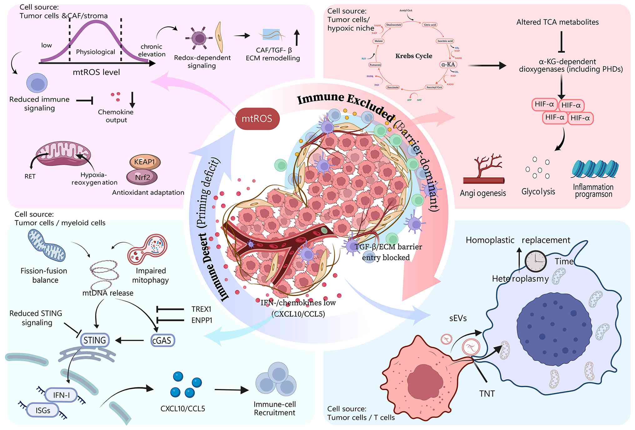 | Figure 3.Mitochondria-associated mechanisms
potentially shaping spatial immune phenotypes in NSCLC.
Mitochondrial alterations may influence spatial immune organization
in NSCLC through multiple interconnected mechanisms, including
redox regulation, metabolic remodeling, mitochondrial danger
signaling, and intercellular mitochondrial communication. The
schematic summarizes candidate mitochondria-associated pathways
that may contribute to immune-excluded and immune-desert
phenotypes. In the upper left panel, altered mtROS levels are
depicted as a context-dependent regulator of cellular signaling.
Moderate mitochondrial redox activity may support physiological
signaling, whereas persistent mtROS elevation may induce
redox-dependent signaling alterations, antioxidant adaptation (for
example, KEAP1-nuclear factor erythroid 2-related factor 2
pathway), and stromal remodeling involving CAFs and ECM components.
These processes may influence immune-cell function and spatial
immune organization. In the upper right panel, tumor-associated
metabolic remodeling, including alterations in TCA cycle
metabolites and α-KG-dependent dioxygenase activity, may regulate
HIF-α stabilization and downstream hypoxia-associated programs,
such as angiogenesis, glycolytic adaptation and inflammatory
transcriptional responses. These metabolic changes represent
potential microenvironmental constraints rather than directly
established drivers of immune exclusion. In the lower left panel,
mitochondrial stress-associated signals, including mtDNA release
caused by altered mitochondrial dynamics or impaired mitophagy, may
activate cGAS-STING-mediated innate immune signaling. Subsequent
induction of type I IFN-related genes and chemokines, including
CXCL10 and CCL5, may facilitate immune-cell recruitment. However,
the spatial consequences of mtDNA sensing remain context dependent
and may vary according to tumor type, immune composition and
signaling status. In the lower right panel, intercellular
mitochondrial communication mediated by TNTs or sEVs is illustrated
as a potential mechanism influencing mitochondrial heterogeneity
and immune-cell fitness. Current evidence for mitochondrial
transfer in cancer immunity is predominantly derived from
experimental models, and its contribution to spatial immune
phenotypes in human NSCLC requires further investigation. The
illustrated pathways represent a combination of relatively
established mitochondrial signaling mechanisms, preclinical
observations and hypothesis-generating models. The connections
between mitochondrial alterations and spatial immune phenotypes
should therefore be interpreted as potential regulatory
relationships rather than universally validated causal pathways.
NSCLC, non-small cell lung cancer; mtROS, mitochondrial reactive
oxygen species; CAFs, cancer-associated fibroblasts; ECM,
extracellular matrix; TCA, tricarboxylic acid; α-ketoglutarate;
α-KG, α-ketoglutarate; HIF-α, hypoxia-inducible factor-α; IFN,
interferon; TNTs, tunneling nanotubes; sEVs, small extracellular
vesicles. |
Tumor-cell mitochondrial metabolism
and redox stress signaling
NSCLC cells do not uniformly rely on glycolysis
alone. Spatial mapping studies have revealed substantial
intratumoral heterogeneity in mitochondrial organization and
bioenergetic activity. Better-perfused regions tend to exhibit
greater oxidative metabolic capacity, whereas poorly perfused and
hypoxic regions are more commonly associated with glycolytic
programs (20,21). Metabolic analyses of patient samples
have further shown that glucose-derived carbon can contribute
substantially to the TCA cycle in biologically aggressive NSCLC
(22). These findings indicate that
tumor-cell mitochondrial metabolism varies spatially and may create
regional differences in the demand for oxygen and carbon
substrates.
In tumor cells with relatively high OXPHOS activity,
electron transport chain (ETC) flux supports ATP production,
biosynthesis and adaptation to metabolic stress. Respiratory
activity is also required to sustain biosynthetic pathways
essential for lung cancer growth and survival (21,23).
Moreover, lactate can modulate ETC activity independently of its
direct oxidation as a carbon substrate, further illustrating the
metabolic plasticity of the respiratory chain (24). Under glucose-limited conditions,
NSCLC cells, including drug-resistant cell models, can activate
adaptive metabolic programs that sustain survival and proliferation
(25). Collectively, these findings
support the existence of tumor-cell states characterized by both
high metabolic demand and metabolic plasticity.
These observations, however, do not in themselves
demonstrate direct substrate competition between tumor and immune
cells in human NSCLC. Although the cited studies establish spatial
bioenergetic heterogeneity, metabolic adaptation and altered
substrate utilization, they do not simultaneously measure
tumor-cell OXPHOS activity, local substrate depletion, and
mitochondrial dysfunction in neighboring T cells within the same
tissue regions (20–25). Tumor-cell substrate consumption
should therefore be regarded as a candidate upstream metabolic
pressure rather than an established proximal cause of immune-desert
or immune-excluded architecture. The potential effects of these
tumor-cell metabolic states on mitochondrial fitness in neighboring
T cells are discussed in the following section.
In addition to regulating bioenergetic demand,
tumor-cell mitochondria participate in redox signaling. mtROS arise
primarily from electron leakage at respiratory-chain complexes I
and III. Their production is influenced by membrane potential,
substrate redox state and changes in oxygen availability, including
hypoxia-reoxygenation (25,26). Depending on their abundance,
duration and cellular context, mtROS may act as signaling mediators
or cause oxidative damage (27,28).
Accordingly, the proposed ‘biphasic threshold’ of mtROS is best
regarded as a conceptual framework rather than a universally
established spatial rule. Moderate redox signaling may facilitate
cellular adaptation or inflammatory signaling, whereas sustained or
excessive oxidative stress can damage macromolecules and reinforce
stress-response programs.
The consequences of tumor-cell redox signaling are
further shaped by tumor-intrinsic antioxidant capacity and
molecular context. Broad manipulation of oxidative stress or
antioxidant pathways has yielded inconsistent effects across
experimental cancer models and may, in some contexts, promote
rather than inhibit tumor progression (29). In NSCLC, transcriptional states that
phenocopy deleterious Kelch-Like ECH-associated protein 1 (KEAP1)
alterations have been associated with clinical outcomes after
immunotherapy (30), whereas
KEAP1/STK11 alterations also define distinct stress responses and
therapeutic vulnerabilities (31).
These findings underscore that redox biology cannot be interpreted
independently of tumor genotype and cellular state. Importantly,
they do not establish either low or high tumor-cell mtROS as a sole
determinant of any specific spatial immune phenotype.
Overall, tumor-cell mitochondrial activity may
contribute to spatially heterogeneous metabolic and redox
constraints through changes in oxygen consumption, substrate
demand, respiratory-chain activity and oxidative stress. Current
evidence from human NSCLC supports the existence and clinical
relevance of regional metabolic heterogeneity, but direct causal
links among tumor-cell mitochondrial states, immune-cell
positioning, and ICI response remain insufficiently defined. Future
studies should integrate spatial bioenergetic and metabolomic
mapping (18,20) with multiplexed immune imaging
(19) and cell type-resolved
measurements of mitochondrial function to determine whether
tumor-cell metabolic states act as upstream drivers, amplifiers, or
concomitant features of spatial immune organization.
T-cell mitochondrial fitness and
dysfunction
The activation, effector activity, and long-term
persistence of CD8+ T cells depend on tightly
coordinated mitochondrial bioenergetics and quality-control
mechanisms. Beyond ATP generation, mitochondrial OXPHOS, spare
respiratory capacity, membrane potential, and redox homeostasis
collectively support cytokine production, cytotoxicity, and
sustained T-cell function under conditions of metabolic stress
(32). Together, these
characteristics define mitochondrial fitness, a functional property
that is conceptually distinct from T-cell abundance or the extent
of spatial infiltration.
Evidence from lung cancer further indicates
substantial heterogeneity in the mitochondrial states of
tumor-infiltrating CD8+ T cells. A CD38-high
CD8+ T-cell subset exhibiting features of mitochondrial
adaptation has been identified in lung cancer, suggesting that
certain T-cell populations may preserve bioenergetic programs that
facilitate persistence within the TME (5). However, the identification of such
subsets does not suggest that mitochondrial fitness is uniformly
maintained in inflamed tumors, nor does it establish any single
T-cell marker as a universal indicator of functional competence
across patients.
Within the TME, T cells are simultaneously exposed
to nutrient deprivation, hypoxia, extracellular acidification and
immunosuppressive metabolites. Experimental evidence indicates that
glucose restriction can reshape mitochondrial signaling and alter
T-cell differentiation programs (33). Studies in a non-NSCLC model have
additionally shown that selective suppression of glutamine
metabolism in tumor cells can enhance antitumor T-cell activity,
supporting the broader principle that competition for metabolic
substrates between malignant and immune cells can influence immune
function (34). Nevertheless,
direct evidence demonstrating glutamine competition between tumor
cells and T cells within spatially resolved regions of human NSCLC
remains scarce.
Lactate and adenosine constitute two additional
metabolic constraints capable of compromising T-cell mitochondrial
fitness and effector activity. Lactate accumulation and
extracellular acidification can inhibit T-cell glycolysis, cytokine
production and migration, although the magnitude of these effects
varies according to metabolite concentration, duration of exposure,
and T-cell differentiation state (35,36).
In parallel, extracellular ATP released by stressed, hypoxic, or
dying cells can be sequentially converted to adenosine by
ecto-nucleotidases expressed across tumor, stromal and immune
compartments. In NSCLC, CAFs and other cellular populations may
contribute to CD73-dependent adenosine production and signaling
(37,38). Activation of the A2A receptor by
adenosine can suppress T-cell metabolic activity and chemotaxis,
whereas experimental blockade or disruption of this pathway can
partially restore T-cell function (36,39).
However, because some of these functional observations were
obtained in models other than NSCLC, they should be regarded as
mechanistic evidence supporting biological plausibility rather than
as direct demonstrations of spatial immune regulation in human
NSCLC.
These metabolic stressors are unlikely to operate in
isolation. Nutrient limitation, lactate accumulation, extracellular
acidification, adenosine signaling, hypoxia and high
tumor-intrinsic metabolic activity frequently converge within the
same tissue regions. Their combined effects may impair T-cell
OXPHOS, reduce spare respiratory capacity and ATP generation,
disturb redox homeostasis, and ultimately compromise functional
persistence. To date, however, studies of human NSCLC have not
directly established a complete causal sequence in which tumor-cell
substrate consumption creates localized nutrient depletion,
subsequently induces mitochondrial dysfunction in adjacent T cells,
and thereby determines their spatial distribution or responsiveness
to immune checkpoint inhibition (6,40).
Metabolic competition should therefore be viewed as a biologically
plausible and experimentally testable framework rather than an
established patient-level mechanism.
Importantly, spatial infiltration and mitochondrial
competence represent interconnected but non-equivalent dimensions
of antitumor immunity. An inflamed tumor may harbor abundant
CD8+ T cells while those cells remain metabolically
compromised, functionally exhausted, or unable to sustain cytotoxic
activity. Conversely, low T-cell density may arise from defective
priming or impaired recruitment rather than from an intrinsic
mitochondrial defect in the T cells that do reach the tumor.
Accordingly, mitochondrial fitness is best considered a potential
determinant of sustained T-cell function after immune-cell
recruitment, rather than a universal mechanism governing the
initial formation of a particular spatial immune phenotype.
DC and myeloid mitochondrial
sensing
Effective antitumor immunity requires more than the
physical presence of T cells; it also depends on efficient antigen
presentation, DC activation, costimulatory signaling, and
productive T-cell priming. DC functional states can influence the
magnitude of benefit derived from PD-L1 blockade, suggesting that
immune checkpoint inhibition is less likely to generate durable
adaptive immunity when local priming circuits are inadequate
(41). Mitochondrial stress may
intersect with these processes through the release of mtDNA and
subsequent engagement of cytosolic DNA-sensing pathways in tumor
and myeloid compartments.
When released from mitochondria into the cytosol,
mtDNA can act as a damage-associated molecular pattern.
Experimental studies have demonstrated that mitochondrial
fragmentation, altered membrane permeability, disrupted cristae
architecture, mitochondrial permeability transition, and defective
mitophagy can promote the cytosolic accumulation of mtDNA or other
damaged mitochondrial components (42–46).
Much of this mechanistic evidence, however, has been generated in
non-NSCLC settings, including other malignancies and inflammatory
disease models. These findings establish the biological
plausibility of stress-induced mtDNA release but do not, in
themselves, demonstrate that this mechanism is a dominant
determinant of immune organization in human NSCLC.
Once cytosolic, double-stranded DNA can be
recognized by cyclic GMP-AMP synthase (cGAS), which catalyzes the
production of cyclic GMP-AMP (cGAMP). cGAMP subsequently activates
stimulator of IFN genes (STING), triggering downstream signaling
through mediators such as TANK-binding kinase 1 (TBK1) and inducing
type I IFNs, IFN-stimulated genes, and chemokines including C-X-C
motif chemokine ligand 10 (CXCL10) and C-C motif chemokine ligand 5
(CCL5). These outputs can promote innate immune activation and
facilitate immune-cell recruitment. Importantly, however, the
cytosolic DNA that activates this pathway cannot automatically be
assumed to originate from mitochondria. Nuclear DNA damage,
chromosomal instability, replication stress and therapy-induced DNA
damage can likewise generate cytosolic DNA and activate cGAS-STING
signaling (47–50). This distinction is particularly
important in the setting of radiotherapy and other DNA-damaging
treatments, in which mitochondrial and nuclear DNA may
simultaneously contribute to pathway activation (48).
The cellular compartment in which cGAS-STING
signaling occurs is equally important. Tumor cells may sense
cytosolic DNA and produce cGAMP, whereas downstream STING
activation and IFN production may occur predominantly in
neighboring DCs or macrophages. Cancer-cell-intrinsic cGAS activity
has been associated with tumor immunogenicity (49), while lung cancer models have
demonstrated immune-dependent antitumor effects mediated by STING
activation in trans (51).
Collectively, these findings support a compartmentalized signaling
model in which tumor-derived signals are transferred to myeloid
cells that execute downstream innate immune responses.
Nevertheless, the relative contributions of tumor-cell cGAS,
tumor-cell STING, DC STING, macrophage STING, and intercellular
cGAMP transfer differ across experimental systems and remain
insufficiently defined in human NSCLC specimens.
NSCLC-specific studies provide several
complementary, although mechanistically distinct, lines of
evidence. In preclinical NSCLC models, rocaglamide (RocA) activated
cGAS-STING-associated signaling, increased the expression of CXCL10
and CCL5, and enhanced natural killer-cell infiltration and
antitumor activity (52,53). These findings provide mechanistic
support for the ability of mitochondrial damage-associated
signaling to promote immune recruitment; however, because the
evidence remains preclinical, it should not be interpreted as
clinical validation of either RocA or mtDNA-directed therapeutic
strategies. Separately, loss or reduced expression of STING has
been associated with clinical and prognostic characteristics in
NSCLC (54,55). STK11/LKB1-deficient NSCLC has
likewise been linked to attenuated STING/type I IFN/CD8+
T-cell programs and diminished responsiveness to immunotherapy
(54,55). Together, these observations support
the clinical relevance of impaired innate immune signaling but do
not establish mtDNA release as the initiating event in individual
tumors.
Epigenetic regulation and other tumor-intrinsic
mechanisms may further modulate the magnitude of pathway
activation. Reduced STING expression, transcriptional silencing,
negative regulators of cGAS-STING signaling, and extracellular
degradation of cGAMP can each attenuate downstream IFN and
chemokine production (50,53). Consequently, comparable levels of
mitochondrial or nuclear DNA stress may produce markedly different
immune consequences depending on the functional competence of the
cGAS-STING pathway in tumor and myeloid cells.
Overall, defective tumor-myeloid innate sensing may
contribute to inadequate immune priming and recruitment in a subset
of immune-desert NSCLC. However, spatially resolved studies that
simultaneously define the source of cytosolic DNA, cell
type-specific activation of cGAS and STING, DC maturation,
chemokine production, T-cell recruitment and clinical response to
ICIs remain lacking. The mtDNA-cGAS-STING axis should therefore be
regarded as a high-priority candidate mechanism supported by
molecular and preclinical evidence, rather than as a universal
proximal driver of the immune-desert phenotype (Fig. 4).
 | Figure 4.Defective tumor-DC cGAS-STING
coupling as a candidate mechanism of priming failure in
immune-desert NSCLC. Schematic illustration of a proposed model
describing how impaired tumor-DC cGAS-STING coupling may contribute
to immune-desert phenotypes in NSCLC. In a functional priming
state, mitochondrial stress-associated mtDNA release may contribute
to cytosolic DNA sensing by cGAS and subsequent cGAMP production in
tumor cells. Tumor-derived cGAMP can then be transferred to DCs
through proposed intercellular communication mechanisms, including
gap-junction-mediated transfer, leading to STING activation, type I
interferon signaling, DC maturation and productive T-cell priming.
By contrast, disruption of this tumor-DC innate sensing axis,
potentially caused by reduced mtDNA/cGAS signaling, impaired cGAMP
transfer, or defective downstream STING activation, may result in
insufficient DC activation and reduced T-cell priming. This
schematic represents a conceptual framework rather than an
established causal mechanism, and the contribution of mtDNA-driven
cGAS-STING signaling to immune-desert formation in human NSCLC
requires further spatial and functional validation. DC, dendritic
cell; cGAMP, cyclic GMP-AMP; cGAS, cyclic GMP-AMP synthase; STING,
stimulator of interferon genes; NSCLC, non-small cell lung cancer;
mtDNA, mitochondrial DNA; MHC, major histocompatibility complex;
TCR, T cell receptor. |
Stromal and vascular mitochondrial
context
Direct evidence linking stromal-cell- or
endothelial-cell-intrinsic mitochondrial programs to the spatial
organization of antitumor immunity in human NSCLC remains scarce.
By contrast, the spatial distribution of CAFs, ECM organization,
and vascular and hypoxic features has been more directly associated
with impaired T-cell infiltration (7). Mitochondrial processes within these
compartments are therefore better regarded as context-dependent
modulators of stromal and vascular barriers rather than established
primary drivers of the immune-excluded phenotype.
CAFs in lung cancer exhibit substantial phenotypic
and spatial heterogeneity, with distinct fibroblast states
contributing to different microenvironmental conditions and
therapeutic responses (55).
Spatial analyses of human lung tumors have further demonstrated
that CAF localization and matrix-associated programs at the
tumor-stroma interface are associated with T-cell exclusion
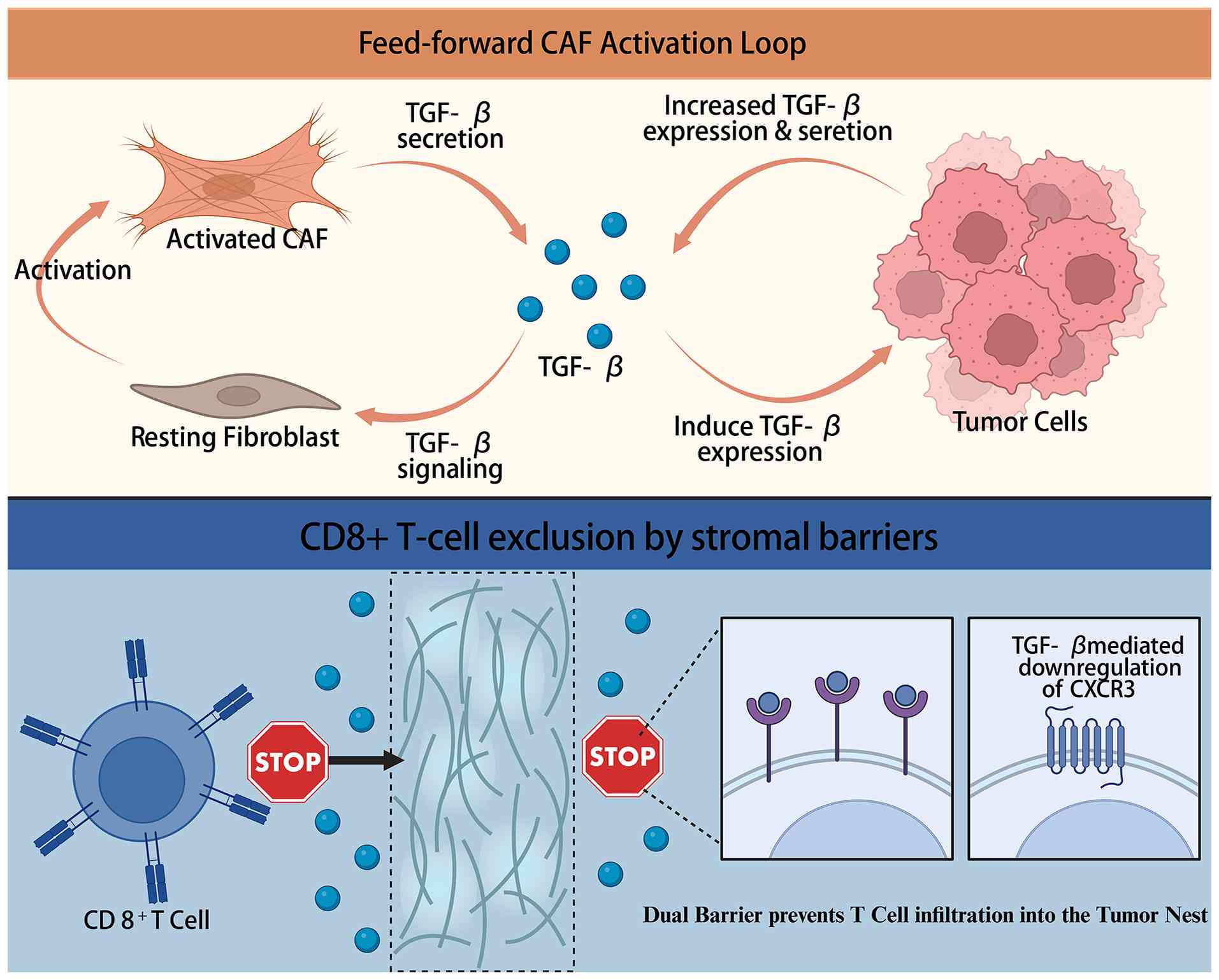
(17). Transforming growth factor-β
(TGF-β) signaling can promote fibroblast activation and ECM
remodeling, whereas experimental inhibition of TGF-β-associated
stromal programs can enhance the efficacy of PD-L1-directed therapy
in selected models (56). These
observations are consistent with a self-reinforcing relationship in
which TGF-β drives fibroblast activation, while activated
fibroblasts in turn maintain TGF-β-rich, matrix-dense
microenvironments.
The resulting stromal barrier may operate through
two complementary mechanisms. First, collagen-rich, remodeled ECM
can impose a physical constraint on CD8+ T-cell
penetration and migration from stromal regions into tumor-cell
nests (17,57). Second, TGF-β-associated signaling
can directly impair T-cell proliferation and effector function and
may disrupt migration-related programs, thereby diminishing T-cell
responsiveness to chemotactic cues. Together, these structural and
signaling barriers may confine immune cells to the tumor margin or
stromal compartments and prevent effective contact with malignant
cells (Fig. 5).
Abnormal vasculature and hypoxia may impose an
additional layer of spatial restriction. Dysregulation of
hypoxia-inducible factor-1α and vascular endothelial growth
factor-dependent pathways has been documented in NSCLC, including
treatment-resistant and epidermal growth factor receptor
(EGFR)-mutant disease (58). Poor
perfusion and regional hypoxia may coincide with extracellular
acidification, lactate accumulation and adenosine-generating
pathways involving tumor, stromal and immune-cell populations
(35,37,38).
Collectively, these conditions can intensify metabolic stress and
may further compromise immune-cell trafficking and sustained
effector function. However, direct evidence showing that
endothelial-cell mitochondrial dysfunction itself is sufficient to
drive immune exclusion in human NSCLC is currently unavailable.
Mitochondrial redox signaling may further interact
with these stromal and vascular processes. Chronic oxidative stress
can accompany hypoxia, impaired perfusion, lactate accumulation and
fibroblast activation, while mtROS exert strongly context- and cell
type-dependent effects (28). In
experimental systems, oxidative stress can promote stress-response
pathways and tissue-remodeling programs. In patient tumors,
however, oxidative stress, CAF accumulation, ECM remodeling,
hypoxia and vascular abnormalities frequently occur simultaneously,
making their temporal sequence and causal relationships difficult
to resolve.
Accordingly, mtROS and other mitochondrial metabolic
alterations should not be portrayed as universal proximal causes of
stromal immune exclusion. A more evidence-based interpretation is
that mitochondrial redox and metabolic states may reinforce or
sustain pre-existing barriers associated with CAFs, ECM remodeling,
hypoxia, or vascular dysfunction in selected tumors. At present,
the strongest evidence from human lung cancer supports a spatial
association between CAF/matrix programs and T-cell exclusion
(17), whereas the contribution of
cell type-specific mitochondrial dysfunction remains largely
mechanistic and hypothesis-generating.
Future investigations should integrate spatially
resolved assessments of CAF states, ECM architecture, vascular
organization, hypoxia, and immune-cell localization with cell
type-specific measurements of mitochondrial function and redox
status. Longitudinal and treatment-paired analyses will be
particularly important for determining whether mitochondrial
alterations precede stromal barrier formation, arise as secondary
adaptations, or predominantly contribute to maintenance of the
immune-excluded state.
TCA metabolites and epigenetic
regulation as hypothesis-generating links
Among the proposed mitochondrial mechanisms linking
tumor metabolism to spatial immune phenotypes, the relationship
among TCA cycle metabolites, epigenetic regulation and immune
architecture remains particularly speculative in human NSCLC.
Although the biochemical coupling between mitochondrial metabolism
and chromatin regulation is well established, direct evidence
linking defined metabolite alterations to specific spatial immune
states in patient tumors remains limited.
α-Ketoglutarate (α-KG) functions as an essential
cofactor for multiple α-KG-dependent dioxygenases, including
enzymes involved in DNA and histone demethylation and the
regulation of hypoxia-inducible factors (59). Reduced α-KG availability, or
relative accumulation of metabolites such as succinate and
fumarate, can inhibit these enzymes and thereby alter chromatin
organization and hypoxia-responsive signaling.
JmjC-domain-containing histone demethylases constitute one
important class of mediators through which mitochondrial metabolic
state may be coupled to transcriptional regulation (60–63).
In NSCLC, however, current evidence more strongly
supports broad alterations in metabolic flux, hypoxic signaling,
and epigenetic reprogramming than a recurrent, canonical
oncometabolite-driven phenotype. Patient-derived studies have shown
that glucose-derived carbon can make a substantial contribution to
the TCA cycle in aggressive NSCLC (22), while multimodal and multi-omics
analyses have revealed extensive epigenetic and transcriptional
reprogramming across lung tumors (62,63).
These observations support the biological plausibility of
metabolism-chromatin coupling but do not demonstrate that any
specific TCA metabolite directly determines an inflamed,
immune-desert, or immune-excluded phenotype.
A more cautious framework is that altered
TCA-associated metabolic states may reinforce pre-existing
transcriptional and microenvironmental programs rather than
initiate them. By modulating α-KG-dependent enzymes, chromatin
accessibility, and hypoxia-responsive pathways, changes in TCA
metabolites could promote the persistence of angiogenic,
glycolytic, inflammatory, or stromal-remodeling states. Epigenomic
state transitions have also been observed during lung
adenocarcinoma progression in experimental models (64), supporting the broader concept that
chromatin programs can stabilize tumor-cell phenotypes over time.
Whether TCA metabolite alterations function as upstream drivers,
downstream consequences, or parallel adaptations of these states,
however, remains unresolved.
The cellular compartment in which these effects
occur is another major source of uncertainty. Metabolite-dependent
epigenetic regulation may operate in tumor cells, fibroblasts,
endothelial cells, myeloid cells, or T cells, and the functional
consequences are unlikely to be equivalent across these
populations. Bulk-tissue measurements cannot reliably distinguish
whether a given metabolite or epigenetic signature originates from
malignant or non-malignant compartments. Associations between
TCA-related signatures and immune phenotypes should therefore not
be interpreted as evidence of a tumor-cell-intrinsic causal
mechanism in the absence of cell type-resolved validation.
At present, large-scale studies that simultaneously
quantify TCA metabolites, α-KG-dependent enzyme activity, chromatin
states, and the spatial distribution of immune cells within the
same NSCLC tissue regions are lacking (62–66).
Most available human evidence is cross-sectional and correlative,
with few longitudinal or interventional datasets demonstrating that
manipulation of a TCA metabolite-epigenetic axis is sufficient to
reprogram a defined spatial immune phenotype.
TCA metabolites are therefore best regarded as
potential stabilizers or modifiers of established immune and
microenvironmental states rather than confirmed initiating drivers
of cold-tumor formation in NSCLC. This framework provides a
biologically plausible and experimentally testable link between
mitochondrial metabolism and epigenetic persistence, but its
relevance to spatial immune organization will require direct
validation in cell type-resolved, spatially annotated human
specimens.
Intercellular mitochondrial transfer
as an emerging and context-dependent mechanism
Intercellular mitochondrial transfer has emerged as
a form of non-cell-autonomous metabolic communication within the
TME. Unlike suppression mediated by soluble metabolites, this
process entails the transfer of intact mitochondria, mitochondrial
components, or mtDNA-containing material between donor and
recipient cells. Its biological effects are neither inherently
beneficial nor detrimental but instead depend on the direction of
transfer, the identities of the donor and recipient cells, the
functional integrity of the transferred mitochondria, the route of
transfer, and the capacity of recipient cells to maintain
mitochondrial quality control.
The most direct evidence relevant to NSCLC comes
from Ikeda et al (67), who
identified shared mtDNA mutations in cancer cells and TILs from
clinical specimens and subsequently demonstrated the transfer of
cancer-cell-derived mitochondria to T cells in experimental
systems. Their findings implicated both direct transfer through
tunneling nanotubes (TNTs) and indirect transfer via small
extracellular vesicles (EVs). Disruption of direct cell contact,
TNT formation, or small-EV release reduced mitochondrial transfer,
suggesting that these routes may operate in parallel rather than as
functionally interchangeable pathways (67). However, pharmacological inhibition
of TNT- or EV-associated pathways can also perturb other cellular
processes, and EVs carry a broad range of additional
immunomodulatory cargo. Thus, route-inhibition experiments alone
cannot ascribe all downstream immune effects specifically to
mitochondrial transfer.
In the same study, the acquisition of mitochondria
harboring pathogenic mtDNA variants was associated with reduced
mitochondrial membrane potential (ΔΨm), altered ROS production,
metabolic dysregulation, senescence-associated phenotypes, impaired
effector activity, and defective memory formation in recipient T
cells (67). Over time, transferred
mutant mtDNA could shift from heteroplasmy toward homoplasmic
replacement. This progression appeared to reflect not only repeated
transfer but also differences in mitochondrial quality control:
Endogenous T-cell mitochondria remained susceptible to
stress-induced mitophagy, whereas cancer-cell-derived mitochondria
were comparatively resistant to clearance. Notably, the
mitophagy-inhibitory molecule implicated in this process was USP30
rather than ubiquitin-specific protease 27. USP30 was
co-transferred with cancer-cell mitochondria and antagonized
Parkin-dependent mitophagy, while pharmacological inhibition or
RNA-interference-mediated suppression of USP30 partially reduced
the persistence of transferred mitochondria and their progression
toward homoplasmy (67).
These findings provide mechanistic support for a
model in which tumor-to-T-cell mitochondrial transfer contributes
to impaired antitumor immunity. In melanoma and NSCLC cohorts,
tumor mtDNA mutations were also associated with poorer outcomes
after PD-1 blockade (66). However,
tumor mtDNA mutation status should not be interpreted as a specific
biomarker of mitochondrial transfer, because such mutations may
affect tumor biology through multiple independent mechanisms.
Furthermore, the prevalence, spatial distribution and quantitative
contribution of tumor-to-T-cell mitochondrial transfer across human
NSCLC remain incompletely defined. Current clinical evidence
therefore supports an association and a biologically plausible
mechanism but does not establish mitochondrial transfer as a
universal cause of T-cell dysfunction or resistance to immune
checkpoint inhibition.
Mitochondrial exchange between cancer and immune
cells may also occur in the reverse direction. A single-cell
genomic study reported predominantly T-cell-to-cancer-cell
mitochondrial transfer and inferred donor-recipient relationships
using mitochondrial single-nucleotide variants, raising the
possibility that cancer cells can acquire mitochondrial material
from neighboring T cells to enhance their metabolic fitness
(67). Because genomic inference
does not directly visualize the movement of intact organelles in
each transfer event, these observations should be distinguished
from transfer demonstrated through imaging and functional
perturbation. Nevertheless, when considered alongside the
tumor-to-T-cell transfer described by Ikeda et al (67), these findings suggest that
mitochondrial exchange within the TME can be bidirectional and
strongly context dependent.
The functional competence of the transferred
mitochondria is also a key determinant of biological outcome.
Baldwin et al (68) showed
that nanotube-mediated transfer of metabolically competent
mitochondria from bone-marrow stromal cells to CD8+ T
cells increased mitochondrial respiration and spare respiratory
capacity and promoted T-cell expansion, tumor infiltration,
resistance to exhaustion, and antitumor activity in experimental
models. By contrast, transfer of dysfunctional mitochondria failed
to confer comparable benefits (68). These observations indicate that
mitochondrial transfer can either compromise or enhance T-cell
function depending on the cellular source and bioenergetic quality
of the transferred organelles. The biological significance of
mitochondrial transfer therefore cannot be inferred solely from its
occurrence.
Several methodological limitations should also be
considered when interpreting evidence of intercellular
mitochondrial transfer. Shared mtDNA variants can facilitate
donor-recipient lineage tracing but may be confounded by cellular
admixture, sequencing depth, clonal expansion, or technical
contamination. Fluorescent mitochondrial labels can identify
transferred mitochondrial material without necessarily establishing
the movement of intact, functionally competent organelles.
Likewise, TNT and EV-release inhibitors lack complete pathway
specificity, and distinct EV populations may transport
mitochondrial proteins, mtDNA fragments, partial organelles, or
other bioactive cargo. Future studies should therefore integrate
mtDNA lineage tracing, live-cell imaging, ultrastructural
confirmation, functional assessment of mitochondrial activity, and
route-specific genetic perturbation rather than relying on any
single detection strategy.
Overall, intercellular mitochondrial transfer is
best regarded as an emerging, context-dependent mechanism capable
of modifying T-cell mitochondrial fitness and antitumor immunity in
selected tumors. Current evidence is strongest for the experimental
feasibility of transfer and its functional consequences, whereas
spatially resolved data defining its prevalence and causal
contribution in human NSCLC remain limited. Therapeutic strategies
aimed at disrupting TNT- or EV-mediated transfer, overcoming
USP30-dependent resistance to mitophagy, or supplying metabolically
competent mitochondria to therapeutic T cells are conceptually
promising but remain preclinical. These approaches should therefore
be considered separately from established ICI therapies in the
translational discussion (Table
SII).
Mitochondrial regulation across spatial
immune phenotypes in NSCLC
The forementioned mitochondrial processes are
unlikely to function independently. Within individual NSCLC
lesions, tumor-cell metabolism, immune-cell mitochondrial fitness,
innate immune sensing, stromal architecture, vascular function and
intercellular mitochondrial communication may interact to influence
both the spatial distribution and functional state of antitumor
immune cells. The relative importance of these processes is likely
to differ not only between patients but also among spatially
distinct regions within the same tumor.
Within this framework, mitochondrial alterations can
be considered in relation to the three commonly recognized spatial
immune phenotypes: Immune-desert, immune-excluded and inflamed
tumors. These phenotypes are not necessarily discrete or mutually
exclusive, and transitions between them may occur during tumor
progression or in response to therapy. The following sections
therefore examine how mitochondrial and microenvironmental
mechanisms may contribute to each spatial phenotype without
assuming a fixed one-to-one correspondence between any single
biological process and a particular immune state.
Immune-desert phenotype
The immune-desert phenotype is characterized by
sparse immune-cell infiltration within both tumor nests and the
surrounding stromal compartment. Biologically, this pattern is more
consistent with inadequate initiation of the cancer-immunity cycle
than with a simple inability of already activated T cells to enter
the tumor. Deficient antigen presentation, attenuated type I IFN
and chemokine signaling, and limited DC abundance or functional
maturation may collectively impair T-cell priming and subsequent
recruitment (14,16,41).
The functional state of intratumoral DCs is
particularly relevant to the efficacy of immune checkpoint
blockade. PD-L1 blockade can enhance DC-mediated co-stimulation by
releasing CD80 from PD-L1-dependent sequestration, thereby
promoting CD28 signaling and T-cell priming. In patients with NSCLC
or renal cell carcinoma (RCC) treated with atezolizumab, a
DC-related gene signature was associated with improved overall
survival (41). These observations
suggest that PD-(L)1 blockade is more likely to be effective when a
functional antigen-presenting and costimulatory circuit is already
present, at least to some extent. Conversely, checkpoint inhibition
alone may have limited efficacy when tumors contain few functional
DCs and lack an established pool of tumor-reactive T cells.
Mitochondria-related innate sensing may intersect
with this priming defect through the cGAS-STING pathway. Cytosolic
DNA sensing in tumor or myeloid cells can induce type I IFNs and
chemokines, thereby promoting DC activation and immune-cell
recruitment. Preclinical NSCLC studies have shown that
pharmacological or treatment-induced activation of STING-associated
signaling can increase the expression of chemokines such as CXCL10
and CCL5 and enhance immune-cell infiltration (51,52).
Conversely, reduced STING expression has been associated with
adverse clinical features in NSCLC (55). However, as aforementioned, cytosolic
DNA can arise from mitochondrial damage, nuclear DNA damage,
chromosomal instability, or replication stress. Attenuated STING
signaling should therefore not be attributed automatically to
insufficient mtDNA release.
Tumor genotype provides one example of how this
pathway can be disrupted. In KRAS-driven lung cancer models, loss
of STK11/LKB1 induced epigenetic silencing of STING and impaired
responses to cytosolic double-stranded DNA. Restoration of STING
reactivated downstream TBK1-IRF3 signaling in these models
(69). Subsequent clinical and
translational studies have linked STK11/LKB1 deficiency to impaired
STING, type I IFN and CD8+ T-cell programs, as well as
reduced benefit from immunotherapy (54,69,70).
Nevertheless, this represents a genotype-specific mechanism of
immune attenuation rather than a universal explanation for the
immune-desert phenotype in NSCLC.
The contribution of mtROS is less clearly defined.
Moderate redox signaling can participate in inflammatory and innate
immune responses, whereas excessive antioxidant buffering or
altered mitochondrial signaling could, in principle, attenuate
these pathways. However, direct spatial evidence demonstrating that
reduced mtROS in tumor or myeloid cells causes an immune-desert
phenotype in human NSCLC is lacking. The proposed relationship
between diminished mitochondrial danger signaling and defective
immune initiation should therefore remain hypothesis-generating
rather than being framed as a defined mtROS threshold.
Taken together, the immune-desert phenotype likely
results from multiple convergent deficiencies, including limited
tumor-antigen recognition, inadequate DC abundance or maturation,
attenuated IFN and chemokine signaling, tumor-intrinsic suppression
of innate immune sensing, and insufficient T-cell recruitment.
Impairment of the tumor-myeloid cGAS-STING axis may contribute to
selected tumors, but current evidence does not establish mtDNA
release or mitochondrial dysfunction as the dominant initiating
cause of this spatial phenotype.
Immune-excluded phenotype
In immune-excluded tumors, lymphocytes are present
at the invasive margin or within stromal compartments but exhibit
limited penetration into malignant cell nests. This spatial pattern
indicates that immune-cell recruitment has occurred to some extent,
whereas subsequent trafficking through the tumor stroma and
productive interactions with cancer cells remain restricted
(14,17). Compared with immune-desert tumors,
the dominant constraint is therefore less likely to reflect a
complete failure of immune priming and more likely to arise from
the organization and functional properties of the stromal and
vascular interface.
Among the mechanisms associated with immune
exclusion, the strongest spatial evidence in human lung tumors
relates to CAF organization and ECM programs. Integrated
single-cell and multiplex imaging analyses have identified distinct
CAF populations positioned around or within tumor structures and
associated with dense, aligned ECM deposition and peripheral
localization of T cells (17).
These findings indicate that the effects of CAFs cannot be inferred
from their overall abundance alone; their molecular states, spatial
distribution, and relationship to matrix architecture are also
important determinants of immune organization. The primary human
lung tumor study supporting this association is included as
reference (17).
TGF-β signaling may reinforce this spatial barrier
by promoting fibroblast activation and matrix remodeling while
simultaneously suppressing antitumor immune responses. Activated
CAFs can sustain a self-reinforcing stromal state through the
production of TGF-β, ECM components and additional regulatory
mediators (55,56). The resulting barrier has both
structural and functional dimensions: Dense or highly aligned
matrix can restrict T-cell migration, whereas TGF-β-rich conditions
can suppress T-cell proliferation and effector activity and reduce
responsiveness to local chemotactic signals. Together, these
processes provide a plausible basis for the accumulation of
CD8+ T cells outside, rather than within, tumor-cell
nests.
Abnormal vasculature and regional hypoxia may
further strengthen this excluded state. Disorganized or
dysfunctional tumor vessels can impair perfusion, promote hypoxia
and acidosis, and disrupt the conditions required for leukocyte
adhesion, extravasation and tissue entry. HIF-1α- and
VEGF-associated vascular programs have been described in NSCLC,
including treatment-resistant and EGFR-mutant contexts (58). A previous analysis of two human
NSCLC cohorts further linked vascular leakage to spatially distinct
stromal niches, immune evasion and poor responses to immunotherapy
(71). These findings support the
clinical relevance of vascular abnormalities in immune exclusion,
although they do not establish endothelial mitochondrial
dysfunction as the direct causal mechanism.
Metabolic conditions within poorly perfused stromal
regions may impose an additional functional barrier. Hypoxia,
lactate accumulation, extracellular acidification and CD73-mediated
adenosine signaling can impair T-cell migration and the maintenance
of effector function (35,37,38).
These metabolic pressures may therefore reduce the likelihood that
T cells retained near the tumor boundary remain sufficiently motile
and functionally competent to traverse the stromal compartment.
Nevertheless, metabolic dysfunction alone does not account for the
characteristic spatial geometry of immune exclusion, which is more
directly supported by CAF organization, matrix architecture and
vascular abnormalities.
Mitochondrial redox signaling should likewise be
interpreted within this broader microenvironmental context. Chronic
oxidative stress may coexist with hypoxia, lactate accumulation,
fibroblast activation, and vascular dysfunction and may reinforce
tissue-remodeling or immunosuppressive programs in selected tumors
(28,58). However, these processes are highly
interdependent in patient tissues. Current evidence is insufficient
to determine whether altered mtROS precedes CAF activation and
matrix remodeling, arises secondarily from hypoxia and impaired
perfusion, or primarily contributes to maintaining an already
established stromal barrier.
The immune-excluded phenotype is therefore best
understood as the product of interacting stromal, vascular,
metabolic and immune constraints. Evidence from human lung tumors
most directly supports the spatial association between specific
CAF/matrix programs and T-cell marginalization (17), whereas mitochondrial and redox
processes are more appropriately regarded as potential modifiers or
amplifiers of this architecture. Defining their causal contribution
will require spatially matched assessments of CAF identity, ECM
organization, vascular function, tissue oxygenation, mitochondrial
activity and T-cell localization before and after therapeutic
intervention.
Inflamed phenotype
The inflamed phenotype is characterized by the
accumulation of immune cells, particularly CD8+ T cells,
within or in close proximity to tumor-cell nests. This spatial
pattern is frequently accompanied by active antigen-presentation
machinery, IFN-associated signaling, and chemokine programs that
promote immune-cell recruitment and retention (14,16,19,40,72).
Compared with immune-desert and immune-excluded tumors, inflamed
tumors are generally more likely to harbor a pre-existing antitumor
immune response that can be further enhanced by immune checkpoint
inhibition. Nevertheless, the presence of intratumoral T cells does
not necessarily indicate that these cells remain metabolically
competent or capable of sustaining effective tumor control.
Maintenance of an inflamed state requires
coordinated and persistent interactions among tumor-antigen
recognition, DC activation, T-cell co-stimulation, local chemokine
production and effector-cell persistence. In human NSCLC, increased
tumor-cell calreticulin (CRT) expression has been associated with
greater infiltration by mature DCs and effector-memory T cells, as
well as with favorable clinical outcomes (73). These findings support the importance
of immunogenic tumor-cell signals and effective antigen
presentation in establishing a T-cell-rich microenvironment.
However, CRT expression should be regarded as an associated feature
rather than a sufficient marker defining the inflamed
phenotype.
DCs may also determine whether a pre-existing immune
response can be effectively amplified by PD-L1 blockade. PD-L1
expressed on DCs can sequester CD80 through cis interactions,
thereby reducing the availability of CD80 for CD28-mediated T-cell
co-stimulation. Blocking PD-L1 can relieve this constraint and
enhance DC-dependent T-cell priming (41). Thus, the therapeutic responsiveness
of an inflamed tumor depends not only on the abundance of
infiltrating T cells but also on the preservation of effective
antigen presentation and costimulatory signaling. Mechanistically,
the relevant interaction is CD80-CD28 rather than CD75-CD25.
Mitochondrial fitness constitutes an additional
functional dimension of the inflamed phenotype. Activated
CD8+ T cells require coordinated OXPHOS, mitochondrial
quality control, redox homeostasis, and metabolic flexibility to
sustain cytokine production, cytotoxicity, and persistence under
tumor-associated stress (32).
However, these properties cannot be assumed to be uniformly
preserved among T cells within an inflamed tumor. Spatial
infiltration and mitochondrial competence are related but distinct
biological features, and substantial metabolic heterogeneity may
exist even among neighboring T-cell populations.
A lung cancer study identified CD38hi and
CD38int CD8+ T-cell populations with distinct
mitochondrial and functional characteristics (5). CD38hi CD8+ T
cells exhibited higher expression of exhaustion-associated markers
together with dysregulated mitochondrial bioenergetics. Their
abundance also differed between peripheral and central tumor
regions, and their association with response to anti-PD-1 therapy
varied according to spatial location. These observations indicate
that, even in T-cell-infiltrated lung tumors, mitochondrial status
cannot be inferred solely from T-cell density or expression of a
single surface marker. Instead, spatial context should be
integrated with direct functional assessment of mitochondria when
interpreting the biological significance of individual T-cell
subsets.
A recent clinical study involving patients with
NSCLC and clear cell RCC further illustrates the complexity of
mitochondrial functional readouts. Tumor-associated CD8+
T-cell clones with high ΔΨm exhibited increased OXPHOS-related
signatures but simultaneously expressed stress- and
exhaustion-associated programs. Moreover, the corresponding
TMRE-high signature was negatively associated with clinical benefit
from ICIs (74). Although the study
was not confined to NSCLC, these findings caution against
interpreting elevated ΔΨm or increased oxidative activity as a
direct surrogate for superior T-cell fitness.
T cells within inflamed tumors also remain subject
to substrate limitation, hypoxia, lactate accumulation,
extracellular acidification and adenosine-mediated suppression
(33,35–39).
These metabolic constraints can reduce spare respiratory capacity,
disrupt mitochondrial redox homeostasis, and compromise sustained
effector function without necessarily preventing T-cell
infiltration. Consequently, some inflamed tumors may retain
abundant intratumoral lymphocytes while exhibiting impaired
cytokine production, defective memory formation, or progressive
T-cell exhaustion. This distinction provides a biological basis for
the clinical observation that an inflamed spatial phenotype does
not invariably translate into durable benefit from immune
checkpoint inhibition.
Intercellular mitochondrial transfer may provide an
additional mechanism through which an initially inflamed
microenvironment becomes functionally compromised. Tumor-derived
mitochondria harboring pathogenic mtDNA variants can be transferred
to TILs, leading to metabolic dysfunction, senescence-associated
phenotypes, and impaired effector and memory functions (66,67).
In melanoma and NSCLC cohorts, tumor mtDNA mutations were also
associated with poorer outcomes following immune checkpoint
inhibition (66). However, tumor
mtDNA mutation status is not a specific surrogate for mitochondrial
transfer, and the prevalence, spatial distribution and quantitative
contribution of this process in human NSCLC remain incompletely
characterized. Mitochondrial transfer should therefore be
considered an emerging mechanism that may contribute to dysfunction
in selected T-cell-infiltrated tumors rather than a universal
feature of the inflamed phenotype.
Overall, the inflamed phenotype is best
conceptualized as a spatial state in which immune-cell recruitment
and tumor entry have occurred, but the functional quality of the
infiltrating immune compartment remains heterogeneous. Preserved DC
activity, antigen presentation, costimulatory signaling, and T-cell
mitochondrial competence may sustain effective antitumor immunity,
whereas metabolic stress, progressive exhaustion, and the
acquisition of dysfunctional mitochondria may undermine an
otherwise established immune response. Mitochondrial processes are
therefore more likely to regulate the persistence and functional
efficacy of the inflamed state than to determine T-cell
infiltration per se.
Translational opportunities and
limitations
Phenotype-matched therapeutic
strategies
The therapeutic relevance of spatial immune
phenotypes lies in their potential to identify the stage of the
antitumor immune response that is predominantly constrained. Within
this framework, immune-desert tumors are primarily characterized by
inadequate immune initiation and recruitment, immune-excluded
tumors by restricted access of immune cells to malignant cell
nests, and inflamed tumors by variable maintenance and functional
competence of an already established immune infiltrate. Therapeutic
priorities may therefore differ across these spatial states.
However, spatial immune phenotypes have not yet been validated as
stand-alone biomarkers for treatment selection in NSCLC. They
should instead be considered alongside tumor stage, histological
and molecular features, PD-L1 expression, genomic alterations,
prior therapies, and the patient's overall clinical condition
rather than applied as a rigid treatment algorithm (Fig. 6).
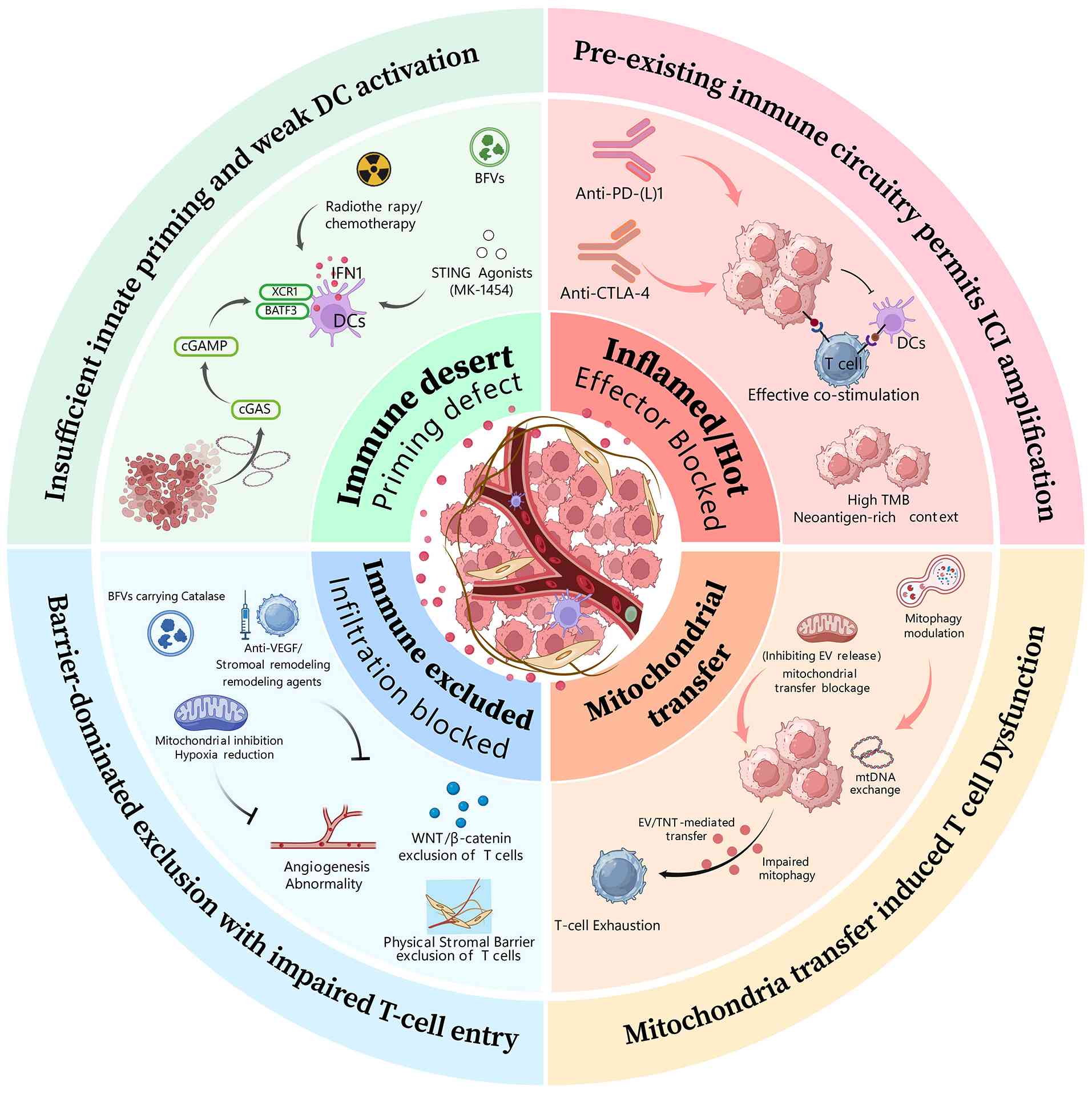 | Figure 6.Conceptual framework linking spatial
immune phenotypes to potential mitochondria-related intervention
directions in NSCLC. This schematic summarizes potential
therapeutic strategies associated with distinct spatial immune
phenotypes in NSCLC. In immune-desert tumors, impaired innate
immune priming and insufficient DC activation may contribute to
limited T-cell recruitment. Therefore, approaches targeting innate
immune sensing pathways, including cGAS-STING activation and
treatment-induced immune stimulation, represent potential
strategies to enhance immune initiation. In immune-excluded tumors,
stromal barriers, abnormal angiogenesis, hypoxia-associated
metabolic alterations, and impaired immune-cell infiltration may
restrict T-cell access to tumor nests. Strategies targeting stromal
remodeling, vascular abnormalities, or metabolic constraints have
therefore been proposed as potential approaches to improve
immune-cell penetration, although most remain at preclinical or
early translational stages. In inflamed tumors with pre-existing
immune activity, immune checkpoint blockade may further enhance
antitumor responses by reinforcing existing immune effector
functions. However, the clinical benefit of ICIs depends on
multiple tumor-intrinsic and microenvironmental factors rather than
spatial phenotype alone. The mitochondrial transfer-related module
represents an emerging and exploratory mechanism that may
contribute to immune dysfunction through mitochondrial exchange
between tumor and immune cells, including extracellular vesicle- or
tunneling nanotube-mediated processes. These mechanisms remain
hypothesis-generating, and their therapeutic implications require
further experimental and clinical validation. Overall, this figure
provides a conceptual overview of potential mitochondria-related
intervention directions across spatial immune contexts rather than
a validated phenotype-guided therapeutic algorithm. The maturity of
these approaches varies substantially, ranging from clinically
established immunotherapy to preclinical and emerging
mitochondrial-targeted strategies. NSCLC, non-small cell lung
cancer; DC, dendritic cells; ICI, immune checkpoint inhibitor; DCs,
dendritic cells; mtDNA, mitochondrial DNA; PD-(L)1, programmed cell
death protein 1; CTLA-4, cytotoxic t-lymphocyte associated protein
4; TMB, tumor mutational burden; EV, endothelial vesicle. |
Immune-desert tumors: Restoring immune
priming and recruitment
In immune-desert tumors, the immediate therapeutic
objective is to initiate an effective antitumor immune response
rather than merely release inhibitory constraints on a response
that is largely absent. PD-(L)1 blockade can amplify pre-existing
T-cell activity, but its efficacy may remain limited when DC
abundance or maturation, antigen presentation, costimulatory
signaling, IFN responses, and chemokine production are inadequate
(41). Therapeutic strategies for
this phenotype should therefore focus on restoring antigen
availability, innate immune activation, DC function, and subsequent
T-cell recruitment before or in conjunction with checkpoint
inhibition.
Radiotherapy and selected cytotoxic agents may
facilitate immune initiation by inducing tumor-cell damage,
increasing antigen release, and generating cytosolic DNA and other
damage-associated signals. These effects can activate
cGAS-STING-related pathways and enhance type I IFN and chemokine
production, although the magnitude of the response depends on
radiation dose and fractionation, tumor genotype, DNA-degradation
pathways, and the functional competence of cGAS-STING signaling in
both tumor and myeloid cells (48–50).
Clinical experience with chemoradiotherapy followed by checkpoint
inhibition demonstrates that immune activation can be successfully
integrated with checkpoint blockade in specific NSCLC treatment
settings. However, these data do not establish radiotherapy as a
universal strategy for converting all immune-desert tumors into
immune-responsive states.
More direct restoration of innate immune sensing
represents another potential strategy. In preclinical NSCLC models,
RocA-induced mitochondrial damage and activation of cGAS-STING
signaling increased CXCL10 and CCL5 expression and promoted
immune-cell recruitment (52).
Treatment-induced STING activation in trans has also contributed to
immune-dependent antitumor effects in lung cancer models (51). These findings, however, remain
predominantly preclinical, and RocA should not be regarded as a
clinically validated immune-sensitizing agent for NSCLC.
Early clinical studies of intratumoral STING
agonists have demonstrated the feasibility of pharmacological STING
activation. Ulevostinag, administered either alone or in
combination with pembrolizumab in patients with advanced solid
tumors, induced pharmacodynamic changes consistent with STING
pathway engagement and showed a manageable safety profile. However,
the expansion cohorts were not designed to establish efficacy
specifically in immune-desert NSCLC, and the clinical benefit of
STING agonism remains uncertain (75). STING agonists and related approaches
should therefore be regarded as investigational strategies aimed at
initiating or strengthening immune priming, with subsequent
checkpoint blockade potentially amplifying the resulting antitumor
response rather than independently mediating immune conversion.
DC-directed interventions may provide an additional
means of restoring immune priming. Potential approaches include
expanding or recruiting cross-presenting DC populations, DC
vaccination, inducing immunogenic tumor-cell death, and enhancing
antigen presentation (41,76). These strategies, however, differ
substantially in their degree of clinical development. Moreover,
the presence of a DC-related signature does not itself establish
that a patient will benefit from a specific DC-targeted
intervention. Biomarker development should therefore distinguish DC
abundance from functional maturation, spatial proximity to tumor
cells and T cells, antigen-presenting capacity, and effective
CD80-CD28 costimulatory signaling.
Immune-excluded tumors: Relieving
stromal and vascular barriers
In immune-excluded tumors, immune priming and
recruitment have occurred to some extent, but T cells remain
concentrated at the invasive margin or within stromal compartments
and fail to establish sufficient contact with malignant cells. The
therapeutic priority is therefore to improve intratumoral access
rather than simply enhance peripheral T-cell activation. Strategies
targeting vascular abnormalities, hypoxia, CAF-associated programs,
TGF-β signaling, and ECM organization may increase the likelihood
that recruited immune cells can penetrate tumor nests and maintain
their functional capacity after entry (17,55–58).
Vascular normalization is particularly relevant
because abnormal angiogenesis and impaired perfusion can
simultaneously restrict immune-cell trafficking, exacerbate
hypoxia, and intensify metabolic and immunosuppressive stress. The
clinical efficacy of atezolizumab combined with bevacizumab and
chemotherapy in metastatic non-squamous NSCLC provides a precedent
for integrating VEGF inhibition with immune checkpoint blockade and
cytotoxic therapy (77). However,
patients in this trial were not selected on the basis of a
spatially defined immune-excluded phenotype. These findings
therefore demonstrate the clinical feasibility of combining
antiangiogenic therapy with immunotherapy but do not validate
vascular normalization as a phenotype-specific treatment for immune
exclusion.
CAF- and matrix-directed interventions may
complement vascular strategies. TGF-β-associated fibroblast
activation and ECM remodeling can impose both structural and
signaling barriers to T-cell entry (17,55–56).
Potential approaches include modulation of TGF-β-related pathways,
specific CAF states, collagen organization, matrix crosslinking,
lysyl oxidase-associated processes, and focal adhesion signaling
(56,57). Nevertheless, CAF populations are
highly heterogeneous and can exert context-dependent
tumor-promoting, immunoregulatory, or tissue-supportive functions.
Consequently, indiscriminate fibroblast depletion or non-specific
disruption of the ECM may produce unintended biological
effects.
Clinical experience further indicates that
biologically plausible stromal targeting does not necessarily
translate into improved clinical outcomes. In a randomized phase
III trial involving treatment-naive patients with PD-L1-high
advanced NSCLC, the bifunctional TGF-β trap/PD-L1 inhibitor
bintrafusp alfa failed to demonstrate superior efficacy over
pembrolizumab (78). This finding
does not exclude a relevant role for TGF-β in selected
immune-excluded tumors; rather, it indicates that simultaneous
systemic inhibition of TGF-β and PD-L1 is insufficient in the
absence of appropriate biological and spatial patient selection.
Future trials should therefore evaluate stromal interventions in
tumors with directly documented TGF-β-, CAF-, or ECM-associated
immune exclusion rather than assuming that all NSCLC tumors share a
common stromal barrier.
Modulation of tumor respiration and hypoxia may
provide another means of improving the immune-excluded
microenvironment. In a window-of-opportunity study involving
patients with NSCLC, atovaquone reduced hypoxic tumor volume as
measured by hypoxia positron-emission tomography and altered
transcriptional programs associated with hypoxia and metabolism
(79). These findings provide
evidence of a biological effect on tumor oxygenation in patients
but do not demonstrate improved responsiveness to ICIs. Atovaquone
should therefore be regarded as a clinical proof of principle for
hypoxia modulation rather than as an established immune-sensitizing
therapy.
Overall, effective treatment of immune-excluded
tumors may require coordinated alleviation of vascular, stromal and
metabolic barriers. The optimal intervention is unlikely to be
identical across tumors dominated by abnormal angiogenesis,
TGF-β-rich fibroblast programs, dense collagen architecture, or
severe hypoxia. Spatial profiling may therefore be particularly
useful for identifying the predominant barrier requiring
intervention and determining whether its modification is followed
by increased intratumoral T-cell infiltration.
Inflamed tumors: Amplifying immunity
while preserving T-cell function
In inflamed tumors, immune cells have already
penetrated tumor nests, making checkpoint inhibition a more direct
therapeutic strategy than in immune-desert or immune-excluded
states. Nevertheless, an inflamed spatial phenotype does not
guarantee durable tumor control. Infiltrating T cells may remain
constrained by persistent antigen stimulation, inhibitory receptor
signaling, metabolic stress, adenosine accumulation, mitochondrial
dysfunction, or inadequate antigen presentation and costimulatory
signaling (32,36–41).
PD-(L)1 blockade remains the principal strategy for
amplifying a pre-existing antitumor immune response, whereas
cytotoxic t-lymphocyte associated protein 4 (CTLA-4)-containing
regimens may enhance T-cell priming and clonal expansion in
selected clinical or molecular contexts. For example, CTLA-4
blockade has shown the potential to overcome KEAP1/STK11-associated
resistance to PD-(L)1 inhibition (80). However, KEAP1 or STK11 status should
not be considered equivalent to an inflamed, immune-excluded, or
immune-desert spatial phenotype. Molecular genotype, spatial
architecture, and immune function should instead be evaluated as
complementary dimensions of tumor biology (54,69,70,80).
Metabolic interventions may be considered when
immune infiltration is present, but T-cell function remains
compromised. The CD73-adenosine-A2A receptor axis can impair T-cell
metabolic fitness, migration and effector activity (36–39),
providing a rationale for combining adenosine-pathway inhibition
with checkpoint blockade. However, such strategies remain
investigational and will require biomarkers demonstrating that
adenosine signaling constitutes an active suppressive mechanism
within the tumor being treated.
Similarly, preserving or restoring T-cell
mitochondrial fitness represents a potential complementary
therapeutic objective rather than an established treatment
category. Strategies that enhance mitochondrial quality control,
substrate utilization, redox homeostasis, or the ex vivo
metabolic fitness of therapeutic T cells may help sustain effector
function (32,81). Conversely, tumor-to-T-cell transfer
of dysfunctional mitochondria may contribute to impaired antitumor
immunity and treatment resistance in selected tumors (66). Interventions targeting mitochondrial
transfer and mitochondrial quality control are discussed separately
below because they remain predominantly preclinical.
Taken together, phenotype-matched therapy should
target the predominant biological bottleneck: Restoring immune
priming in immune-desert tumors, facilitating tissue entry in
immune-excluded tumors, and sustaining effective antitumor function
in inflamed tumors. Because these spatial states may coexist within
the same lesion and evolve during treatment, therapeutic selection
should ideally be guided by spatially resolved baseline assessment
and, where feasible, longitudinal reassessment.
Emerging mitochondria-directed
interventions and modular combination strategies
Given the multifaceted roles of mitochondria in
tumor metabolism, immune activation and intercellular
communication, mitochondria-related processes have emerged as
potential therapeutic targets for reshaping spatial immune
phenotypes in NSCLC. However, the strength of evidence supporting
mitochondria-directed interventions varies substantially across
strategies, ranging from early clinical observations of metabolic
modulation to predominantly preclinical evidence for mitochondrial
regulation of antitumor immunity. Rather than serving as
stand-alone therapeutic targets, mitochondrial interventions are
more likely to function as complementary modules integrated with
established immunotherapeutic strategies. Their therapeutic value
may depend on the specific mitochondrial process targeted, the
cellular compartment affected, and the predominant spatial immune
barrier within the tumor (Table
SIII).
Modulation of tumor mitochondrial
metabolism and hypoxia
Tumor mitochondrial metabolism contributes not only
to cancer-cell survival but also to the formation of metabolically
restrictive immune niches through oxygen consumption, nutrient
utilization and the accumulation of immunosuppressive metabolites
(18,20). Modulating mitochondrial respiration
may therefore provide a means of alleviating metabolic constraints
within the TME.
Atovaquone provides an example of clinically
investigated modulation of mitochondrial metabolism in NSCLC. By
inhibiting mitochondrial complex III, atovaquone reduces cellular
oxygen consumption and intratumoral hypoxia. A
window-of-opportunity study in patients with NSCLC demonstrated
that short-term atovaquone treatment reduced hypoxic tumor volume
and altered hypoxia-associated transcriptional programs (79). These findings provide human evidence
that modulation of mitochondrial respiration can alter the tumor
metabolic state; however, they do not establish whether these
changes are sufficient to enhance immune-cell infiltration or
improve the efficacy of ICIs.
Mitochondrial metabolic intervention should
therefore currently be regarded as a strategy for modifying tumor
metabolic constraints rather than as an established approach for
sensitizing tumors to immunotherapy.
Exploiting mitochondrial danger
signaling pathways
Mitochondrial damage can generate immunogenic
signals through the release of mtDNA, activation of cGAS-STING
signaling, and induction of type I IFN-associated immune responses
(50–52). On this basis, pharmacological
induction of mitochondrial stress has been proposed as a strategy
for enhancing innate immune activation and promoting immune-cell
recruitment.
In lung cancer models, RocA-induced mitochondrial
dysfunction activated cGAS-STING signaling, increased CXCL10 and
CCL5 expression, and promoted immune-cell infiltration (52). Similarly, STING activation in trans
has been shown to enhance immune-dependent tumor control through
activation of innate immune responses (51).
These findings provide mechanistic and preclinical
support for targeting mitochondria-associated danger-sensing
pathways. However, STING responses are strongly influenced by tumor
genotype, pathway competence, immune-cell composition and treatment
context. Mitochondria-associated STING activation should therefore
currently be considered an investigational immunomodulatory
strategy rather than a clinically validated approach for converting
immunologically cold tumors into inflamed tumors.
Targeting pathogenic intercellular
mitochondrial transfer
Beyond intracellular mitochondrial regulation,
emerging evidence indicates that mitochondria can function as
transferable biological units mediating communication between tumor
and immune cells.
Tumor-derived dysfunctional mitochondria harboring
pathogenic mtDNA alterations can be transferred to T cells through
mechanisms such as tunneling nanotubes (TNTs) and EVs, resulting in
mitochondrial dysfunction, impaired metabolic adaptation, and
reduced immune-cell function (66,67).
From a therapeutic perspective, disrupting
pathogenic mitochondrial transfer or restoring mitochondrial
quality control represents a potential intervention strategy.
Candidate approaches include inhibition of TNT formation,
modulation of EV-mediated mitochondrial trafficking, and
manipulation of mitophagy-associated pathways.
Among these mechanisms, mitochondrial
quality-control regulators such as USP30 may provide potential
therapeutic targets by influencing the clearance of dysfunctional
mitochondria. However, these strategies remain largely confined to
experimental models, and selectively disrupting pathological
mitochondrial transfer without interfering with physiological
intercellular mitochondrial communication remains a major
challenge.
Enhancing T-cell mitochondrial
fitness
Because mitochondrial function is closely linked to
T-cell persistence, memory formation and sustained effector
activity, restoring T-cell mitochondrial fitness represents another
potential therapeutic strategy (32).
Experimental studies have investigated approaches
such as metabolic optimization of adoptive cell therapies and the
delivery of functional mitochondria to immune cells. Transfer of
metabolically competent mitochondria has been shown to enhance
T-cell respiratory capacity, persistence and functional activity in
experimental systems (81).
Although these findings provide a biological
rationale for improving immune-cell metabolic fitness, clinical
translation remains constrained by challenges related to delivery
efficiency, manufacturing feasibility, long-term safety, and
identification of patients most likely to benefit.
Modular combination therapy guided by
spatial immune barriers
Given the complex and context-dependent roles of
mitochondria in both tumor and immune cells, mitochondria-directed
interventions are unlikely to replace established immunotherapies.
Instead, they may be incorporated as modular components of
phenotype-guided combination strategies.
Conceptually, distinct mitochondrial interventions
may address different spatial immune barriers. In immune-desert
tumors, strategies that enhance mitochondrial danger signaling may
promote innate immune activation and immune-cell recruitment. In
immune-excluded tumors, metabolic and hypoxia-modulating approaches
may help alleviate environmental constraints that limit immune-cell
infiltration. In inflamed tumors, interventions designed to
preserve T-cell mitochondrial fitness may enhance immune-cell
persistence and sustain antitumor activity.
Several emerging platforms, including
multifunctional bioengineered vesicles, have been developed to
simultaneously modulate immune activation, hypoxia and tumor-cell
susceptibility (7). However, these
approaches remain experimental, and their clinical translation will
require spatially resolved biomarkers capable of identifying the
predominant mitochondrial abnormality and guiding selection of the
most appropriate intervention modules.
Clinical translation, evidence gaps
and future experimental priorities
Although mitochondria-related mechanisms provide a
potentially useful framework for understanding spatial immune
heterogeneity and resistance to immunotherapy in NSCLC, several
challenges must be addressed before these concepts can be
translated into clinically actionable strategies. Importantly,
current evidence linking mitochondrial alterations to spatial
immune phenotypes is derived largely from mechanistic studies,
experimental models and correlative analyses. Mitochondrial
remodeling should therefore currently be regarded as a potential
regulator or modifier of spatial immune organization rather than an
established determinant of immune phenotype.
Successful clinical translation will require not
only deeper mechanistic understanding but also standardized spatial
biomarkers, cell type-specific functional measurements, and
prospective validation in human NSCLC cohorts.
Limited causal evidence in human
NSCLC
A major challenge is the paucity of direct causal
evidence demonstrating that mitochondrial alterations actively
drive the establishment of specific spatial immune phenotypes in
human NSCLC. Although mitochondrial dysfunction has been associated
with immune exclusion, T-cell dysfunction and resistance to ICIs,
these associations do not establish whether mitochondrial
remodeling is an initiating event, an adaptive response, or a
downstream consequence of tumor evolution, immune pressure, or
treatment exposure (3,5,12).
For example, mtROS accumulation, mtDNA release and
metabolic remodeling have been proposed to modulate immune-cell
recruitment and function. However, the biological effects of these
mitochondrial alterations are highly dependent on cellular context,
spatial localization, magnitude and duration. Accordingly, models
of mtROS-mediated immune regulation or mitochondrial stress-induced
immune remodeling should currently be regarded as mechanistic
hypotheses rather than definitive explanations for the development
of spatial immune phenotypes in human NSCLC.
Lack of spatially resolved
mitochondrial biomarkers
Current clinical decision-making for NSCLC
immunotherapy relies primarily on PD-L1 expression, TMB, actionable
genomic alterations and clinical characteristics. Although these
biomarkers provide valuable predictive information, they do not
fully capture spatial immune organization, metabolic interactions,
or cell type-specific mitochondrial states within the TME.
An important unmet need is therefore the
development of biomarkers that integrate mitochondrial function
with spatial immune architecture. Emerging technologies, including
spatial transcriptomics, multiplex imaging, spatial metabolomics
and single-cell multi-omics, provide opportunities to characterize
tumor ecosystems at increasingly high spatial and cellular
resolution. However, the incorporation of functional mitochondrial
measurements into routine clinical workflows remains technically
challenging.
Future biomarker strategies should prioritize cell
type-resolved mitochondrial features rather than bulk mitochondrial
signatures, which may mask biologically opposing effects across
tumor, immune and stromal cell populations.
Importantly, spatial immune phenotypes should
currently be viewed as complementary to established biomarkers such
as PD-L1 and TMB rather than as replacements, because prospective
clinical evidence demonstrating their superiority is not yet
available (19,82).
Cell-type specificity and therapeutic
trade-offs
Another major challenge stems from the shared
dependence of tumor, immune and stromal cells on mitochondrial
processes. The biological consequences of mitochondrial alterations
cannot be reduced to a universal state of ‘mitochondrial
dysfunction’, because distinct cellular compartments may exhibit
markedly different, or even opposing, responses.
In tumor cells, enhanced mitochondrial metabolism
may promote proliferation, invasion and adaptation to metabolic
stress. By contrast, mitochondrial oxidative metabolism is
essential for CD8+ T-cell persistence, memory formation
and sustained effector function (32). Broad suppression of mitochondrial
activity could therefore inhibit tumor metabolism while
simultaneously compromising antitumor immunity.
Similarly, mitochondrial remodeling in DCs,
macrophages, CAFs and endothelial cells may affect immune
recruitment, stromal organization and vascular function through
distinct cell type-specific mechanisms.
Future therapeutic strategies should therefore
prioritize selective modulation of disease-associated mitochondrial
pathways in defined cellular compartments rather than generalized
inhibition of mitochondrial function.
Technical and methodological
challenges
The clinical interpretation of mitochondrial
alterations is further complicated by substantial technical and
methodological heterogeneity. Mitochondrial parameters are highly
sensitive to tissue acquisition procedures, ischemia time,
preservation conditions, tissue dissociation and analytical
platforms. These preanalytical and analytical variables can
substantially affect measurements of mitochondrial mass, membrane
potential, oxygen consumption and ROS production.
Moreover, individual mitochondrial parameters
reflect distinct biological processes and should not be interpreted
interchangeably. For example, increased mitochondrial abundance
does not necessarily indicate enhanced oxidative capacity, whereas
elevated ΔΨm may reflect either metabolic adaptation or cellular
stress, depending on the biological context.
Standardized analytical frameworks that integrate
spatial localization with functional mitochondrial assessment are
therefore required before mitochondria-associated signatures can be
reliably incorporated into clinical stratification strategies.
Future experimental priorities to
establish causality
Several experimental priorities should be addressed
to move beyond association and establish causal relationships
between mitochondrial processes and spatial immune phenotypes.
First, longitudinal studies incorporating
pretreatment, on-treatment and post-treatment samples are needed to
determine whether mitochondrial remodeling precedes changes in
spatial immune organization or instead emerges as a consequence of
therapeutic selection.
Second, cell type-specific perturbation approaches
are essential for distinguishing the contributions of mitochondrial
alterations in tumor, immune and stromal cells. Selective
manipulation of mitochondrial pathways in tumor cells and
CD8+ T cells, for example, could help determine whether
observed associations are driven primarily by tumor-cell
adaptation, immune-cell dysfunction, or interactions between the
two compartments.
Third, spatially resolved functional platforms that
integrate spatial transcriptomics, multiplex imaging, metabolomics
and measurements of mitochondrial activity will be necessary to
directly link mitochondrial states to immune architecture.
Finally, prospective clinical trials incorporating
both mitochondrial biomarkers and spatial immune profiling will be
required to determine whether mitochondria-associated features can
improve patient stratification and inform the selection of
combination therapies.
Conclusion
ICIs have provided substantial clinical benefit to
a subset of patients with advanced NSCLC, but their overall
efficacy remains constrained by the spatial organization of the
TIME. In general, inflamed tumors are more frequently associated
with treatment sensitivity, whereas immune-desert and
immune-excluded phenotypes are more commonly associated with
primary or acquired resistance. Compared with individual molecular
biomarkers, spatial immune phenotypes may provide a more integrated
framework for understanding the structural and cellular constraints
that shape ICI responsiveness.
At the mitochondrial level, energy production,
substrate availability, redox homeostasis, innate immune signaling,
and metabolic regulation of gene expression are closely
interconnected. Mitochondrial processes may therefore influence
T-cell recruitment, infiltration, and functional maintenance
through multiple context-dependent mechanisms. Organizing these
mechanisms into distinct modules, including substrate competition,
mtROS and redox homeostasis, mtDNA release and cGAS-STING
signaling, TCA cycle metabolites and epigenetic regulation,
mitochondrial transfer, and mitochondrial quality control, may
facilitate the development of testable models linking specific
mitochondrial programs to features of immune-desert,
immune-excluded, and inflamed tumor.
However, the available evidence in human NSCLC
remains predominantly correlative or is derived from experimental
model systems. In some biological contexts, mitochondrial
abnormalities may act as upstream regulators; in others, they may
amplify pre-existing immune barriers or simply accompany broader
tumor-state changes. Mitochondrial alterations should therefore be
interpreted according to the strength and type of supporting
evidence, ranging from observational findings in human tumors to
functional perturbation and interventional validation. Neither a
single biopsy nor an individual biomarker is currently sufficient
to establish temporal directionality or causality. A more
informative approach will require the integration of spatial
multi-omics with cell type-resolved measurements of mitochondrial
function in window-of-opportunity studies and serial-biopsy
designs, thereby enabling temporal relationships between
mitochondrial remodeling and spatial immune phenotypes to be
experimentally defined.
From a therapeutic perspective, cold-to-hot tumor
conversion should not be treated as a single-pathway problem.
Instead, therapeutic selection should be guided by identification
of the dominant biological barrier and subsequent matching of that
barrier to an appropriate intervention. Strategies for
immune-desert tumors may need to prioritize restoration of immune
priming and recruitment, whereas immune-excluded tumors may require
disruption of vascular and stromal barriers before effective
ICI-based combinations can be achieved. Inflamed tumors, in turn,
may benefit from approaches that preserve T-cell metabolic fitness
and sustained immune effector function. At the same time,
mitochondria-targeted interventions must be evaluated with careful
consideration of their context-dependent and potentially
bidirectional effects, because systemic modulation of mitochondrial
respiration may affect both tumor cells and antitumor immune
populations.
Overall, mitochondrial biology does not provide a
singular explanation for the development of cold tumors or
therapeutic resistance in NSCLC. Rather, it offers a potential
framework for integrating spatial immune organization, metabolic
adaptation and treatment response. Future studies combining spatial
profiling, functional perturbation and longitudinal sampling will
be essential to determine whether mitochondrial alterations
function as causal drivers, secondary adaptations, or
context-dependent modifiers of immune phenotypes. Such efforts may
help address the fundamental questions of why cold tumors arise,
why they resist therapy, and how they can be converted into more
treatment-responsive states, ultimately providing a stronger
mechanistic foundation for precision immunotherapy in NSCLC.
Supplementary Material
Supporting Data
Acknowledgements
Not applicable.
Funding
The present study was supported by the National Key R&D
Program of China (grant no. 2023YFC2508604), the Science and
Technology Innovation Team Cultivation Project of the First
Affiliated Hospital of Nanchang University (grant no.
YFYKCTDPY202201) and the Jiangxi Provincial Natural Science
Foundation (grant no. 20212ACB206014).
Availability of data and materials
The data generated in the present study are
included in the figures and/or tables of this article.
Authors' contributions
HYL, KS and MSL conceptualized the study and wrote
the manuscript. HYL, KS, MSL, KS AND XL reviewed the manuscript.
MSL and XL supervised the study. All authors read and approved the
final version of the manuscript. Data authentication is not
applicable.
Ethics approval and consent to
participate
Not applicable.
Patient consent for publication
Not applicable.
Competing interests
The authors declare that they have no competing
interests.
Glossary
Abbreviations
Abbreviations:
|
A2AR
|
adenosine A2A receptor
|
|
ATP
|
adenosine triphosphate
|
|
α-KG
|
alpha-ketoglutarate
|
|
CAFs
|
cancer-associated fibroblasts
|
|
cGAMP
|
cyclic GMP-AMP
|
|
cGAS
|
cyclic GMP-AMP synthase
|
|
CRT
|
calreticulin
|
|
CTLA-4
|
cytotoxic t-lymphocyte associated
protein 4
|
|
CXCL
|
C-X-C motif chemokine ligands
|
|
DC
|
dendritic cell
|
|
ECM
|
extracellular matrix
|
|
EGFR
|
epidermal growth factor receptor
|
|
ETC
|
electron transport chain
|
|
EVs
|
extracellular vesicles
|
|
HIF-α
|
hypoxia-inducible factor alpha
subunit
|
|
ICIs
|
immune checkpoint inhibitors
|
|
IFN
|
interferon
|
|
IFN-I
|
type I IFN
|
|
IRF8
|
IFN regulatory factor 8
|
|
KEAP1
|
Kelch-Like ECH-associated protein
1
|
|
mtDNA
|
mitochondrial DNA
|
|
ROS
|
reactive oxygen species
|
|
mtROS
|
mitochondrial ROS
|
|
NSCLC
|
non-small cell lung cancer
|
|
OXPHOS
|
oxidative phosphorylation
|
|
PD-(L)1
|
programmed cell death protein
1/programmed death-ligand 1
|
|
RocA
|
rocaglamide
|
|
STING
|
stimulator of IFN genes
|
|
STK11
|
serine/threonine kinase 11
|
|
TCA
|
tricarboxylic acid
|
|
TBK1
|
TANK-binding kinase 1
|
|
TCR
|
T-cell receptor
|
|
TGF-β
|
transforming growth factor beta
|
|
TILs
|
tumor-infiltrating lymphocytes
|
|
TMB
|
tumor mutational burden
|
|
TME
|
tumor microenvironment
|
|
TNF
|
tumor necrosis factor
|
|
TNTs
|
tunneling nanotubes
|
|
TRAIL
|
TNF-related apoptosis-inducing
ligand
|
|
ΔΨm
|
mitochondrial membrane potential
|
References
|
1
|
Siegel RL, Kratzer TB, Wagle NS, Sung H
and Jemal A: Cancer statistics, 2026. CA Cancer J Clin.
76:e700432026.PubMed/NCBI
|
|
2
|
Backman M, La Fleur L, Kurppa P,
Djureinovic D, Elfving H, Brunnström H, Mattsson JSM, Lindberg A,
Pontén V, Eltahir M, et al: Infiltration of NK and plasma cells is
associated with a distinct immune subset in non-small cell lung
cancer. J Pathol. 255:243–256. 2021. View Article : Google Scholar : PubMed/NCBI
|
|
3
|
Tsang AP, Krishnan SN, Eliason JN, McGue
JJ, Qin A, Frankel TL and Rao A: Assessing the tumor immune
landscape across multiple spatial scales to differentiate
immunotherapy response in metastatic non-small cell lung cancer.
Lab Invest. 104:1021482024. View Article : Google Scholar : PubMed/NCBI
|
|
4
|
Engblom C and Lundeberg J: Putting cancer
immunotherapy into spatial context in the clinic. Nat Biotechnol.
43:471–476. 2025. View Article : Google Scholar : PubMed/NCBI
|
|
5
|
Lv LL, Zhai JW, Wu JJ, Fan GQ, Zhang YX,
Shen Y, Qu QX and Chen C: High CD38 expression defines a
mitochondrial function-adapted CD8+ T cell subset with
implications for lung cancer immunotherapy. Cancer Immunol
Immunother. 74:492025. View Article : Google Scholar : PubMed/NCBI
|
|
6
|
Zheng Y, Sadée C, Ozawa M, Howitt BE and
Gevaert O: Single-cell multimodal analysis reveals tumor
microenvironment predictive of treatment response in non-small cell
lung cancer. Sci Adv. 11:eadu21512025. View Article : Google Scholar : PubMed/NCBI
|
|
7
|
Park S, Ock CY, Kim H, Pereira S, Park S,
Ma M, Choi S, Kim S, Shin S, Aum BJ, et al: Artificial
intelligence-powered spatial analysis of tumor-infiltrating
lymphocytes as complementary biomarker for immune checkpoint
inhibition in non-small-cell lung cancer. J Clin Oncol.
40:1916–1928. 2022. View Article : Google Scholar : PubMed/NCBI
|
|
8
|
Sprenger HG, MacVicar T, Bahat A, Fiedler
KU, Hermans S, Ehrentraut D, Ried K, Milenkovic D, Bonekamp N,
Larsson NG, et al: Cellular pyrimidine imbalance triggers
mitochondrial DNA-dependent innate immunity. Nat Metab. 3:636–650.
2021. View Article : Google Scholar : PubMed/NCBI
|
|
9
|
Luzwick JW, Dombi E, Boisvert RA, Roy S,
Park S, Kunnimalaiyaan S, Goffart S, Schindler D and Schlacher K:
MRE11-dependent instability in mitochondrial DNA fork protection
activates a cGAS immune signaling pathway. Sci Adv. 7:eabf94412021.
View Article : Google Scholar : PubMed/NCBI
|
|
10
|
Wang Q, Yu Y, Zhuang J, Liu R and Sun C:
Demystifying the cGAS-STING pathway: Precision regulation in the
tumor immune microenvironment. Mol Cancer. 24:1782025. View Article : Google Scholar : PubMed/NCBI
|
|
11
|
Bryant JD, Lei Y, VanPortfliet JJ, Winters
AD and West AP: Assessing mitochondrial DNA release into the
cytosol and subsequent activation of innate immune-related pathways
in mammalian cells. Curr Protoc. 2:e3722022. View Article : Google Scholar : PubMed/NCBI
|
|
12
|
Kim J, Yong SH, Jang G, Kim Y, Park R, Koh
HH, Kim S, Oh CM and Lee SH: Spatial profiling of non-small cell
lung cancer provides insights into tumorigenesis and immunotherapy
response. Commun Biol. 7:9302024. View Article : Google Scholar : PubMed/NCBI
|
|
13
|
Parra ER, Zhang J, Jiang M, Tamegnon A,
Pandurengan RK, Behrens C, Solis L, Haymaker C, Heymach JV, Moran
C, et al: Immune cellular patterns of distribution affect outcomes
of patients with non-small cell lung cancer. Nat Commun.
14:23642023. View Article : Google Scholar : PubMed/NCBI
|
|
14
|
Sorin M, Karimi E, Rezanejad M, Yu MW,
Desharnais L, McDowell SAC, Doré S, Arabzadeh A, Breton V, Fiset B,
et al: Single-cell spatial landscape of immunotherapy response
reveals mechanisms of CXCL13 enhanced antitumor immunity. J
Immunother Cancer. 11:e0055452023. View Article : Google Scholar : PubMed/NCBI
|
|
15
|
Goliwas KF, Hough KP, Sivan S, Single SL,
Deshmukh SS, Berry JL, Khalil M, Wei B, Boumber Y, Athar M, et al:
Patient-derived three-dimensional lung tumor models to evaluate
response to immunotherapy. bioRxiv [Preprint]. 2025.05.19.654890.
2025.
|
|
16
|
Horn LA, Chariou PL, Gameiro SR, Qin H,
Iida M, Fousek K, Meyer TJ, Cam M, Flies D, Langermann S, et al:
Remodeling the tumor microenvironment via blockade of LAIR-1 and
TGF-β signaling enables PD-L1-mediated tumor eradication. J Clin
Invest. 132:e1551482022. View Article : Google Scholar : PubMed/NCBI
|
|
17
|
Hu H, Piotrowska Z, Hare PJ, Chen H,
Mulvey HE, Mayfield A, Noeen S, Kattermann K, Greenberg M, Williams
A, et al: Three subtypes of lung cancer fibroblasts define distinct
therapeutic paradigms. Cancer Cell. 39:1531–1547.e10. 2021.
View Article : Google Scholar : PubMed/NCBI
|
|
18
|
Grout JA, Sirven P, Leader AM, Maskey S,
Hector E, Puisieux I, Steffan F, Cheng E, Tung N, Maurin M, et al:
Spatial positioning and matrix programs of cancer-associated
fibroblasts promote T-cell exclusion in human lung tumors. Cancer
Discov. 12:2606–2625. 2022. View Article : Google Scholar : PubMed/NCBI
|
|
19
|
Vaccaro A, Rahal Z, Kadara H and Cascone
T: A roadmap to precision immunotherapy for early-stage non-small
cell lung cancer. Cancer Discov. 15:884–889. 2025. View Article : Google Scholar : PubMed/NCBI
|
|
20
|
Chen X, Lin P, Lu Y, Zheng J, Lin Y, Zhou
Z, Cui L and Zhao X: Mitochondrial regulation of CD8+ T cells:
Mechanisms and therapeutic modulation. Adv Sci (Weinh).
12:e030952025. View Article : Google Scholar : PubMed/NCBI
|
|
21
|
Shen J, Sun N, Zens P, Kunzke T, Buck A,
Prade VM, Wang J, Wang Q, Hu R, Feuchtinger A, et al: Spatial
metabolomics for evaluating response to neoadjuvant therapy in
non-small cell lung cancer patients. Cancer Commun (Lond).
42:517–535. 2022. View Article : Google Scholar : PubMed/NCBI
|
|
22
|
Han M, Bushong EA, Segawa M, Tiard A, Wong
A, Brady MR, Momcilovic M, Wolf DM, Zhang R, Petcherski A, et al:
Spatial mapping of mitochondrial networks and bioenergetics in lung
cancer. Nature. 615:712–719. 2023. View Article : Google Scholar : PubMed/NCBI
|
|
23
|
Cararo Lopes E, Shi F, Sawant A, Ibrahim
M, Gomez-Jenkins M, Hu Z, Manchiraju P, Bhatt V, Wang W, Hinrichs
CS, et al: Respiration defects limit serine synthesis required for
lung cancer growth and survival. Nat Commun. 16:76212025.
View Article : Google Scholar : PubMed/NCBI
|
|
24
|
Cai X, Ng CP, Jones O, Fung TS, Ryu KW, Li
D and Thompson CB: Lactate activates the mitochondrial electron
transport chain independently of its metabolism. Mol Cell.
83:3904–3920.e7. 2023. View Article : Google Scholar : PubMed/NCBI
|
|
25
|
Eltayeb K, Alfieri R, Fumarola C, Bonelli
M, Galetti M, Cavazzoni A, Digiacomo G, Galvani F, Vacondio F,
Lodola A, et al: Targeting metabolic adaptive responses induced by
glucose starvation inhibits cell proliferation and enhances cell
death in osimertinib-resistant non-small cell lung cancer (NSCLC)
cell lines. Biochem Pharmacol. 228:1161612024. View Article : Google Scholar : PubMed/NCBI
|
|
26
|
He J, Shangguan X, Zhou W, Cao Y, Zheng Q,
Tu J, Hu G, Liang Z, Jiang C, Deng L, et al: Glucose limitation
activates AMPK coupled SENP1-Sirt3 signalling in mitochondria for T
cell memory development. Nat Commun. 12:43712021. View Article : Google Scholar : PubMed/NCBI
|
|
27
|
Edwards DN, Ngwa VM, Raybuck AL, Wang S,
Hwang Y, Kim LC, Cho SH, Paik Y, Wang Q, Zhang S, et al: Selective
glutamine metabolism inhibition in tumor cells improves antitumor T
lymphocyte activity in triple-negative breast cancer. J Clin
Invest. 131:e1401002021. View Article : Google Scholar : PubMed/NCBI
|
|
28
|
Cai L, Hammond NG, Tasdogan A, Alsamraae
M, Yang C, Cameron RB, Quan P, Solmonson A, Gu W, Pachnis P, et al:
High glucose contribution to the TCA cycle is a feature of
aggressive non-small cell lung cancer in patients. Cancer Discov.
15:702–716. 2025. View Article : Google Scholar : PubMed/NCBI
|
|
29
|
Liao ZX, Kempson IM, Hsieh CC, Tseng SJ
and Yang PC: Potential therapeutics using tumor-secreted lactate in
nonsmall cell lung cancer. Drug Discov Today. 26:2508–2514. 2021.
View Article : Google Scholar : PubMed/NCBI
|
|
30
|
O'Connor RA, Chauhan V, Mathieson L,
Titmarsh H, Koppensteiner L, Young I, Tagliavini G, Dorward DA,
Prost S, Dhaliwal K, et al: T cells drive negative feedback
mechanisms in cancer associated fibroblasts, promoting expression
of co-inhibitory ligands, CD73 and IL-27 in non-small cell lung
cancer. Oncoimmunology. 10:19406752021. View Article : Google Scholar : PubMed/NCBI
|
|
31
|
Briceño P, Rivas-Yañez E, Rosemblatt MV,
Parra-Tello B, Farías P, Vargas L, Simon V, Cárdenas C, Lladser A,
Salazar-Onfray F, et al: CD73 ectonucleotidase restrains CD8+ T
cell metabolic fitness and anti-tumoral activity. Front Cell Dev
Biol. 9:6380372021. View Article : Google Scholar : PubMed/NCBI
|
|
32
|
Newton HS, Chimote AA, Arnold MJ,
Wise-Draper TM and Conforti L: Targeted knockdown of the adenosine
A2A receptor by lipid NPs rescues the chemotaxis of head
and neck cancer memory T cells. Mol Ther Methods Clin Dev.
21:133–143. 2021. View Article : Google Scholar : PubMed/NCBI
|
|
33
|
Saigí M, Mesía-Carbonell O, Barbie DA and
Guillamat-Prats R: Unraveling the intricacies of CD73/adenosine
signaling: The pulmonary immune and stromal microenvironment in
lung cancer. Cancers (Basel). 15:57062023. View Article : Google Scholar : PubMed/NCBI
|
|
34
|
Federico L, McGrail DJ, Bentebibel SE,
Haymaker C, Ravelli A, Forget MA, Karpinets T, Jiang P, Reuben A,
Negrao MV, et al: Distinct tumor-infiltrating lymphocyte landscapes
are associated with clinical outcomes in localized non-small-cell
lung cancer. Ann Oncol. 33:42–56. 2022. View Article : Google Scholar : PubMed/NCBI
|
|
35
|
Okoye CN, Stevens D and Kamunde C:
Modulation of mitochondrial site-specific hydrogen peroxide efflux
by exogenous stressors. Free Radic Biol Med. 164:439–456. 2021.
View Article : Google Scholar : PubMed/NCBI
|
|
36
|
Pagacz J, Broniec A, Wolska M, Osyczka A
and Borek A: ROS signaling capacity of cytochrome bc1:
Opposing effects of adaptive and pathogenic mitochondrial
mutations. Free Radic Biol Med. 163:243–254. 2021. View Article : Google Scholar : PubMed/NCBI
|
|
37
|
Nigam M, Punia B, Dimri DB, Mishra AP,
Radu AF and Bungau G: Reactive oxygen species: A double-edged sword
in the modulation of cancer signaling pathway dynamics. Cells.
14:12072025. View Article : Google Scholar : PubMed/NCBI
|
|
38
|
Abascal J, Oh MS, Liclican EL, Dubinett
SM, Salehi-Rad R and Liu B: Dendritic cell vaccination in non-small
cell lung cancer: Remodeling the tumor immune microenvironment.
Cells. 12:24042023. View Article : Google Scholar : PubMed/NCBI
|
|
39
|
Li H, Liu Z, Liu L, Zhang H, Han C, Girard
L, Park H, Zhang A, Dong C, Ye J, et al: AXL targeting restores
PD-1 blockade sensitivity of STK11/LKB1 mutant NSCLC through
expansion of TCF1+ CD8 T cells. Cell Rep Med.
3:1005542022. View Article : Google Scholar : PubMed/NCBI
|
|
40
|
Wang H, Cheng X, Yang F, Chen L, Zhang A,
Zhong L, Long H, Zhu B and Wang Z: Subsequent strategies and
underlying mechanism of acquired resistance to PD-1 axis inhibitors
in advanced non-small cell lung cancer. Chin Med J (Engl).
137:880–882. 2024. View Article : Google Scholar : PubMed/NCBI
|
|
41
|
Palma FR, Gantner BN, Sakiyama MJ, Kayzuka
C, Shukla S, Lacchini R, Cunniff B and Bonini MG: ROS production by
mitochondria: Function or dysfunction? Oncogene. 43:295–303. 2024.
View Article : Google Scholar : PubMed/NCBI
|
|
42
|
Monkman J, Moradi A, Yunis J, Ivison G,
Mayer A, Ladwa R, O'Byrne K and Kulasinghe A: Spatial insights into
immunotherapy response in non-small cell lung cancer (NSCLC) by
multiplexed tissue imaging. J Transl Med. 22:2392024. View Article : Google Scholar : PubMed/NCBI
|
|
43
|
Elliot TAE, Jennings EK, Lecky DAJ,
Thawait N, Flores-Langarica A, Copland A, Maslowski KM, Wraith DC
and Bending D: Antigen and checkpoint receptor engagement
recalibrates T cell receptor signal strength. Immunity.
54:2481–2496.e6. 2021. View Article : Google Scholar : PubMed/NCBI
|
|
44
|
Nazari A, Osati P, Seifollahy Fakhr S,
Faghihkhorasani F, Ghanaatian M, Faghihkhorasani F, Rezaei-Tazangi
F, Pazhouhesh Far N, Shourideh A, Ebrahimi N and Aref AR: New
emerging therapeutic strategies based on manipulation of the redox
regulation against therapy resistance in cancer. Antioxid Redox
Signal: ars20230491. 2024.(Epub ahead of print). View Article : Google Scholar : PubMed/NCBI
|
|
45
|
Schmidt S, Qiao X and Bergö MO: Effects of
antioxidants on cancer progression. EMBO Mol Med. 17:1896–1901.
2025. View Article : Google Scholar : PubMed/NCBI
|
|
46
|
Scalera S, Ricciuti B, Marinelli D,
Mazzotta M, Cipriani L, Bon G, Schiavoni G, Terrenato I, Di
Federico A, Alessi JV, et al: Transcriptional phenocopies of
deleterious KEAP1 mutations correlate with survival outcomes in
lung cancer treated with immunotherapy. Clin Cancer Res.
30:4397–4411. 2024. View Article : Google Scholar : PubMed/NCBI
|
|
47
|
Galan-Cobo A, Vokes NI, Qian Y, Molkentine
D, Ramkumar K, Paula AG, Pisegna M, McGrail DJ, Poteete A, Cho S,
et al: KEAP1 and STK11/LKB1 alterations enhance vulnerability to
ATR inhibition in KRAS mutant non-small cell lung cancer. Cancer
Cell. 43:1530–1548.e9. 2025. View Article : Google Scholar : PubMed/NCBI
|
|
48
|
Raven KD and Kapetanovic R: Mitochondrial
dynamics: Regulating cell metabolism, homoeostasis, health and
disease. Semin Cell Dev Biol. 161-162:20–21. 2024. View Article : Google Scholar : PubMed/NCBI
|
|
49
|
Li Y, Yang Q, Chen H, Yang X, Han J, Yao
X, Wei X, Si J, Yao H, Liu H, et al: TFAM downregulation promotes
autophagy and ESCC survival through mtDNA stress-mediated STING
pathway. Oncogene. 41:3735–3746. 2022. View Article : Google Scholar : PubMed/NCBI
|
|
50
|
Willemsen J, Neuhoff MT, Hoyler T, Noir E,
Tessier C, Sarret S, Thorsen TN, Littlewood-Evans A, Zhang J, Hasan
M, et al: TNF leads to mtDNA release and cGAS/STING-dependent
interferon responses that support inflammatory arthritis. Cell Rep.
37:1099772021. View Article : Google Scholar : PubMed/NCBI
|
|
51
|
Zhong W, Rao Z, Xu J, Sun Y, Hu H, Wang P,
Xia Y, Pan X, Tang W, Chen Z, et al: Defective mitophagy in aged
macrophages promotes mitochondrial DNA cytosolic leakage to
activate STING signaling during liver sterile inflammation. Aging
Cell. 21:e136222022. View Article : Google Scholar : PubMed/NCBI
|
|
52
|
Li Y, Chen H, Yang Q, Wan L, Zhao J, Wu Y,
Wang J, Yang Y, Niu M, Liu H, et al: Increased Drp1 promotes
autophagy and ESCC progression by mtDNA stress mediated cGAS-STING
pathway. J Exp Clin Cancer Res. 41:762022. View Article : Google Scholar : PubMed/NCBI
|
|
53
|
Yan X, Yao C, Fang C, Han M, Gong C, Hu D,
Shen W, Wang L, Li S and Zhu S: Rocaglamide promotes the
infiltration and antitumor immunity of NK cells by activating
cGAS-STING signaling in non-small cell lung cancer. Int J Biol Sci.
18:585–598. 2022. View Article : Google Scholar : PubMed/NCBI
|
|
54
|
Vicencio JM, Evans R, Green R, An Z, Deng
J, Treacy C, Mustapha R, Monypenny J, Costoya C, Lawler K, et al:
Osimertinib and anti-HER3 combination therapy engages immune
dependent tumor toxicity via STING activation in trans. Cell Death
Dis. 13:2742022. View Article : Google Scholar : PubMed/NCBI
|
|
55
|
Lohinai Z, Dora D, Caldwell C, Rivard CJ,
Suda K, Yu H, Rivalland G, Ellison K, Rozeboom L, Dziadziuszko R,
et al: Loss of STING expression is prognostic in non-small cell
lung cancer. J Surg Oncol. 125:1042–1052. 2022. View Article : Google Scholar : PubMed/NCBI
|
|
56
|
Bakhoum SF and Cantley LC: The
multifaceted role of chromosomal instability in cancer and its
microenvironment. Cell. 174:1347–1360. 2018. View Article : Google Scholar : PubMed/NCBI
|
|
57
|
Yang C, Liang Y, Liu N and Sun M: Role of
the cGAS-STING pathway in radiotherapy for non-small cell lung
cancer. Radiat Oncol. 18:1452023. View Article : Google Scholar : PubMed/NCBI
|
|
58
|
Zheng S, Wang W, Shen L, Yao Y, Xia W and
Ni C: Tumor battlefield within inflamed, excluded or desert immune
phenotypes: The mechanisms and strategies. Exp Hematol Oncol.
13:802024. View Article : Google Scholar : PubMed/NCBI
|
|
59
|
Wu P, Liu Z, Zheng L, Du Y, Zhou Z, Wang W
and Lu C: Comprehensive multimodal and multiomic profiling reveals
epigenetic and transcriptional reprogramming in lung tumors. Commun
Biol. 8:5272025. View Article : Google Scholar : PubMed/NCBI
|
|
60
|
No authors listed. Unraveling the role of
succinyl-CoA in tumor immunity. Nat Genet. 57:496–497. 2025.
View Article : Google Scholar : PubMed/NCBI
|
|
61
|
Baksh SC and Finley LWS: Metabolic
coordination of cell fate by α-ketoglutarate-dependent
dioxygenases. Trends Cell Biol. 31:24–36. 2021. View Article : Google Scholar : PubMed/NCBI
|
|
62
|
Wang X and Zhao K: Reactivating antitumor
immunity by inhibiting JMJD1C. Nat Immunol. 25:390–391. 2024.
View Article : Google Scholar : PubMed/NCBI
|
|
63
|
Zhang L, Chen Y, Li Z, Lin C, Zhang T and
Wang G: Development of JmjC-domain-containing histone demethylase
(KDM2-7) inhibitors for cancer therapy. Drug Discov Today.
28:1035192023. View Article : Google Scholar : PubMed/NCBI
|
|
64
|
Mikkelsen SU, Gillberg L, Lykkesfeldt J
and Grønbæk K: The role of vitamin C in epigenetic cancer therapy.
Free Radic Biol Med. 170:179–193. 2021. View Article : Google Scholar : PubMed/NCBI
|
|
65
|
LaFave LM, Kartha VK, Ma S, Meli K, Del
Priore I, Lareau C, Naranjo S, Westcott PMK, Duarte FM, Sankar V,
et al: Epigenomic state transitions characterize tumor progression
in mouse lung adenocarcinoma. Cancer Cell. 43:981–984. 2025.
View Article : Google Scholar : PubMed/NCBI
|
|
66
|
Ren Y, Liu Z, Yue Y, Weng S, Jia H, Li J,
Li P, Lu C, Han X and Zhang G: Multi-omics analysis of epigenetic
dysregulation reveals clinical heterogeneity and evaluates the
immunotherapeutic potential of lung adenocarcinoma. Genes Dis.
12:1015612025. View Article : Google Scholar : PubMed/NCBI
|
|
67
|
Ikeda H, Kawase K, Nishi T, Watanabe T,
Takenaga K, Inozume T, Ishino T, Aki S, Lin J, Kawashima S, et al:
Immune evasion through mitochondrial transfer in the tumour
microenvironment. Nature. 638:225–236. 2025. View Article : Google Scholar : PubMed/NCBI
|
|
68
|
Baldwin JG, Heuser-Loy C, Saha T, Schelker
RC, Slavkovic-Lukic D, Strieder N, Hernandez-Lopez I, Rana N,
Barden M, Mastrogiovanni F, et al: Intercellular nanotube-mediated
mitochondrial transfer enhances T cell metabolic fitness and
antitumor efficacy. Cell. 187:6614–6630.e21. 2024. View Article : Google Scholar : PubMed/NCBI
|
|
69
|
Baldari CT: Nanotube-mediated
mitochondrial transfer: Power to the T cells! Trends Immunol.
45:917–919. 2024. View Article : Google Scholar : PubMed/NCBI
|
|
70
|
Mayoux M, Roller A, Pulko V, Sammicheli S,
Chen S, Sum E, Jost C, Fransen MF, Buser RB, Kowanetz M, et al:
Dendritic cells dictate responses to PD-L1 blockade cancer
immunotherapy. Sci Transl Med. 12:eaav74312020. View Article : Google Scholar : PubMed/NCBI
|
|
71
|
Maekawa H, Inoue T, Ouchi H, Jao TM, Inoue
R, Nishi H, Fujii R, Ishidate F, Tanaka T, Tanaka Y, et al:
Mitochondrial damage causes inflammation via cGAS-STING signaling
in acute kidney injury. Cell Rep. 29:1261–1273.e6. 2019. View Article : Google Scholar : PubMed/NCBI
|
|
72
|
Nilsson MB, Robichaux J, Herynk MH,
Cascone T, Le X, Elamin Y, Patel S, Zhang F, Xu L, Hu L, et al:
Altered regulation of HIF-1α in naive- and drug-resistant
EGFR-mutant NSCLC: Implications for a vascular endothelial growth
factor-dependent phenotype. J Thorac Oncol. 16:439–451. 2021.
View Article : Google Scholar : PubMed/NCBI
|
|
73
|
Rømer AMA, Thorseth ML and Madsen DH:
Immune modulatory properties of collagen in cancer. Front Immunol.
12:7914532021. View Article : Google Scholar : PubMed/NCBI
|
|
74
|
Fucikova J, Becht E, Iribarren K, Goc J,
Remark R, Damotte D, Alifano M, Devi P, Biton J, Germain C, et al:
Calreticulin expression in human non-small cell lung cancers
correlates with increased accumulation of antitumor immune cells
and favorable prognosis. Cancer Res. 76:1746–1756. 2016. View Article : Google Scholar : PubMed/NCBI
|
|
75
|
Hellmann MD, Nathanson T, Rizvi H, Creelan
BC, Sanchez-Vega F, Ahuja A, Ni A, Novik JB, Mangarin LMB,
Abu-Akeel M, et al: Genomic features of response to combination
immunotherapy in patients with advanced non-small-cell lung cancer.
Cancer Cell. 33:843–852.e4. 2018. View Article : Google Scholar : PubMed/NCBI
|
|
76
|
Yu Y, Zeng D, Ou Q, Liu S, Li A, Chen Y,
Lin D, Gao Q, Zhou H, Liao W and Yao H: Association of survival and
immune-related biomarkers with immunotherapy in patients with
non-small cell lung cancer: A meta-analysis and individual
patient-level analysis. JAMA Netw Open. 2:e1968792019. View Article : Google Scholar : PubMed/NCBI
|
|
77
|
Kraemer AI, Chong C, Huber F, Pak H,
Stevenson BJ, Müller M, Michaux J, Altimiras ER, Rusakiewicz S,
Simó-Riudalbas L, et al: The immunopeptidome landscape associated
with T cell infiltration, inflammation and immune editing in lung
cancer. Nat Cancer. 4:608–628. 2023. View Article : Google Scholar : PubMed/NCBI
|
|
78
|
Schadt L, Sparano C, Schweiger NA, Silina
K, Cecconi V, Lucchiari G, Yagita H, Guggisberg E, Saba S,
Nascakova Z, et al: Cancer-cell-intrinsic cGAS expression mediates
tumor immunogenicity. Cell Rep. 29:1236–1248.e7. 2019. View Article : Google Scholar : PubMed/NCBI
|
|
79
|
Zou SS, Qiao Y, Zhu S, Gao B, Yang N, Liu
YJ and Chen J: Intrinsic strategies for the evasion of cGAS-STING
signaling-mediated immune surveillance in human cancer: How therapy
can overcome them. Pharmacol Res. 166:1055142021. View Article : Google Scholar : PubMed/NCBI
|
|
80
|
Rakaee M, Andersen S, Giannikou K, Paulsen
EE, Kilvaer TK, Busund LR, Berg T, Richardsen E, Lombardi AP, Adib
E, et al: Machine learning-based immune phenotypes correlate with
STK11/KEAP1 co-mutations and prognosis in resectable NSCLC: A
sub-study of the TNM-I trial. Ann Oncol. 34:578–588. 2023.
View Article : Google Scholar : PubMed/NCBI
|
|
81
|
Zhao L, Liu P, Kepp O and Kroemer G:
Mitochondrial DNA transfer between malignant cells and T
lymphocytes shapes the cancer-immunity dialogue. Oncoimmunology.
14:25121092025. View Article : Google Scholar : PubMed/NCBI
|
|
82
|
Gandhi MM, Elkrief A, Moore CG, Ricciuti
B, Alessi JV, Richards AL, Tischfield S, Williams J, Lamberti G,
Pecci F, et al: Gene copy deletion of STK11, KEAP1, and SMARCA4:
Clinicopathologic features and association with the outcomes of
immunotherapy with or without chemotherapy in nonsquamous NSCLC. J
Thorac Oncol. 20:725–738. 2025. View Article : Google Scholar : PubMed/NCBI
|