Introduction
Tumor immune escape is a major contributor to
treatment failure and disease progression in ovarian cancer
(1). Aberrant immune checkpoint
expression and infiltration by immunosuppressive cells are central
to this process (2). Immune
checkpoint molecules negatively regulate T cell activation,
proliferation, and cytokine secretion, thereby dampening antitumor
immune responses (3–5). To date, immune checkpoint inhibitors
targeting programmed cell death protein 1/programmed death-ligand 1
(PD-1/PD-L1) and cytotoxic T-lymphocyte-associated protein 4 have
demonstrated substantial clinical activity in multiple solid
tumors. However, the objective response rate in ovarian cancer
remains 10–15%, suggesting that additional, incompletely
characterized immunoregulatory mechanisms operate in the tumor
microenvironment (TME) (6,7). V-domain Ig suppressor of T cell
activation (VISTA), a novel B7 family immune checkpoint molecule,
has emerged as an immune checkpoint of increasing interest
(8). VISTA directly inhibits T cell
activation and proliferation by engaging receptors on T cells and
also modulates the function of antigen-presenting cells such as
dendritic cells and macrophages, thereby broadly contributing to
immune tolerance and tumor immune escape (9–11).
Previous studies have confirmed that VISTA is highly expressed in
various solid tumors, including lung cancer, melanoma and gastric
cancer and its expression level is closely associated with tumor
stage, lymph node metastasis and patient prognosis; most studies
indicate that high VISTA expression predicts poorer clinical
outcomes (12,13). In ovarian cancer, accumulating
evidence confirms VISTA expression in tumor tissues, although its
prognostic significance remains inconsistent. Some studies
associate high VISTA expression with unfavorable outcomes in
patients with ovarian cancer (14–16),
while others report that high VISTA expression is associated with
improved survival in high-grade serous ovarian carcinoma (17). This discrepancy may partly reflect
the predominantly bulk-tissue assessment of VISTA in previous
studies, which has not resolved its distribution across specific
cell populations or the cellular mechanisms through which it
remodels the ovarian cancer immune microenvironment. Whether VISTA
exerts immunosuppressive effects through specific immune cell
subsets in ovarian cancer, and how its expression changes across
disease stages, remain poorly characterized.
Among immunosuppressive cells in the TME, follicular
regulatory T (Tfr) cells are increasingly recognized for their
distinct immunoregulatory functions. As a specialized subset of
regulatory T cells (Tregs), Tfr-like cells co-express the
transcription factor forkhead box P3 (FOXP3), which mediates
immunosuppressive function, and the chemokine receptor CXCR5, which
drives homing to lymphoid follicles; these features enable their
localization to secondary lymphoid follicles and tumor-associated
lymphoid structures. Under physiological conditions, Tfr-like cells
maintain humoral immune homeostasis by restraining excessive
activation of follicular helper T cells, thereby regulating B-cell
proliferation, antibody secretion, and germinal-center reactions
(18–20). In the TME, however, Tfr-like cell
function may become dysregulated. Increased Tfr-like cell
infiltration has been reported in breast cancer, colorectal cancer,
and other malignancies, in which these cells may reinforce
immunosuppression through secretion of anti-inflammatory cytokines
such as IL-10 and TGF-β1 and thereby promote tumor progression
(21–23). However, the infiltration patterns,
functional properties, and regulatory mechanisms of Tfr-like cells
in ovarian cancer remain poorly defined. The regulatory interaction
between VISTA and Tfr-like cells in ovarian cancer, as well as the
clinical significance of VISTA-positive Tfr-like cells across
ovarian cancer stages, therefore remains to be established
(24,25).
Aberrant activation of intracellular signaling
pathways is a common molecular feature of dysfunction in both tumor
and immune cells (26). The protein
kinase B (AKT) and extracellular signal-regulated kinase 1/2
(ERK1/2) signaling pathways, which coordinate cell proliferation,
differentiation, and immune function, are important regulators of
immune-cell activity (27,28). Previous studies suggest that AKT
phosphorylation can impair the immunosuppressive function of Treg
cells, whereas ERK1/2 inhibition may enhance Treg cell stability by
increasing FOXP3 expression (29,30).
AKT phosphorylation has been reported to correlate negatively with
the abundance of CXCR5+FOXP3+ cells, a characteristic phenotype of
Tfr-like cells, suggesting that the AKT/MAPK pathway is involved in
the functional regulation of Tfr-like cells. However, whether VISTA
modulates Tfr-like cell proliferation, cytokine secretion and
immunosuppressive function via the AKT/MAPK pathway and thereby
contributes to immune escape in ovarian cancer, has not been
experimentally established.
To address these questions, the present study
characterized the VISTA-Tfr-like cell axis in ovarian cancer,
focusing on VISTA expression in Tfr-like cells, its
stage-associated accumulation, and downstream AKT/MAPK signaling.
The findings may clarify how VISTA contributes to immune
microenvironment remodeling in ovarian cancer and provides a basis
for further evaluation of VISTA-targeted immunotherapeutic
strategies.
Materials and methods
Clinical samples
Surgically resected ovarian cancer tissues and
non-malignant control ovarian tissues were consecutively collected
at the Affiliated Cancer Hospital of Xinjiang Medical University
between September 2024 and August 2025. A total of 71 female
subjects were enrolled in this study, and the age of patients with
ovarian cancer ranged from 45–65 years old. The ovarian cancer
cohort (n=51) was stratified by FIGO stage into stage I/II (n=21),
stage III (n=21) and stage IV (n=9). Non-malignant controls (n=20)
were histologically non-malignant ovarian tissues from patients
with benign gynecological diseases. The final sample size was based
on the availability of eligible surgical specimens. The small stage
IV subgroup reflects clinical practice, in which primary surgical
resection is uncommon for stage IV ovarian cancer. A post hoc power
analysis for the primary endpoint (the difference in VISTA protein
expression between advanced ovarian cancer and non-malignant
controls) indicated that the sample size provided >80%
statistical power at α=0.05. Baseline clinical characteristics are
provided in Table SI. The present
study was approved by Affiliated Tumor Hospital of Xinjiang Medical
University ethics committee (approval No. K-2025116), and all
participants provided written informed consent. Eligible
participants had pathologically confirmed ovarian cancer without
prior antitumor therapy; controls had no history of malignancy or
autoimmune disease. Participants with other malignancies, severe
infection, or incomplete clinical data were excluded.
After rinsing with physiological saline, the
surgically resected tissues were divided into three aliquots for
sectioning and immunofluorescence staining, reverse
transcription-quantitative (RT-q) PCR and western blot analysis.
All samples were processed within 30 min of collection and either
frozen at −80°C or fixed in 4% paraformaldehyde at room temperature
for 24 h. Patient information, including age, disease stage, and
histologic type, was recorded. All samples were coded and managed
under blinded conditions while maintaining sample traceability.
Cell culture
Peripheral blood mononuclear cells (PBMCs) were
isolated from peripheral blood samples obtained from three
independent healthy adult donors (two men and one woman; aged 25–35
years) with no history of malignancy, autoimmune disease, or acute
or chronic infection. Written informed consent was obtained from
all donors, and the protocol was approved by the Ethics Committee
of the Affiliated Cancer Hospital of Xinjiang Medical University.
All datasets reported as n=3 comprised three biological replicates,
each derived from a different donor.
Human peripheral blood lymphocyte separation medium
(Beijing Solarbio Science & Technology Co., Ltd.; cat. no.
P8610) was used for PBMC isolation. Fresh anticoagulated whole
blood was diluted 1:1 with RPMI 1640 medium (Gibco; Thermo Fisher
Scientific, Inc.; cat. no. 11875101). Diluted blood (6 ml) was
gently layered over 3 ml of separation medium in a 15-ml centrifuge
tube (ExCell Bio; cat. no. CS015-0001). After centrifugation at 400
× g (Shanghai Feige; DK-80) for 20 min at room temperature, cells
from the intermediate buffy-coat layer were collected, washed twice
with 5 ml PBS (300 × g, 5 min, room temperature), and resuspended
in RPMI 1640 medium for subsequent use.
Naive CD4+ T cells were isolated using the Naive
CD4+ T Cell Isolation Kit II (Miltenyi Biotec GmbH; cat. no.
130-094-131). The cells were resuspended in 10 µl of PBMCs and 40
µl of isolation buffer. The biotin antibody mixture (10 µl) and
magnetic beads (20 µl) were added sequentially, and the suspension
was incubated for 5 min at room temperature. LS Columns (Miltenyi
Biotec GmbH; cat. no. 130-042-401) were placed in an
OctoMACS™ Separator (Miltenyi Biotec GmbH; cat. no.
130-042-108), rinsed with 3 ml of separation buffer, and the cell
suspension was then added. Unlabeled cells were collected and
rinsed with 3 ml of separation buffer. The retained cells were
treated with 1 ml of Bead Release Reagent and subsequently washed
with PBS for later use. Naive CD4+ T cells were differentiated into
FOXP3+CXCR5+ T cells by culture in RPMI 1640 medium supplemented
with fetal bovine serum (Excell Bio; cat. no. FND500),
penicillin-streptomycin (Gibco; Thermo Fisher Scientific, Inc.;
cat. no. 15070-063), CD3 monoclonal antibody [MultiSciences (Lianke
Biotech); cat. no. F1100300], CD28 antibody (Santa Cruz
Biotechnology, Inc.; cat. no. SC-53876; 0.5 µg/ml), IL-2
(Proteintech Group, Inc.; HZ-1015, 5 ng/ml), TGF-β1 (Proteintech
Group, Inc.; HZ-1011, 5 ng/ml), and recombinant IL-23 (Proteintech
Group, Inc.; cat. no. HZ-1254, 25 ng/ml), for 5 days. For flow
cytometric (BD FACSCanto II; BD Biosciences) sorting of Tfr-like
cells, cells were suspended in serum-free medium at 1×10⁷ cells/ml
and first stained with Zombie NIR fixable viability dye (BioLegend,
Inc.; cat. no. 423106) for dead cell exclusion, followed by surface
staining with CD25 (BD Biosciences; cat. no. 557753), CD127 (BD
Biosciences; cat. no. 557938), CXCR5 (Thermo Fisher Scientific,
Inc.; cat. no. 17-9185-42), and VISTA (Thermo Fisher Scientific,
Inc.; cat. no. 25-1088-42) antibodies. The cells were incubated at
4°C in the dark for 30 min. Compensation controls were prepared
using UltraComp eBeads (Invitrogen; Thermo Fisher Scientific, Inc.;
cat. no. 01-2222-42) stained with each individual fluorochrome, and
compensation matrices were calculated using BD FACSuite software
v1.6 (BD Biosciences) prior to each experiment.
Fluorescence-minus-one controls were set up for each fluorochrome
to define accurate gating boundaries, particularly for the VISTA
channel with relatively dim expression. The sequential gating
strategy was applied as follows: Lymphocytes were first identified
by forward scatter area (FSC-A) vs. side scatter area (SSC-A);
single cells were then discriminated using FSC-height (FSC-H) vs.
FSC-A; dead cells were excluded based on viability dye negativity;
CD25+CD127-CXCR5+ Tfr-like cells were further gated and subdivided
into VISTA+ and VISTA- subsets for sorting. CD25+CD127-CXCR5+VISTA+
and CD25+CD127-CXCR5+VISTA- cells were sorted using a flow
cytometer (BD FACSCanto II; BD Biosciences). Post-sort purity was
verified by re-analyzing an aliquot of the sorted cell suspension
and consistently exceeded 95% across all experiments. The gating
strategy and post-sort purity results are shown in Fig. S1.
Construction of Tfr-like cells with
VISTA overexpression or silencing
Lentivirus particles were packaged via a
third-generation lentiviral vector system and 293T packaging cells
(ATCC; cat. no. CRL-3216) were used for viral production. The
lentiviral vectors used for VISTA overexpression and silencing were
purchased from Shanghai GeneChem Co., Ltd. Then three groups were
set up: Group M (conventional medium), Group A (supplemented with
HiTransG A), and Group P (supplemented with HiTransG P), to compare
the effects of different infection conditions. For lentiviral
transduction, a cell suspension at 5×10⁴ cells/ml was seeded into a
96-well plate at 100 µl per well (including three control wells),
and cultured at 37°C for 24 h until the reported confluence reached
30%. The negative control virus was diluted to concentrations of
2×10⁷, 1×10⁷ and 1×10⁶ TU/ml, and added to the corresponding
systems at multiplicities of infection (MOIs) of 1, 10, and 20,
respectively (Group M: 90 µl medium + 10 µl virus; Groups A/P: 86
µl medium + 10 µl virus + 4 µl infection solution). After 12 h of
culture, the medium was replaced with normal medium. At 72 h, cells
were examined microscopically and the condition and MOI yielding
~80% infection efficiency with acceptable cell morphology were
selected for subsequent experiments. For establishment of stably
transduced Tfr-like cell lines, puromycin at a final concentration
of 2 µg/ml was used for continuous screening over 7 days; the
stable cell pool was maintained in complete culture medium
supplemented with 0.5 µg/ml puromycin during routine passaging. The
targeting sequences of short hairpin (sh)RNA used in the present
study were: sh-VISTA-1: CCCTGACTCTCCAAACTTTGA; sh-VISTA-2:
CCGTATTCCCTGTATGTCTGT; sh-NC: TTCTCCGAACGTGTCACGT. Only the
gene-targeting fragment sequences are presented.
Assessment of cell proliferation
The induced Tfr-like cells were sorted by flow
cytometry to obtain VISTA+ Tfr-like cells and VISTA- Tfr-like
cells. For VISTA- Tfr-like cells, three groups were set up: blank
group, OE negative control group (transduced with negative control
virus), and VISTA overexpression group (transduced with LV–VISTA
overexpression lentivirus). For VISTA+ Tfr-like cells, three groups
were set up: blank group, shRNA negative control group (transduced
with negative control virus), and VISTA-silencing group (transduced
with VISTA-RNAi). Prior to lentiviral transduction, cells in each
group were stained with the CellTrace™
5,6-carboxyfluorescein diacetate succinimidyl ester (CFSE) cell
proliferation kit (Thermo Fisher Scientific, Inc.; cat. no. C34570)
at 0.5 µM with a cell density of 1×10⁶ cells/ml. and the staining
reaction was incubated at 37°C for 20 min in the dark. After
lentiviral transduction, cells in each group were cultured
individually. At the end of culture, cells in each group were
collected, and cell viability was evaluated by trypan blue
exclusion to determine whether cell death confounded the
proliferation findings. Total cell apoptosis rate was calculated as
the sum percentage of early apoptotic cells and late apoptotic
cells. No statistically significant difference in cell viability
was observed among the groups. Subsequently, cells were washed once
with PBS, resuspended in 500 µl PBS, and CFSE fluorescence
intensity was measured by flow cytometry using a BD FACSCanto II
flow cytometer (BD Biosciences). Three independent experiments were
performed, and all replicates were included in the statistical
analysis. Flow cytometric raw data were acquired and analyzed using
BD FACSDiva software v8.0 (BD Biosciences).
Enzyme-linked immunosorbent assay
(ELISA)
Induced VISTA+ Tfr-like cells and VISTA- Tfr-like
cells were sorted by flow cytometry and assigned to blank control,
negative control, and target intervention groups as aforementioned.
After lentiviral transduction, cells in each group were cultured
separately. At the end of culture, the culture supernatant from
each group was collected, and cytokine concentrations were measured
by ELISA using a microplate reader (Bio-Rad Laboratories, Inc.;
xMark™). For each monoculture group, IL-10, TGF-β1,
IL-21 and granzyme B were detected (detection kits from Multi
Sciences; cat. nos. EK110, EK981, EK121 and EK158), and the assays
were performed according to the manufacturers' instructions. Three
independent experiments were performed, and all replicates were
included in the statistical analysis.
Western blotting
For the extraction of total protein from ovarian
cancer tissues, RIPA lysis buffer (Wuhan Boster Biological
Technology, Ltd.; cat. no. AR0105) containing a protease inhibitor
(Wuhan Boster Biological Technology, Ltd.; cat. no. AR1178) was
used. The tissues were ground in liquid nitrogen and mixed with
prechilled lysis buffer. The mixture was incubated at 4°C for 60
min, followed by centrifugation at 13,500 × g, 4°C (Shanghai Lishen
Scientific Equipment Co., Ltd.; Neofuge) for 15 min to collect the
supernatant. Protein concentration was determined using the Easy II
Protein Quantitative Kit (TransGen Biotech Co., Ltd.; cat. no.
DQ111-01), and total protein quantification was performed via the
bicinchoninic acid (BCA) colorimetric assay. The absorbance was
measured at 562 nm with a microplate reader (Bio-Rad Laboratories,
Inc.; xMark), and the protein concentration was calculated
accordingly. Protein samples were mixed with SDS-PAGE loading
buffer (containing β-mercaptoethanol; Amresco, LLC; cat. no. 0482),
denatured at 100°C for 5 min, and centrifuged at 13,500 × g, 4°C
for 5 min. Then, 50 µg of protein per well and a prestained protein
marker (Beijing Solarbio Science & Technology Co., Ltd.; cat.
no. PR1910) were loaded onto 10% separating SDS-PAGE gel with 4%
stacking gel. Following electrophoresis, proteins were transferred
to a 0.45 µm PVDF membrane (MilliporeSigma; cat. no. IPVH00010)
using a Mini-PROTEAN Tetra System transfer apparatus (Bio-Rad
Laboratories, Inc.). The membrane was blocked with 5% non-fat milk
blocking solution at room temperature for 1 h. All TBST washing
buffer contained 0.05% (v/v) Tween-20. After rinsing with TBST, the
membrane was incubated with primary antibodies separately:
anti-β-actin (SinoBiological, cat. no. 100166-MM10, 1:1,000
dilution) and anti-VISTA (Abcam; cat. no. ab300042, 1:1,000
dilution) at 4°C overnight. After rinsing with TBST, the
corresponding secondary antibodies were added: Goat anti-mouse IgG
H&L (HRP) (Abcam; cat. no. ab205719, 1:10,000 dilution) and
goat anti-rabbit IgG H&L (HRP) (Abcam; cat. no. ab205718,
1:5,000 dilution), followed by incubation at room temperature for 1
h. After rinsing with TBST, the membrane was developed with
SuperSignal West Pico PLUS Chemiluminescent Substrate (Thermo
Fisher Scientific, Inc.; cat. no. 34580), and imaged using a
ChemiScope 3000 chemiluminescence imaging system (Clinx Science
Instruments Co., Ltd.). For quantitative analysis, Band intensities
were measured by manually drawing rectangular regions of interest
around each target band in ImageJ, band gray value quantification
was completed using ImageJ software v1.54f (National Institutes of
Health); total protein levels were normalized to β-actin as the
loading control; relative phosphorylation levels of AKT and ERK1/2
were calculated as the ratio of phosphorylated (p-) protein to the
corresponding total protein (p-AKT/total AKT, p-ERK1/2/total
ERK1/2). Three independent experiments were performed, and all
replicates were included in the statistical analysis.
Reverse transcription-quantitative
(RT-q) PCR
Total RNA extraction from human ovarian tissues was
performed using TRIzol® Reagent (Thermo Fisher
Scientific, Inc.; cat. no. 15596026), and all procedures for RNA
isolation, cDNA reverse transcription and quantitative real-time
PCR strictly followed the standard protocols provided by each
reagent manufacturer. TRIzol® Reagent was used as the
dedicated lysis/extraction buffer for total RNA isolation, which
contains phenol, guanidinium isothiocyanate and other proprietary
components for simultaneous RNA, DNA and protein separation. Tissue
(50 mg) was ground in liquid nitrogen, then 1 ml of TRIzol was
added. The mixture was incubated at room temperature for more than
15 min, followed by addition of 200 µl of chloroform, thorough
mixing, and incubation for 5 min. Centrifugation was conducted at
13,500 × g, 4°C (Thermo Fisher Scientific, Inc.; Heraeus Multifuge
X1R) for 15 min. An equal volume of isopropanol was added to the
supernatant, and the mixture was placed at −20°C for 60 min,
followed by centrifugation at 13,500 × g, 4°C for 15 min. The
precipitate was washed twice with 75% ethanol (centrifugation at
13,500 × g, 4°C for 15 min each time) then dissolved in RNase-free
water. RNA concentration and purity were measured using a
micro-spectrophotometer (Hangzhou Allsheng Instrument Co., Ltd.;
Nano-100), with an A260/A280 ratio of 1.8–2.1 considered
acceptable. RNA integrity was assessed by agarose gel
electrophoresis (agarose: Sangon Biotech Co., Ltd.; cat. no.
A610013-0250; electrophoresis power supply: Beijing Baygene Biotech
Co., Ltd.; BG-Power600i; electrophoresis tank: Beijing Liuyi
Biotechnology; cat. no. DYCZ-21) combined with a gel imaging system
(Tanon Science and Technology Co., Ltd.; 2500), with clear 28S and
18S bands considered acceptable. First-strand cDNA synthesis was
performed using 5X All-In-One RT MasterMix (Applied Biological
Materials; cat. no. G492). The reaction system contained 1,000 ng
of total RNA, 4 µl of 5X RT MasterMix, and RNase-free water to make
up the total volume to 20 µl. The reaction was carried out in a PCR
instrument (Bio-Rad Laboratories, Inc.; MyCycler Thermal Cycler)
under the following conditions: 37°C for 15 min, 60°C for 10 min,
and 95°C for 3 min. The synthesized cDNA was stored at −80°C and
diluted 1:1 before use. For qPCR, EvaGreen Express 2XqPCR
MasterMix-Low Rox (Applied Biological Materials; cat. no. G891) was
used. The primers were VSIR-F (TGTACCCATCCTCCTCCCAG),
VSIR-R (GAATGTTGCTGTCCATCCGC), GAPDH-F
(TGTTGCCATCAATGACCCCTT), and GAPDH-R (CTCCACGACGTACTCAGCG).
The qPCR reaction system contained 10 µl of 2XMasterMix, 0.5 µl of
each forward and reverse primer, 2 µl of cDNA, and RNase-free water
to adjust the total volume to 20 µl. The reaction was run on a
Real-Time PCR instrument (Applied Biosystems; Thermo Fisher
Scientific, Inc.; 7500 Fast) with the following program:
Pre-denaturation at 95°C for 3 min, followed by 40 cycles of
denaturation at 95°C for 15 sec and annealing/extension at 60°C for
1 min. The relative expression level of the target gene was
calculated using the 2−ΔΔCq method (31). Results were obtained from three
independent experiments, with three technical replicates for each
gene.
Immunofluorescence staining
Human ovarian tissue sections were incubated at 65°C
in a constant-temperature incubator (Shanghai Jinghong; Model
DNP-9272) for 1.5–2 h. All ovarian tissue specimens were fixed in
4% paraformaldehyde at room temperature for 24 h before paraffin
embedding and sectioning. Subsequently, dewaxing was performed with
xylene twice, followed by hydration through absolute ethanol I and
II, 95, 90, 80 and 70% ethanol and distilled water. The sections
were placed in 0.01 M citrate buffer (pH 6.0) and heated in a
pressure cooker; heating was discontinued after steam release. The
sections were soaked for 10 min, then the lid was opened to cool to
room temperature. PBS was used as the uniform washing reagent for
all subsequent washing steps throughout the immunofluorescence
staining procedure. After washing with PBS, the tissue boundaries
were marked with a hydrophobic barrier pen (Fuzhou Maxim; cat. no.
PEN-0002). Then, 3% H2O2 was added dropwise
to cover the tissue and the sections were incubated at room
temperature in the dark for 25 min to inactivate endogenous
peroxidase. After washing with PBS, excess buffer was removed and
3% goat serum (Wuhan Servicebio Technology Co., Ltd.; cat. no.
G1209) was added dropwise for blocking. Diluted primary antibodies
were applied sequentially: anti-VISTA antibody (Abcam; cat. no.
ab300042; 1:2,000 dilution), anti-CD4 antibody (Abcam; cat. no.
ab133616; 1:2,000 dilution), CXCR5 antibody (Abcam; cat. no.
ab254415; 1:2,000 dilution), and FOXP3 antibody (Santa Cruz
Biotechnology, Inc.; cat. no. sc-53876; 1:1,000 dilution). The
sections were incubated overnight at 4°C in a humidified chamber,
then washed with PBS. HRP-conjugated goat anti-rabbit or anti-mouse
secondary antibodies were applied as appropriate, followed by
incubation at room temperature for 50 min. The sections were then
washed with PBS. Corresponding tyramide conjugates (100 µl each)
were added dropwise: iF440-Tyramide (Wuhan Servicebio Technology
Co., Ltd. cat. no. G1250), iF546-Tyramide (Wuhan Servicebio
Technology Co., Ltd.; cat. no. G1251), iF488-Tyramide (Wuhan
Servicebio Technology Co., Ltd.; cat. no. G1231), and
iF647-Tyramide (Wuhan Servicebio Technology Co., Ltd.; cat. no.
G1232) (all at 1:500 dilution). The sections were incubated at room
temperature in the dark for 10 min, then washed with PBS. DAPI
staining solution (Wuhan Boster Biological Technology, Ltd.; cat.
no. AR1176) was added dropwise, and the sections were incubated at
room temperature in the dark for 10 min, followed by washing with
PBS. Antifade mounting medium (Wuhan Boster Biological Technology,
Ltd.; cat. no. AR0036) was added dropwise for mounting. Images were
acquired using a laser confocal microscope (Nikon Corporation;
Model AIRHD25).
Statistical analysis
Statistical analysis was performed using SPSS 19.0
software (IBM Corp.). Unless otherwise specified, in vitro
data were derived from three independent experiments. The
Shapiro-Wilk test was used to assess data normality, and Levene's
test was used to assess homogeneity of variance across groups.
Normally distributed continuous data were summarized as the mean ±
standard deviation. One-way analysis of variance (ANOVA) was used
for overall comparisons among multiple groups, followed by pairwise
comparisons using Tukey's multiple comparisons test (for
homogeneous variance) or Dunnett's T3 test (for heterogeneous
variance). Non-normally distributed continuous data were summarized
as the median (interquartile range) [M
(P25-P75)]. The Kruskal-Wallis H test was
used for overall comparisons among multiple groups, and pairwise
post-hoc comparisons were performed using Dunn's test with false
discovery rate correction for multiple testing, which is consistent
with the analytical workflow implemented in GraphPad Prism software
(Dotmatics). Spearman rank correlation was used to assess the
association between VISTA expression and Tfr-like cell infiltration
density. Exact P-values are reported for all statistical
comparisons. P<0.05 was considered to indicate a statistically
significant difference (Table SII,
Table SIII, Table SIV, Table SV, Table SVI, Table SVII, Table SVIII, Table SIX). Graphs were generated using
GraphPad Prism 9.0 software (Dotmatics).
Results
VISTA expression increased with
ovarian cancer progression
Ovarian tissue sections from non-malignant controls
and patients with different pathological stages (Stage I/II, III,
and IV) of ovarian cancer were collected, and CD4, FOXP3, CXCR5,
and VISTA expression was assessed by immunofluorescence staining
(Fig. 1A). CD4 protein (red
fluorescence, primarily localized to the cell membrane) showed the
highest mean fluorescence intensity in the non-malignant controls
group, which gradually decreased with advancing ovarian cancer
stage (Stage I/II → III → IV). By contrast, the expression of FOXP3
(yellow fluorescence, nuclear localization), CXCR5 (green
fluorescence, membrane localization) and VISTA (cyan fluorescence,
membrane localization) showed the opposite pattern, with low
expression in non-malignant controls and progressive increases
across disease stages. A small population of VISTA-positive
Tfr-like cells coexpressing all four proteins was also observed in
ovarian tissues, and their apparent abundance and fluorescence
intensity increased with advancing tumor stage. Compared with the
non-malignant controls group, CD4 expression was markedly decreased
in all stages of ovarian cancer, while the other markers were
markedly increased (Fig. 1B-F).
Differences between adjacent stages were also statistically
significant, suggesting that changes in these four markers and
VISTA+ Tfr-like cells may be associated with ovarian cancer
progression.
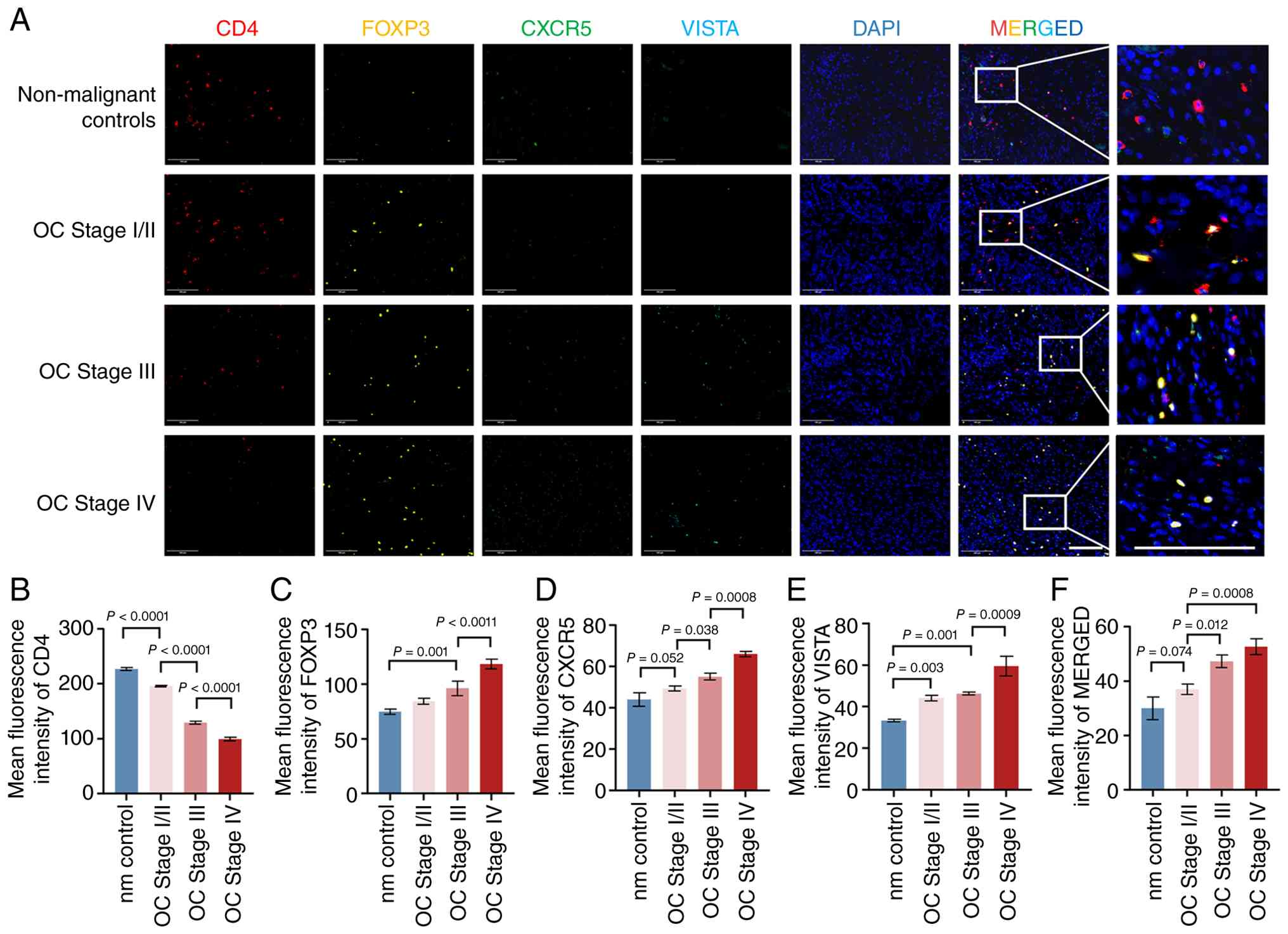 | Figure 1.Expression of VISTA in ovarian cancer
tissues. (A) Immunofluorescence staining images of ovarian tissue
sections from the non-malignant control group and patients with
ovarian cancer at different FIGO stages (Stage I/II, Stage III and
Stage IV). CD4 was visualized in red, FOXP3 in yellow, CXCR5 in
green, VISTA in cyan, and nuclei were stained with DAPI in blue.
Scale bar, 100 µm. Quantitative bar graphs of the fluorescence
intensity of each protein: (B) CD4, (C) FOXP3, (D) CXCR5, (E) VISTA
and (F) merged signals (n=3). Data are presented as the mean ± SD.
VISTA, V-domain Ig suppressor of T cell activation; FIGO,
International Federation of Gynecology and Obstetrics; CD4, cluster
of differentiation 4; FOXP3, forkhead box P3; CXCR5, C-X-C motif
chemokine receptor 5; DAPI, 4′,6-diamidino-2-phenylindole; NM,
non-malignant control; OC, ovarian cancer; SD, standard
deviation. |
Association of VISTA mRNA and protein
expression with ovarian cancer stage
qPCR and western blotting were used to examine VISTA
expression across the clinical groups. VISTA mRNA expression was
low in non-malignant control tissues (Fig. 2A). Across ovarian cancer stages,
VISTA mRNA expression showed an overall upward trend from stage
I/II to stages III and IV. However, one-way ANOVA indicated that
the between-group differences were not statistically significant.
Nevertheless, some individual stage III/IV samples showed mRNA
levels approximately threefold higher than those in non-malignant
controls, indicating substantial interindividual variability. By
contrast, changes in VISTA protein levels were more pronounced.
VISTA protein expression differed markedly among the stage I/II,
stage III, and stage IV groups and associated positively with FIGO
stage. These findings indicate that upregulation of VISTA protein
expression may be associated with ovarian cancer progression
(Fig. 2B and C).
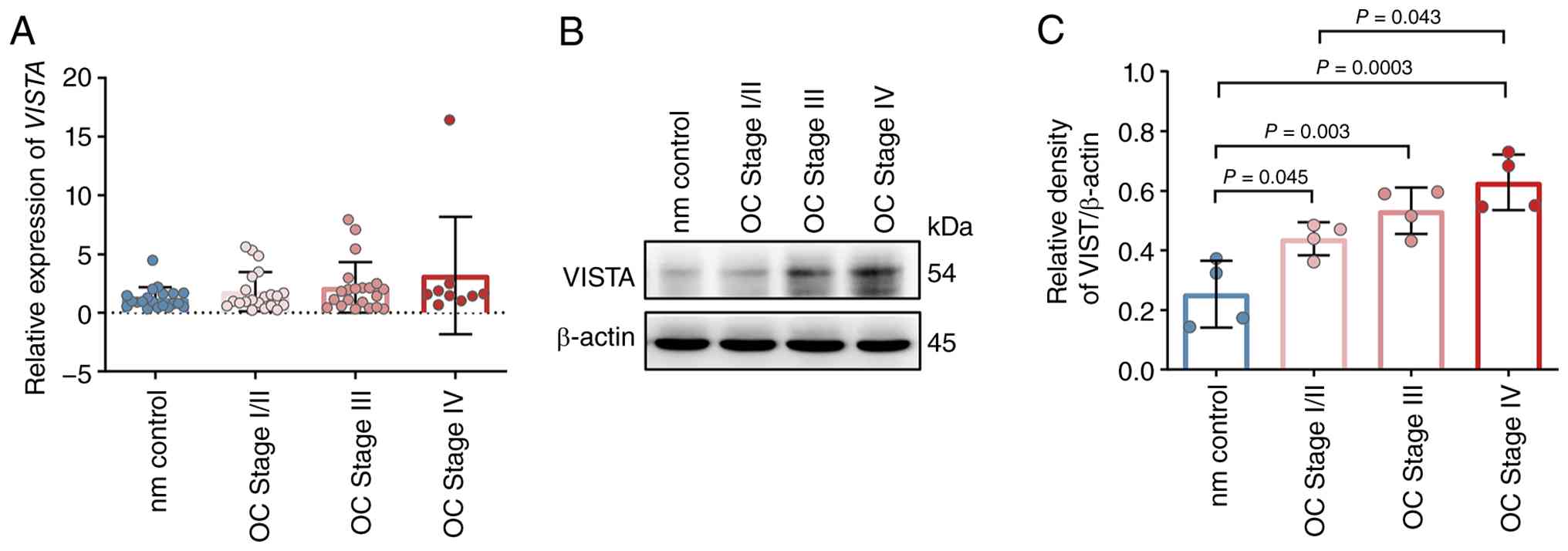 | Figure 2.VISTA mRNA and protein expression in
non-malignant control and ovarian cancer tissues across
pathological stages. (A) Quantification of VISTA mRNA expression in
tissues from the non-malignant controls group (n=20), ovarian
cancer Stage I/II (n=21), Stage III (n=21), and Stage IV (n=9),
measured by qPCR. (B) Western blot images of VISTA protein in
ovarian tissues from the non-malignant controls group and ovarian
cancer tissues with different pathological stages. (C) Quantitative
analysis of VISTA protein expression was performed using ImageJ,
and the results were normalized to the expression level of β-actin
(n=4). Data are presented as the mean ± SD. VISTA, V-domain Ig
suppressor of T cell activation; mRNA, messenger RNA; qPCR,
quantitative real-time polymerase chain reaction; NM, non-malignant
control; OC, ovarian cancer; SD, standard deviation. |
In vitro induction and sorting of
Tfr-like cells and validation of VISTA modulation
Naive CD4+ T cells were isolated using the Naive
CD4+ T Cell Isolation Kit II according to the manufacturer's
instructions. The isolated naive CD4+ T cells were stimulated with
CD3 monoclonal antibody, CD28 monoclonal antibody, recombinant
IL-2, recombinant TGF-β1, and recombinant IL-23 for 5 days to
induce differentiation (Fig. 3A).
After induction, phenotypic characterization and sorting were
performed using flow cytometry. CD25+CD127- cells were selected,
and the Tfr-like subset was further identified by CXCR5 expression.
VISTA+ Tfr-like cells (CD25+CD127-CXCR5+VISTA+) and VISTA- Tfr-like
cells (CD25+CD127-CXCR5+VISTA-) were sorted (Fig. 3B). To investigate the functional
role of VISTA, VISTA expression was manipulated by lentiviral
transduction: VISTA+ Tfr-like cells were transduced with
VISTA-shRNA lentivirus (knockdown group), and VISTA- Tfr-like cells
were transduced with LV–VISTA overexpression lentivirus
(overexpression group). Blank control and negative control virus
groups were also included. qPCR results showed that VISTA mRNA
levels were decreased in the knockdown group and increased in the
overexpression group compared with the control groups (Fig. 3C and F). Western blot analysis
confirmed corresponding changes at the protein level, supporting
effective modulation of VISTA expression (Fig. 3D, E, G and H).
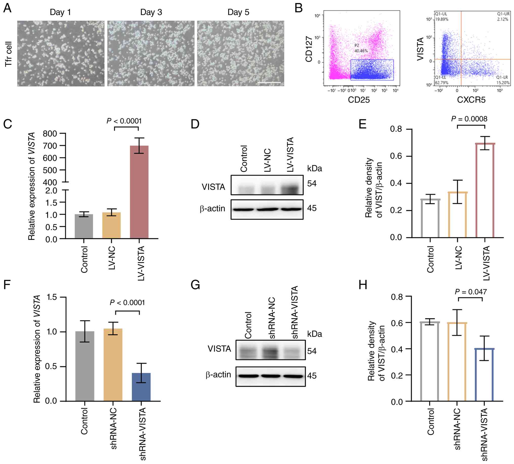 | Figure 3.Tfr-like cell differentiation and
validation of VISTA overexpression and silencing models. (A)
Microscopic morphology of naive CD4+ T cells after induction for 1
day, 3 days, and 5 days. Naive CD4+ T cells were treated with a CD3
monoclonal antibody, a CD28 monoclonal antibody, recombinant IL-2,
recombinant TGF-β1, and recombinant IL-23. Scale bar, 100 µm. (B)
VISTA+ Tfr-like cells (CD25+CD127-CXCR5+VISTA+) and VISTA- Tfr-like
cells (CD25+CD127-CXCR5+VISTA-) were sorted by flow cytometry.
VISTA knockdown was performed in VISTA+ Tfr-like cells, and VISTA
overexpression was performed in VISTA- Tfr-like cells by lentiviral
transduction. (C) VISTA mRNA expression in VISTA- Tfr-like cells
after lentivirus-mediated overexpression, measured by qPCR. Western
blot analysis of VISTA protein expression in VISTA- Tfr-like cells
after lentivirus-mediated overexpression: (D) Representative
western blot image and (E) relative expression. (F) VISTA mRNA
expression in VISTA- Tfr-like cells after lentivirus-mediated
overexpression, measured by qPCR. Western blot analysis of VISTA
protein expression in VISTA- Tfr-like cells after
lentivirus-mediated overexpression: (G) Representative western blot
image and (H) relative expression. Data are presented as the mean ±
SD. Tfr, follicular regulatory T cell; VISTA, V-domain Ig
suppressor of T cell activation; CD4, cluster of differentiation 4;
CD3, cluster of differentiation 3; CD28, cluster of differentiation
28; IL, interleukin; CD25, cluster of differentiation 25; CD127,
cluster of differentiation 127; CXCR5, C-X-C motif chemokine
receptor 5; mRNA, messenger RNA; qPCR, quantitative polymerase
chain reaction; SD, standard deviation; TGF-β1, transforming growth
factor β1; LV–VISTA, VISTA-overexpression lentiviral vector; sh,
short interfering. |
Effects of VISTA on Tfr-like cell
proliferation and cytokine secretion
CFSE fluorescence decreases by approximately half
with each cell division, allowing flow-cytometric assessment of
proliferation under 488-nm excitation. Proliferation assays showed
that the CFSE fluorescence decay ratio was markedly higher in
VISTA+ Tfr-like cells than in VISTA- Tfr-like cells, indicating
greater proliferative activity in the VISTA+ subset (Fig. 4A and B). Following VISTA
overexpression in VISTA- Tfr-like cells, the CFSE fluorescence
decay ratio was markedly higher than in the blank control and OE
negative control groups (Fig. 4C).
Conversely, silencing VISTA in VISTA+ Tfr-like cells resulted in a
markedly lower ratio than in the blank control and shRNA negative
control groups (Fig. 4D). These
findings indicate that VISTA promotes Tfr-like cell proliferation,
whereas VISTA silencing reduces proliferation. ELISA results
revealed that the levels of IL-10 and TGF-β1 in the supernatant of
VISTA+ Tfr-like cells were markedly higher than those in VISTA-
Tfr-like cells, whereas the levels of IL-21 and granzyme B were
markedly lower (Fig. S2A-D). After
VISTA overexpression in VISTA- Tfr-like cells, IL-10 and TGF-β1
levels markedly increased, while IL-21 and granzyme B levels
markedly decreased (Fig. 4E). By
contrast, silencing VISTA in VISTA+ Tfr-like cells led to markedly
reduced IL-10 and TGF-β1 levels and markedly elevated IL-21 and
granzyme B levels (Fig. 4F). These
findings indicate that VISTA promotes anti-inflammatory cytokine
secretion and suppresses immune effector molecules, producing a
profile consistent with enhanced immunosuppressive function in
Tfr-like cells.
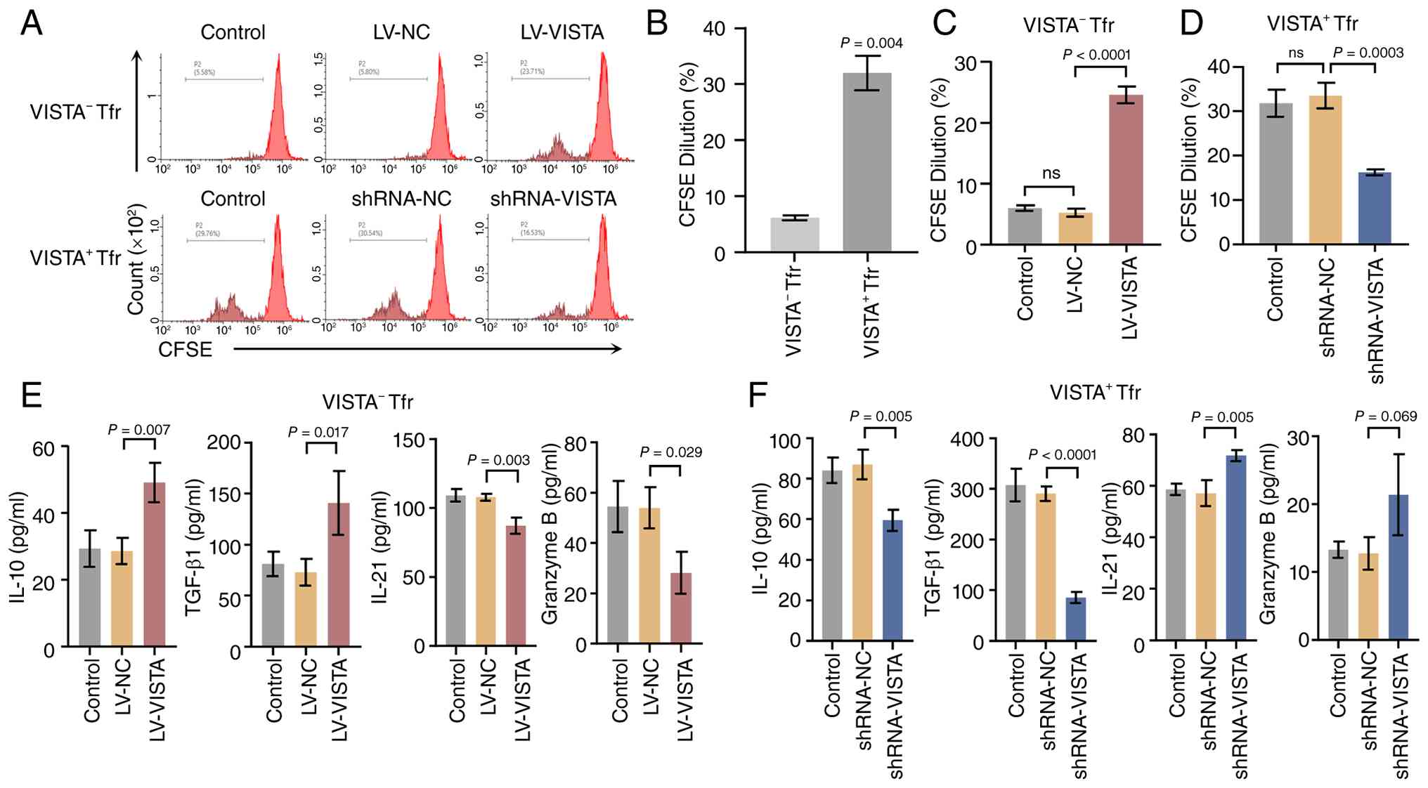 | Figure 4.Effects of VISTA on Tfr-like cell
proliferation and cytokine secretion. (A) Representative
flow-cytometry histograms from the CFSE proliferation assay. The
x-axis represents CFSE fluorescence intensity, and the y-axis
represents cell count (n=3). (B) Quantification of CFSE
fluorescence intensity in VISTA- Tfr-like cells and VISTA+ Tfr-like
cells. (C) Quantification of CFSE fluorescence intensity in VISTA-
Tfr-like cells after VISTA overexpression. (D) Quantification of
CFSE fluorescence intensity in VISTA+ Tfr-like cells after VISTA
silencing (VISTA-shRNA). (E and F) Levels of IL-10, TGF-β1, IL-21,
and granzyme B in the culture supernatants of VISTA- Tfr-like cells
(E) and VISTA+ Tfr-like cells (F), detected by ELISA (n=3). Data
are presented as the mean ± SD. VISTA, V-domain Ig suppressor of T
cell activation; Tfr, follicular regulatory T cell; CFSE,
5,6-carboxyfluorescein diacetate succinimidyl ester; ELISA,
enzyme-linked immunosorbent assay; TGF-β1, transforming growth
factor β1; IL, interleukin; LV–VISTA, VISTA-overexpression
lentiviral vector; SD, standard deviation; shRNA, short hairpin
RNA. |
VISTA-associated suppression of AKT
and ERK1/2 phosphorylation in Tfr-like cells
Following VISTA overexpression in VISTA- Tfr-like
cells, the levels of p-AKT and p-ERK1/2 were markedly lower than in
the blank control and negative control groups. Conversely,
silencing VISTA in VISTA+ Tfr-like cells using VISTA shRNA resulted
in markedly higher p-AKT and p-ERK1/2 levels than in the blank and
negative control groups (Figs.
5A-I, S2E-H). By contrast,
total AKT and total ERK1/2 protein levels showed no significant
differences across groups, suggesting that VISTA alters pathway
phosphorylation without affecting total AKT or ERK1/2
abundance.
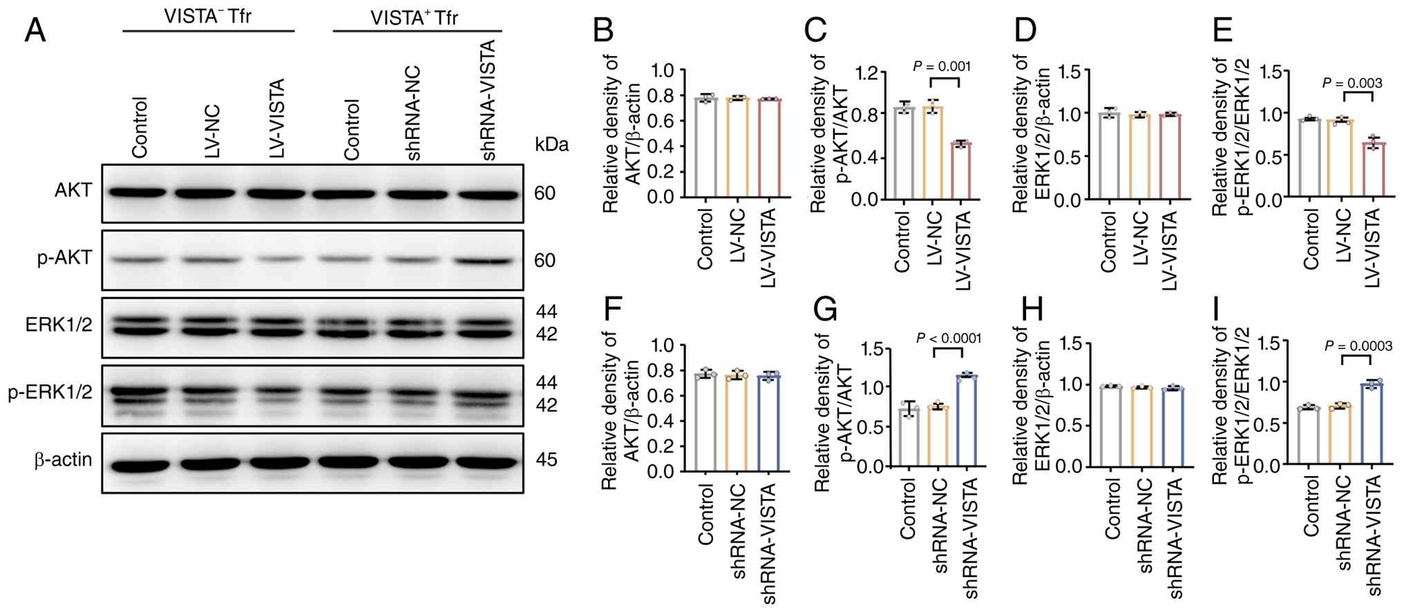 | Figure 5.Effects of VISTA expression on AKT
and ERK1/2 pathway proteins in Tfr-like cells. (A) Western blot
images showing the protein expression of AKT, p-AKT, ERK1/2 and
p-ERK1/2 in VISTA- Tfr-like cells, VISTA+ Tfr-like cells, VISTA-
Tfr-like cells with VISTA overexpression and VISTA+ Tfr-like cells
with VISTA silencing. (B-I) Quantitative analysis of protein
expression levels was performed using ImageJ. Total AKT and total
ERK1/2 protein levels were normalized to β-actin, while the
relative phosphorylation levels of p-AKT and p-ERK1/2 were
calculated as the ratio of phosphorylated protein to the
corresponding total protein (n=3). Data are presented as the mean ±
SD. VISTA, V-domain Ig suppressor of T cell activation; AKT,
protein kinase B; ERK1/2, extracellular signal-regulated kinase
1/2; p-, phosphorylated; SD, standard deviation; Tfr, follicular
regulatory T cell. |
Previous studies suggest that AKT phosphorylation
can inhibit Treg function31 and may correlate negatively
with the abundance of CXCR5+FOXP3+ cells, a characteristic Tfr-like
phenotype, in a model of acute colitis (32). In addition, ERK inhibition can
promote FOXP3 expression (33) and
FOXP3 is a key transcriptional regulator of Treg and Tfr-like cell
function. Together, these observations are consistent with an
inverse relationship between AKT/ERK1/2 phosphorylation and
Tfr-like cell proliferation and immunosuppressive activity. These
data revealed that altered VISTA expression coincided with changes
in AKT and ERK1/2 phosphorylation alongside shifts in Tfr-like cell
proliferation and immunosuppressive activity. These observations
identified correlative changes between VISTA levels, AKT/ERK1/2
phosphorylation and Tfr-like cell functional phenotypes.
Discussion
The present study characterized VISTA expression in
ovarian cancer tissues and examined how VISTA may regulate Tfr-like
cell function in the context of tumor immune escape. To the best of
the authors' knowledge, the findings identified a previously
underexplored role for the VISTA-Tfr-like cell axis in ovarian
cancer progression. The findings provided experimental evidence
relevant to remodeling of the ovarian cancer immune
microenvironment and may inform further development of
immunotherapeutic strategies.
The high mortality rate of ovarian cancer is closely
associated with its complex tumor immune escape mechanisms and the
abnormal expression of immune checkpoint molecules is a central
component of this process (34–37).
VISTA protein expression increased across FIGO stages and
associated positively with the infiltration density of FOXP3+CXCR5+
Tfr-like cells, suggesting that VISTA may be a candidate
stage-associated marker in ovarian cancer. This finding was
consistent with reports linking high VISTA expression to more
aggressive disease in several solid tumors (17,29).
The present data further suggested that accumulation of VISTA+
Tfr-like cells may contribute to the association between VISTA
expression and ovarian cancer stage (17). Notably, the expression trends of
VISTA at the mRNA and protein levels were not completely
consistent: VISTA protein expression showed a clearer association
with stage, whereas mRNA expression varied substantially among
individuals (38). This discordance
may reflect post-translational modification of VISTA, such as
glycosylation, or regulation of protein stability (39,40).
VISTA activity in ovarian cancer may therefore be influenced by
post-transcriptional or post-translational regulation rather than
transcription alone, a possibility that warrants further
investigation (41).
As regulators linking cellular and humoral immunity,
Tfr-like cells may contribute to tumor immune escape when
dysregulated (18,42,43).
The present findings indicated that VISTA may enhance the
immunosuppressive function of Tfr-like cells by promoting
proliferation and secretion of the anti-inflammatory cytokines
IL-10 and TGF-β1 while reducing release of the immune effector
molecules IL-21 and granzyme B. This finding was consistent with
breast cancer studies in which Tfr-like cells have been implicated
in tumor progression through anti-inflammatory cytokine secretion
(21). The present study extended
those observations by implicating VISTA as a regulator of this
process. At the mechanistic level, the findings suggested that
VISTA-associated changes in Tfr-like cell function are accompanied
by reduced AKT and ERK1/2 phosphorylation. Previous studies have
confirmed that AKT phosphorylation can inhibit the
immunosuppressive function of Treg cells, while ERK1/2 inhibitors
can enhance Treg cell stability by upregulating FOXP3 expression
(32–34). In the present study, after VISTA
silencing, the levels of p-AKT and p-ERK1/2 increased, accompanied
by decreased Tfr-like cell proliferation and reduced secretion of
anti-inflammatory factors. These observations suggested that VISTA
may support Tfr-like cell activity by attenuating AKT/MAPK pathway
phosphorylation. This proposed mechanism expands the signaling
framework for VISTA in immune cells and offers a perspective on
molecular regulation of Tfr-like cell function. However, the
present study focused on verifying the involvement of AKT and
ERK1/2 core kinases in this regulatory process, while the upstream
initiating molecules and downstream transcriptional targets of this
pathway require investigation in future studies.
The observed discordance between VISTA mRNA and
protein expression trends indicates that post-translational
regulation may contribute to the upregulation of VISTA protein
during ovarian cancer progression. Ubiquitin-proteasome-mediated
degradation is a recognized regulator of VISTA protein homeostasis.
VISTA undergoes constitutive K48-linked polyubiquitination in T
cells, contributing to a short protein half-life. Protective
modifications such as TRIM25-mediated K63-linked ubiquitination may
counteract this degradation. TRIM25 knockdown increases K48-linked
VISTA ubiquitination and accelerates protein turnover, providing
mechanistic evidence that post-translational regulation can
dissociate VISTA protein abundance from mRNA expression (44,45).
Glycosylation is another post-translational modification of
B7-family immune checkpoints that can influence protein stability,
membrane localization and receptor-binding affinity (45). Studies in clear cell renal cell
carcinoma have shown that B7-H5 (VISTA) is highly glycosylated and
that mutations at specific N-glycosylation sites alter its
abundance and subcellular distribution (46–48).
The UniProt entry for VISTA also lists experimentally validated
N-glycosylation sites, supporting the presence of this
modification. On this basis, aberrant glycosylation could partly
explain the discrepancy between VISTA mRNA and protein expression
in ovarian cancer. However, this hypothesis has not been
experimentally validated in the current study and the specific
regulatory mechanism warrants dedicated investigation in future
work. Coexpression of VISTA with other immune checkpoints, such as
PD-L1, also warrants investigation. Multiple immune checkpoints can
be coordinately upregulated in solid tumors, contributing to an
immunosuppressive microenvironment. Concurrent VISTA and PD-L1
upregulation in ovarian cancer could intensify Tfr-like
cell-mediated immunosuppression (49–51).
This possibility provides a rationale for evaluating combined VISTA
and PD-1/PD-L1 targeting. Future studies should assess coexpression
in clinical samples and evaluate combined intervention in animal
models.
Certain limitations of the present study should be
acknowledged. First, this was a single-center exploratory study
with potential selection bias and no independent external
validation cohort. The small sample size of the Stage IV subgroup
(n=9) may limit the statistical robustness of stage-stratified
subgroup analyses; non-parametric tests were therefore applied for
all small-subgroup comparisons to reduce the risk of false-positive
results. In addition, control tissues were non-malignant ovarian
samples from patients with benign gynecological diseases rather
than from healthy individuals, and long-term follow-up survival
data were unavailable, so the prognostic value of VISTA and VISTA+
Tfr-like cells could not be evaluated. Second, functional assays
were limited to in vitro cell culture with induced Tfr-like
cells, which were identified only by a limited marker panel
(CD4+FOXP3+CXCR5+). Additional Tfr lineage markers such as BCL6,
PD-1 and ICOS were not assessed, and thus the cells should be
interpreted as Tfr-like cells rather than definitively confirmed
bona fide Tfr cells; in vivo models are further required to
evaluate the role of the VISTA-Tfr-like axis in ovarian cancer
growth and immune microenvironment remodeling. Third, the molecular
mechanism underlying the discordance between VISTA mRNA and protein
expression in ovarian cancer was not experimentally verified; the
glycosylation-based hypothesis was derived from prior literature
and requires further functional validation. Fourth, the mechanistic
investigation of the present study focused on the core kinases AKT
and ERK1/2; upstream regulators and downstream transcriptional
effectors including FOXP3 and Blimp-1 were not examined. Notably,
formal pathway rescue experiments were not been performed to
establish a definitive causal link between VISTA-mediated AKT/MAPK
modulation and functional changes in Tfr-like cells, nor to exclude
potential off-target effects of lentiviral intervention. Despite
these limitations, the findings characterized VISTA expression in
ovarian cancer and its association with Tfr-like cell function,
providing a basis for further mechanistic and translational
studies.
In summary, the present study characterized VISTA
expression in ovarian cancer and its potential regulation of
Tfr-like cell function. VISTA expression increased with advancing
tumor stage and was positively associated with Tfr-like cell
infiltration. In vitro, VISTA promoted Tfr-like cell
proliferation and anti-inflammatory cytokine secretion while
reducing immune effector molecule release; these functional changes
were accompanied by reduced AKT and ERK1/2 phosphorylation. These
findings only revealed correlative changes among VISTA levels,
AKT/MAPK phosphorylation status and the immunosuppressive phenotype
of Tfr-like cells. Overall, the results highlighted a potential
role for the VISTA-Tfr-like cell axis in regulating the ovarian
cancer immune microenvironment and provide a basis for further
evaluation of VISTA-targeted immunotherapeutic strategies.
Supplementary Material
Supporting Data
Supporting Data
Acknowledgements
Not applicable.
Funding
This work was supported by National Natural Science Foundation
of China-Regional Project (project name: Study on the Role and
Mechanism of Vista-Mediated Follicular Regulatory T Cell Activation
in Ovarian Cancer Immune Evasion, project number: 82160302); Key
Research and Development Project of Xinjiang Uygur Autonomous
Region (project name: Research on the Construction of an
Intelligent and Precise Early Screening and Treatment System for
Ovarian Cancer in Xinjiang Women, project number: 2024B03037).
Availability of data and materials
The data generated in the present study may be
requested from the corresponding author.
Authors' contributions
LL conceptualized the study and designed the
research protocol. CY and HW acquired the experimental data. YM and
YX analyzed the data, drafted the initial manuscript and
contributed to manuscript preparation. LL critically revised the
manuscript for important intellectual content. YM and YX confirm
the authenticity of all the raw data. All authors read and approved
the final manuscript and agree to be accountable for all aspects of
the work, ensuring that questions concerning the accuracy or
integrity of any part of the work are appropriately investigated
and resolved.
Ethics approval and consent to
participate
The present study was conducted in accordance with
the Declaration of Helsinki and approved by the Ethics Committee of
the Affiliated Tumor Hospital of the Third Clinical Medical
College, Xinjiang Medical University (approval no: K-2025116).
Written informed consent was obtained from all participants
(patients with ovarian cancer for tumor tissues, individuals with
benign diseases for non-malignant ovarian tissues, and healthy
donors for peripheral blood) prior to sample collection. Patient
information was de-identified and managed confidentially to protect
privacy.
Patient consent for publication
Not applicable.
Competing interests
The authors declare that they have no competing
interests.
Glossary
Abbreviations
Abbreviations:
|
AKT
|
protein kinase B
|
|
CFSE
|
5,6-carboxyfluorescein diacetate
succinimidyl ester
|
|
ELISA
|
enzyme-linked immunosorbent assay
|
|
ERK1/2
|
extracellular signal-regulated kinase
1/2
|
|
FIGO
|
International Federation of Gynecology
and Obstetrics
|
|
FOXP3
|
forkhead box P3
|
|
IL
|
interleukin
|
|
MAPK
|
mitogen-activated protein kinase
|
|
PBMC
|
peripheral blood mononuclear cell
|
|
PD-1
|
programmed cell death protein 1
|
|
PD-L1
|
programmed death-ligand 1
|
|
qPCR
|
quantitative PCR
|
|
Tfr
|
follicular regulatory T cell
|
|
TGF-β1
|
transforming growth factor beta 1
|
|
Treg
|
regulatory T cell
|
|
VISTA
|
V-domain Ig suppressor of T cell
activation
|
References
|
1
|
Gupta R, Kumar R, Penn CA and Wajapeyee N:
Immune evasion in ovarian cancer: Implications for immunotherapy
and emerging treatments. Trends Immunol. 46:166–181. 2025.
View Article : Google Scholar : PubMed/NCBI
|
|
2
|
Lheureux S, Braunstein M and Oza AM:
Epithelial ovarian cancer: Evolution of management in the era of
precision medicine. CA Cancer J Clin. 69:280–304. 2019.PubMed/NCBI
|
|
3
|
Ghisoni E, Morotti M, Sarivalasis A, Grimm
AJ, Kandalaft L, Dangaj Laniti D and Coukos G: Immunotherapy for
ovarian cancer: Towards a tailored immunophenotype-based approach.
Nat Rev Clin Oncol. 21:801–817. 2024. View Article : Google Scholar : PubMed/NCBI
|
|
4
|
Frentzas S, Austria Mislang AR, Lemech C,
Nagrial A, Underhill C, Wang W, Wang ZM, Li B, Xia Y and Coward
JIG: Phase 1a dose escalation study of ivonescimab (AK112/SMT112),
an anti-PD-1/VEGF-A bispecific antibody, in patients with advanced
solid tumors. J Immunother Cancer. 12:e0080372024. View Article : Google Scholar : PubMed/NCBI
|
|
5
|
Sun Y, Patterson-Fortin J, Han S, Li Z,
Nowicka Z, Hirohashi Y, Kilgas S, Yi JK, Spektor A, Fendler W, et
al: 53BP1 loss elicits cGAS-STING-dependent antitumor immunity in
ovarian and pancreatic cancer. Nat Commun. 15:66762024. View Article : Google Scholar : PubMed/NCBI
|
|
6
|
Bray F, Laversanne M, Sung H, Ferlay J,
Siegel RL, Soerjomataram I and Jemal A: Global cancer statistics
2022: GLOBOCAN estimates of incidence and mortality worldwide for
36 cancers in 185 countries. CA Cancer J Clin. 74:229–263.
2024.PubMed/NCBI
|
|
7
|
Murakami R, Hamanishi J, Brown JB, Abiko
K, Yamanoi K, Taki M, Hosoe Y, Yamaguchi K, Baba T, Matsumura N, et
al: Combination of gene set signatures correlates with response to
nivolumab in platinum-resistant ovarian cancer. Sci Rep.
11:114272021. View Article : Google Scholar : PubMed/NCBI
|
|
8
|
Flies DB and Chen L: The new B7s: Playing
a pivotal role in tumor immunity. J Immunother. 30:251–260. 2007.
View Article : Google Scholar : PubMed/NCBI
|
|
9
|
Gao Y, He Y, Tang Y, Chen ZS and Qu M:
VISTA: A novel checkpoint for cancer immunotherapy. Drug Discov
Today. 29:1040452024. View Article : Google Scholar : PubMed/NCBI
|
|
10
|
Slater BT, Han X, Chen L and Xiong Y:
Structural insight into T cell coinhibition by PD-1H (VISTA). Proc
Natl Acad Sci USA. 117:1648–1657. 2020. View Article : Google Scholar : PubMed/NCBI
|
|
11
|
Zhang RJ and Kim TK: VISTA-mediated immune
evasion in cancer. Exp Mol Med. 56:2348–2356. 2024. View Article : Google Scholar : PubMed/NCBI
|
|
12
|
Martin AS, Molloy M, Ugolkov A, von
Roemeling RW, Noelle RJ, Lewis LD, Johnson M, Radvanyi L and
Martell RE: VISTA expression and patient selection for immune-based
anticancer therapy. Front Immunol. 14:10861022023. View Article : Google Scholar : PubMed/NCBI
|
|
13
|
Huang X, Zhang X, Li E, Zhang G, Wang X,
Tang T, Bai X and Liang T: VISTA: An immune regulatory protein
checking tumor and immune cells in cancer immunotherapy. J Hematol
Oncol. 13:832020. View Article : Google Scholar : PubMed/NCBI
|
|
14
|
Wan C, Keany MP, Dong H, Al-Alem LF,
Pandya UM, Lazo S, Boehnke K, Lynch KN, Xu R, Zarrella DT, et al:
Enhanced efficacy of simultaneous PD-1 and PD-L1 immune checkpoint
blockade in high-grade serous ovarian cancer. Cancer Res.
81:158–173. 2021. View Article : Google Scholar : PubMed/NCBI
|
|
15
|
Ma Y, Xu Y, Yu C, Wu H and Li L:
VISTA+ follicular regulatory T cells modulate the
function of effector immune cells: implications for ovarian cancer
immune escape. Front Immunol. 16:17040482025. View Article : Google Scholar : PubMed/NCBI
|
|
16
|
Liu S, Zhou Q, Ji F, Zhu T, Ding B, Xu J,
Zhang T, Lin H and Shen Y: VISTA promotes ovarian cancer
progression through activation of NF-κB pathway and tumor
microenvironment reprogramming. J Ovarian Res. 19:1652026.
View Article : Google Scholar : PubMed/NCBI
|
|
17
|
Zong L, Zhou Y, Zhang M, Chen J and Xiang
Y: VISTA expression is associated with a favorable prognosis in
patients with high-grade serous ovarian cancer. Cancer Immunol
Immunother. 69:33–42. 2020. View Article : Google Scholar : PubMed/NCBI
|
|
18
|
Tan D, Yin W, Guan F, Zeng W, Lee P,
Candotti F, James LK, Saraiva Camara NO, Haeryfar SMM, Chen Y, et
al: B cell-T cell interplay in immune regulation: A focus on
follicular regulatory T and regulatory B cell functions. Front Cell
Dev Biol. 10:9918402022. View Article : Google Scholar : PubMed/NCBI
|
|
19
|
Song H, Liu A, Liu G, Wu F and Li Z: T
follicular regulatory cells suppress Tfh-mediated B cell help and
synergistically increase IL-10-producing B cells in breast
carcinoma. Immunol Res. 67:416–423. 2019. View Article : Google Scholar : PubMed/NCBI
|
|
20
|
Dreisinger M, Roškar Z, Goropevšek A,
Zakelšek A, Čurič S, Živko N, Bevc S and Homšak E: Increased STAT3
phosphorylation in CD4+ T-cells of treated patients with
chronic lymphocytic leukemia and changes in circulating regulatory
T-cell subsets relative to tumor mass distribution value and
disease duration. Biomedicines. 13:12042025. View Article : Google Scholar : PubMed/NCBI
|
|
21
|
Clark NM, Martinez LM, Murdock S, deLigio
JT, Olex AL, Effi C, Dozmorov MG and Bos PD: Regulatory T cells
support breast cancer progression by opposing IFN-γ-dependent
functional reprogramming of myeloid cells. Cell Rep. 33:1084822020.
View Article : Google Scholar : PubMed/NCBI
|
|
22
|
Tan SN, Hao J, Ge J, Yang Y, Liu L, Huang
J, Lin M, Zhao X, Wang G, Yang Z, et al: Regulatory T cells
converted from Th1 cells in tumors suppress cancer immunity via
CD39. J Exp Med. 222:e202404452025. View Article : Google Scholar : PubMed/NCBI
|
|
23
|
Wang B, Zhang Z, Liu W and Tan B:
Targeting regulatory T cells in gastric cancer: Pathogenesis,
immunotherapy, and prognosis. Biomed Pharmacother. 158:1141802023.
View Article : Google Scholar : PubMed/NCBI
|
|
24
|
Chap BS, Rayroux N, Grimm AJ, Ghisoni E
and Dangaj Laniti D: Crosstalk of T cells within the ovarian cancer
microenvironment. Trends Cancer. 10:1116–1130. 2024. View Article : Google Scholar : PubMed/NCBI
|
|
25
|
Suzuki S, Ogawa T, Sano R, Takahara T,
Inukai D, Akira S, Tsuchida H, Yoshikawa K, Ueda R and Tsuzuki T:
Immune-checkpoint molecules on regulatory T-cells as a potential
therapeutic target in head and neck squamous cell cancers. Cancer
Sci. 111:1943–1957. 2020. View Article : Google Scholar : PubMed/NCBI
|
|
26
|
Hanahan D: Hallmarks of cancer: New
dimensions. Cancer Discov. 12:31–46. 2022. View Article : Google Scholar : PubMed/NCBI
|
|
27
|
Xu X, Shang B, Wu H, Jin X, Wang J, Li J,
Li D, Liang B, Wang X, Su L, et al: FXR shapes an immunosuppressive
microenvironment in PD-L1lo/-non-small cell lung cancer by
upregulating HVEM. JCI Insight. 10:e1907162025. View Article : Google Scholar : PubMed/NCBI
|
|
28
|
Yuan Y, Peng Z and Yucheng S: The
CXCL9-CXCR3 axis mediates tumor progression and immune checkpoint
regulation in colorectal cancer. Mol Cell Probes. 89:1020782026.
View Article : Google Scholar : PubMed/NCBI
|
|
29
|
Shekari N, Shanehbandi D, Kazemi T,
Zarredar H, Baradaran B and Jalali SA: VISTA and its ligands: The
next generation of promising therapeutic targets in immunotherapy.
Cancer Cell Int. 23:2652023. View Article : Google Scholar : PubMed/NCBI
|
|
30
|
Yasinska IM, Meyer NH, Schlichtner S,
Hussain R, Siligardi G, Casely-Hayford M, Fiedler W, Wellbrock J,
Desmet C, Calzolai L, et al: Ligand-receptor interactions of
galectin-9 and VISTA suppress human T lymphocyte cytotoxic
activity. Front Immunol. 11:5805572020. View Article : Google Scholar : PubMed/NCBI
|
|
31
|
Livak KJ and Schmittgen TD: Analysis of
relative gene expression data using real-time quantitative PCR and
the 2(−Delta Delta C(T)) method. Methods. 25:402–408. 2001.
View Article : Google Scholar : PubMed/NCBI
|
|
32
|
Pompura SL and Dominguez-Villar M: The
PI3K/AKT signaling pathway in regulatory T-cell development,
stability, and function. J Leukoc Biol. 103:1065–1076. 2018.
View Article : Google Scholar
|
|
33
|
Jin J, Zhong Y, Long J, Wu T, Jiang Q,
Wang H, Ge W, Zhao H and Liu D: Ginsenoside Rg1 relieves
experimental colitis by regulating balanced differentiation of
Tfh/Treg cells. Int Immunopharmacol. 100:1081332021. View Article : Google Scholar : PubMed/NCBI
|
|
34
|
Liu H, Yao S, Dann SM, Qin H, Elson CO and
Cong Y: ERK differentially regulates Th17- and Treg-cell
development and contributes to the pathogenesis of colitis. Eur J
Immunol. 43:1716–1726. 2013. View Article : Google Scholar : PubMed/NCBI
|
|
35
|
Tang Q, Chen Y, Li X, Long S, Shi Y, Yu Y,
Wu W, Han L and Wang S: The role of PD-1/PD-L1 and application of
immune-checkpoint inhibitors in human cancers. Front Immunol.
13:9644422022. View Article : Google Scholar : PubMed/NCBI
|
|
36
|
Galassi C, Chan TA, Vitale I and Galluzzi
L: The hallmarks of cancer immune evasion. Cancer Cell.
42:1825–1863. 2024. View Article : Google Scholar : PubMed/NCBI
|
|
37
|
Nie H, Saini P, Miyamoto T, Liao L,
Zielinski RJ, Liu H, Zhou W, Wang C, Murphy B, Towers M, et al:
Targeting branched N-glycans and fucosylation sensitizes ovarian
tumors to immune checkpoint blockade. Nat Commun. 15:28532024.
View Article : Google Scholar : PubMed/NCBI
|
|
38
|
Yang L, Li L, Ma J, Yang S, Zou C and Yu
X: miRNA and mRNA integration network construction reveals novel
key regulators in left-sided and right-sided colon adenocarcinoma.
Biomed Res Int. 2019:71492962019.PubMed/NCBI
|
|
39
|
Chatham JC, Zhang J and Wende AR: Role of
O-linked N-acetylglucosamine protein modification in cellular
(patho)physiology. Physiol Rev. 101:427–493. 2021. View Article : Google Scholar : PubMed/NCBI
|
|
40
|
Hsieh HC, Ling LL and Wang YC:
Post-translational modifications of immune checkpoints: Molecular
mechanisms, tumor microenvironment remodeling, and therapeutic
implications. J Biomed Sci. 33:32026. View Article : Google Scholar : PubMed/NCBI
|
|
41
|
Schwanhäusser B, Busse D, Li N, Dittmar G,
Schuchhardt J, Wolf J, Chen W and Selbach M: Global quantification
of mammalian gene expression control. Nature. 473:337–342. 2011.
View Article : Google Scholar : PubMed/NCBI
|
|
42
|
Eschweiler S, Clarke J, Ramírez-Suástegui
C, Panwar B, Madrigal A, Chee SJ, Karydis I, Woo E, Alzetani A,
Elsheikh S, et al: Intratumoral follicular regulatory T cells
curtail anti-PD-1 treatment efficacy. Nat Immunol. 22:1052–1063.
2021. View Article : Google Scholar : PubMed/NCBI
|
|
43
|
Li Z, Zhang Y, Hao H, Chen L, Lv T, Zhang
X, Qi Y and Wang Z: Esophageal cancer cell-derived small
extracellular vesicles decrease circulating Tfh/Tfr via sEV-PDL1 to
promote immunosuppression. Cancer Immunol Immunother. 72:4249–4259.
2023. View Article : Google Scholar : PubMed/NCBI
|
|
44
|
Xiao L, Guan X, Xiang M, Wang Q, Long Q,
Yue C, Chen L, Liu J and Liao C: B7 family protein glycosylation:
Promising novel targets in tumor treatment. Front Immunol.
13:10885602022. View Article : Google Scholar : PubMed/NCBI
|
|
45
|
Sun Y, Zhang Z, Li H, Bu X, Chen L, Wang
X, Fan L, Chen B, Kong L, Dai P, et al: Destruction of VISTA by
TRIM25 ablation in T cells potentiates cancer immunotherapy. Cell
Res. 35:1003–1020. 2025. View Article : Google Scholar : PubMed/NCBI
|
|
46
|
Emaldi M, Alamillo-Maeso P, Rey-Iborra E,
Mosteiro L, Lecumberri D, Pulido R, López JI and Nunes-Xavier CE: A
functional role for glycosylated B7-H5/VISTA immune checkpoint
protein in metastatic clear cell renal cell carcinoma. iScience.
27:1105872024. View Article : Google Scholar : PubMed/NCBI
|
|
47
|
Zapała Ł, Kunc M, Sharma S, Pęksa R,
Popęda M, Biernat W and Radziszewski P: Immune checkpoint receptor
VISTA on immune cells is associated with expression of T-cell
exhaustion marker TOX and worse prognosis in renal cell carcinoma
with venous tumor thrombus. J Cancer Res Clin Oncol. 149:4131–4139.
2023. View Article : Google Scholar : PubMed/NCBI
|
|
48
|
Hong S, Yuan Q, Xia H, Zhu G, Feng Y, Wang
Q, Zhang Z, He W, Lu J, Dong C and Ni L: Analysis of VISTA
expression and function in renal cell carcinoma highlights VISTA as
a potential target for immunotherapy. Protein Cell. 10:840–845.
2019. View Article : Google Scholar : PubMed/NCBI
|
|
49
|
Miyashita H, Bevins NJ, Thangathurai K,
Lee S, Pabla S, Nesline MK, Glenn ST, Conroy JM, DePietro P, Rubin
E, et al: The transcriptomic expression pattern of immune
checkpoints shows heterogeneity between and within cancer types. Am
J Cancer Res. 14:2240–2252. 2024. View Article : Google Scholar : PubMed/NCBI
|
|
50
|
Nygaard V, Ree AH, Dagenborg VJ,
Børresen-Dale AL, Edwin B, Fretland ÅA, Grzyb K, Haugen MH,
Mælandsmo GM and Flatmark K: A PRRX1 signature identifies TIM-3 and
VISTA as potential immune checkpoint targets in a subgroup of
microsatellite stable colorectal cancer liver metastases. Cancer
Res Commun. 3:235–244. 2023. View Article : Google Scholar : PubMed/NCBI
|
|
51
|
Tarhini AA, Hedges D, Tan AC, Rodriguez P,
Sukrithan V, Ratan A, McCarter MD, Carpten J, Colman H, Ikeguchi
AP, et al: Differences in co-expression of T cell co-inhibitory and
co-stimulatory molecules with PD-1 across different human cancers.
J Oncol Res Ther. 9:102242024.PubMed/NCBI
|

















