Introduction
Cancer development results from interactions among
genetic abnormalities, epigenetic changes and alterations in
cellular regulatory networks, which collectively shape tumor
initiation and progression (1).
Aberrations in genomic non-coding regions can influence
transcriptional control and participate in malignant transformation
and tumor evolution (2). Among
non-coding RNAs (ncRNAs), microRNAs (miRNAs or miRs) constitute key
post-transcriptional regulators that have attracted considerable
research interest due to their involvement in physiological
regulation and pathological processes (3,4).
Typically consisting of 17–25 nucleotides, miRNAs regulate gene
expression through sequence-dependent interactions with target
mRNAs, typically involving binding to the 3′-untranslated regions
(UTRs) and altering downstream protein production. These
interactions result in suppression of protein translation or
degradation of target transcripts, affecting diverse cellular
functions (4). In cancers, miRNAs
may display tumor-suppressive or -promoting activities by
modulating specific molecular targets and signaling networks,
thereby influencing tumorigenesis, tumor growth, metastasis and
drug resistance (5–7). The emerging diagnostic, prognostic and
therapeutic potential of miRNAs has generated increasing interest
in their clinical translation in oncology (7,8).
The MIR324 gene is located on chromosome 17p13.1
(9), and miR-324-5p is processed
through the canonical miRNA biogenesis pathway. The primary miRNA
transcript undergoes sequential cleavage by Drosha and Dicer to
generate mature miR-324-5p (10).
miR-324-5p exhibits variable expression across malignancies, with
both upregulation and downregulation (11–13).
Specifically, miR-324-5p expression is upregulated in several types
of cancer, including gastric cancer (GC) (14,15),
lung adenocarcinoma (LUAD) (16,17)
and papillary thyroid carcinoma (PTC) (18,19).
Conversely, downregulation of miR-324-5p has been observed in
colorectal cancer (CRC) (20), live
cancer (21,22), glioblastoma (GBM) (23,24)
and melanoma (12). Furthermore,
miR-324-5p modulates tumorigenesis and cancer progression by
influencing diverse molecular pathways, including JAK2/STAT3
signaling, rho-associated kinase 1 (ROCK1) and ETS proto-oncogene
1, transcription factor (ETS1)/Sp1 transcription factor
(SP1)-mediated regulatory mechanisms (25–27).
The tissue-specific and cancer-type-dependent expression patterns
of miR-324-5p underscore its potential clinical value. Accumulating
evidence has revealed the biological functions of miR-324-5p across
cancer contexts (14,16,20,22,24).
The present review summarizes the functional roles of miR-324-5p in
cancer types, highlighting its regulatory mechanisms, biomarker
potential and therapeutic implications in oncology.
Cancer associated with miR-324-5p
The Cancer Genome Atlas (TCGA) is a large-scale
cancer research initiative that provides multidimensional molecular
profiling datasets from diverse tumor cohorts, including genomic,
transcriptomic, epigenomic and copy-number information (28). To characterize miR-324-5p expression
patterns across distinct tumor entities, differential expression
analysis was conducted using TCGA cancer cohorts (Supplementary Material 1; Table SI). Mature miRNA isoform expression
data for hsa-miR-324-5p (miRBase accession no. MIMAT0000761)
(29,30) were retrieved from the TCGA/National
Cancer Institute Genomic Data Commons (GDC) repository using the
TCGAbiolinks R package (version 2.34.1; Bioconductor; accessed on
July 21, 2026) (31).
hsa-miR-324-5p expression was normalized to counts per million
mature miRNA reads (CPM) and transformed as log2(CPM + 1).
Tumor-normal expression differences were assessed using a two-sided
unpaired Wilcoxon rank-sum test.
Significant increases in miR-324-5p expression were
observed in TCGA tumor cohorts, including head and neck squamous
cell carcinoma, LUAD, uterine corpus endometrial carcinoma, lung
squamous cell carcinoma (LUSC) and bladder urothelial carcinoma
(BLCA; Fig. 1; Table SI). Conversely, decreased
miR-324-5p expression was observed in colorectal adenocarcinoma
(COAD), rectum adenocarcinoma (READ), pancreatic adenocarcinoma
(PAAD) and kidney chromophobe. By contrast, no significant
tumor-normal difference was detected in skin cutaneous melanoma and
kidney renal papillary cell carcinoma.
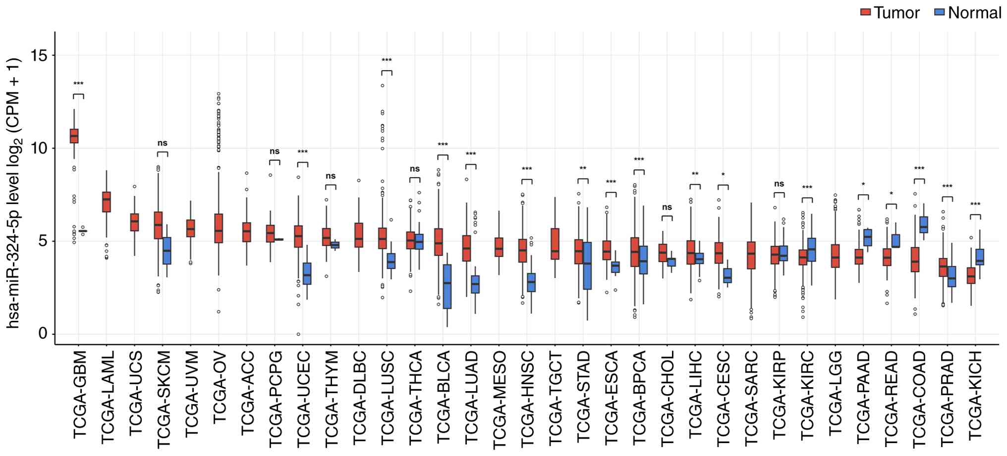 | Figure 1.hsa-miR-324-5p expression between
tumor and normal samples across TCGA cancer cohorts. hsa-miR-324-5p
expression levels were normalized to CPM and transformed as
log2(CPM+1). Samples were compared using a two-sided Wilcoxon
rank-sum test when both groups contained ≥2 samples. Statistical
significance was based on unadjusted P-values. *P<0.05;
**P<0.01; ***P<0.001. CPM, counts per million; ns, not
significant; TCGA, The Cancer Genome Atlas; LAML, acute myeloid
leukemia; UCS, uterine carcinosarcoma; SKCM, skin cutaneous
melanoma; UVM, uveal melanoma; ACC, adrenocortical carcinoma; OV,
ovarian serous cystadenocarcinoma; PCPG, pheochromocytoma and
paraganglioma; UCEC, uterine corpus endometrial carcinoma; DLBC,
lymphoid neoplasm diffuse large B cell lymphoma; THYM, thymoma;
THCA, thyroid carcinoma; LUSC, lung squamous cell carcinoma; BLCA,
bladder urothelial carcinoma; MESO, mesothelioma; TGCT, testicular
germ cell tumors; LUAD, lung adenocarcinoma; HNSC, head and neck
squamous cell carcinoma; ESCA, esophageal carcinoma; LIHC, liver
hepatocellular carcinoma; CHOL, cholangiocarcinoma; BRCA, breast
invasive carcinoma; CESC, cervical squamous cell carcinoma and
endocervical adenocarcinoma; STAD, stomach adenocarcinoma; KIRP,
kidney renal papillary cell carcinoma; LGG, brain lower grade
glioma; SARC, sarcoma; READ, rectum adenocarcinoma; PAAD,
pancreatic adenocarcinoma; KIRC, kidney renal clear cell carcinoma;
COAD, colon adenocarcinoma; PRAD, prostate adenocarcinoma; KICH,
kidney chromophobe; GBM, glioblastoma multiforme. |
Biological functions of miR-324-5p
across malignancies
Oncogenic effects of miR-324-5p upregulation in
cancer
Lung cancer (LC)
LC is a significant public health challenge, with
~2.6 million new cases and 1.9 million deaths worldwide in 2024,
accounting for 12.8% of all cancer cases and 19.1% of cancer
deaths, respectively (32).
Clinical studies have demonstrated altered circulating miR-324-5p
levels in patients with non-small cell lung cancer (NSCLC),
including increased plasma levels in one cohort (33,34).
F-box protein 11 (FBXO11), the mammalian ortholog of the
Caenorhabditis elegans E-box protein DRE-1, is a member of
the E3 ubiquitin ligase complex and is involved in multiple
biological processes, including apoptosis, DNA replication and
transcriptional regulation (35,36).
Mechanistically, long ncRNA (lncRNA) GATA6-AS1 acts as a competing
endogenous RNA (ceRNA) by sequestering miR-324-5p, thereby
restoring FBXO11 expression and suppressing LC cell proliferation
and invasiveness (17). Conversely,
elevated miR-324-5p expression negatively regulates the
tumor-suppressive activity of FBXO11, leading to decreased
cisplatin sensitivity and enhanced chemoresistance in LC cells
(16). miR-324-5p may contribute to
benzo[a]pyrene-7,8-diol-9,10-epoxide-induced malignant
transformation of human bronchial epithelial cells via modulation
of the circ_0067716-mediated miR-324-5p-DNA damage-regulated
autophagy modulator 1 (DRAM1) axis (37). Additionally, analysis of TCGA
sequencing data showed higher miR-324-5p expression in LUAD and
LUSC than in normal tissue samples, while cell-based experiments
showed that miR-324-5p upregulation promotes proliferation and
invasion in A549 human lung adenocarcinoma cells (38). However, another cell-based study
found that miR-324-5p upregulation decreases prostate
cancer-associated transcript 1 (PCAT-1)-induced proliferation of
A549 cells and promotes apoptosis, potentially through the
PCAT-1/miR-324-5p/RAP1A (RAP1A member of RAS oncogene family) axis
(39).
GC
GC ranks fifth worldwide in both incidence and
mortality, with ~980,000 new cases and 642,000 deaths estimated
globally in 2024 (32). Evidence
indicates elevated miR-324-5p expression in both GC tissues and
cell lines (15). Mechanistically,
inhibition of miR-324-5p suppresses GC cell proliferation and
invasive behavior while promoting apoptosis, potentially through
relief of miR-324-5p-mediated suppression of PTEN, a downstream
target negatively regulated by miR-324-5p (15). Moreover, circ_0049447 modulates GC
progression through sequestration of miR-324-5p, potentially
involving a protein tyrosine phosphatase receptor Δ (PTPRD)-related
mechanism (40). miR-324-5p has
been identified as a component of a plasma small extracellular
vesicle (sEV)-miRNA signature associated with distant lymph node
metastasis (41). However, another
study found that miR-324-5p expression is lower in five paired
clinical GC than in adjacent normal tissues (14). Mechanistically, inhibition of
miR-324-5p reverses the antitumor effects induced by circ_0091994
knockdown (14).
Breast cancer (BC)
In 2024, ~2.4 million new breast cancer cases and
694,000 BC-related deaths were estimated worldwide, accounting for
11.8% of all new cancer cases and 7.1% of all cancer deaths,
respectively (32). miR-324-5p
contributes to BC progression (42), with increased expression associated
with unfavorable clinical outcomes (43). TCGA transcriptomic profiling
combined with multi-omics analyses predicted miR-324-5p as a
potential regulator of Friend leukemia virus integration 1 (FLI1)
in breast invasive carcinoma (BRCA) (44). miR-324-5p expression shows an
inverse association with FLI1 levels, while increased miR-324-5p
abundance is associated with poorer overall survival (OS) (44). Furthermore, FLI1 was implicated in
the regulation of the BRCA immune microenvironment in a
cell-type-specific manner (44). In
addition, the Multicase-Control Spain (MCC-Spain) project reported
elevated circulating miR-324-5p levels in BC cases compared with
controls (45). Mechanistically,
miR-324-5p promotes BC progression by suppressing glutathione
peroxidase 3, thereby enhancing cellular proliferation, migration
and invasiveness (46).
Additionally, miR-324-5p regulates BC cell motility and
invasiveness through mechanisms involving NF-κB signaling, a
pathway involved in epithelial-mesenchymal transition (EMT) and
metastatic dissemination (47).
miR-324-5p downregulation results in increased CUE
domain-containing 2 (CUEDC2) expression, which inhibits IκB kinase
(IKK) phosphorylation and decreases NF-κB activity, ultimately
limiting BC cell invasion and migration (47). These findings demonstrate the
intricate regulatory mechanisms by which miR-324-5p influences BC
progression.
PTC
PTC is the predominant histological subtype of
thyroid cancer. In 2024, ~959,000 new thyroid cancer cases and
48,000 deaths were estimated worldwide (32). Increased miR-324-5p expression in
PTC tissue is associated with unfavorable clinicopathological
characteristics, including lymph node metastasis (LNM), greater
tumor size and advanced clinical stages (19). Molecular studies indicate that
miR-324-5p promotes PTC cell proliferation, migration and invasion
(18,19). Mechanistically, miR-324-5p directly
targets PTPRD and suppresses its protein expression, leading to
CCAAT enhancer-binding protein δ (CEBPD) upregulation (18). Elevated CEBPD expression enhances
the migratory and invasive capacities of human umbilical vein
endothelial cells within the tumor microenvironment and promotes
macrophage polarization toward an M2-like phenotype (18). Additionally, enhanced CEBPD levels
reinforce M2 macrophage polarization by increasing IL-4 and IL-13
production (18), thereby
contributing to PTC progression. Collectively, these observations
highlight the contribution of miR-324-5p to PTC development and
progression.
Pancreatic cancer (PC)
PC is characterized by an unfavorable prognosis. In
2024, ~531,000 new cases and 491,000 deaths from PC were estimated
worldwide (32), reflecting its
high mortality burden. miRNAs participate in PC development by
regulating molecular targets involved in tumor proliferation and
apoptosis, including KLF3 (48,49).
Evidence has implicated miR-324-5p in PC progression (48,49).
Increased miR-324-5p expression is detected in PC cell lines and
patient-derived tumor samples (49). Inhibition of miR-324-5p suppresses
PC cell proliferation and induces apoptosis, whereas restoration of
miR-324-5p expression enhances proliferative activity and
attenuates apoptotic responses (49). Mechanistically, Krüppel-like factor
3 (KLF3) is a direct downstream target of miR-324-5p. By
suppressing KLF3 expression, miR-324-5p promotes PC cell
proliferation and decreases apoptosis (49). Collectively, these data demonstrate
that miR-324-5p contributes to PC progression through regulation of
the KLF3-mediated molecular pathway. In addition, a recent study
implicates miR-324-5p in trace element metabolism (50,51).
In pancreatic ductal adenocarcinoma (PDAC) specimens, miR-324-5p
abundance is negatively associated with selenium, manganese and
zinc levels, suggesting a potential role for miR-324-5p in trace
element homeostasis (50).
Bladder cancer
Bladder cancer is a common malignant neoplasm of the
urinary system. In 2024, ~635,000 new bladder cancer cases and
228,000 deaths were estimated worldwide (32). Elevated urinary miR-324-5p levels
have been detected in patients with bladder cancer (52), while increased miR-324-5p abundance
has also been observed in surgically resected bladder cancer
tissues compared with matched adjacent tumor tissues (53). Mechanistic investigations have
implicated miR-324-5p in bladder cancer progression through
regulatory associations involving regulator of calcineurin 1
(RCAN1), FosB proto-oncogene, AP-1 transcription factor subunit
(FOSB) and eyes absent homolog 4 (EYA4) (52,53).
In particular, suppression of RCAN1 and EYA4 expression was
associated with enhanced bladder cancer cell proliferation, colony
formation and invasive behavior (53). Taken together, these findings
indicate that miR-324-5p may contribute to bladder cancer
progression by promoting oncogenic processes.
Oncogenic effects of miR-324-5p downregulation in
cancer
CRC
In 2024, >2 million new CRC cases and ~918,000
deaths were estimated worldwide, making CRC the third most commonly
diagnosed cancer and the second leading cause of cancer death
globally (32). TCGA analysis
identified decreased miR-324-5p levels in COAD and READ samples
compared with corresponding normal tissue. One study reported
decreased miR-324-5p levels in CRC cells (20). Overexpression of miR-324-5p
inhibited CRC cell proliferation, migration and invasion (20). Mechanistically, embryonic lethal
abnormal vision Drosophila-like protein 1 (ELAVL1) was a direct
target of miR-324-5p. Through regulation of invasion-associated
effectors, including MMP9, urokinase-type plasminogen activator
(uPA) and uPA receptor (uPAR), ELAVL1 influences CRC tumor growth
and metastatic potential (20).
Moreover, hsa_circ_0079662 serves as a ceRNA that sequesters
miR-324-5p, preventing miRNA-mediated repression of homeobox A9
(HOXA9) and enhancing tumor cell proliferation and oxaliplatin
resistance (54). Additionally,
miR-324-5p directly regulates superoxide dismutase 2 (SOD2),
resulting in decreased stemness characteristics of CRC cells and
improved responsiveness to chemotherapy with folinic acid,
oxaliplatin and fluorouracil (FOLFOX) (55). Supplementation with dietary n-3
polyunsaturated fatty acids experimentally elevates miR-324-5p
levels, counteracting carcinogen-induced suppression and protecting
against colorectal carcinogenesis (56). These effects may involve
Dicer-mediated miRNA biogenesis. Reduced Dicer expression is
associated with advanced CRC and poorer overall survival (57), whereas experimental Dicer deficiency
disrupts miRNA maturation, promotes cytoskeletal remodeling and
intestinal inflammation, and increases tumor formation (58). Decreased miR-324-5p levels are
reported in intestinal epithelial cells from Dicer-deficient mice
and in Dicer-deficient CRC cell lines (58). Mechanistically, miR-324-5p is a
downstream component of the Dicer-mediated miRNA network,
regulating high-mobility group box-containing protein 3 (HMGXB3)
and WAS protein family member 2 (WASF-2). This regulatory pathway
contributes to preservation of intestinal epithelial integrity,
decreased inflammatory responses and inhibited tumor initiation
(58). Furthermore, one study in
COAD revealed that augmenting miR-324-5p levels or suppressing the
lncRNA VPS9D1-AS1 limits tumor cell proliferation (59). VPS9D1-AS1 serves as a ceRNA by
sequestering miR-324-5p, thereby decreasing miR-324-5p-mediated
repression of integrin subunit α2 (ITGA2) and driving COAD
progression (59). Kang et
al (60) observed decreased
miR-324-5p expression in F-box and WD repeat domain-containing 7
(FBXW7)-mutant rectal neuroendocrine tumors (NETs), where
miR-324-5p was among seven miRNAs identified through
NanoString-based profiling of tumor samples. The altered expression
pattern of miR-324-5p may reflect FBXW7-associated molecular
changes and the biological heterogeneity of rectal NETs (60).
Hepatocellular carcinoma (HCC)
In 2024, ~843,000 new liver cancer cases and 732,000
deaths were estimated worldwide (32). HCC represents the predominant
histological subtype of primary liver cancer (61). Recent studies have highlighted
miR-324-5p-associated regulatory networks involved in HCC
progression, immune regulation and prognosis (62,63).
Notably, miR-324-5p expression is reduced in hepatitis C virus
(HCV)-infected HCC cells but is restored following IFN-α treatment,
indicating its involvement in the molecular interplay between HCV
infection and the response to IFN-α therapy (64). Similarly, HCC tissue and cell-based
models exhibit diminished miR-324-5p expression (27). Conversely, increasing miR-324-5p
expression inhibits the invasive capacity and metastatic properties
of HCC cells (27).
Mechanistically, miR-324-5p regulates ETS1 and SP1, resulting in
the downregulation of MMP2 and MMP9, and decreased HCC cell
invasion and migration (27). In
addition to direct target regulation, miR-324-5p activity in HCC is
modulated by multiple lncRNAs (27). For example, LINC00491 serves as a
ceRNA that binds miR-324-5p, thereby increasing ROCK1 expression
and promoting liver cancer progression (22). Similarly, lnc RP11-85G21.1, is
abundantly expressed in HCC cells and cell-derived exosomes, where
it promotes malignant phenotypes by modulating the functional
availability of miR-324-5p (65).
LINC01134 interacts with miR-324-5p and attenuates its suppressive
effect on insulin-like growth factor 2 mRNA binding protein 1
(IGF2BP1) (13). Through
stabilization of Yin Yang-1 (YY1) transcripts, IGF2BP1 increases
YY1 expression, which promotes LINC01134 transcription and creates
a positive feedback mechanism contributing to HCC progression
(13). Moreover, evidence suggests
an association between C-type lectin domain family 4s (CLEC4s)
member expression in HCC tissue and infiltration of immune cells
(66). miR-324-5p, a potential
regulator of CLEC4s, may modulate the HCC immune microenvironment
by targeting CLEC4s expression (66). In HCC, exosomal circGSE1 interacts
with miR-324-5p and modulates the transforming growth factor β
receptor 1 (TGFBR1)/Smad3 pathway, resulting in enhanced regulatory
T cell (Treg) expansion and immune evasion (21). In addition,
(1E,4E)-1,5-bis(2-bromophenyl) penta-1,4-dien-3-one (GL63), a
synthetic derivative of curcumin, displays antitumor activity,
partially by modulating circZNF83, a ceRNA within the miR-324-5p
regulatory network, thereby attenuating malignant phenotypes and
promoting apoptosis in HCC cells (25). Collectively, these data elucidate
the intricate molecular pathways involving miR-324-5p in HCC
pathogenesis and therapy.
Cervical cancer (CC)
In 2024, ~604,000 new CC cases and 280,000 deaths
were estimated worldwide (32).
Accumulating evidence implicates miR-324-5p in the regulation of CC
progression and treatment response through diverse molecular
pathways (11,67). Decreased expression of miR-324-5p is
reported in CC tissue, indicating its potential involvement in
tumor development (11).
Mechanistically, LINC00511 attenuates miR-324-5p activity through
competitive binding, resulting in DRAM1 dysregulation and altered
proliferative and migratory behavior of CC cells (11). Moreover, re-expression of miR-324-5p
increases the radiosensitivity of CC cells by enhancing apoptosis
and limiting migratory capacity (67). This radiosensitizing effect is
mediated, at least partially, by directly targeting ELAVL1,
followed by modulation of apoptosis-associated proteins, including
Bcl-2 and Bax (67). Therefore,
miR-324-5p may be a critical regulator of CC pathogenesis and
treatment-associated responses.
Ovarian cancer (OC)
In 2024, ~331,000 new ovarian cancer cases and
204,000 deaths were estimated worldwide (32). Dysregulated miRNA profiles
contribute to OC progression and therapeutic response (68). Survival analyses of serous OC
cohorts reveal that patients with lower miR-324-5p levels exhibit
poorer OS (69,70) and, in one cohort, with shorter
progression-free intervals (69).
Notably, although miR-324-5p downregulation is associated with
olaparib resistance, inhibition of the ataxia telangiectasia and
Rad3-related/checkpoint kinase 1 (ATR/CHK1) pathway suppresses
miR-324-5p expression and enhances olaparib sensitivity in
BRCA2-mutant olaparib-resistant ovarian cancer cells (PEO1-OR)
(69). Computational network
analysis has identified G2/M checkpoint-associated genes, including
tyrosine 3-monooxygenase/tryptophan 5-monooxygenase activation
protein θ, as potential downstream effectors of miR-324-5p,
suggesting miR-324-5p may influence cell cycle regulation during
the development of olaparib resistance (69). Furthermore, circZNF609 promotes OC
progression by sequestering miR-324-5p and relieving
miR-324-5p-mediated repression of voltage-dependent anion channel 1
expression (71). Conversely,
enforced miR-324-5p expression impairs OC cell proliferative and
invasive capacity and decreases xenograft tumor growth,
highlighting its tumor-suppressive function in OC (71).
Multiple myeloma (MM)
MM is a hematological malignancy driven by the
uncontrolled expansion of clonal plasma cells in the bone marrow
microenvironment, which is associated with clinical complications,
including renal dysfunction, hypercalcemia, anemia and osteolytic
bone lesions (72). Increased
miR-324-5p expression may attenuate MM progression by suppressing
cell proliferation, viability and cancer stem-like characteristics
(73,74). miR-324-5p enhances the therapeutic
response to bortezomib (BTZ) by regulating multidrug
resistance-associated proteins and apoptosis-associated members of
the Bcl-2 family (73). At the
molecular level, circATIC participates in a ceRNA regulatory
network by sequestering miR-324-5p, thereby relieving
miR-324-5p-mediated suppression of hepatocyte growth factor (HGF)
expression. This regulatory pathway enhances proliferation,
migration, invasion and glycolytic reprogramming while attenuating
apoptosis in MM cells (74).
Collectively, these results identify miR-324-5p as a key target
downstream of circATIC and regulator of MM biology.
GBM
GBM is a highly aggressive primary brain malignancy
(75). Despite multimodal
treatment, the median overall survival is ~15 months, and the
5-year survival rate remains ~5.8% (76). In GBM tissue, decreased miR-324-5p
abundance is observed with upregulation of circ_0079593 and X-box
binding protein 1 (XBP1) (77).
Mechanistically, miR-324-5p directly binds XBP1 and suppresses its
expression, thereby limiting GBM cell proliferation (77). By contrast, circ_0079593 promotes
GBM progression by competitive binding with miR-324-5p, thereby
increasing XBP1 expression (77).
Furthermore, lncRNA NEAT1 contributes to GBM progression through a
ceRNA mechanism, in which it binds miR-324-5p and prevents
miR-324-5p-mediated suppression of potassium channel
tetramerization domain-containing 20 (KCTD20). Restoration of
KCTD20 expression promotes GBM cell proliferation and attenuates
apoptosis (78). circSERPINE2
contributes to GBM progression by competitively binding miR-324-5p
and miR-361-3p, thereby relieving miRNA-mediated suppression of
Bcl-2 and promoting malignant cell survival (79). Conversely, miR-324-5p exerts
inhibitory effects on GBM by targeting enhancer of zeste homolog 2
(EZH2), thereby limiting tumor growth and increasing temozolomide
(TMZ) responsiveness (23).
Therefore, miR-324-5p appears to function as a negative regulator
of GBM progression.
Hematological malignancy
Hematological malignancy comprise neoplastic
disorders involving molecular alterations and exhibiting
substantial variability in treatment response. A circulating miRNA
study in diffuse large B cell lymphoma (DLBCL) have linked
miR-324-5p to responsiveness to rituximab, cyclophosphamide,
doxorubicin, vincristine and prednisone (80). Tumor miRNA profiling demonstrated an
association between miR-324-5p expression and survival outcomes in
patients with DLBCL (81).
Circulating and tissue-derived miR-324-5p display divergent
expression patterns in DLBCL, with elevated serum levels observed
in refractory patients but decreased expression detected in tumors
from non-responsive cases (80).
Re-expression of miR-324-5p impairs DLBCL cell survival through
thioredoxin reductase 1 regulation and ferroptosis induction,
providing functional evidence for its context-dependent antitumor
activity (80). Bioinformatics
suggests a potential role for miR-324-5p in acute myeloid leukemia
(AML) (82). A miRNA profiling
study identified miR-324-5p among the miRNAs upregulated in primary
AML samples compared with normal bone marrow (83). A bioinformatic analysis predicted a
hsa_circ_0058058/miR-324-5p regulatory axis involving
adaptor-associated protein complex 1 γ1 subunit (AP1G1) and SP1,
suggesting this network may contribute to AML progression (82). Collectively, the aforementioned
studies indicate that miR-324-5p has context-dependent roles in
hematological malignancy with its biological effects varying across
disease entities and regulatory networks. The expression patterns,
regulatory mechanisms and biological functions of miR-324-5p across
different cancer types are summarized in Table I.
 | Table I.Expression, regulatory mechanisms and
biological functions of miR-324-5p in cancer. |
Table I.
Expression, regulatory mechanisms and
biological functions of miR-324-5p in cancer.
| A, LC |
|---|
|
|---|
| Regulation | Biological
effects | Regulatory
pathway/targets | Role | (Refs.) |
|---|
| Up | Contributes to LC
initiation |
circ_0067716/miR-324-5p/DRAM1 |
Tumor-promoting | (37) |
| Up | Enhances
proliferation and invasiveness of LC cells via FBXO11
regulation |
GATA6-AS1/miR-324-5p/FBXO11 |
Tumor-promoting | (17) |
| Up | Promotes LC
progression by suppressing FBXO11-mediated tumor inhibition and
decreasing cisplatin sensitivity |
miR-324-5p/FBXO11 |
Tumor-promoting | (16) |
| Up | Promotes LC cell
proliferation and invasion | GPX3, RCAN1 and
MGAT3 |
Tumor-promoting | (38) |
| Not assessed | Suppresses LC cell
proliferation and promotes apoptosis by targeting RAP1A |
PCAT-1/miR-324-5p/RAP1A |
Tumor-suppressive | (39) |
|
| B, GC |
|
| Up | Enhances metastatic
behavior by regulating GC cell migration and invasion |
circ_0049447/miR-324-5p/PTPRD |
Tumor-promoting | (40) |
| Down | Suppresses GC cell
viability and promotes apoptosis via HMGA1 regulation |
hsa_circ_0091994/miR-324-5p/HMGA1 |
Tumor-suppressive | (14) |
| Up | Promotes GC cell
proliferation, migration and invasion while inhibiting apoptosis
via PTEN regulation |
miR-324-5p/PTEN |
Tumor-promoting | (15) |
|
| C, Breast
cancer |
|
| Up | Promotes tumor
progression by enhancing proliferation, migration, invasion and
unfavorable clinical outcomes |
miR-324-5p/GPX3 |
Tumor-promoting | (46) |
| Up | Elevated
circulating miR-324-5p contributes to diagnostic prediction of
breast cancer | Circulating
miR-324-5p; seven-miRNA signature | Biomarker | (45) |
| Up | Regulates cell
motility and invasiveness via modulation of NF-κB signaling and
EMT-associated processes |
miR-324-5p/CUEDC2/NF-κB |
Tumor-promoting | (47) |
|
| D, PTC |
|
| Up | Promotes
proliferation, migration and invasion of thyroid cancer cells and
remodels the tumor microenvironment via endothelial activation and
M2 macrophage polarization | miR-324-5p/PTPRD;
miR-324-5p/PTPRD/CEBPD |
Tumor-promoting | (18) |
|
| E, PC |
|
| Up | Promotes PC cell
proliferation and suppresses apoptosis, thereby facilitating tumor
progression |
miR-324-5p/KLF3 |
Tumor-promoting | (49) |
|
| F, Bladder
cancer |
|
| Up | Promotes
proliferation, colony formation and invasion of bladder cancer
cells | miR-324-5p/RCAN1
and EYA4 |
Tumor-promoting | (53) |
|
| G, CRC |
|
|
Regulation | Biological
effects | Regulatory
pathway/targets | Role | (Refs.) |
|
| Down | Restores
miR-324-5p-mediated inhibition of tumor growth and invasion |
miR-324-5p/ELAVL1 |
Tumor-suppressive | (20) |
| Down | Suppresses CRC cell
proliferation and invasion by regulating invasion-associated
proteins |
miR-324-5p/ELAVL1/MMP9/uPA/uPAR |
Tumor-suppressive | (18) |
| Down | Promotes tumor cell
proliferation and oxaliplatin resistance by sequestering
miR-324-5p, thereby decreasing miR-324-5p-mediated suppression of
HOXA9 |
hsa_circ_007966/miR-324-5p/HOXA9 | Tumor-promoting
(ceRNA-mediated) | (54) |
| Down | Restores miR-324-5p
expression and suppresses colorectal carcinogenesis by
counteracting carcinogen-induced miRNA downregulation | Dietary n-3
PUFA/miR-324-5p |
Tumor-suppressive | (55) |
| Down | Decreases cancer
stem cell-like properties and enhances FOLFOX sensitivity via SOD2
regulation |
miR-324-5p/SOD2 |
Tumor-suppressive | (55) |
| Down | Loss of
Dicer-mediated miR-324-5p regulation promotes intestinal
inflammation, barrier disruption and tumor initiation |
Dicer/miR-324-5p/HMGXB3/WASF-2 |
Tumor-suppressive | (58) |
| Down | Inhibits COAD cell
proliferation by restoring miR-324-5p activity |
VPS9D1-AS1/miR-324-5p/ITGA2 |
Tumor-suppressive | (59) |
|
| H, HCC |
|
| Down | Suppresses invasion
and migration by inhibiting extracellular matrix remodeling | miR-324-5p/ETS1,
SP1/MMP2 and MMP9 |
Tumor-suppressive | (27) |
| Down | Enhances
proliferation and migration via modulation of miR-324-5p
availability |
lnc85/miR-324-5p |
Tumor-suppressive | (65) |
| Down | Facilitates tumor
progression by activation of an IGF2BP1/YY1 positive feedback
loop |
LINC01134/miR-324-5p/IGF2BP1/YY1 | Tumor-promoting
(ceRNA-mediated) | (13) |
| Down | May regulate the
immune microenvironment via modulation of CLEC4s expression and
immune cell infiltration |
miR-324-5p/CLEC4s |
Context-dependent | (66) |
| Down | Promotes immune
evasion by enhancing Treg expansion via TGF-β signaling
activation | Exosomal
circGSE1/miR-324-5p/TGFBR1/Smad3 | Tumor-promoting
(immune regulation) | (21) |
| Down | GL63 inhibits tumor
growth by suppressing the JAK2/STAT3 pathway |
GL63/circZNF83/miR-324-5p/CDK16/JAK2/STAT3 |
Tumor-suppressive | (25) |
|
| I, Gallbladder
cancer |
|
| Down | Inhibits migration,
invasion and EMT of gallbladder cancer cells |
miR-324-5p/TGFB2 |
Tumor-suppressive | (90) |
|
| J, CC |
|
|
Regulation | Biological
effects | Regulatory
pathway/targets | Role | (Refs.) |
|
| Down | Promotes
proliferation and migration via regulation of DRAM1 expression | RNA
LINC00511/ceRNA/miR-324-5p/DRAM1 |
Tumor-suppressive | (11) |
| Down | Enhances
radiosensitivity by promoting apoptosis and suppressing
migration |
miR-324-5p/ELAVL1/Bax/Bcl-2 |
Tumor-suppressive | (67) |
|
| K, Endometrial
cancer |
|
| Down | Inhibits the
proliferation, migration and invasion of endometrial cancer
cells |
miR-324-5p/HMGA1 |
Tumor-suppressive | (91) |
|
| L, OC |
|
| Down | Low miR-324-5p
expression is associated with poor OS and shorter PFI and
influences olaparib resistance | ATR/CHK1 |
Context-dependent | (68) |
| Down | Modulates G2/M
checkpoint regulation and olaparib response via YWHAQ-associated
mechanisms |
miR-324-5p/YWHAQ |
Tumor-suppressive | (68) |
| Down | Suppresses
proliferation, migration, invasion and tumor growth by inhibiting
VDAC1 expression |
circZNF609/miR-324-5p/VDAC1 |
Tumor-suppressive | (71) |
|
| M, MM |
|
| Down | Suppresses
proliferation, viability and cancer stem-like properties and
enhances bortezomib sensitivity | miR-324-5p/Bcl-2
family and multidrug resistance-associated proteins |
Tumor-suppressive | (73) |
| Down | Promotes
proliferation, migration, invasion and glycolytic reprogramming via
HGF activation following miR-324-5p sequestration |
circATIC/miR-324-5p/HGF | Tumor-promoting
(ceRNA-mediated) | (74) |
|
| N, GBM |
|
| Down | Suppresses
proliferation by directly targeting XBP1 |
miR-324-5p/XBP1 |
Tumor-suppressive | (77) |
| Down | Promotes
proliferation by sequestering miR-324-5p, thereby decreasing
miR-324-5p-mediated suppression of XBP1 |
circ_0079593/miR-324-5p/XBP1 | Tumor-promoting
(ceRNA-mediated) | (77) |
| Down | Enhances
proliferation and decreases apoptosis through KCTD20
activation |
NEAT1/miR-324-5p/KCTD20 | Tumor-promoting
(ceRNA-mediated) | (78) |
| Down | Promotes malignant
cell survival via Bcl-2 upregulation |
circSERPINE2/miR-324-5p/miR-361-3p/Bcl-2 | Tumor-promoting
(ceRNA-mediated) | (79) |
| Down | Inhibits tumor
growth and increases temozolomide sensitivity via EZH2
suppression |
miR-324-5p/EZH2 |
Tumor-suppressive | (23) |
|
| O,
DLBCL |
|
|
Regulation | Biological
effects | Regulatory
pathway/targets | Role | (Refs.) |
|
| Down (tumor
tissue); up (serum) | Inhibits lymphoma
cell survival and promotes ferroptosis via TXNRD1 suppression |
miR-324-5p/TXNRD1 |
Context-dependent | (80) |
miR-324-5p mediates tumor-associated
pathways
The major tumor-associated signaling pathways
involving miR-324-5p are summarized in Fig. 2.
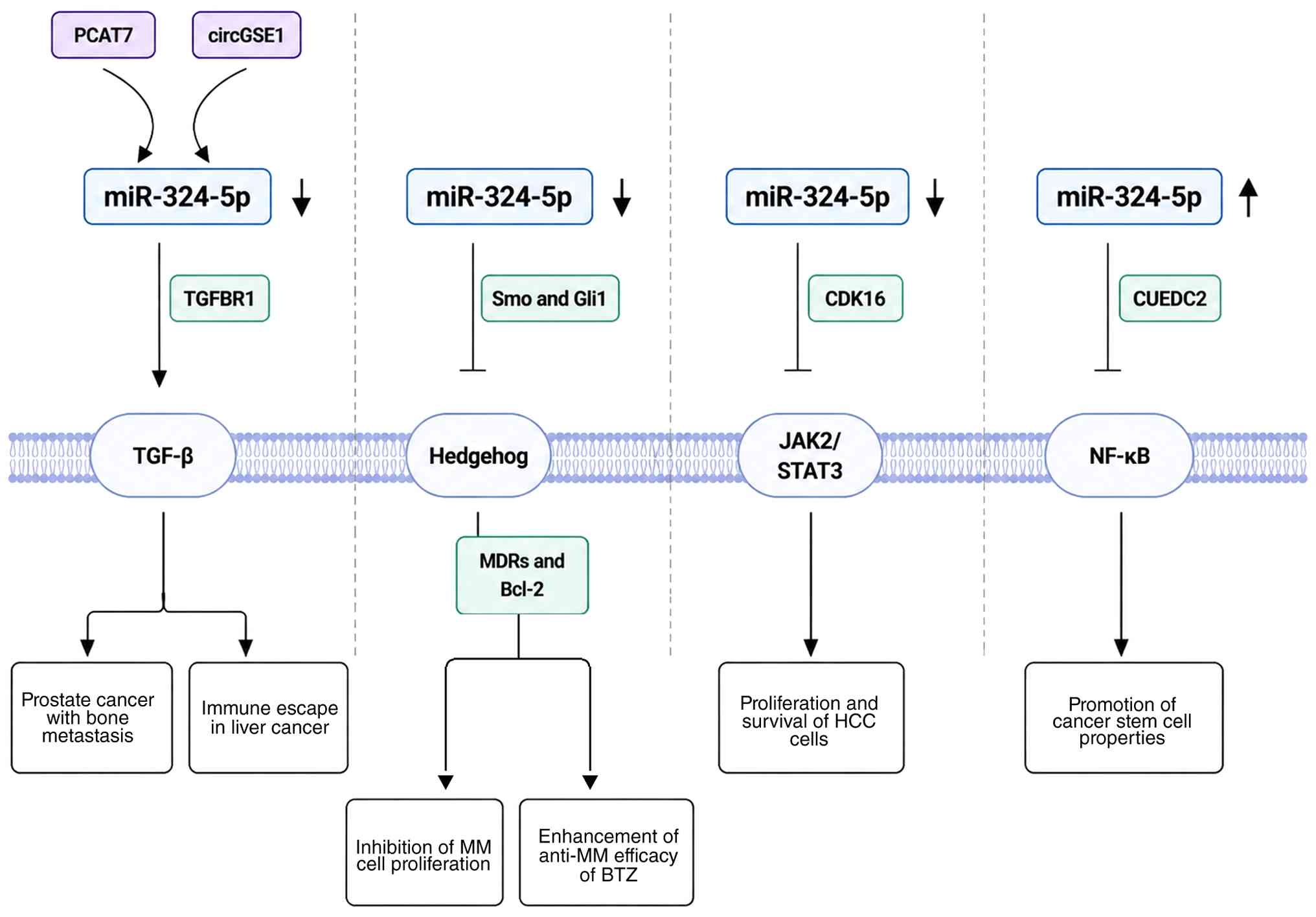 | Figure 2.miR-324-5p-mediated tumor pathway.
miR-324-5p-associated regulatory networks modulate TGFBR1
expression and TGF-β signaling. In MM, miR-324-5p suppresses the
Hedgehog pathway through Smo and Gli, decreases MDR protein and
Bcl-2 expression and enhances bortezomib sensitivity. In HCC,
increased miR-324-5p activity suppresses CDK16 expression and
attenuates JAK2/STAT3 signaling. miR-324-5p downregulates CUEDC2,
thereby affecting NF-κB-associated cancer stem-like phenotypes.
CUEDC2, CUE domain-containing 2; TGFBR1, transforming growth factor
β receptor 1; Smo, smoothened; Gli1, Gli family zinc finger 1; MM,
multiple myeloma; CDK16, cyclin-dependent kinase 16; HCC,
hepatocellular carcinoma; circ, circular; miR, microRNA. |
TGF-β pathway
TGF-β signaling is extensively involved in tumor
biology and influences multiple cell programs, including
differentiation, cell motility, EMT and metastatic spread (84). In prostate cancer, lncRNA PCAT7
promotes TGFBR1 expression by competitively interacting with
miR-324-5p, thereby increasing TGF-β pathway activity. This
PCAT7/miR-324-5p/TGFBR1 regulatory pathway contributes to prostate
cancer bone metastasis (85).
Conversely, exosome-derived circGSE1 in HCC decreases miR-324-5p
availability, resulting in TGFBR1 upregulation, Smad3-dependent
signaling activation and increased Treg infiltration, thereby
promoting tumor immune escape (21). The influence of miR-324-5p on TGF-β
signaling is shaped by tumor-specific ceRNA networks, resulting in
distinct functional outcomes across cancer types.
Hedgehog (Hh) pathway
Hh signaling is key to developmental patterning and
the maintenance of tissue homeostasis in adulthood, whereas
aberrant pathway activity is associated with tumor progression
(86). In MM, the miR-324-5p locus
resides within a chromosomal region typically affected by 17p
deletion. Patients with del(17p) frequently exhibit decreased
miR-324-5p expression, resulting in enhanced Hh pathway activity
(73). Experimental findings show
miR-324-5p negatively regulates the Hh pathway by targeting Gli
family zinc finger 1 and Smoothened, with consequent inhibition of
MM cell proliferation, survival and stem-like behavior (73). Moreover, restoration of miR-324-5p
expression disrupts the supportive interactions between MM and bone
marrow stromal cells (73).
Increased miR-324-5p levels also regulate multidrug
resistance-associated proteins and Bcl-2 family members via Hh
pathway inhibition, thereby improving the therapeutic response to
BTZ in MM (73). Overall,
miR-324-5p influences MM progression and treatment sensitivity
through Hh pathway regulation, suggesting this miRNA may represent
a candidate for therapeutic intervention in MM.
JAK2/STAT3 pathway
JAK2/STAT3 signaling coordinates cellular activities
and has been implicated in both tumorigenesis and malignant
progression (87). Zhao et
al (25) reported that GL63, a
curcumin derivative, suppresses circZNF83 expression, thereby
restoring miR-324-5p availability and relieving circZNF83-mediated
sequestration of miR-324-5p. Increased miR-324-5p activity
suppresses cyclin-dependent kinase 16 (CDK16), leading to decreased
JAK2/STAT3 signaling activity and suppression of malignant
phenotypes in HCC (25). Thus, GL63
may inhibit JAK2/STAT3 signaling in HCC via the
circZNF83/miR-324-5p/CDK16 regulatory pathway (25).
NF-κB pathway
NF-κB signaling regulates tumor cell biology,
including proliferation, inflammatory activity, survival and immune
escape (88). Persistent
disturbance of this pathway may promote malignant transformation,
invasion and metastatic dissemination. Elevated miR-324-5p levels
are detected in NSCLC cells and other malignancies carrying
gain-of-function TP53 mutations (89). At the molecular level, mutant p53
enhances miR-324-5p transcription via the c-Myc signaling pathway,
resulting in CUEDC2 downregulation and NF-κB activation. This
regulatory pathway promotes key cancer stem-like properties,
including increased proliferation, survival and chemotherapy
resistance (89). Conversely,
miR-324-5p silencing restores CUEDC2 expression, suppresses NF-κB
signaling activity and attenuates cancer-like phenotypes (89). Available evidence implicates the
TP53/c-Myc/miR-324-5p/CUEDC2 pathway in coupling miR-324-5p
activity with NF-κB signaling and cancer stem-like properties
(89).
miR-324-5p affects tumor growth and metastasis in
animal models
Several in vivo studies have evaluated the
tumor-regulatory functions of miR-324-5p in cancer models (Table II) (14,22,49,58,59,79,90,91).
These studies indicate that miR-324-5p-related interventions can
influence tumor growth, metastasis and treatment-associated
phenotypes through mechanisms involving direct target gene
regulation, EMT-associated changes, ceRNA networks and intestinal
barrier-associated pathways. These in vivo findings
complement cell-based evidence and provide additional support for
the context-dependent role of miR-324-5p in tumor regulation.
| Table II.Animal models investigating
miR-324-5p-mediated tumor progression and regulatory
mechanisms. |
Table II.
Animal models investigating
miR-324-5p-mediated tumor progression and regulatory
mechanisms.
| Cancer | Animal model | Treatment | Intervention | Key in vivo
phenotypic outcomes |
miR-324-5p-associated pathways | (Refs.) |
|---|
| Gastric cancer | BALB/c nude
xenograft model | Not applicable | hsa_circ_0091994
knockdown | Decreased tumor
growth; miR-324-5p restoration |
hsa_circ_0091994/miR-324-5p/HMGA1 | (14) |
| Liver cancer | Nude mouse
xenograft and metastasis models | Xenograft tumors
and lung metastasis | LINC00491
overexpression | Increased tumor
growth and metastasis and proliferation markers |
LINC00491/miR-324-5p/ROCK1 | (22) |
| PDAC | BALB/c nude
subcutaneous xenograft model | miR-324-5p
antagomir (5 nmol, every 5 days for 35 days) | miR-324-5p
inhibition | Decreased tumor
volume/weight and PCNA-positive cells |
miR-324-5p/KLF3 | (49) |
| Colorectal
cancer | Intestinal
epithelial Dicer-deficient mouse model (DSS/AOM-DSS CRC
models) | miR-324-5p agomiR
(10 nmol); 3% DSS-induced colitis and AOM/DSS-induced CRC
models | Dicer deletion to
decrease miR-324-5p; miR-324-5p agomiR rescue treatment in DSS
model | Dicer deficiency
promotes intestinal injury and tumorigenesis; miR-324-5p rescue
restores intestinal protection |
Dicer/miR-324-5p/HMGXB3/WASF-2 | (58) |
| COAD | Nude mouse
xenograft | Transfection before
implantation | VPS9D1-AS1
knockdown | Decreased tumor
growth |
VPS9D1-AS1/miR-324-5p/ITGA2 ceRNA | (59) |
| Gliobla stoma | Nude mouse
xenograft model | ~1×10⁶ cells
injected subcutaneously | circSERPINE2
knockdown | Decreased tumor
growth and proliferation; increased apoptosis |
circSERPINE2/miR-361-3p/miR-324-5p/BCL2 | (79) |
| GBC | BALB/c nude
xenograft model | 1×10⁶
SGC-996 cells | miR-324-5p
overexpression or TGFB2 knockdown | Decreased incidence
of liver metastasis and fewer liver metastatic foci | TGFB2 downstream of
miR-324-5p | (90) |
| GBC | BALB/c nude
xenograft model | Stable SGC-996
×enograft model | miR-324-5p
overexpression | Decreased liver
metastasis and metastatic foci |
miR-324-5p/TGFB2/EMT-associated
metastasis | (90) |
| Endometrial
carcinoma | BALB/c nude
xenograft model; circ_0067835 knockdown | Cell-based shRNA
xenograft model | circ_0067835
knockdown | Decreased tumor
growth, proliferation and EMT; increased apoptosis |
circ_0067835/miR-324-5p/HMGA1 | (91) |
| RMS | SCID xenograft
model | 1×10⁶ RD
cells injected into the gastro-cnemius muscle; doxycycline (1
mg/ml) initiated 1 week later | miR-324-5p
overexpression | Decreased tumor
growth and ITGA9/Ki-67/CD31 |
miR-324-5p/ITGA9/FAK-associated
signaling | (94) |
miR-324-5p regulates tumor growth, invasion and
metastasis through target-gene-associated pathways
Integrin and focal adhesion kinase (FAK) signaling
pathways are key regulators of tumor invasion and metastatic
progression (92,93). ITGA9 is directly regulated by
miR-324-5p through interaction with the ITGA9 3′UTR (94). In rhabdomyosarcoma (RMS),
miR-324-5p-mediated suppression of ITGA9 limits proliferative and
invasive behavior in vitro and decreases tumor growth in
mouse xenograft models (94).
Decreased ITGA9 levels are accompanied by reduced FAK
phosphorylation, indicating that the miR-324-5p/ITGA9 pathway may
regulate RMS progression via FAK-related signaling (94). In a COAD xenograft model, enforced
miR-324-5p expression and VPS9D1-AS1 silencing markedly inhibits
tumor growth. VPS9D1-AS1 regulates the miR-324-5p/ITGA2 pathway by
limiting miR-324-5p activity, thereby favoring ITGA2 expression and
COAD progression (59).
Beyond integrin-associated pathways, miR-324-5p is
associated with EMT regulation and metastatic dissemination. In
gallbladder carcinoma (GBC), elevated miR-324-5p expression
decreases cell motility, invasion and EMT-related phenotypes,
leading to fewer hepatic metastatic lesions in vivo
(90). Conversely, miR-324-5p is
downregulated in liver cancer tissue and cells models relative to
corresponding non-tumor controls and its restoration suppresses
tumor-associated phenotypes (22,27).
Mechanistically, miR-324-5p limits tumor expansion and metastatic
potential by modulating ROCK1 (22).
miR-324-5p regulates tumor growth by modulating
transcriptional factors. In endometrial cancer xenografts,
miR-324-5p inhibits subcutaneous tumor formation by suppressing
high mobility group AT-hook 1 (HMGA1) expression (91). In a PC xenograft model, miR-324-5p
promotes xenograft tumor growth by targeting KLF3. Conversely,
inhibition of miR-324-5p markedly attenuates tumor growth (49).
miR-324-5p regulates tumor progression via ceRNA
networks
circRNAs and lncRNAs modulate miR-324-5p activity in
ceRNA networks, thereby affecting tumor-associated phenotypes. In a
GBM xenograft model, loss of circSERPINE2 markedly inhibits tumor
growth (79). Mechanistically,
circSERPINE2 sequesters miR-324-5p and miR-361-3p, decreasing
suppression of Bcl-2 and favoring GBM progression (79). In GC xenografts, higher miR-324-5p
abundance is associated with smaller and lighter tumors (14). Silencing hsa_circ_0091994 increases
miR-324-5p availability, resulting in decreased HMGA1 expression
and suppression of GC progression (14).
miR-324-5p regulates tumorigenesis through
cytoskeletal remodeling and intestinal barrier maintenance
Altered Dicer expression is implicated in CRC
(57), and experimental Dicer
deficiency reduces miR-324-5p expression, supporting a role for
Dicer-mediated miRNA processing in the regulation of miR-324-5p
(58). In a CRC model, Dicer
deficiency markedly decreases miR-324-5p expression in mice,
resulting in abnormal cytoskeletal alterations in intestinal
epithelial cells, compromised barrier function, enhanced
inflammatory response and increased tumor formation (58). Conversely, intraperitoneal delivery
of miR-324-5p agonist alleviates dextran sulfate sodium-induced
intestinal inflammation and tissue injury, restores epithelial
barrier integrity and decreases HMGXB3 and WASF-2 expression,
thereby suppressing CRC progression (58).
miR-324-5p as a potential cancer
biomarker
Application of miR-324-5p in cancer
diagnosis
miRNAs, which are detectable in both intracellular
and extracellular environments, have attracted attention for their
potential utility in cancer detection, prognostic evaluation and
assessment of therapeutic response (95). Combined RNA sequencing and reverse
transcription-quantitative (RT-q)PCR have detected enrichment of
miR-324-5p in plasma-derived exosomes from patients with NSCLC,
indicating potential biomarker relevance (33). Similarly, elevated miR-324-5p levels
are observed in urine specimens from individuals with bladder
cancer and show an inverse association with the expression of the
lncRNA miR-497-HG (52). These
clinical observations are complemented by a separate functional
study showing that miR-324-5p promotes bladder cancer cell
proliferation, colony formation, migration and invasion (53). In PTC, higher miR-324-5p levels are
linked to LNM, greater tumor burden and more advanced disease
stage, supporting its potential utility for patient stratification
and risk assessment (19).
Gómez-Acebo et al (45)
reported higher circulating miR-324-5p levels in patients with
breast cancer than in healthy controls and incorporated this miRNA
into a seven-miRNA diagnostic signature (OR=1.60, 95% CI:
1.08–2.37), supporting its potential value as a diagnostic marker.
Collectively, evidence points to a potential diagnostic application
for miR-324-5p, although validation is needed to establish its
clinical biomarker utility.
Association between miR-324-5p levels
and patient prognosis
miR-324-5p expression patterns are associated with
clinical outcomes across several types of malignancies. In glioma,
miR-324-5p has been identified as an independent prognostic
indicator (23). In GC, RT-qPCR has
revealed elevated miR-324-5p levels in tumor tissue and cell lines,
which are associated with lymphatic metastasis, advanced disease
stage and poor survival (15).
Another study profiled miRNAs in plasma-derived small extracellular
vesicles from patients with GC using sEV-miRNA sequencing followed
by RT-qPCR validation and found that miR-324-5p abundance is
associated with organ-specific metastatic patterns, suggesting
potential prognostic relevance (41). By contrast, lower miR-324-5p levels
in GBC are linked to adverse clinical outcomes (90). This association may be partly
explained by miR-324-5p-mediated regulation of TGFB2, which affects
GBC cell migration, invasion and EMT (90). In triple-negative breast cancer,
next-generation sequencing of 51 specimens showed that elevated
miR-324-5p levels were associated with shorter OS (43). Overall, these observations indicate
that miR-324-5p may have prognostic value across multiple tumor
types.
Potential of miR-324-5p as a therapeutic
target
Accumulating evidence indicates miRNAs participate
in carcinogenesis by controlling target transcripts (95,96).
Therapeutic alteration of miRNA function has been investigated as a
strategy to reshape oncogenic signaling networks (97). Given its context-dependent functions
across malignancies, miR-324-5p may be therapeutically modulated
through lncRNA/circRNA regulatory networks or by exogenous miRNA
mimics or inhibitors (12,20,22,59,74,77,78,91).
In addition, miR-324-5p is implicated in cancer-specific responses
to chemotherapy and radiotherapy, as well as in the regulation of
tumor-associated immune processes (16,21,23,54,55,66,67,69,73)
(Fig. 3). Further development of
such strategies requires tumor-specific determination of whether
miR-324-5p activity should be restored or inhibited.
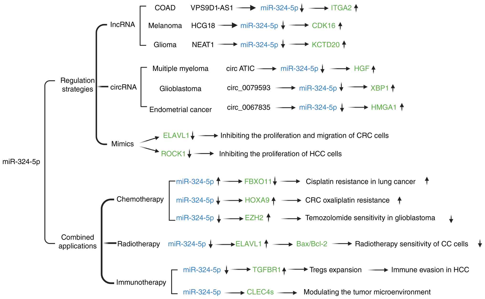 | Figure 3.miR-324-5p-mediated tumor therapeutic
strategies. miR-324-5p is therapeutically modulated through
lncRNA/circRNA regulatory networks or by exogenous miRNA mimics and
inhibitors. These strategies may alter tumor growth, invasion and
apoptosis and may modify responses to chemotherapy, radiotherapy
and tumor-associated immune regulation through cancer-specific
downstream targets and signaling pathways. ITGA2, integrin subunit
α2; CDK16, cyclin-dependent kinase 16; KCTD20, potassium channel
tetramerization domain containing 20; HGF, hepatocyte growth
factor; XBP1, X-box binding protein 1; HMGA1, high mobility group
A1; ELAVL1, embryonic lethal abnormal vision Drosophila-like
protein 1; CRC, colorectal cancer; ROCK1, rho-associated kinase 1;
HCC, hepatocellular carcinoma; FBXO11, F-box protein 11; HOXA9,
homeobox A9; EZH2, zeste homolog 2; TGFBR1, transforming growth
factor β receptor 1; CC, cervical cancer; CLEC4S, C-type lectin 4s;
circ, circular RNA; miR, microRNA; lnc, long non-coding; COAD,
colon adenocarcinoma. |
Strategies for regulating
miR-324-5p
lncRNA strategies
lncRNAs interact with miRNAs through regulatory
networks involved in tumor initiation and progression (98). Several lncRNAs have been reported to
alter the availability or activity of miR-324-5p, with effects on
malignant phenotypes (12,59,78).
In GBM, lncRNA NEAT1 sequesters miR-324-5p, thereby weakening its
inhibitory effect on KCTD20 and facilitating tumor cell
proliferation (78). A similar
regulatory mechanism operates in COAD, where lncRNA VPS9D1-AS1
overexpression suppresses miR-324-5p activity and elevates ITGA2
expression. The VPS9D1-AS1/miR-324-5p/ITGA2 pathway promotes COAD
cell proliferation and decreases apoptosis (59). In melanoma, HLA complex group 18
(HCG18) expression is elevated in both tumor specimens and cultured
cells, and modulates the miR-324-5p/CDK16 pathway (12). By decreasing miR-324-5p-mediated
suppression of CDK16, HCG18 enhances malignant cell proliferation,
motility and invasiveness while decreasing apoptotic activity
(12). Although these lncRNAs act
in different tumor settings, they converge on miR-324-5p and
regulate malignant phenotypes through distinct downstream
effectors, including KCTD20, ITGA2 and CDK16 (12,59,78).
Whether these networks are independent or share common upstream
regulatory features remains unclear. Comparison of their expression
patterns, intracellular localization and interaction with
miR-324-5p may clarify this issue.
circRNA-based strategies
circRNAs influence gene expression through several
mechanisms, including functional interaction with miRNAs (99). One mechanism involves sequestration
of miRNAs, thereby decreasing miRNA-mediated suppression of
downstream targets and influencing tumor development (100). Interactions between circRNAs and
miR-324-5p are implicated in the regulation of multiple
tumor-associated phenotypes (74,77,91).
In MM, circATIC binds miR-324-5p, thereby increasing HGF expression
and promoting cell proliferation, invasion, migration and
glycolysis while suppressing apoptosis (74). In GBM, circ_0079593 is upregulated
and associated with decreased miR-324-5p expression (77). This circRNA is implicated in GBM
progression via the miR-324-5p/XBP1 regulatory pathway (77). Similarly, circ_0067835 is
upregulated in endometrial carcinoma and negatively associated with
miR-324-5p levels (91). By serving
as a ceRNA, circ_0067835 increases HMGA1 levels and promotes
endometrial carcinoma progression (91). Although these circRNAs are involved
in different tumor contexts, they converge on miR-324-5p and
influence malignant phenotypes through distinct downstream
effectors (74,77,91).
To the best of our knowledge, whether these regulatory events occur
independently or share common upstream determinants has not been
fully explored. Future studies comparing miR-324-5p interactions
and upstream regulatory mechanisms may clarify this. Further
investigation may reveal whether individual circRNAs or broader
miR-324-5p regulatory networks represent feasible therapeutic
targets.
Therapeutic application of miR-324-5p
overexpression or inhibition
Therapeutic modulation of miR-324-5p using mimics
or antagonists has demonstrated potential in preclinical cancer
models (20,22,58).
In CRC cell systems, miR-324-5p mimics directly target ELAVL1,
resulting in decreased levels of invasion-associated proteins,
including MMP-9, uPA and uPAR, and suppressing CRC cell
proliferation, migration and invasive behavior (20). Conversely, inhibition of miR-324-5p
increases ELAVL1 expression together with invasion-associated
factors, thereby promoting CRC cell growth and invasiveness
(20). This supports the
possibility that restoring miR-324-5p activity may represent a
therapeutic strategy for CRC. Similarly, in liver cancer, treatment
with miR-324-5p mimics suppresses the proliferative, migratory and
invasive capacity of HUH-7 and SK-Hep1 cells while inducing
apoptosis, whereas miR-324-5p inhibition produces opposing cell
responses (22). Mechanistically,
LINC00491 promotes HCC progression by serving as a ceRNA that
sequesters miR-324-5p, thereby decreasing miR-324-5p-mediated
suppression of ROCK1 (22).
Restoration of miR-324-5p through mimics counteracts the oncogenic
effects associated with LINC00491, supporting the therapeutic
relevance of miR-324-5p modulation (22). Taken together, the aforementioned
preclinical studies suggest that miR-324-5p-based interventions may
have therapeutic value in certain cancer contexts. However,
clinical translation of miRNA mimics remains limited by challenges,
including delivery efficiency, molecular stability,
immune-associated responses and off-target effects, which require
further optimization before clinical application (101).
Regulation of miR-324-5p in combination
with chemotherapy, radiotherapy and immunotherapy
miR-324-5p is implicated in treatment response
across cancer, particularly in relation to chemotherapy,
radiotherapy and tumor-associated immune regulation (16,21,23,54,55,66,67,69,73).
Chemotherapy
Resistance to chemotherapy is a key barrier
limiting the effectiveness of anticancer treatment. Increasing
evidence indicates that miRNAs contribute to chemotherapy
sensitivity and resistance through post-transcriptional regulation
of target genes (95,102). miR-324-5p influences chemotherapy
responses across multiple cancer types. In LC, increased miR-324-5p
expression decreases FBXO11 levels, thereby decreasing cisplatin
sensitivity and promoting chemoresistance (16). In OC, miR-324-5p is implicated in
the response to olaparib: In olaparib-resistant PEO1-OR cells,
changes in miR-324-5p expression are observed following combined
treatment with olaparib and ATR/CHK1 pathway inhibitors, which were
associated with resensitization to olaparib (69). In CRC, miR-324-5p suppresses cell
proliferation through negative regulation of HOXA9 (54). Conversely, hsa_circ_0079662
modulates the miR-324-5p/HOXA9 pathway by sequestering miR-324-5p,
thereby relieving suppression of HOXA9 and enhancing colorectal
tumor growth and oxaliplatin resistance (54). miR-324-5p decreases chemoresistance
and cancer stem-like properties in CRC by directly targeting SOD2,
thereby increasing sensitivity to FOLFOX (55). In GBM, elevated miR-324-5p
expression suppresses EZH2, inhibits cell proliferation and
enhances TMZ sensitivity (23).
Overall, evidence links miR-324-5p to chemotherapy responsiveness
through cancer-specific regulatory mechanisms and suggests
potential therapeutic relevance in drug-resistant disease (16,23,54,55,69).
Radiotherapy
Increased miR-324-5p expression sensitizes CC cells
to irradiation-induced apoptosis and inhibits cell migration
(67). Mechanistically, miR-324-5p
directly targets ELAVL1, leading to alterations in the apoptotic
protein profile, including Bax and Bcl-2, thereby increasing cell
sensitivity to irradiation (67).
Immune regulation
Previous studies have established that miRNAs
participate in tumor immune surveillance and immune escape through
regulation of immune-associated pathways (99,100,103). Studies have linked
miR-324-5p-associated regulatory networks with features of the
tumor immune microenvironment (44,104).
In HCC, exosomal circGSE1 modulates the miR-324-5p/TGFBR1/Smad3
pathway, thereby promoting Treg expansion and immune evasion
(21). CLEC receptors contribute to
immune cell migration and tumor-associated processes (105). miR-324-5p has been suggested to
regulate CLEC4s family members, thereby influencing the tumor
microenvironment and immune responses in HCC (66). Collectively, evidence links
miR-324-5p-associated regulatory networks to tumor immune
modulation, including immune cell infiltration, Treg expansion and
immune escape, although the direct therapeutic relevance remains to
be established (21,44,104).
Taken together, the aforementioned studies
highlight the complex regulatory role of miR-324-5p across cancers,
involving diverse expression patterns, signaling pathways, in
vivo phenotypes, biomarker applications and therapeutic
implications. Several miR-324-5p-associated target genes or
downstream effectors recur across different malignancies, but their
functional consequences are not always consistent across tumor
contexts. These recurrent targets, together with their reported
functional outcomes and evidence categories, are summarized in
Table III.
| Table III.miR-324-5p cross-cancer targets. |
Table III.
miR-324-5p cross-cancer targets.
| Target gene | Cancer | Association with
miR-324-5p | Functional
effect | Study-specific
evidence | (Refs.) |
|---|
| CDK16 | Melanoma | Directly
suppressed | Promotes
proliferation, migration and invasion and suppresses apoptosis | Dual-luciferase
validation; CDK16 regulation; rescue experiments | (12) |
|
| Hepatocellular
carcinoma | Directly
suppressed | Promotes
proliferation, migration and invasion; inhibits apoptosis | Dual-luciferase
validation; CDK16 regulation; rescue experiments; pathway-level
xenograft support | (25) |
| HMGA1 | Gastric cancer | Directly
suppressed | Promotes GC cell
viability and suppresses apoptosis | Dual-luciferase
validation; HMGA1 rescue; pathway-level xenograft support | (14) |
|
| Endometrial
carcinoma | Directly
suppressed | Promotes
proliferation, migration and invasion | Dual-luciferase
validation; pathway rescue; pathway-level xenograft support | (91) |
| ELAVL1 | Colorectal
cancer | Directly
suppressed | Promotes
proliferation, migration and invasion | Dual-luciferase
validation; ELAVL1 expression regulation; downstreamuPA/uPAR/MMP-9
changes | (20) |
|
| Cervical
cancer | Directly
suppressed | Promotes
radioresistance and malignant behavior under IR | Dual-luciferase
validation; ELAVL1 expression regulation; ELAVL1 rescue under
IR | (67) |
| TGFBR1 | Hepatocellular
carcinoma | Directly
suppressed | Promotes Treg
expansion and immune escape | Dual-luciferase
validation; TGFBR1 regulation; miR-324-5p/TGFBR1/Smad3 rescue;
pathway-level in vivo support | (21) |
| TGFBR1 | Prostate
cancer | Directly
suppressed | Promotes migration,
invasion and bone metastasis | Dual-luciferase
validation; TGFBR1 regulation; TGFBR1 rescue; pathway-level
bone-metastasis support | (85) |
| ROCK1 | Liver cancer | Directly
suppressed | Promotes
proliferation, migration and invasion | Dual-luciferase
validation; mRNA/protein regulation; rescue with miR-324-5p mimic
or ROCK1 knockdown | (22) |
| ROCK1 | Laryngeal squamous
cell cancer | Directly
suppressed | Tumor-promoting;
supports cell viability and cell cycle progression | Dual-luciferase
validation; ROCK1 expression reguated by miR-324-5p; ROCK1
rescue | (26) |
| CUEDC2 | Breast cancer | Suppressed | Inhibits
NF-κB-dependent migration and invasion | CUEDC2 regulation;
miR-324-5p mimic rescue; NF-κB pathway evidence | (47) |
|
| NSCLC | Suppressed | Suppresses
NF-κB-dependent cancer stemness | CUEDC2 regulation;
complementation rescue; miR-324-5p inhibition rescue; CCLE/TCGA
support | (89) |
| CUEDC2 | Colon
cancer/TAMs | Directly
suppressed | Restrains
inflammatory macrophage dysfunction and CAC development | 3′UTR luciferase
validation; CUEDC2 rescue; Cuedc2-KO CAC model | (108) |
Challenges and prospects
Despite its potential diagnostic and therapeutic
value in cancer, the clinical translation of miR-324-5p remains
challenging because of its complex regulatory behavior and
context-specific biological functions. miR-324-5p displays
divergent effects among different types of malignancies. For
example, miR-324-5p exhibits tumor-suppressive functions and
decreased expression in CC (11)
and HCC (27), whereas elevated
expression and tumor-promoting effects have been reported in LC and
breast cancer (33,43). miR-324-5p may exert different
biological effects within the same cancer type. In PDAC tissue and
cells, miR-324-5p expression is elevated and exerts an oncogenic
role through KLF3 regulation (49).
By contrast, a bioinformatics analysis of PAAD suggested decreased
miR-324-5p expression, with higher expression associated with a
more favorable prognosis, and predicted a regulatory axis involving
the lnc AL049555.1, miR-324-5p and ring finger protein 169 (RNF169)
(106). This evidence indicates
that the functional output of miR-324-5p may be influenced by the
tumor-specific molecular context and its associated regulatory
networks, rather than solely by its expression. Therefore,
elucidating the molecular determinants that govern miR-324-5p
function is key for accurately assessing its diagnostic and
therapeutic relevance.
At the molecular level, specific downstream
targets, ceRNA regulatory networks and genetic variants represent
three molecular dimensions that warrant consideration when
interpreting the biological outcomes of miR-324-5p regulation. For
example, KLF3 in pancreatic cancer (49), ROCK1 in liver cancer (22), CUEDC2 in breast cancer (47) and ITGA9 in RMS (94) represent distinct downstream
effectors involved in different malignant processes, emphasizing
the importance of tumor-specific molecular environments in
determining miR-324-5p function. ceRNA interactions mediated by
lncRNAs and circRNAs regulate miR-324-5p availability and activity
of miR-324-5p (74,78). Moreover, genetic variations
affecting miRNA-binding regions contribute to functional
variability (107). Abid et
al (107) identified PRKCG
3′UTR variants that alter binding sites for cancer-associated
miRNAs, including miR-324-5p, suggesting genetic backgrounds
influence miRNA-mediated regulation. Incorporating molecular
information into future biomarker and therapeutic studies may
facilitate more precise patient stratification and evaluation of
miR-324-5p clinical potential.
The variable biological effects of miR-324-5p
across tumor types complicate the interpretation of its clinical
value. Inconsistent findings have also been reported within the
same cancer type, particularly in LC (17,38,39)
and GC (14,15,40).
In LC, studies have suggested a tumor-promoting role for miR-324-5p
(38,39). Upregulation of miR-324-5p promotes
A549 cell proliferation and invasion (38), while miR-324-5p mimic attenuates the
inhibitory effects of GATA6-AS1 on proliferation and invasion in
NCI-H1975 and A549 cells (17).
However, another cell-based study showed that miR-324-5p
upregulation decreases PCAT-1-induced proliferation in A549 cells
and promotes apoptosis (39).
Although all aforementioned studies involved A549 cells, the
reported effects of miR-324-5p on proliferation were not
consistent, indicating that cell line identity alone is
insufficient to explain this discrepancy.
Table IV compares
the tumor type/subtype, sample and clinical background,
miR-324-5p-associated findings, experimental models and major
methods, as well as regulatory targets or ceRNA axes. The studies
differed in study design and evidence source (Table IV). Lin et al (38) combined population-level expression
analysis with cell functional experiments, Wang et al
(17) used paired clinical tissue
together with ceRNA and rescue experiments, whereas Huang et
al (39) conducted a cell-based
mechanistic study without an independent patient cohort. These
differences may affect the comparability between studies. The
regulatory context also differed between studies. Wang et al
(17) focused on the
GATA6-AS1/miR-324-5p/FBXO11 regulatory association, whereas Huang
et al (39) investigated the
PCAT-1/miR-324-5p/RAP1A regulatory association. Each study
validated its respective regulatory axis, but FBXO11- and
RAP1A-associated pathways were not directly compared under matched
experimental conditions in A549 cells. Therefore, the available
evidence does not establish that differences in regulatory context
account for the opposite effects on proliferation.
| Table IV.miR-324-5p in LC and GC. |
Table IV.
miR-324-5p in LC and GC.
| Tumor type | Sample cohort | Clinical
context/treatment | miR-324-5p
effects | Experimental
model | Methods | Target/ceRNA | (Refs.) |
|---|
| GC | Paired GC/adjacent
tissue (n=5); GEO/TCGA datasets | Stage not
reported | Downregulated in
paired GC tissue; associated with
circ_0091994/miR-324-5p/HMGA1-associated functional effects | AGS, MKN45, HGC-27;
xenograft | GEO/TCGA analysis;
RT-qPCR; CCK-8, apoptosis, scratch and luciferase assay;
xenograft |
circ_0091994/miR-324-5p/HMGA1 | (14) |
| GC | 122 primary GC with
adjacent normal tissue | TNM I–IV | Upregulated in GC;
promotes proliferation, migration and invasion and inhibits
apoptosis; higher expression associated with poorer prognosis | AGS, MKN45 | RT-qPCR;
gain/loss-of-function experiments; proliferation/apoptosis,
Transwell and luciferase assay |
miR-324-5p/PTEN | (15) |
| LC | Paired LC/adjacent
tissue (n=100) | Clinical cohort
includes mixed histology | miR-324-5p mimic
attenuates GATA6-AS1-mediated inhibition of proliferation and
invasion | NCI-H1975,
A549 | RT-qPCR; CCK-8;
Transwell assay and dual-luciferase assay; rescue experiments |
GATA6-AS1/miR-324-5p/FBXO11 | (17) |
| LUAD; LUSC | TCGA: LUAD, 458
tumor/46 normal; LUSC, 342 tumor/45 normal | Not applicable | Upregulated in
LUAD/LUSC; overexpression increases A549 proliferation and
invasion | A549 cells | TCGA small RNA
profiling; RT-qPCR; miR mimic; proliferation/invasion assay | GPX3, RCAN1, MGAT3:
predicted/ association- supported candidates | (38) |
| LC | Cell-based
study | Not applicable | miR-324-5p
attenuates PCAT-1-associated proliferation and promotes
apoptosis | A549, SPC-A-1,
NCI-H1734 and H460 cells; 16HBE cells; subsequent mechanistic
experiments performed in A549 cells | RT-qPCR; RNA
pull-down, luciferase and viability/apoptosis assay |
PCAT-1/miR-324-5p/RAP1A | (39) |
| GC | Paired GC/adjacent
tissue (n=80); miR-324-5p subset: 30 pairs | pTNM I–III | Upregulated in the
30-pair GC tissue comparison | AGS, HGC-27 and
MGC-803 cells; xenograft model | RT-qPCR; CCK-8,
Transwell and luciferase assay; xenograft |
circ_0049447/miR-324-5p; PTPRD-associated
mechanism | (40) |
Similarly, inconsistent findings have also been
reported in GC: Certain studies show elevated miR-324-5p expression
in GC tissue (15,40), whereas another study reported
decreased expression in GC tissues (14). Functionally, miR-324-5p exerts
tumor-promoting effects, with higher expression associated with an
unfavorable prognosis (15),
whereas another study suggested a tumor-suppressive role (14).
With respect to patient cohorts and disease stage,
Zheng et al (15) included
122 patients with TNM stage I–IV disease, Tang et al
(40) used 30 paired samples for
the miR-324-5p expression comparison, whereas Xie et al
(14) analyzed five paired clinical
tissue samples and did not report detailed staging information
(14). Mechanistically, Zheng et
al (15) showed that inhibition
of miR-324-5p suppressed GC cell proliferation, migration and
invasion, and identified PTEN as a direct target negatively
regulated by miR-324-5p. Tang et al (40) reported that circ_0049447 may
modulate GC progression via sequestration of miR-324-5p,
potentially involving a PTPRD-related mechanism. By contrast, Xie
et al (14) found that
inhibition of miR-324-5p reversed the antitumor effects induced by
circ_0091994 knockdown. Differences in cohort size, stage and
mechanistic context may affect the comparability of these findings.
Because the aforementioned studies did not perform direct
comparisons using matched clinical samples or experimental models
or standardized detection and analytical conditions, the available
evidence remains insufficient to explain the opposite results
reported across studies.
Establishing standardized workflows for sample
collection, miRNA detection, data normalization and interpretation
at the clinical level, together with validation in independent
prospective cohorts, is essential for determining the clinical
utility of miR-324-5p as a biomarker (109). At the mechanistic level,
establishing standardized functional models, matched experimental
conditions and direct pathway comparisons is essential for
clarifying the context-dependent function of miR-324-5p and
assessing mechanistic reproducibility. Integrating miR-324-5p with
other molecular markers and multi-omics datasets may provide a more
comprehensive understanding of its regulatory context.
Computational approaches, including machine learning-based
analyses, may provide additional support for interpreting
multidimensional datasets (110).
The development of miR-324-5p-based therapeutic
strategies is constrained by challenges associated with miRNA
stability and delivery. Rapid degradation, systemic clearance,
limited tumor targeting and potential immune activation compromise
therapeutic efficacy and contribute to unintended biological
effects (111). Delivery
platforms, including exosomes (112), poly (lactic-co-glycolic
acid)-based nanoparticles (113)
and targeted liposome systems (114), have been explored to protect miRNA
stability and facilitate intracellular delivery. Stimuli-responsive
approaches, such as pH- or enzyme-sensitive materials (115), and tumor-directed strategies,
including CD44-targeted delivery systems (116), may improve delivery efficiency and
specificity. However, most of these approaches remain in the
preclinical stage.
From a translational perspective, combining
miR-324-5p-based approaches, including mimics or inhibitors, with
chemotherapy, radiotherapy or immunotherapy may enable simultaneous
modulation of complementary cancer-associated pathways and improve
treatment sensitivity (117,118). However, the feasibility of such
combinations may depend on tumor type, molecular subtype, dominant
downstream targets of miR-324-5p and the direction of its
dysregulation. Future studies should identify patient subsets that
may benefit from miR-324-5p restoration or inhibition. Studies
should prioritize validation using standardized preclinical models
and comprehensive evaluation of therapeutic efficacy and safety
(118). Although clinical
experience from other miRNA-based therapeutics provides valuable
guidance (119), to the best of
our knowledge, direct clinical evidence supporting miR-324-5p-based
therapy remains unavailable. Carefully designed multicenter and
prospective studies are necessary before miR-324-5p-based
strategies can be integrated into precision oncology
approaches.
Supplementary Material
Supporting Data
Acknowledgements
Not applicable.
Funding
Funding: No funding was received.
Availability of data and materials
The data generated in the present study are
included in the figures and/or tables of this article.
Authors' contributions
KZ wrote the manuscript and constructed figures. ZX
performed the literature review and wrote the manuscript. YBL
performed the literature review. CQL revised the manuscript. YQZ
conceived the study. KZ and YQZ confirm the authenticity of all the
raw data. All authors have read and approved the final
manuscript.
Ethics approval and consent to
participate
Not applicable.
Patient consent for publication
Not applicable.
Competing interests
The authors declare that they have no competing
interests.
References
|
1
|
Zhu Y, Wang C, Becker SA, Hurst K,
Nogueira LM, Findlay VJ and Camp ER: miR-145 antagonizes
SNAI1-Mediated stemness and radiation resistance in colorectal
cancer. Mol Ther. 26:744–754. 2018. View Article : Google Scholar : PubMed/NCBI
|
|
2
|
Wang X, Geng H, Yao Z, Jiang Y, Chen C, Lu
Z, Tian S, Zhang M, Liu R, Feng C, et al: Mapping genetic
regulation of transcription to identify functional variants and
genes associated with pancreatic cancer risk. Adv Sci (Weinh).
13:e171842026. View Article : Google Scholar : PubMed/NCBI
|
|
3
|
Chang M, Papazyan T, Pons-Tostivint E and
Fradin D: Unlocking the power of non-coding RNAs: Toward real-time
cancer monitoring in precision oncology. Mol Cancer. 25:342026.
View Article : Google Scholar : PubMed/NCBI
|
|
4
|
Leng X, Zhang M, Xu Y, Wang J, Ding N, Yu
Y, Sun S, Dai W, Xue X, Li N, et al: Non-coding RNAs as therapeutic
targets in cancer and its clinical application. J Pharm Anal.
14:1009472024. View Article : Google Scholar : PubMed/NCBI
|
|
5
|
Calin GA and Croce CM: MicroRNA signatures
in human cancers. Nat Rev Cancer. 6:857–866. 2006. View Article : Google Scholar : PubMed/NCBI
|
|
6
|
Ventura A and Jacks T: MicroRNAs and
cancer: Short RNAs go a long way. Cell. 136:586–591. 2009.
View Article : Google Scholar : PubMed/NCBI
|
|
7
|
Fadaka AO, Akinsoji T, Klein A, Madiehe
AM, Meyer M, Keyster M, Sikhwivhilu LM and Sibuyi NRS:
Stage-specific treatment of colorectal cancer: A
microRNA-nanocomposite approach. J Pharm Anal. 13:1235–1251. 2023.
View Article : Google Scholar : PubMed/NCBI
|
|
8
|
Lim SY, Boyd SC, Diefenbach RJ and Rizos
H: Circulating MicroRNAs: Functional biomarkers for melanoma
prognosis and treatment. Mol Cancer. 24:992025. View Article : Google Scholar : PubMed/NCBI
|
|
9
|
Kadkhoda S, Hussen BM, Eslami S and
Ghafouri-Fard S: A review on the role of miRNA-324 in various
diseases. Front Genet. 13:9501622022. View Article : Google Scholar : PubMed/NCBI
|
|
10
|
Wei X, Ke H, Wen A, Gao B, Shi J and Feng
Y: Structural basis of microRNA processing by Dicer-like 1. Nat
Plants. 7:1389–1396. 2021. View Article : Google Scholar : PubMed/NCBI
|
|
11
|
Zhang X, Wang Y, Zhao A, Kong F, Jiang L
and Wang J: Long non-coding RNA LINC00511 accelerates proliferation
and invasion in cervical cancer through targeting miR-324-5p/DRAM1
axis. Onco Targets Ther. 13:10245–10256. 2020. View Article : Google Scholar : PubMed/NCBI
|
|
12
|
Zhang C, Lv H, Zhang F and Ji A: LncRNA
HCG18 facilitates melanoma progression by modulating
miR-324-5p/CDK16 axis. Am J Transl Res. 14:1246–1257.
2022.PubMed/NCBI
|
|
13
|
Rong Z, Wang Z, Wang X, Qin C and Geng W:
Molecular interplay between linc01134 and YY1 dictates
hepatocellular carcinoma progression. J Exp Clin Cancer Res.
39:612020. View Article : Google Scholar : PubMed/NCBI
|
|
14
|
Xie Y, Liu Z and Zhu H: Knockdown of
hsa_circ_0091994 constrains gastric cancer progression by
suppressing the miR-324- 5p/HMGA1 axis. Aging (Albany NY).
13:20598–20608. 2021. View Article : Google Scholar : PubMed/NCBI
|
|
15
|
Zheng Z, Li J, An J, Feng Y and Wang L:
High miR-324-5p expression predicts unfavorable prognosis of
gastric cancer and facilitates tumor progression in tumor cells.
Diagn Pathol. 16:52021. View Article : Google Scholar : PubMed/NCBI
|
|
16
|
Ba Z, Zhou Y, Yang Z, Xu J and Zhang X:
miR-324-5p upregulation potentiates resistance to cisplatin by
targeting FBXO11 signalling in non-small cell lung cancer cells. J
Biochem. 166:517–527. 2019. View Article : Google Scholar : PubMed/NCBI
|
|
17
|
Wang Z, Pan L, Yang L, Lv P, Mai S and
Wang Y: Long non-coding RNA GATA6-AS1 sponges miR-324-5p to inhibit
lung cancer cell proliferation and invasion. Onco Targets Ther.
13:9741–9751. 2020. View Article : Google Scholar : PubMed/NCBI
|
|
18
|
Yang Y, Xia S, Zhang L, Wang W, Chen L and
Zhan W: MiR-324-5p/PTPRD/CEBPD axis promotes papillary thyroid
carcinoma progression via microenvironment alteration. Cancer Biol
Ther. 21:522–532. 2020. View Article : Google Scholar : PubMed/NCBI
|
|
19
|
Yang Y, Xia S, Ni X, Zhang L, Wang W, Kong
Y, Wang Y, Ye L and Zhan W: MiR-324-5p assists ultrasonography in
predicting lymph node metastasis of unifocal papillary thyroid
microcarcinoma without extracapsular spread. Oncotarget.
8:83802–83816. 2017. View Article : Google Scholar : PubMed/NCBI
|
|
20
|
Gu C, Zhang M, Sun W and Dong C:
Upregulation of miR-324-5p inhibits proliferation and invasion of
colorectal cancer cells by targeting ELAVL1. Oncol Res. 27:515–524.
2019. View Article : Google Scholar : PubMed/NCBI
|
|
21
|
Huang M, Huang X and Huang N: Exosomal
circGSE1 promotes immune escape of hepatocellular carcinoma by
inducing the expansion of regulatory T cells. Cancer Sci.
113:1968–1983. 2022. View Article : Google Scholar : PubMed/NCBI
|
|
22
|
Wang W, Yang T, Li D, Huang Y, Bai G and
Li Q: LINC00491 promotes cell growth and metastasis through
miR-324-5p/ROCK1 in liver cancer. J Transl Med. 19:5042021.
View Article : Google Scholar : PubMed/NCBI
|
|
23
|
Zhi T, Yu T, Pan M, Nie E, Wu W, Wang X,
Liu N, You Y, Wang Y and Zhang J: EZH2 alteration driven by
microRNA-524-5p and microRNA-324-5p promotes cell proliferation and
temozolomide resistance in glioma. Oncotarget. 8:96239–96248. 2017.
View Article : Google Scholar : PubMed/NCBI
|
|
24
|
Xu HS, Zong HL, Shang M, Ming X, Zhao JP,
Ma C and Cao L: MiR-324-5p inhibits proliferation of glioma by
target regulation of GLI1. Eur Rev Med Pharmacol Sci. 18:828–832.
2014.PubMed/NCBI
|
|
25
|
Zhao JA, Nie W, Dong L, Liu W and Wei W: A
curcumin analog GL63 inhibits the malignant behaviors of
hepatocellular carcinoma by inactivating the JAK2/STAT3 signaling
pathway via the circular RNA zinc finger protein
83/microRNA-324-5p/cyclin-dependent kinase 16 axis. J Gastroenterol
Hepatol. 36:2967–2977. 2021. View Article : Google Scholar : PubMed/NCBI
|
|
26
|
Liu F, Xiao Y, Ma L and Wang J: Regulating
of cell cycle progression by the lncRNA CDKN2B-AS1/miR-324-5p/ROCK1
axis in laryngeal squamous cell cancer. Int J Biol Markers.
35:47–56. 2020. View Article : Google Scholar : PubMed/NCBI
|
|
27
|
Cao L, Xie B, Yang X, Liang H, Jiang X,
Zhang D, Xue P, Chen D and Shao Z: MiR-324-5p suppresses
hepatocellular carcinoma cell invasion by counteracting ECM
degradation through Post-transcriptionally downregulating ETS1 and
SP1. PLoS One. 10:e01330742015. View Article : Google Scholar : PubMed/NCBI
|
|
28
|
Danaher P, Warren S, Lu R, Samayoa J,
Sullivan A, Pekker I, Wallden B, Marincola FM and Cesano A:
Pan-cancer adaptive immune resistance as defined by the Tumor
Inflammation Signature (TIS): Results from the cancer genome atlas
(TCGA). J Immunother Cancer. 6:632018. View Article : Google Scholar : PubMed/NCBI
|
|
29
|
Kim J, Krichevsky A, Grad Y, Hayes GD,
Kosik KS, Church GM and Ruvkun G: Identification of many microRNAs
that copurify with polyribosomes in mammalian neurons. Proc Natl
Acad Sci USA. 101:360–365. 2004. View Article : Google Scholar : PubMed/NCBI
|
|
30
|
Kozomara A, Birgaoanu M and
Griffiths-Jones S: miRBase: From microRNA sequences to function.
Nucleic Acids Res. 47:D155–D162. 2019. View Article : Google Scholar : PubMed/NCBI
|
|
31
|
Colaprico A, Silva TC, Olsen C, Garofano
L, Cava C, Garolini D, Sabedot TS, Malta TM, Pagnotta SM,
Castiglioni I, et al: TCGAbiolinks: An R/Bioconductor package for
integrative analysis of TCGA data. Nucleic Acids Res. 44:e712016.
View Article : Google Scholar : PubMed/NCBI
|
|
32
|
Sung H, Filho AM, Laversanne M, Ferlay J,
Siegel RL, Soerjomataram I, Jemal A and Bray F: Global cancer
statistics 2024: GLOBOCAN estimates of incidence and mortality
worldwide for 34 cancers in 186 countries. CA Cancer J Clin.
76:e700902026.PubMed/NCBI
|
|
33
|
Ni Y, Zhang W, Mu G, Gu Y, Wang H, Wei K,
Xia Y, Xie X, Ge Q, Tan T and Wang J: Extracellular RNA profiles in
non-small cell lung cancer plasma. J Thorac Dis. 15:2742–2753.
2023. View Article : Google Scholar : PubMed/NCBI
|
|
34
|
Zaporozhchenko IA, Morozkin ES,
Ponomaryova AA, Rykova EY, Cherdyntseva NV, Zheravin AA,
Pashkovskaya OA, Pokushalov EA, Vlassov VV and Laktionov PP:
Profiling of 179 miRNA expression in blood plasma of lung cancer
patients and Cancer-Free individuals. Sci Rep. 8:63482018.
View Article : Google Scholar : PubMed/NCBI
|
|
35
|
Skaar JR, Pagan JK and Pagano M:
Mechanisms and function of substrate recruitment by F-box proteins.
Nat Rev Mol Cell Biol. 14:369–381. 2013. View Article : Google Scholar : PubMed/NCBI
|
|
36
|
Fielenbach N, Guardavaccaro D, Neubert K,
Chan T, Li D, Feng Q, Hutter H, Pagano M and Antebi A: DRE-1: An
evolutionarily conserved F box protein that regulates C. elegans
developmental age. Dev Cell. 12:443–455. 2007. View Article : Google Scholar : PubMed/NCBI
|
|
37
|
Zhao S, Xiao M, Li L, Zhang H, Shan M, Cui
S, Zhang L, Zhang G, Wu S, Jin C, et al: A unique
circ_0067716/EIF4A3 double-negative feedback loop impacts malignant
transformation of human bronchial epithelial cells induced by
benzo(a)pyrene. Sci Total Environ. 923:1713492024. View Article : Google Scholar : PubMed/NCBI
|
|
38
|
Lin MH, Chen YZ, Lee MY, Weng KP, Chang
HT, Yu SY, Dong BJ, Kuo FR, Hung LT, Liu LF, et al: Comprehensive
identification of microRNA arm selection preference in lung cancer:
miR-324-5p and −3p serve oncogenic functions in lung cancer. Oncol
Lett. 15:9818–9826. 2018.PubMed/NCBI
|
|
39
|
Huang N, Dai W, Li Y, Sun J, Ma C and Li
W: LncRNA PCAT-1 upregulates RAP1A through modulating miR-324-5p
and promotes survival in lung cancer. Arch Med Sci. 16:1196–1206.
2020. View Article : Google Scholar : PubMed/NCBI
|
|
40
|
Tang KW, Guo ZX, Wu ZH, Zhou C, Sun J,
Wang X, Song YX and Wang ZN: Circ_0049447 acts as a tumor
suppressor in gastric cancer through reducing proliferation,
migration, invasion, and epithelial-mesenchymal transition. Chin
Med J (Engl). 134:1345–1355. 2021. View Article : Google Scholar : PubMed/NCBI
|
|
41
|
Zhang C, Yang J, Chen Y, Jiang F, Liao H,
Liu X, Wang Y, Kong G, Zhang X, Li J, et al: miRNAs derived from
plasma small extracellular vesicles predict organo-tropic
metastasis of gastric cancer. Gastric Cancer. 25:360–374. 2022.
View Article : Google Scholar : PubMed/NCBI
|
|
42
|
Feng Y, Spezia M, Huang S, Yuan C, Zeng Z,
Zhang L, Ji X, Liu W, Huang B, Luo W, et al: Breast cancer
development and progression: Risk factors, cancer stem cells,
signaling pathways, genomics, and molecular pathogenesis. Genes
Dis. 5:77–106. 2018. View Article : Google Scholar : PubMed/NCBI
|
|
43
|
Turashvili G, Lightbody ED, Tyryshkin K,
SenGupta SK, Elliott BE, Madarnas Y, Ghaffari A, Day A and Nicol
CJB: Novel prognostic and predictive microRNA targets for
triple-negative breast cancer. FASEB J. fj201800120R2018.PubMed/NCBI
|
|
44
|
Pei J, Peng Y, Ma K, Lan C, Zhang T, Li Y,
Chen X and Gao H: Integrated analysis reveals FLI1 regulates the
tumor immune microenvironment via its cell-type-specific expression
and transcriptional regulation of distinct target genes of immune
cells in breast cancer. BMC Genomics. 25:2502024. View Article : Google Scholar : PubMed/NCBI
|
|
45
|
Gómez-Acebo I, Llorca J, Alonso-Molero J,
Díaz-Martínez M, Pérez-Gómez B, Amiano P, Belmonte T, Molina AJ,
Burgui R, Castaño-Vinyals G, et al: Circulating miRNAs signature on
breast cancer: the MCC-Spain project. Eur J Med Res. 28:4802023.
View Article : Google Scholar : PubMed/NCBI
|
|
46
|
Lou W, Ding B, Wang S and Fu P:
Overexpression of GPX3, a potential biomarker for diagnosis and
prognosis of breast cancer, inhibits progression of breast cancer
cells in vitro. Cancer Cell Int. 20:3782020. View Article : Google Scholar : PubMed/NCBI
|
|
47
|
Song L, Liu D, Zhao Y, He J, Kang H, Dai
Z, Wang X, Zhang S and Zan Y: Sinomenine inhibits breast cancer
cell invasion and migration by suppressing NF-κB activation
mediated by IL-4/miR-324-5p/CUEDC2 axis. Biochem Biophys Res
Commun. 464:705–710. 2015. View Article : Google Scholar : PubMed/NCBI
|
|
48
|
Fuller RN, Morcos A, Bustillos JG, Molina
DC and Wall NR: Small non-coding RNAs and pancreatic ductal
adenocarcinoma: Linking diagnosis, pathogenesis, drug resistance,
and therapeutic potential. Biochim Biophys Acta Rev Cancer.
1879:1891532024. View Article : Google Scholar : PubMed/NCBI
|
|
49
|
Wan Y, Luo H, Yang M, Tian X, Peng B, Zhan
T, Chen X, Ding Y, He J, Cheng X, et al: miR-324-5p contributes to
cell proliferation and apoptosis in pancreatic cancer by targeting
KLF3. Mol Ther Oncolytics. 18:432–442. 2020. View Article : Google Scholar : PubMed/NCBI
|
|
50
|
Pisano A, Sabalic A, Forte G, Fenu G,
Bocca B, Etzi F, Tutedde D, Trignano C, Fiorito G, Massányi P and
Madeddu R: Relationship between heavy metals and miRNAs in
pancreatic ductal adenocarcinoma. J Trace Elem Med Biol.
92:1277542025. View Article : Google Scholar : PubMed/NCBI
|
|
51
|
Fenu G, Griñán-Lisón C, Pisano A,
Hernández-Camarero P, López-Ruiz E, Griñán-Lisón C, Giovannetti E,
Marchal JA and Perán M: Unveiling the microRNA landscape in
pancreatic ductal adenocarcinoma patients and cancer cell models.
BMC Cancer. 24:13082024. View Article : Google Scholar : PubMed/NCBI
|
|
52
|
Eissa S, Safwat M, Matboli M, Zaghloul A,
El-Sawalhi M and Shaheen A: Measurement of urinary level of a
specific competing endogenous RNA network (FOS and RCAN
mRNA/miR-324-5p, miR-4738-3p, /lncRNA miR-497-HG) Enables diagnosis
of bladder cancer. Urol Oncol. 37:292.e19–292.e27. 2019. View Article : Google Scholar : PubMed/NCBI
|
|
53
|
Tsai KW, Kuo WT and Jeng SY: microRNA-324
plays an oncogenic role in bladder cancer cell growth and motility.
Transl Cancer Res. 9:707–716. 2020. View Article : Google Scholar : PubMed/NCBI
|
|
54
|
Lai M, Liu G, Li R, Bai H, Zhao J, Xiao P
and Mei J: Hsa_circ_0079662 induces the resistance mechanism of the
chemotherapy drug oxaliplatin through the TNF-α pathway in human
colon cancer. J Cell Mol Med. 24:5021–5027. 2020. View Article : Google Scholar : PubMed/NCBI
|
|
55
|
Bamodu OA, Yang CK, Cheng WH, Tzeng DTW,
Kuo KT, Huang CC, Deng L, Hsiao M, Lee WH and Yeh CT:
4-Acetyl-Antroquinonol B Suppresses SOD2-enhanced cancer stem
Cell-Like phenotypes and chemoresistance of colorectal cancer cells
by Inducing hsa-miR-324 re-Expression. Cancers (Basel). 10:2692018.
View Article : Google Scholar : PubMed/NCBI
|
|
56
|
Davidson LA, Wang N, Shah MS, Lupton JR,
Ivanov I and Chapkin RS: n-3 Polyunsaturated fatty acids modulate
carcinogen-directed non-coding microRNA signatures in rat colon.
Carcinogenesis. 30:2077–2084. 2009. View Article : Google Scholar : PubMed/NCBI
|
|
57
|
Faggad A, Kasajima A, Weichert W,
Stenzinger A, Elwali NE, Dietel M and Denkert C: Down-regulation of
the microRNA processing enzyme Dicer is a prognostic factor in
human colorectal cancer. Histopathology. 61:552–561. 2012.
View Article : Google Scholar : PubMed/NCBI
|
|
58
|
Sun LN, Xing C, Zhi Z, Liu Y, Chen LY,
Shen T, Zhou Q, Liu YH, Gan WJ, Wang JR, et al: Dicer suppresses
cytoskeleton remodeling and tumorigenesis of colorectal epithelium
by miR-324-5p mediated suppression of HMGXB3 and WASF-2.
Oncotarget. 8:55776–55789. 2017. View Article : Google Scholar : PubMed/NCBI
|
|
59
|
Huang G, Yang Y, Lv M, Huang T, Zhan X,
Kang W and Hou J: VPS9D1-AS1, a novel long-non-coding RNA, acts as
a tumor promoter by regulating the miR-324-5p/ITGA2 axis in colon
adenocarcinoma. Am J Transl Res. 14:955–966. 2022.PubMed/NCBI
|
|
60
|
Kang HS, Park HY, Lim H, Son IT, Kim MJ,
Kim NY, Kim MJ, Nam ES, Cho SJ and Kwon MJ: Different miRNAs
related to FBXW7 mutations or high mitotic indices contribute to
rectal neuroendocrine tumors: A pilot study. Int J Mol Sci.
24:63292023. View Article : Google Scholar : PubMed/NCBI
|
|
61
|
Forner A, Reig M and Bruix J:
Hepatocellular carcinoma. Lancet. 391:1301–1314. 2018. View Article : Google Scholar : PubMed/NCBI
|
|
62
|
Wang C, Zhang X, Yu J, Bu J, Gu X, Wang Y,
Zhu X and Lin J: Spotlights on extracellular vesicles in
hepatocellular carcinoma diagnosis and treatment: An update review.
Front Bioeng Biotechnol. 11:12155182023. View Article : Google Scholar : PubMed/NCBI
|
|
63
|
You J, Xia H, Huang Z, He R, Zhao X, Chen
J, Liu S, Xu Y and Cui Y: Research progress of circulating
non-coding RNA in diagnosis and treatment of hepatocellular
carcinoma. Front Oncol. 13:12047152023. View Article : Google Scholar : PubMed/NCBI
|
|
64
|
Zhang X, Daucher M, Armistead D, Russell R
and Kottilil S: MicroRNA expression profiling in HCV-infected human
hepatoma cells identifies potential anti-viral targets induced by
interferon-α. PLoS One. 8:e557332013. View Article : Google Scholar : PubMed/NCBI
|
|
65
|
Huang X, Sun L, Wen S, Deng D, Wan F, He
X, Tian L, Liang L, Wei C, Gao K, et al: RNA sequencing of plasma
exosomes revealed novel functional long noncoding RNAs in
hepatocellular carcinoma. Cancer Sci. 111:3338–3349. 2020.
View Article : Google Scholar : PubMed/NCBI
|
|
66
|
Zhang Y, Wei H, Fan L, Fang M, He X, Lu B
and Pang Z: CLEC4s as potential therapeutic targets in
hepatocellular carcinoma microenvironment. Front Cell Dev Biol.
9:6813722021. View Article : Google Scholar : PubMed/NCBI
|
|
67
|
Fan MJ, He PJ, Lin XY, Yang CR, Li CZ and
Xing LG: MicroRNA-324-5p affects the radiotherapy response of
cervical cancer via targeting ELAV-like RNA binding protein 1.
Kaohsiung J Med Sci. 36:965–972. 2020. View Article : Google Scholar : PubMed/NCBI
|
|
68
|
Chen SN, Chang R, Lin LT, Chern CU, Tsai
HW, Wen ZH, Li YH, Li CJ and Tsui KH: MicroRNA in ovarian cancer:
Biology, pathogenesis, and therapeutic opportunities. Int J Environ
Res Public Health. 16:15102019. View Article : Google Scholar : PubMed/NCBI
|
|
69
|
Biegała Ł, Kołat D, Gajek A,
Szymczak-Pajor I, Marczak A, Śliwińska A and Rogalska A: Uncovering
miRNA-mRNA regulatory networks related to olaparib resistance and
resensitization of BRCA2(MUT) ovarian cancer PEO1-OR cells with the
ATR/CHK1 pathway inhibitors. Cells. 13:8672024. View Article : Google Scholar : PubMed/NCBI
|
|
70
|
Kuznetsov VA, Tang Z and Ivshina AV:
Identification of common oncogenic and early developmental pathways
in the ovarian carcinomas controlling by distinct prognostically
significant microRNA subsets. BMC Genomics. 18:6922017. View Article : Google Scholar : PubMed/NCBI
|
|
71
|
Ren L, Huo X and Zhao Y:
CircZNF609/miR-324-5p/voltage-dependent anion channel 1 axis
promotes malignant progression of ovarian cancer cells. iScience.
27:1108612024. View Article : Google Scholar : PubMed/NCBI
|
|
72
|
Cowan AJ, Green DJ, Kwok M, Lee S, Coffey
DG, Holmberg LA, Tuazon S, Gopal AK and Libby EN: Diagnosis and
management of multiple myeloma: A review. JAMA. 327:464–477. 2022.
View Article : Google Scholar : PubMed/NCBI
|
|
73
|
Tang B, Xu A, Xu J, Huang H, Chen L, Su Y,
Zhang L, Li J, Fan F, Deng J, et al: MicroRNA-324-5p regulates
stemness, pathogenesis and sensitivity to bortezomib in multiple
myeloma cells by targeting hedgehog signaling. Int J Cancer.
142:109–120. 2018. View Article : Google Scholar : PubMed/NCBI
|
|
74
|
Wu B, Wang F, Wang Y, Deng X and Wu W:
CircATIC contributes to multiple myeloma progression via
miR-324-5p-Dependent regulation of HGF. Biochem Genet.
60:2515–2532. 2022. View Article : Google Scholar : PubMed/NCBI
|
|
75
|
Lapointe S, Perry A and Butowski NA:
Primary brain tumours in adults. Lancet. 392:432–446. 2018.
View Article : Google Scholar : PubMed/NCBI
|
|
76
|
Tan AC, Ashley DM, López GY, Malinzak M,
Friedman HS and Khasraw M: Management of glioblastoma: State of the
art and future directions. CA Cancer J Clin. 70:299–312.
2020.PubMed/NCBI
|
|
77
|
Wang P, Wang T, Dong L, Xu Z, Guo S and
Chang C: Circular RNA circ_0079593 facilitates glioma development
via modulating miR-324-5p/XBP1 axis. Metab Brain Dis. 37:2389–2403.
2022. View Article : Google Scholar : PubMed/NCBI
|
|
78
|
Zhang J, Li Y, Liu Y, Xu G, Hei Y, Lu X
and Liu W: Long non-coding RNA NEAT1 regulates glioma cell
proliferation and apoptosis by competitively binding to
microRNA-324-5p and upregulating KCTD20 expression. Oncol Rep.
46:1252021. View Article : Google Scholar : PubMed/NCBI
|
|
79
|
Li D, Li L, Chen X, Yang W and Cao Y:
Circular RNA SERPINE2 promotes development of glioblastoma by
regulating the miR-361-3p/miR-324-5p/BCL2 signaling pathway. Mol
Ther Oncolytics. 22:483–494. 2021. View Article : Google Scholar : PubMed/NCBI
|
|
80
|
Regazzo G, Vari G, Marchesi F, Díaz Méndez
AB, Di Giuliani M, Sacconi A, Palombi F, Lulli V, Goeman F, Novello
M, et al: A predictive serum miRNA signature impacts diffuse large
B-cell lymphoma cell viability via inhibition of EGLN1 and TXNRD1
regulators of ferroptosis. Br J Haematol. 207:802–812. 2025.
View Article : Google Scholar : PubMed/NCBI
|
|
81
|
Lim EL, Trinh DL, Scott DW, Chu A,
Krzywinski M, Zhao Y, Robertson AG, Mungall AJ, Schein J, Boyle M,
et al: Comprehensive miRNA sequence analysis reveals survival
differences in diffuse large B-cell lymphoma patients. Genome Biol.
16:182015. View Article : Google Scholar : PubMed/NCBI
|
|
82
|
Misir S, Ozer Yaman S, Petrovic N, Šami A,
Akidan O, Hepokur C and Aliyazicioglu Y: Identification of a Novel
hsa_circ_0058058/miR-324-5p axis and Prognostic/Predictive
molecules for acute myeloid leukemia outcome by
bioinformatics-based analysis. Biology (Basel).
13:4872024.PubMed/NCBI
|
|
83
|
Dixon-McIver A, East P, Mein CA, Cazier
JB, Molloy G, Chaplin T, Andrew Lister T, Young BD and Debernardi
S: Distinctive patterns of microRNA expression associated with
karyotype in acute myeloid leukaemia. PLoS One. 3:e21412008.
View Article : Google Scholar : PubMed/NCBI
|
|
84
|
Tauriello DVF, Sancho E and Batlle E:
Overcoming TGFβ-mediated immune evasion in cancer. Nat Rev Cancer.
22:25–44. 2022. View Article : Google Scholar : PubMed/NCBI
|
|
85
|
Lang C, Dai Y, Wu Z, Yang Q, He S, Zhang
X, Guo W, Lai Y, Du H, Wang H, et al: SMAD3/SP1 complex-mediated
constitutive active loop between lncRNA PCAT7 and TGF-β signaling
promotes prostate cancer bone metastasis. Mol Oncol. 14:808–828.
2020. View Article : Google Scholar : PubMed/NCBI
|
|
86
|
Jing J, Wu Z, Wang J, Luo G, Lin H, Fan Y
and Zhou C: Hedgehog signaling in tissue homeostasis, cancers, and
targeted therapies. Signal Transduct Target Ther. 8:3152023.
View Article : Google Scholar : PubMed/NCBI
|
|
87
|
Yu H, Lee H, Herrmann A, Buettner R and
Jove R: Revisiting STAT3 signalling in cancer: New and unexpected
biological functions. Nat Rev Cancer. 14:736–746. 2014. View Article : Google Scholar : PubMed/NCBI
|
|
88
|
Taniguchi K and Karin M: NF-κB,
inflammation, immunity and cancer: Coming of age. Nat Rev Immunol.
18:309–324. 2018. View Article : Google Scholar : PubMed/NCBI
|
|
89
|
Ghatak D, Datta A, Roychowdhury T,
Chattopadhyay S and Roychoudhury S: MicroRNA-324-5p-CUEDC2 axis
mediates gain-of-function mutant p53-driven cancer stemness. Mol
Cancer Res. 19:1635–1650. 2021. View Article : Google Scholar : PubMed/NCBI
|
|
90
|
Zhang X, Zhang L, Chen M and Liu D:
miR-324-5p inhibits gallbladder carcinoma cell metastatic
behaviours by downregulation of transforming growth factor beta 2
expression. Artif Cells Nanomed Biotechnol. 48:315–324. 2020.
View Article : Google Scholar : PubMed/NCBI
|
|
91
|
Liu Y, Chang Y and Cai Y: Circ_0067835
sponges miR-324-5p to induce HMGA1 expression in endometrial
carcinoma cells. J Cell Mol Med. 24:13927–13937. 2020. View Article : Google Scholar : PubMed/NCBI
|
|
92
|
Huang S, Hu J, Hu M, Hou Y, Zhang B, Liu
J, Liu X, Chen Z and Wang L: Cooperation between SIX1 and DHX9
transcriptionally regulates integrin-focal adhesion signaling
mediated metastasis and sunitinib resistance in KIRC. Oncogene.
43:2951–2969. 2024. View Article : Google Scholar : PubMed/NCBI
|
|
93
|
Sun Q, Ye Z, Qin Y, Fan G, Ji S, Zhuo Q,
Xu W, Liu W, Hu Q, Liu M, et al: Oncogenic function of TRIM2 in
pancreatic cancer by activating ROS-related NRF2/ITGB7/FAK axis.
Oncogene. 39:6572–6588. 2020. View Article : Google Scholar : PubMed/NCBI
|
|
94
|
Molist C, Navarro N, Giralt I, Zarzosa P,
Gallo-Oller G, Pons G, Magdaleno A, Moreno L, Guillén G, Hladun R,
et al: miRNA-7 and miRNA-324-5p regulate alpha9-Integrin expression
and exert anti-oncogenic effects in rhabdomyosarcoma. Cancer Lett.
477:49–59. 2020. View Article : Google Scholar : PubMed/NCBI
|
|
95
|
He B, Zhao Z, Cai Q, Zhang Y, Zhang P, Shi
S, Xie H, Peng X, Yin W, Tao Y and Wang X: miRNA-based biomarkers,
therapies, and resistance in Cancer. Int J Biol Sci. 16:2628–2647.
2020. View Article : Google Scholar : PubMed/NCBI
|
|
96
|
Yan Y, Liu S, Wen J, He Y, Duan C, Nabavi
N, Ashrafizadeh M, Sethi G, Liu L and Ma R: Advances in RNA-based
cancer therapeutics: Pre-clinical and clinical implications. Mol
Cancer. 24:2512025. View Article : Google Scholar : PubMed/NCBI
|
|
97
|
Zhang C, Qin C, Dewanjee S, Bhattacharya
H, Chakraborty P, Jha NK, Gangopadhyay M, Jha SK and Liu Q:
Tumor-derived small extracellular vesicles in cancer invasion and
metastasis: Molecular mechanisms, and clinical significance. Mol
Cancer. 23:182024. View Article : Google Scholar : PubMed/NCBI
|
|
98
|
Li C, Lagunas-Rangel FA, Du L, Sun C and
Schiöth HB: LncRNA PVT1 in human cancers: Genomic complexity,
isoforms, functional elements, mechanism of action, subcellular
localization and possible role as a therapeutic target. Mol Cancer.
25:2026. View Article : Google Scholar
|
|
99
|
Ma Y, Wang T, Zhang X, Wang P and Long F:
The role of circular RNAs in regulating resistance to cancer
immunotherapy: Mechanisms and implications. Cell Death Dis.
15:3122024. View Article : Google Scholar : PubMed/NCBI
|
|
100
|
Zhang W, Xu C, Yang Z, Zhou J, Peng W,
Zhang X, Li H, Qu S and Tao K: Circular RNAs in tumor immunity and
immunotherapy. Mol Cancer. 23:1712024. View Article : Google Scholar : PubMed/NCBI
|
|
101
|
Zhang W, Jiang A, Jia BK, Jin Y, Chen Y,
Li Z, Liao Y, Zhang H, Lin Z, Fang X and Wang L: Progress in
RNA-Targeted therapeutics for human diseases. MedComm (2020).
7:e706072026. View Article : Google Scholar : PubMed/NCBI
|
|
102
|
To KKW, Huang Z, Zhang H, Ashby CR Jr and
Fu L: Utilizing non-coding RNA-mediated regulation of ATP binding
cassette (ABC) transporters to overcome multidrug resistance to
cancer chemotherapy. Drug Resist Updat. 73:1010582024. View Article : Google Scholar : PubMed/NCBI
|
|
103
|
Eichmüller SB, Osen W, Mandelboim O and
Seliger B: Immune modulatory microRNAs involved in tumor attack and
tumor immune escape. J Natl Cancer Inst. 1092017.doi:
10.1093/jnci/djx034. PubMed/NCBI
|
|
104
|
Guan L, Hao Q, Shi F, Gao B, Wang M, Zhou
X, Han T and Ren W: Regulation of the tumor immune microenvironment
by cancer-derived circular RNAs. Cell Death Dis. 14:1322023.
View Article : Google Scholar : PubMed/NCBI
|
|
105
|
Hoober JK, van Vliet SJ, Dudziak D and
Eggink LL: Editorial: Sentinel CLECs at Immunological Decision
Nodes. Front Immunol. 11:20662020. View Article : Google Scholar : PubMed/NCBI
|
|
106
|
Chen Y, Wang SX, Mu R, Luo X, Liu ZS,
Liang B, Zhuo HL, Hao XP, Wang Q, Fang DF, et al: Dysregulation of
the miR-324-5p-CUEDC2 axis leads to macrophage dysfunction and is
associated with colon cancer. Cell Rep. 7:1982–1993. 2014.
View Article : Google Scholar : PubMed/NCBI
|
|
107
|
Wang J, Chen H, Deng Q, Chen Y, Wang Z,
Yan Z, Wang Y, Tang H, Liang H and Jiang Y: High expression of
RNF169 is associated with poor prognosis in pancreatic
adenocarcinoma by regulating tumour immune infiltration. Front
Genet. 13:10226262022. View Article : Google Scholar : PubMed/NCBI
|
|
108
|
Abid F, Khan K, Ashraf NM, Badshah Y,
Shabbir M, Trembley JH, Afsar T, Almajwal A and Razak S: Genetic
variants at PRKCG splice and UTR sites promote cancer
susceptibility by disrupting epigenetic and miRNA Regulatory
Network. J Cancer. 15:6644–6657. 2024. View Article : Google Scholar : PubMed/NCBI
|
|
109
|
Fam MK, Ismail NI, Zahari AI, Ghazali NA
and Hassan N: Biosensor technologies in cancer: Tools for early
detection and prognostic monitoring. Biomark Res. 14:82025.
View Article : Google Scholar : PubMed/NCBI
|
|
110
|
Pei J, Perumal NC, Meng P, Long Q and
Palanisamy CP: Extracellular vesicle-based biosensors for
Alzheimer's disease: A new frontier in precision diagnostics.
Ageing Res Rev. 113:1029042026. View Article : Google Scholar : PubMed/NCBI
|
|
111
|
Forterre A, Komuro H, Aminova S and Harada
M: A comprehensive review of cancer MicroRNA therapeutic delivery
strategies. Cancers. 12:18522020. View Article : Google Scholar : PubMed/NCBI
|
|
112
|
Kim YK, Choi Y, Nam GH and Kim IS:
Functionalized exosome harboring bioactive molecules for cancer
therapy. Cancer Letters. 489:155–162. 2020. View Article : Google Scholar : PubMed/NCBI
|
|
113
|
Ramasubramanian L, Bahatyrevich N, Hao D,
Pivetti C, Loll EG, Paxton ZJ, Gidda SS, Kumar P, Farmer DL,
Panitch A, et al: Development of novel targeted, Membrane-Coated
polymer hybrid nanoparticles for endothelial repair following
balloon angioplasty in rat carotid artery model. Circulation.
148:A129312023. View Article : Google Scholar
|
|
114
|
Cheng Z, Fobian SF, Gurrieri E, Amin M,
D'Agostino VG, Falahati M, Zalba S, Debets R, Garrido MJ, Saeed M,
et al: Lipid-based Nanosystems: The next generation of cancer
immune therapy. J Hematol Oncol. 17:532024. View Article : Google Scholar : PubMed/NCBI
|
|
115
|
Ding D and Jiang X: Advances in
immunogenic cell death for cancer immunotherapy. Small Methods.
7:e23003542023. View Article : Google Scholar : PubMed/NCBI
|
|
116
|
Liu X, Adorno-Cruz V, Chang YF, Jia Y,
Kawaguchi M, Dashzeveg NK, Taftaf R, Ramos EK, Schuster EJ,
El-Shennawy L, et al: EGFR inhibition blocks cancer stem cell
clustering and lung metastasis of triple negative breast cancer.
Theranostics. 11:6632–6643. 2021. View Article : Google Scholar : PubMed/NCBI
|
|
117
|
Alexandre D, Baptista PV and Cruz C:
Harnessing microRNAs in lung cancer: The future of diagnosis and
precision therapy. Biochim Biophys Acta Rev Cancer.
1881:1895352026. View Article : Google Scholar : PubMed/NCBI
|
|
118
|
Qiu C, Wu Y, Shi Q, Guo Q, Zhang J, Meng
Y, Wang C, Xia F, Wang J and Xu C: Advanced strategies for nucleic
acids and small-molecular drugs in combined anticancer therapy. Int
J Biol Sci. 19:789–810. 2023. View Article : Google Scholar : PubMed/NCBI
|
|
119
|
Rupaimoole R and Slack FJ: MicroRNA
therapeutics: Towards a new era for the management of cancer and
other diseases. Nat Rev Drug Discov. 16:203–222. 2017. View Article : Google Scholar : PubMed/NCBI
|

















