Introduction
Lung cancer is the most prevalent malignancy
globally, with ~2.5 million newly diagnosed cases constituting
~12.4% of all cancers worldwide. It remains the leading cause of
cancer-related mortality worldwide, accounting for ~18.7% of cancer
deaths (1). Among its subtypes,
lung adenocarcinoma (LUAD) represents the most prevalent
histological form and is often diagnosed at advanced stages,
limiting the effectiveness of conventional therapies (2). Despite recent advances in targeted
therapy and immunotherapy, the overall survival (OS) of patients
with LUAD remains unsatisfactory (3). This highlights the urgent need for the
identification of new biomarkers and therapeutic targets capable of
enabling earlier detection and enhancing treatment efficacy. In
recent years, attention has increasingly shifted towards
mitochondrial proteins, key regulators of cellular processes that
are frequently disrupted in cancer. Within this category, the CHCHD
protein family has emerged as a notable focus because of its
structural conservation and functional relevance in tumor
biology.
Mitochondria are essential for cellular energy
metabolism, apoptosis regulation and redox homeostasis, functions
that are frequently reprogrammed by cancer cells to sustain
uncontrolled proliferation and survival (4,5). CHCHD
proteins are nuclear-encoded mitochondrial factors characterized by
a CHCH domain stabilized by disulfide bonds (6). Among them, CHCHD2 has attracted
particular attention because of its dual mitochondrial and nuclear
functions, as well as its emerging associations in the field of
cancer biology.
CHCHD2 regulates mitochondrial metabolism,
respiratory chain assembly and cell survival signaling. In
neurodegenerative diseases, it modulates integrated mitochondrial
stress responses and apoptosis via the BCL2/BAX pathway (7). More recently, increasing evidence has
linked CHCHD2 to malignant phenotypes across multiple tumor types,
including enhanced proliferation and migration, therapeutic
resistance and altered microenvironmental features (8).
In addition to CHCHD2, other members of the CHCHD
family also play important roles in cancer. CHCHD4, for instance,
enhances tumor proliferation and metabolic activity under both
normoxic and hypoxic conditions by regulating mitochondrial
respiratory chain subunits, particularly complex I, thereby
creating vulnerabilities to complex I inhibition through increased
reactive oxygen species (ROS) production (9). CHCHD3 promotes prostate cancer cell
proliferation and migration via ROS signaling, the stability of
which is regulated by ASB1-mediated K48-linked ubiquitination
(10). Collectively, the
intersection of mitochondrial biology with cancer hallmarks
positions the CHCHD family as a promising interdisciplinary
research focus that integrates oncology, immunology, and
metabolism. However, despite increasing insights from other
malignancies, the specific role and mechanisms of CHCHD2 in lung
cancer, particularly LUAD, remain insufficiently understood.
Although individual studies have reported CHCHD2
upregulation in certain tumors, systematic, pan-cancer-level
analyses that examine expression patterns, intrafamily
correlations, and prognostic implications across diverse cancer
types are lacking. Preliminary evidence suggests that CHCHD2 can be
coamplified with EGFR in non-small cell lung cancer (NSCLC),
enhancing cell migration, proliferation and mitochondrial
respiration (11). Therefore, the
present study was not designed to claim that CHCHD2 is a newly
discovered oncogenic factor in NSCLC but rather to extend these
previous observations through a LUAD-focused and pan-cancer
multi-omics framework. In LUAD, the integrated picture linking
CHCHD2 to clinical outcomes, tumor immune features,
stemness-related phenotypes, drug-response patterns, and
experimentally validated tumor behaviors remains insufficiently
defined. Existing studies often examine isolated aspects of CHCHD2
function, whereas a comprehensive integration of pan-cancer
expression, prognosis, tumor microenvironment (TME) features,
single-cell localization, drug-sensitivity associations, and
LUAD-specific functional validation remains limited. Moreover, the
role of CHCHD2 in modulating the TME and immune subtypes remains
insufficiently explored.
Considering these gaps, the present study was
organized into three sequential parts. First, the pan-cancer
expression and genomic alteration landscape of the CHCHD family
were characterized and the clinical relevance of CHCHD2 in LUAD was
evaluated. Second, CHCHD2-associated transcriptomic pathways, TME
features, stemness indices, immune infiltration patterns,
single-cell expression distributions and drug-response correlations
were examined. Third, CHCHD2 expression and biological function
were validated using LUAD clinical tissues, in vitro gain-
and loss-of-function assays, and an in vivo xenograft model.
This stepwise design links public multi-omics evidence with
LUAD-specific experimental validation and provides an organized
framework for evaluating the potential role of CHCHD2 in LUAD
progression.
Materials and methods
Data collection and preprocessing
RNA sequencing (RNA-seq) expression matrices
(HTSeq-FPKM) and corresponding clinicopathological annotations
(including survival outcomes, immune subtype labels, and stemness
indices) for 33 The Cancer Genome Atlas (TCGA) cancer types were
downloaded from the UCSC Xena platform (https://xena.ucsc.edu/), which aggregates harmonized
TCGA datasets (12,13). The expression values of CHCHD family
members (CHCHD1-7 and CHCHD10) were extracted for downstream
analyses, whereas CHCHD8 was excluded because of annotation and
availability constraints in the downloaded expression matrix
(14). For tumor types lacking
matched normal samples in the TCGA, normal RNA-seq data from the
Genotype-Tissue Expression (GTEx) project (https://gtexportal.org/) were incorporated to
strengthen tumor-normal comparisons (15). Group comparisons between tumor and
normal tissues were performed using the Wilcoxon rank-sum test in R
(version 4.3.1; R Foundation for Statistical Computing; http://www.r-project.org/). The results were
visualized as boxplots and heatmaps using the ‘ggpubr’ and
‘pheatmap’ packages.
Pan-cancer genomic, clinical and
proteomic analyses
Genetic alteration profiles (including somatic
mutations and copy number alterations) of the CHCHD gene family
across TCGA pan-cancer cohorts were characterized using cBioPortal
(16), which provides integrative
visualization and summary statistics for TCGA multi-omics and
clinical datasets. To examine clinicopathological stage-associated
expression patterns, CHCHD2 mRNA levels across pathological stages
(I–IV) were analyzed using the ‘Stage Plot’ module of GEPIA2
(http://gepia2.cancer-pku.cn).
Log2(TPM+1) values were compared among stages by one-way ANOVA, and
P<0.05 was considered to indicate statistical significance
(17).
Protein-level validation was performed using CPTAC
proteomic datasets accessed via the UALCAN portal (http://ualcan.path.uab.edu/), which provides
normalized protein abundance values for tumor and normal tissues.
Differences between the tumor and normal groups were assessed using
Student's t-test (P<0.05), and the results were visualized as
boxplots (18).
Immunohistochemistry (IHC) images of CHCHD2 in tumor and normal
tissues were retrieved from the Human Protein Atlas (HPA;
http://www.proteinatlas.org/), and
staining intensity and distribution patterns were visually compared
across cancer types (19).
The prognostic value of CHCHD2 expression across
cancers was evaluated using the Kaplan-Meier plotter platform
(https://kmplot.com/analysis/). OS was
compared between the high- and low-expression groups using the
platform's default settings, and hazard ratios (HRs) with log-rank
P-values were reported (20).
Identification of genes, pathways and
cellular functions related to CHCHD2 in LUAD
To elucidate the LUAD-specific transcriptomic
features associated with CHCHD2, co-expression analysis was first
performed using TCGA-LUAD RNA-seq data and genes significantly
correlated with CHCHD2 expression were identified under stringent
criteria (|r|>0.4, FDR <0.05); significant correlations were
visualized using Circos plots. TCGA-LUAD samples were then
stratified into high- and low-expression groups, and differentially
expressed genes (DEGs) were identified using thresholds of
|log2FC|>1 and FDR <0.05. The functional annotation of the
DEGs was subsequently conducted via Gene Ontology (GO) enrichment
analysis (21) and Kyoto
Encyclopedia of Genes and Genomes (KEGG) pathway analysis (22) to characterize the biological
processes (BP) and molecular pathways associated with CHCHD2 in
LUAD.
TME and immune analyses
TME infiltration levels were quantified from
TCGA-LUAD RNA-seq data using the ESTIMATE algorithm to calculate
the stromal score, immune score and ESTIMATE score (and infer tumor
purity where applicable) (23).
Stemness indices [RNA-based stemness score (RNAss) and DNA
methylation-based stemness score (DNAss)] were obtained from
previously published OCLR-based stemness models and/or
corresponding TCGA PanCanAtlas stemness resources, and their
associations with CHCHD2 expression were evaluated by Spearman
correlation (24).
TCGA immune subtype annotations (C1-C6) were used to
compare CHCHD2 expression across immune subtypes, following the
pan-cancer immune landscape framework (25). In addition, correlations between
CHCHD2 and immune checkpoint genes were assessed using Spearman
correlation, with multiple-testing correction applied when
appropriate. Relative abundances of tumor-infiltrating immune cells
were estimated using CIBERSORT (LM22 signature matrix), and
differences in immune cell fractions between the CHCHD2-high and
CHCHD2-low groups were compared using Wilcoxon rank-sum tests
(26). Finally, single-cell
RNA-seq-based validation of CHCHD2 expression across malignant and
immune/stromal cell populations was performed using the Tumor
Immune Single-Cell Hub (TISCH/TISCH2) database (26).
Analysis of the association between
CHCHD2 expression and drug sensitivity across multiple cancer
types
Drug sensitivity data were downloaded from the
CellMiner™ database (27,28) (version: 2020.3; database: 2.4.2;
http://discover.nci.nih.gov/cellminer/home.do).
Patient specimens and cell culture
conditions
A total of 9 matched pairs of LUAD tissues and
corresponding adjacent nontumorous tissues were obtained from the
Department of Cardiothoracic Surgery at the Affiliated Hospital of
Nantong University. All specimens were preserved at −80°C for
subsequent western blotting and reverse transcription-quantitative
PCR (RT-qPCR) analyses. The present study was approved by the
Ethics Committee of the Affiliated Hospital of Nantong University
(approval no. 2023-K010-01; Nantong, China). Written informed
consent was obtained from each participant in compliance with the
Declaration of Helsinki.
Human LUAD cell lines (A549, H1975, H1299, PC9 and
H1650) were obtained from the Cell Bank of the Chinese Academy of
Sciences. All the lines were authenticated and confirmed to be
mycoplasma free. Cells were cultured in appropriate basal medium
supplemented with 10% fetal bovine serum and 1% penicillin at 37°C
in a humidified incubator with 5% CO2. Standard
procedures for revival, subculture and cryopreservation were
performed.
Reagents and antibodies
CHCHD2 knockdown and overexpression plasmids were
obtained from Corues Biotechnology (https://www.corues.cn/). Lipofectamine 2000 was
purchased from Invitrogen (Thermo Fisher Scientific, Inc.). TRIzol™
Reagent was obtained from Invitrogen; Thermo Fisher Scientific,
Inc., and HiScript III RT SuperMix and ChamQ SYBR qPCR Master Mix
were obtained from Vazyme Biotech Co., Ltd. Primary antibodies
against CHCHD2 (1:1,000; cat. no. 19424-1-AP; Proteintech Group,
Inc.) and GAPDH (1:10,000; cat. no. AB0037; Abways Technology) were
used for western blotting. For pathway validation, primary
antibodies against phospho-AKT (Ser473) (1:3,000; cat. no.
28731-1-AP; Proteintech Group, Inc.), total AKT (1:10,000; cat. no.
60203-2-Ig; Proteintech Group, Inc.), phospho-ERK1/2
(Thr202/Tyr204) (1:3,000; cat. no. 28733-1-AP; Proteintech Group,
Inc.) and total ERK1/2 (1:8,000; cat. no. 11257-1-AP; Proteintech
Group, Inc.) were used for western blotting. HRP-conjugated
anti-rabbit IgG (1:3,000; cat. no. 7074) and anti-mouse IgG
(1:3,000; cat. no. 7076) secondary antibodies were obtained from
Cell Signaling Technology, Inc. Western blotting consumables,
including RIPA buffer, PMSF, PVDF membranes, and ECL reagents, were
obtained from Beyotime Institute of Biotechnology, MilliporeSigma
and Thermo Fisher Scientific, Inc. Reagents used for functional
assays, including Cell Counting Kit-8, Annexin V-FITC, and
Transwell chambers, were obtained from the aforementioned
suppliers.
Cell transfection
For CHCHD2 knockdown, PC9 and H1975 cells were
transfected with 2.5 µg/well short hairpin RNA (shRNA) plasmids
targeting CHCHD2 (Corues Biotechnology; http://www.corues.cn/) using Lipofectamine 2000
(Invitrogen; Thermo Fisher Scientific, Inc.) in 6-well plates
according to the manufacturer's instructions. The shRNA
oligonucleotides were subsequently cloned and inserted into the
pRNA3-SV40-EGFP(2A)Puro-U6 vector, which contains a U6 promoter, an
EGFP reporter, and a puromycin resistance cassette. A total of 3
CHCHD2-targeting shRNAs and a non-targeting negative-control shRNA
(shNC) were used. The target sequences were as follows: shCHCHD2-1,
5′-CAGTGGAGGAAGTAATGCTGA-3′; shCHCHD2-2,
5′-CCTCTATGAGATCAAACAGTT-3′; shCHCHD2-3,
5′-GAAACAGTGCCGACTTGCAAA-3′; and shNC, 5′-GGAGTACTTTATCCGTGTATT-3′.
Knockdown efficiency was quantitatively evaluated at both the mRNA
and protein levels. Among the three shRNAs tested, shCHCHD2-1
produced the strongest and most consistent reduction in CHCHD2
expression across PC9 and H1975 cells and was therefore selected
for subsequent functional experiments. For CHCHD2 overexpression,
H1299 and H1650 cells were transfected with 2.5 µg/well CHCHD2
expression plasmids (Corues Biotechnology) using Lipofectamine
2000. The knockdown and overexpression efficiencies were confirmed
by RT-qPCR and western blotting, respectively, at 48 h
post-transfection, and the cells were collected at 48–72 h
post-transfection for functional experiments.
RNA extraction, reverse transcription
and RT-qPCR
Total RNA was extracted with TRIzol and quantified
using a NanoDrop spectrophotometer (Thermo Fisher Scientific, Inc.)
by measuring absorbance at 260 nm; RNA purity was assessed using
the A260/A280 ratio. Reverse transcription was performed with
HiScript III RT SuperMix (Vazyme Biotech Co., Ltd.) according to
the manufacturer's instructions. RT-qPCR was conducted on an ABI
7500 system with ChamQ SYBR Master Mix (Vazyme Biotech Co., Ltd.)
according to the manufacturer's instructions. The thermocycling
conditions were as follows: Initial denaturation at 95°C for 30
sec, followed by 40 cycles of denaturation at 95°C for 10 sec and
annealing/extension at 60°C for 30 sec. GAPDH served as the
internal reference gene, and relative CHCHD2 expression was
calculated using the 2−ΔΔCq method (29). The sequences of the primers used
were as follows: CHCHD2 forward, 5′-ACCAGTCGCTCAGCCACCAG-3′ and
reverse, 5′-CTCGCAGGCTCAGCATTACTTCC-3′; and GAPDH forward,
5′-CAGGAGGCATTGCTGATGAT-3′ and reverse,
5′-GAAGGCTGGGGCTCATTT-3′.
Western blotting
Proteins were extracted using RIPA lysis buffer
supplemented with PMSF and phosphatase inhibitors and quantified
using a BCA assay. Equal amounts of protein (20 µg per lane) were
separated by 10% SDS-PAGE and transferred onto PVDF membranes.
After blocking with 5% non-fat milk in TBST for 1 h at room
temperature, the membranes were incubated with the indicated
primary antibodies overnight at 4°C, followed by incubation with
HRP-conjugated secondary antibodies for 1 h at room temperature.
Protein bands were visualized using ECL reagents (Thermo Fisher
Scientific, Inc.) and quantified using ImageJ software (version
1.53; National Institutes of Health). GAPDH was used as the loading
control for CHCHD2 expression. AKT and ERK pathway activation was
evaluated using the p-AKT/total AKT and p-ERK1/2/total ERK1/2
ratios, respectively.
Cell proliferation assay
Cells were plated on 96-well culture plates at a
density of 2,000 cells per well in 100-µl complete medium with
three replicate wells per condition. After a 12-h incubation period
to ensure cell attachment, 10 µl of Cell Counting Kit-8 (CCK-8)
solution (Corning, Inc.) was added to each well at the indicated
time points (24, 48, 72 and 96 h). The plates were then incubated
at 37°C for an additional 2 h, after which the absorbance at 450 nm
was recorded using a Bio-Rad microplate reader. Cell viability was
assessed by constructing growth curves based on OD450 measurements
over time.
Wound healing assay
After 24 h, the cells were seeded in 6-well plates
to 90–100% confluence. Sterile 200-µl pipette tips were used to
create linear scratches (0.5–1 cm intervals). The wells were then
washed three times with PBS, serum-free medium was replenished, and
the wells were incubated at 37°C with 5% CO2. Images of
the scratches were captured at 0 and 24 h (Olympus inverted
microscope), and closure was quantified via ImageJ software
(National Institutes of Health).
Transwell migration assay
Trypsinized cells were centrifuged at 138 × g for 5
min and resuspended at 2–5×10⁴ cells/well. The upper
chambers received 100 µl of the suspension, and the lower chambers
received 600 µl of the complete medium. After 1–2 days, the
contents of the upper chambers were removed, non-migratory cells
were swabbed, and the membranes were fixed in 4% paraformaldehyde
for 30 min, air-dried, stained with 0.1% crystal violet for 20 min,
washed three times, and air-dried. The migratory cells were
microscopically imaged and counted using ImageJ software (version
1.53; National Institutes of Health).
Flow cytometry for apoptosis
Cells (5×10⁶/sample) were trypsinized, incubated
with Annexin V-FITC (20 µg/ml) at 4°C in the dark for 20 min,
resuspended in 400 µl of PBS, stained with PI (50 µg/ml, 1 µl) for
2 min, and analyzed on a CytoFLEX S cytometer (Beckman Coulter,
Inc.; 488 nm excitation, FITC at 515 nm, PI >560 nm). Flow
cytometry data were analysed using CytExpert software (version 2.4;
Beckman Coulter, Inc.). Apoptotic rates were determined by summing
the early and late populations.
Xenograft mouse model
Male BALB/c nude mice (4–6 weeks old; 18–22 g) were
randomly assigned to the shNC and shCHCHD2-1 groups (n=4 per group;
8 mice in total). The mice were housed under specific pathogen-free
conditions at 22±2°C and 50–60% relative humidity under a 12-h
light/dark cycle, with ad libitum access to food and water.
PC9 cells transfected with shCHCHD2-1 or shNC were subcutaneously
injected into the mice. Tumor dimensions were measured every 3 days
using callipers, and tumor volume was calculated as V=(length ×
width2)/2. Animal health and behavior were monitored
daily, including general condition, activity, food and water
intake, and tumor-associated abnormalities. Humane endpoints
included marked deterioration in body condition, inability to
access food or water, severe impairment of activity, tumor
ulceration or necrosis, or excessive tumor burden according to the
institutional animal-welfare protocol. Animals meeting these
criteria would have been euthanized immediately.
The experimental duration was 30 days. No animals
succumbed or required early euthanasia. All mice were euthanized on
Day 30 by CO2 inhalation. CO2 was gradually
introduced from a compressed-gas cylinder into a non-pre-charged
chamber at a displacement rate of 30% of the chamber volume per min
and maintained for 3 min after respiratory arrest. Death was
confirmed by irreversible cessation of respiration and cardiac
activity. No anesthetic agent was used for cell inoculation or
tumor measurements.
Xenograft tumors were harvested, fixed in 4%
paraformaldehyde at room temperature for 24 h, paraffin-embedded,
sectioned at a thickness of 4 µm, and subjected to IHC staining
using an anti-CHCHD2 primary antibody. DAB and hematoxylin were
used for visualization and counterstaining, respectively, and
CHCHD2 staining was quantified by average optical density (AOD)
using ImageJ. All animal procedures were approved by the Animal
Ethics Committee of Nantong University (approval no. P20250307-034;
Nantong, China).
Statistical analysis
Data are presented as the mean ± standard deviation
(SD). Comparisons between two groups were performed using paired
Student's t-test for matched tumor and adjacent non-tumorous
tissues and unpaired Student's t-test for independent experimental
groups, whereas multiple-group comparisons were performed using
one-way ANOVA followed by Tukey's post hoc test. Time-course data
were analyzed using two-way ANOVA or repeated-measures two-way
ANOVA, as appropriate, followed by Sidak's multiple-comparisons
test. Statistical analyses were performed using GraphPad Prism 8.0
(GraphPad Software, Inc.; Dotmatics). P<0.05 was considered to
indicate a statistically significant difference. Unless otherwise
stated, in vitro experiments were conducted in triplicate
and independently repeated at least three times.
Results
Expression levels of CHCHD family
genes across cancers
The present analysis revealed distinct expression
patterns among the CHCHD family members in the pan-cancer dataset.
CHCHD2 was consistently expressed at high levels across a broad
range of cancers, whereas CHCHD1, CHCHD3 and CHCHD10 were expressed
at moderate levels, and CHCHD4-CHCHD7 were expressed at relatively
low levels (Fig. 1A). Further
stratification indicated that CHCHD1 was most highly expressed in
uterine corpus endometrial carcinoma (UCEC), bladder urothelial
carcinoma (BLCA) and cholangiocarcinoma (CHOL). CHCHD2, CHCHD3 and
CHCHD5 expression levels peaked in glioblastoma multiforme, whereas
the highest expression of CHCHD6 was detected in CHOL (Fig. 1B). Correlation analysis revealed
heterogeneity among gene associations: CHCHD5 and CHCHD1 had the
strongest positive correlation (r=0.44), whereas CHCHD3 and CHCHD1
had the weakest correlation (r=−0.01) (Fig. 1C).
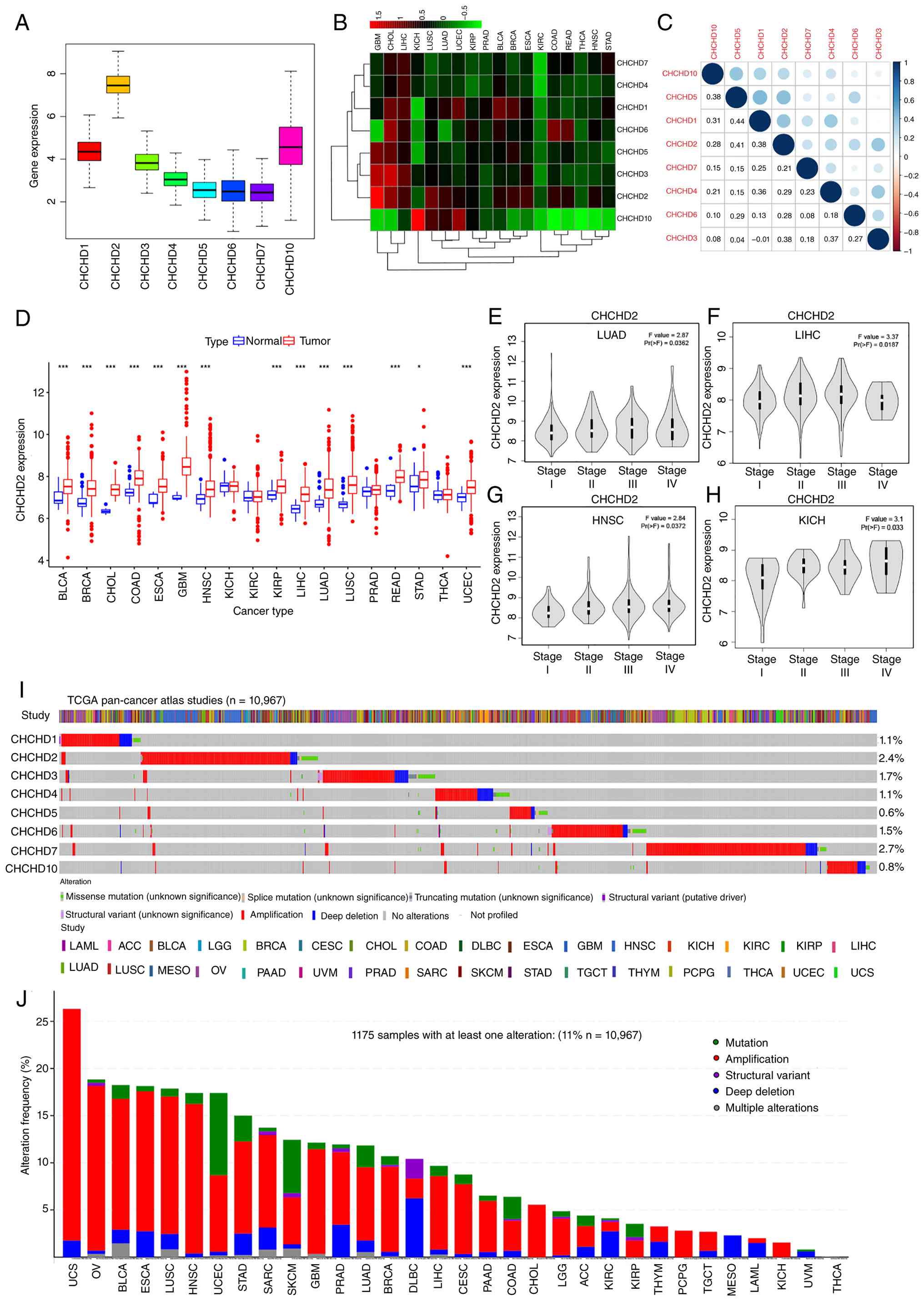 | Figure 1.Pan-cancer expression patterns and
genetic alteration landscape of the CHCHD family. (A) Boxplots
showing the overall mRNA expression distribution of CHCHD family
members (CHCHD1-7 and CHCHD10) across TCGA pan-cancer types. (B)
Heatmap illustrating the relative expression levels of CHCHD family
genes across different cancer types (normalized expression values),
with unsupervised hierarchical clustering applied to both genes and
tumor types. (C) Pairwise expression correlation matrix of CHCHD
family members based on Spearman's correlation analysis; dot size
and color indicate the strength and direction of correlations, with
numerical values representing correlation coefficients. (D)
Differential expression analysis of CHCHD2 between tumor tissues
(red) and corresponding normal tissues (blue) across TCGA cancer
types. (E-H) Pathological stage-associated expression patterns of
CHCHD2 across tumor stages (I–IV), generated using the GEPIA2
‘Stage Plot’ module and evaluated by one-way ANOVA: (E) LUAD, (F)
LIHC, (G) HNSC and (H) KICH. (I) OncoPrint summarizing the spectrum
and frequency of genetic alterations in CHCHD family genes across
10,967 TCGA tumors using cBioPortal. (J) Cancer-type-specific
summary of overall CHCHD family alteration frequencies, indicating
the proportion of tumors harboring at least one CHCHD-family
alteration; copy-number amplification represents the predominant
alteration event. *P<0.05 and ***P<0.001. CHCHD,
coiled-coil-helix-coiled-coil-helix domain; TCGA, The Cancer Genome
Atlas; LUAD, lung adenocarcinoma; LIHC, liver hepatocellular
carcinoma; HNSC, head and neck squamous cell carcinoma; KICH,
kidney chromophobe. |
Next, differential expression analysis comparing
tumor and normal tissues revealed that CHCHD2 expression was
significantly upregulated in multiple cancer types, including BLCA,
breast invasive carcinoma (BRCA) and LUAD (Fig. 1D). The expression levels of other
members, such as CHCHD1, CHCHD3-CHCHD7 and CHCHD10, were also
elevated in most tumor types (Fig.
S1A-G).
Genomic alterations of CHCHD family
genes across cancers
To characterize the genomic alteration landscape of
the CHCHD gene family across cancers, alterations in these genes
were analyzed using cBioPortal based on TCGA data. Across 10,967
tumor samples, the average alteration frequency of CHCHD family
genes was ~1.5%. Among them, CHCHD2 (2.4%) and CHCHD7 (2.7%)
presented relatively high genomic alteration frequencies (Fig. 1I). The 2.4% frequency for CHCHD2
represents its overall genomic alteration frequency, including
mutations and copy number alterations, rather than evidence of
loss-of-function mutations. Overall, 11% of tumors harbored at
least one CHCHD family alteration, with copy-number amplification
being the predominant alteration type. Uterine carcinosarcoma had
the highest overall alteration frequency (26%), whereas thyroid
carcinoma had the lowest (1.5%) (Fig.
1J). Alteration details for individual CHCHD genes are provided
in Figs. S2 and S3.
Differential expression of CHCHD2
across cancers
To further investigate CHCHD2, its relationship with
pathological stage was examined across cancer types using GEPIA. A
positive correlation between CHCHD2 expression and pathological
stage was observed in LUAD (Fig.
1E), liver hepatocellular carcinoma (LIHC) (Fig. 1F), head and neck squamous cell
carcinoma (HNSC) (Fig. 1G) and
kidney chromophobe (Fig. 1H). At
the transcript level, CHCHD2 is consistently overexpressed in
numerous cancers. To assess whether this trend extended to the
protein level, proteomic data from UALCAN were analyzed. CHCHD2
protein levels were significantly elevated in LIHC, UCEC, BRCA,
ovarian serous cystadenocarcinoma, colon adenocarcinoma, lung
squamous cell carcinoma and LUAD (Fig.
S4A). IHC staining from the HPA further confirmed these
findings (Fig. S4B).
Prognostic significance of CHCHD2
expression across cancers
The prognostic significance of CHCHD2 expression
across diverse cancer types was evaluated using the KM Plotter
database. Increased CHCHD2 expression was significantly correlated
with reduced OS in patients with LIHC, HNSC, LUAD, BLCA and ESCA,
as evidenced by HRs uniformly exceeding 1 (Fig. 2A-E; P<0.05). By contrast, higher
CHCHD2 expression in thymoma was associated with a favorable OS
outcome (Fig. 2F, P<0.05).
Collectively, these findings suggest that CHCHD2 is overexpressed
in diverse malignancies and is associated with an unfavorable
prognosis in several tumor types, particularly LUAD. This
highlights its potential clinical utility as both a diagnostic
biomarker and a therapeutic biomarker. Given the strong prognostic
association in LUAD, focus was further addressed on this cancer to
investigate CHCHD2-related genes, pathways and cellular functions,
aiming to delineate its oncogenic mechanisms in greater detail.
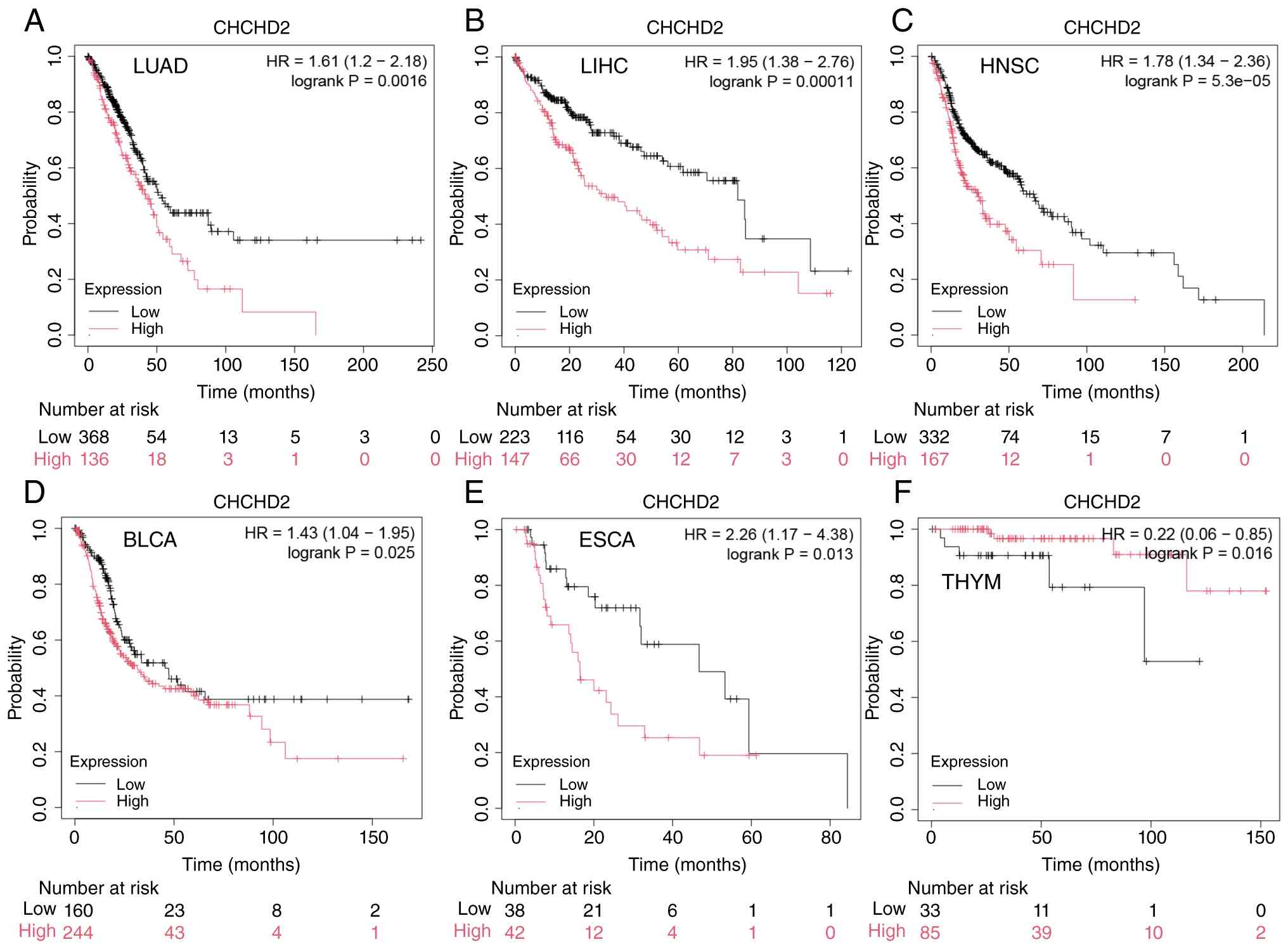 | Figure 2.Association between CHCHD2 expression
and OS across multiple cancers (Kaplan-Meier Plotter). (A) In LUAD,
patients with high CHCHD2 expression (red) exhibited poorer OS than
those with low expression (black). (B) In LIHC, patients with high
CHCHD2 expression (red) exhibited poorer OS than those with low
expression (black). (C) In HNSC, patients with high CHCHD2
expression (red) exhibited poorer OS than those with low expression
(black). (D) In BLCA, patients with high CHCHD2 expression (red)
exhibited poorer OS than those with low expression (black). (E) In
ESCA, patients with high CHCHD2 expression (red) exhibited poorer
OS than those with low expression (black). (F) In THYM, high CHCHD2
expression was associated with improved OS. CHCHD,
coiled-coil-helix-coiled-coil-helix domain; OS, overall survival;
LUAD, lung adenocarcinoma; LIHC, liver hepatocellular carcinoma;
HNSC, head and neck squamous cell carcinoma; BLCA, bladder
urothelial carcinoma; ESCA, esophageal carcinoma; THYM, thymoma;
HR, hazard ratio. |
Identification of genes, pathways and
cellular functions related to CHCHD2 in LUAD
A co-expression analysis was performed using RNA
sequencing data from TCGA and 9,775 genes that were significantly
correlated with CHCHD2 expression were identified, including 5,261
negatively correlated genes and 4,514 positively correlated genes.
The potential interaction network among the 11 genes most strongly
correlated with CHCHD2 is shown in Fig. S5A.
The samples were then stratified into CHCHD2 high-
and low-expression groups, revealing 1,150 DEGs, which comprised
156 significantly upregulated genes and 994 significantly
downregulated genes. A heatmap was generated to visualize the top
100 DEGs (Fig. S5B).
Functional annotation via GO enrichment analysis
revealed that in the BP category there was significant enrichment
in terms such as potassium ion transport and potassium ion
transmembrane transport (Fig.
S5C). In the cellular component category, the enriched terms
included collagen-containing extracellular matrix, neuronal cell
body, and glutamatergic synapse. In the molecular function
category, the most enriched terms were metal ion transmembrane
transporter activity, voltage-gated monoatomic cation channel
activity, and potassium ion transmembrane transporter activity.
KEGG pathway analysis revealed significant enrichment in pathways
such as PI3K-Akt signaling, neuroactive ligand-receptor
interactions, cAMP signaling and MAPK signaling (Fig. S5D), suggesting that these pathways
may be associated with CHCHD2-related transcriptional changes in
LUAD. However, these enrichment results should be interpreted as
hypothesis-generating rather than direct mechanistic evidence.
These pathway associations were subsequently evaluated
experimentally by assessing AKT and ERK phosphorylation following
CHCHD2 knockdown.
Correlations between CHCHD2 expression
and TME/stemness scores in LUAD
Although the role of CHCHD2 in tumor immunity has
not been extensively studied, mitochondria are increasingly
recognized as key regulators of the TME and play important roles in
tumor progression, immune evasion and therapeutic responses
(30). To further elucidate the
potential association between CHCHD2 expression levels and TME
characteristics, as well as stemness scores, Spearman's rank
correlation coefficient was used for nonparametric correlation
analysis. The results revealed a significant positive correlation
between CHCHD2 expression and both the RNAss (R=0.37, P<0.001)
and the DNAss (R=0.13, P<0.001) (Fig. S6A), suggesting that CHCHD2 may play
a role in maintaining or enhancing the stemness phenotype. On the
other hand, CHCHD2 expression was negatively correlated with
several TME-related scoring metrics, including the stromal score
(R=−0.19, P<0.001), immune score (R=−0.17, P<0.001) and the
composite ESTIMATE score (R=−0.19, P<0.001) (Fig. S6A), indicating that increased
CHCHD2 expression may be associated with reduced immune
infiltration and stromal content, further suggesting a potential
immunosuppressive role within the TME.
Association between CHCHD2 expression
and immune subtype in LUAD
To evaluate the differential expression of CHCHD2
across various immune subtypes, the Kruskal-Wallis H test was
applied to LUAD samples, and a non-parametric comparison of CHCHD2
expression among six predefined immune subtypes was conducted (that
is, C1, wound healing; C2, IFN-γ dominant; C3, inflammatory; C4,
lymphocyte-depleted; C5, immunologically quiet; and C6, TGF-β
dominant) (13,25). The statistical results indicated
that CHCHD2 expression was relatively higher in the C1 subtype,
whereas the lowest expression was observed in the C3 subtype
(Fig. S6B), suggesting that CHCHD2
may perform distinct regulatory functions in different
immune-inflammatory microenvironmental contexts.
Correlations between CHCHD2 expression
and immune cell infiltration in LUAD
The relationship between CHCHD2 expression and 50
immune checkpoint-related genes was first analyzed to assess its
potential association with immune checkpoint inhibitors. The
results revealed that the expression of CHCHD2 was significantly
correlated with that of 10 immune checkpoint-related genes
(P<0.001). Specifically, CHCHD2 expression was negatively
correlated with CD40LG, TNFSF15, TNFSF14, CD28, BTLA, ADORA2A,
CD160, TIGIT and NRP1 expression but positively correlated with
TNFSF18 expression (Fig. S6C).
Given the observed correlations with immune checkpoint-related
genes, it was next evaluated whether CHCHD2 expression was also
associated with differences in tumor-infiltrating immune cell
composition.
To further investigate the association between
CHCHD2 and the tumor immune microenvironment, the CIBERSORT
algorithm was used to evaluate immune cell subpopulations in LUAD
samples with different CHCHD2 expression levels. A comparison of
the high- and low-expression groups revealed that CHCHD2 expression
was positively correlated with the numbers of follicular helper T
cells, activated CD4 memory T cells and gamma delta T cells but
negatively correlated with the numbers of resting CD4 memory T
cells and resting mast cells (Fig. S6D
and E).
Single-cell analysis of CHCHD2
expression in an LUAD-derived dataset
Single-cell RNA sequencing analysis was performed
using multiple TISCH2 datasets, with NSCLC_GSE131907 as the primary
LUAD-derived validation cohort. Although categorized under NSCLC in
TISCH2, NSCLC_GSE131907 was derived from patients with
pathologically diagnosed LUAD. In this dataset, CHCHD2 expression
was detectable across multiple cellular compartments, including
epithelial cells, T cells, B cells, fibroblasts and plasma cells
(Fig. S7A-D). Other NSCLC
datasets, including NSCLC_GSE117570, NSCLC_GSE127465, and
NSCLC_GSE99254, were used only as supportive references and were
not interpreted as LUAD-specific evidence.
Drug sensitivity analysis of
CHCHD2
To investigate the potential association between
CHCHD2 expression and drug sensitivity in human cancer cell lines,
correlation analysis was performed using the CellMiner™ database.
The results (Fig. S8A-P) revealed
a significant negative correlation between CHCHD2 expression and
sensitivity to Irofulven. By contrast, CHCHD2 expression was
positively correlated with sensitivity to multiple agents,
including uracil mustard, dexamethasone (Decadron), asparaginase,
acrichine, fluphenazine, vorinostat, PX-316, chelerythrine,
nelarabine, imexon, fenretinide, hydroxyurea, carmustine, lomustine
and ifosfamide (P<0.05).
Experimental validation of CHCHD2
upregulation in LUAD tissues and cell lines
Based on these multi-omics and database-derived
findings, LUAD-specific experimental validation was next performed
at the tissue, cellular and animal levels. Tumor tissues and
matched adjacent normal tissues from nine patients with stage II
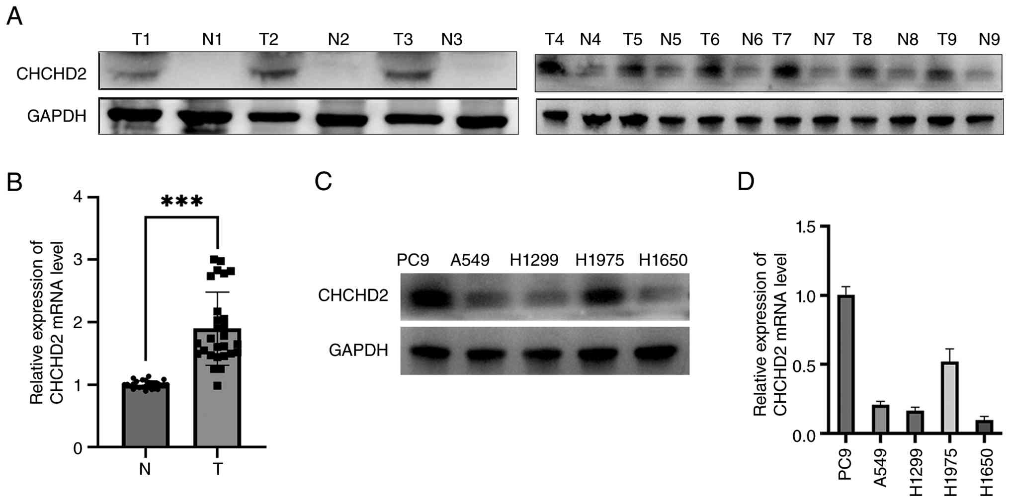
LUAD were used to assess CHCHD2 expression levels. Western blotting
and RT-qPCR results revealed that the protein and mRNA levels of
CHCHD2 were significantly higher in LUAD tissues than in adjacent
normal tissues (Fig. 3A and B),
which is consistent with the findings of the aforementioned
bioinformatics analysis. A total of five LUAD cell lines were
selected to examine CHCHD2 expression levels, and the results
revealed that the protein and mRNA levels of CHCHD2 were markedly
higher in PC9 and H1975 cells than in H1299 and H1650 cells
(Fig. 3C and D).
CHCHD2 knockdown suppresses malignant
phenotypes in PC9 and H1975 cells
Two LUAD cell lines with high CHCHD2 expression, PC9
and H1975, were chosen to establish transient knockdown models
using shRNA. Western blotting and RT-qPCR analyses showed that all
three CHCHD2-targeting shRNAs reduced CHCHD2 expression, with
shCHCHD2-1 producing the strongest and most consistent knockdown
across both cell lines (Fig. 4A-D).
Direct comparisons showed significantly greater protein suppression
by shCHCHD2-1 than by shCHCHD2-2 or shCHCHD2-3 in both PC9 and
H1975 cells. At the mRNA level, shCHCHD2-1 also demonstrated
greater knockdown in PC9 cells and was significantly more effective
than shCHCHD2-2 in H1975 cells, whereas the difference from
shCHCHD2-3 in H1975 cells was not significant. Therefore,
shCHCHD2-1 was selected for subsequent functional assays.
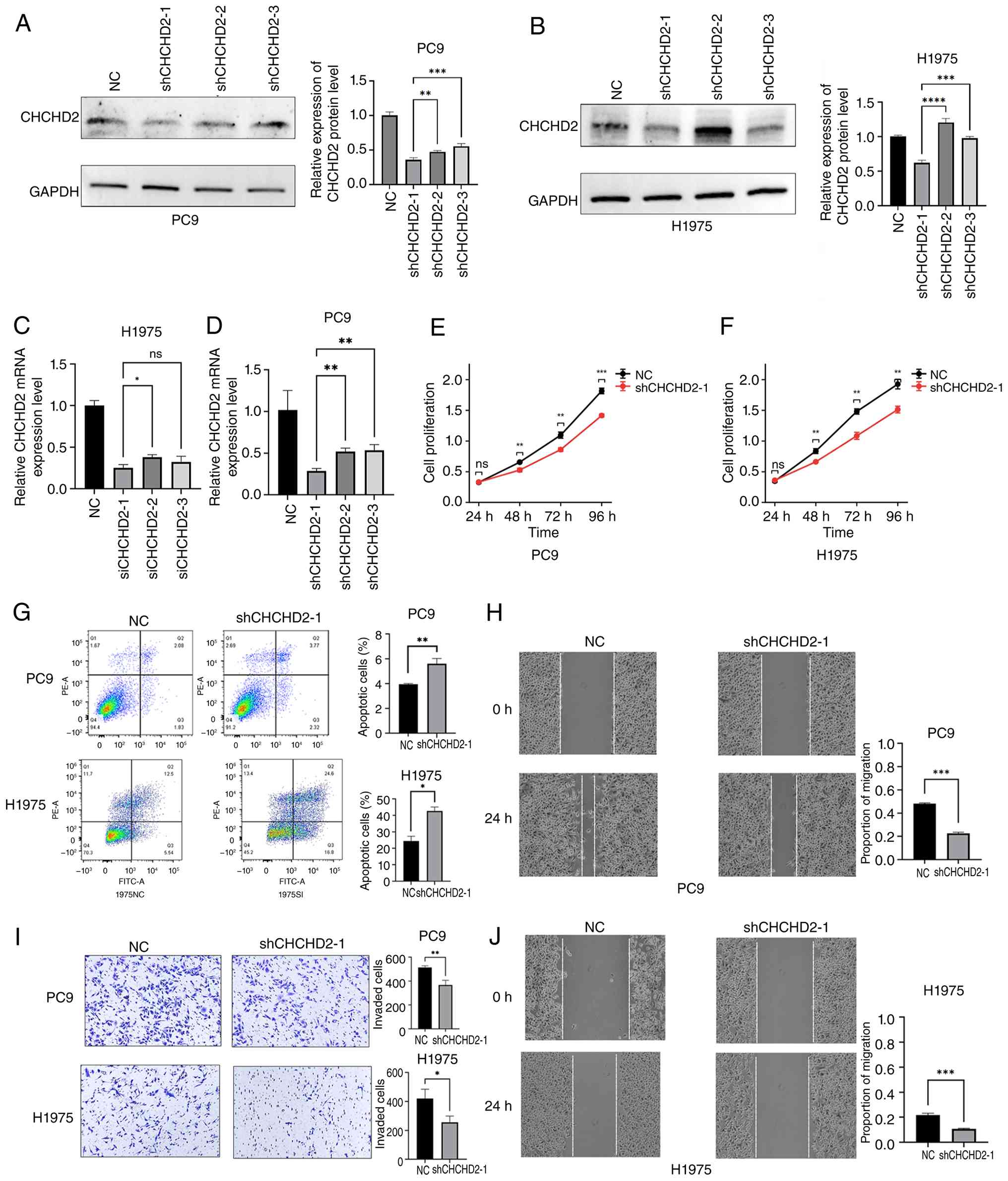 | Figure 4.CHCHD2 knockdown suppresses
proliferation and migration and promotes apoptosis in PC9 and H1975
cells. (A and B) Western blot analyses of CHCHD2 knockdown
efficiency in PC9 (A) and H1975 (B) cells transfected with
shCHCHD2-1, shCHCHD2-2, shCHCHD2-3, or negative-control shRNA
(shNC), with corresponding densitometric quantification of CHCHD2
protein expression. (C and D) Reverse transcription-quantitative
PCR analyses of CHCHD2 mRNA expression following transfection with
shNC or the three CHCHD2-targeting shRNAs in H1975 (C) and PC9 (D)
cells. Direct statistical comparisons among the three shRNAs were
performed to evaluate knockdown efficiency. (E and F) CCK-8 assays
showing that CHCHD2 knockdown reduced cell proliferation in PC9 (E)
and H1975 (F) cells. (G) Flow cytometric analysis showing increased
apoptosis following CHCHD2 knockdown in PC9 and H1975 cells. (H)
Wound-healing assay showing reduced migration after CHCHD2
knockdown in PC9 cells. (I) Transwell migration assays showing
reduced migratory ability after CHCHD2 knockdown in PC9 and H1975
cells. (J) Wound-healing assay showing reduced migration after
CHCHD2 knockdown in H1975 cells. White dashed lines indicate the
wound edges used for wound-width measurement. For the CCK-8
time-course data, statistical significance was assessed using
two-way ANOVA followed by Sidak's multiple-comparisons test. Data
are presented as the mean ± SD. *P<0.05, **P<0.01,
***P<0.001 and ****P<0.0001. CHCHD,
coiled-coil-helix-coiled-coil-helix domain; sh-, short hairpin; NC,
negative control; CCK-8, Cell Counting Kit-8; ns, not
significant. |
CCK-8 assays revealed that CHCHD2 knockdown
significantly reduced cell proliferation in PC9 and H1975 cells
(Fig. 4E and F). Flow cytometry
revealed significantly increased apoptosis following CHCHD2
knockdown (Fig. 4G). Transwell and
wound-healing assays further demonstrated impaired migration in
both cell lines (Fig. 4H-J).
CHCHD2 overexpression enhances
malignant phenotypes in H1299 and H1650 cells
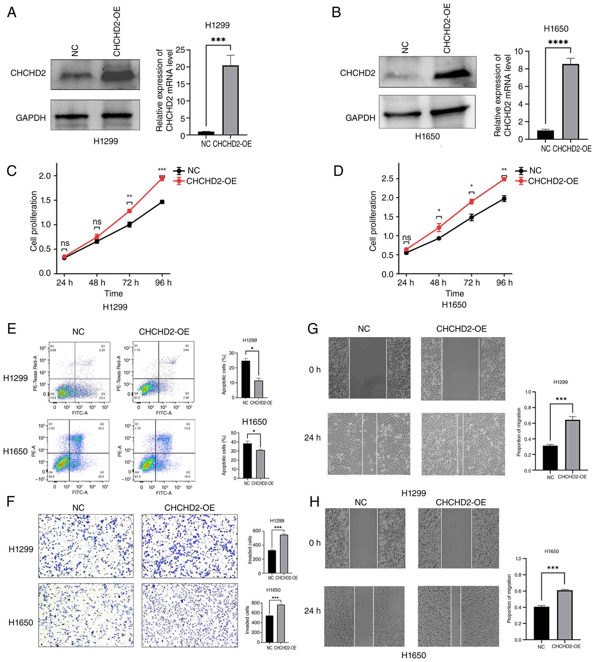
CHCHD2-overexpressing cell models were established
in H1299 and H1650 cells, which exhibit low endogenous CHCHD2
expression. Western blotting and RT-qPCR analyses confirmed a
significant increase in CHCHD2 expression levels in the
overexpression group (Fig. 5A and
B). CCK-8 assays revealed that CHCHD2 overexpression
significantly increased the proliferation rates of H1299 and H1650
cells (Fig. 5C and D). Flow
cytometric analysis revealed that CHCHD2 overexpression
significantly reduced both early and late apoptosis in H1299 and
H1650 cells (Fig. 5E). Wound
healing and Transwell assays revealed that CHCHD2 overexpression
significantly promoted cell migration (Fig. 5F-H). These results indicate that
both knockdown and overexpression of CHCHD2 can alter LUAD cell
proliferation, migration and apoptosis, suggesting a potential
oncogenic role for CHCHD2 in LUAD.
CHCHD2 knockdown reduces AKT/ERK
phosphorylation and suppresses LUAD xenograft tumor growth in
vivo
To further evaluate the signaling pathways suggested
by the KEGG enrichment analysis, AKT and ERK phosphorylation were
examined following CHCHD2 knockdown. Western blot analysis showed
that CHCHD2 knockdown reduced the levels of phosphorylated AKT and
ERK in both PC9 and H1975 cells, as reflected by decreased
p-AKT/t-AKT and p-ERK/t-ERK levels (Fig. 6A and B). These findings provide
experimental support for the involvement of PI3K-AKT and MAPK
signaling in CHCHD2-associated LUAD phenotypes.
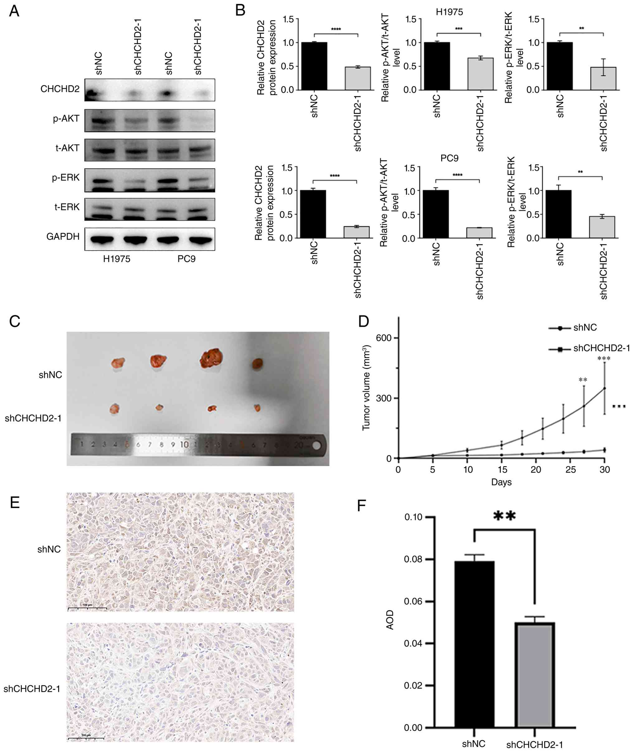 | Figure 6.CHCHD2 knockdown inhibits AKT/ERK
phosphorylation and suppresses lung adenocarcinoma xenograft tumor
growth. (A) Representative western blot analysis of CHCHD2, p-AKT,
t-AKT, p-ERK and t-ERK in H1975 and PC9 cells transfected with shNC
or shCHCHD2-1. GAPDH was used as the loading control. (B)
Densitometric quantification of CHCHD2 expression and the
p-AKT/t-AKT and p-ERK/t-ERK ratios in H1975 and PC9 cells. (C)
Representative images of excised xenograft tumors from the shNC and
shCHCHD2-1 groups. (D) Tumor growth curves showing xenograft tumor
volume changes over time in the shNC and shCHCHD2-1 groups. Tumor
growth curves were analyzed using repeated-measures two-way ANOVA
followed by Sidak's multiple-comparisons test. (E) Representative
IHC images of CHCHD2 staining in xenograft tumor tissues from the
shNC and shCHCHD2-1 groups. Scale bar, 100 µm. (F) Quantification
of CHCHD2 IHC staining by average optical density, showing
decreased CHCHD2 expression in the shCHCHD2-1 group. shNC,
negative-control shRNA; shCHCHD2-1, CHCHD2-targeting shRNA-1. Data
are presented as the mean ± SD. **P<0.01, ***P<0.001 and
****P<0.0001. CHCHD, coiled-coil-helix-coiled-coil-helix domain;
p-, phosphorylated; t-, total; sh-, short hairpin; NC, negative
control; IHC, immunohistochemical. |
To further evaluate the effect of CHCHD2 knockdown
in vivo, PC9-shCHCHD2-1 and PC9-shNC cells were
subcutaneously injected into nude mice. Tumor growth was monitored
every 3 days. At Day 30, xenograft tumors were excised, and
representative tumor images are shown in Fig. 6C. Tumor growth curves demonstrated
that CHCHD2 knockdown significantly reduced xenograft tumor volume
compared with the shNC group (Fig.
6D). IHC staining further showed reduced CHCHD2 expression in
xenograft tumors from the shCHCHD2-1 group (Fig. 6E), which was confirmed by AOD
quantification (Fig. 6F). These
findings indicate that CHCHD2 knockdown suppresses LUAD tumor
growth in vivo and are consistent with the inhibitory
effects observed in vitro.
Discussion
The present study aimed to delineate the expression
landscape and prognostic relevance of the CHCHD family across
cancers, with an emphasis on the biological role and clinical
significance of CHCHD2 in LUAD. It was revealed that CHCHD2 is
broadly upregulated across tumor types and is associated with
adverse outcomes; in LUAD, high expression of CHCHD2 is correlated
with advanced disease stage and poor survival, and it functionally
enhances proliferation and migration, suppresses apoptosis, and
accelerates xenograft growth. Multilayer analyses of the TME,
stemness features, immune subtypes and drug sensitivity support an
oncogenic role for CHCHD2, highlighting its potential as a
biomarker candidate and motivating further mechanistic
investigation. Importantly, because CHCHD2 has previously been
reported to be coamplified with EGFR and to regulate mitochondrial
function and migration in NSCLC, the present findings should be
interpreted as integrative and supportive evidence that extends
prior observations to LUAD-specific multi-omics and experimental
contexts rather than as definitive evidence of a completely new
oncogenic mechanism.
Integrating bioinformatics and experimental
evidence, the present findings indicate that the expression of
CHCHD2, a biomarker linked to prognosis and disease stage, may
facilitate risk stratification and patient management in LUAD.
Pathway enrichment analyses in LUAD revealed associations with
PI3K-AKT and MAPK signaling-related gene sets. Consistent with
these bioinformatic findings, additional western blot analyses
showed that CHCHD2 knockdown reduced AKT and ERK phosphorylation in
both PC9 and H1975 cells, providing experimental support for the
involvement of PI3K-AKT/MAPK signaling in CHCHD2-associated LUAD
phenotypes. Nevertheless, these findings do not establish that
CHCHD2 directly regulates these signaling cascades, and the precise
molecular mechanisms linking CHCHD2 to AKT and ERK activation
remain to be elucidated. Prior studies have reported that CHCHD2
interacts with Bcl-XL and inhibits BAX oligomerization, thereby
attenuating mitochondrial apoptosis (31), which is consistent with the
observation of attenuated apoptotic programs. Moreover, CHCHD2 is
located on chromosome 7p11.2 and is frequently coamplified with
EGFR in NSCLC, where it promotes mitochondrial respiration and
migration (11).
Positive correlations were observed between CHCHD2
and tumor stemness indices (RNAss and DNAss), coupled with lower
ESTIMATE, immune and stromal scores. CHCHD2 expression was higher
in the ‘wound-healing’ (C1) immune subtype and lower in the
‘inflammatory’ (C3) subtype; CIBERSORT deconvolution linked CHCHD2
to the enrichment of follicular helper and activated memory
CD4+ T cells, with depletion of resting immune
compartments. This pattern points to a TME skewed towards
proliferation and matrix remodeling with dampened inflammatory
tonicity, an arrangement aligned with the prognostic and
inflammatory contrasts between C1 and C3 reported by Thorsson et
al (25).
Drug sensitivity analysis revealed that high CHCHD2
expression was negatively correlated with sensitivity to the
DNA-damaging agent Irofulven, suggesting a potential role for
CHCHD2 in modulating cellular responses to this compound. By
contrast, high CHCHD2 expression was positively correlated with
sensitivity to a range of other therapeutic agents, including
alkylating agents (such as uracil mustard, lomustine and
carmustine), metabolic inhibitors (such as asparaginase,
hydroxyurea and fenretinide), and epigenetic regulators (such as
the HDAC inhibitor vorinostat). These preliminary findings suggest
that rather than reflecting a uniform response to DNA-damaging
therapies, CHCHD2 expression may have drug-specific predictive
value. Notably, elevated CHCHD2 levels could predict reduced
sensitivity to Irofulven while being associated with increased
sensitivity to agents with distinct mechanisms of action. Notably,
these observations are derived from the CellMiner database
(28) and warrant further
validation in LUAD-specific pharmacogenomic models and clinical
cohorts.
Multiple cancer-specific studies have linked CHCHD2
to oncogenic phenotypes, such as co-amplification with EGFR and
promotion of migration/respiration in NSCLC (11), upregulation in hepatocellular
carcinoma with angiogenic signaling and poor prognosis (32), and reduced docetaxel sensitivity via
MMP2 activation in breast cancer (33). Against this backdrop, the present
study provides several layers of additional evidence. First, it
places CHCHD2 within a broader pan-cancer framework by integrating
expression, survival, genomic alteration, pathway enrichment, TME,
stemness, single-cell and drug-response analyses. Second, it
focuses on LUAD and combines public multi-omics evidence with
clinical tissue validation, in vitro gain- and
loss-of-function assays, and an in vivo xenograft model.
Third, it reveals potential associations between CHCHD2 expression
and immune/stemness-related features, thereby generating testable
hypotheses for future mechanistic studies. Nevertheless, the
present work should be regarded as an integrative and functional
validation study rather than a complete mechanistic dissection of
CHCHD2 downstream signaling. Further studies are needed to define
the direct molecular interactors and signaling cascades regulated
by CHCHD2 in LUAD. Because functional assays for individual CHCHD2
variants were not performed, the biological consequences of CHCHD2
genomic alterations require further validation.
The limitations of the present study include the
reliance on TCGA data, which may lead to an underestimation of the
representativeness of diverse ethnicities and patients with
advanced disease stage, thereby affecting the generalizability of
pan-cancer inferences; however, the integration of GTEx and CPTAC
data partially mitigates the deficiencies in normal tissue
comparisons. The experimental models primarily employed cell lines
and xenograft mice, which do not fully capture patient
heterogeneity and in situ TME dynamics, although single-cell
and in vivo data compensate for this limitation to some
extent. Although the present western blot findings provide
experimental support for the involvement of PI3K-AKT/MAPK
signaling, pathway-rescue experiments and studies of direct
molecular interactions are still required to establish causality
and clarify the precise molecular mechanisms by which CHCHD2
modulates these signaling pathways. In addition, despite expansion
of the validation cohort to nine paired LUAD specimens, the sample
size remains relatively small and is derived from a single center,
which may limit statistical power and generalizability. Validation
in larger, independent clinical cohorts is therefore warranted.
These shortcomings do not undermine the core conclusions but
indicate the need for further validation of its translational
potential in syngeneic models and prospective studies.
In conclusion, the results of the present study
support the role of CHCHD2 as a potential oncogenic regulator in
LUAD. Pan-cancer and LUAD-focused analyses linked CHCHD2 to adverse
prognosis, genomic alterations, TME features, stemness-related
indices, single-cell expression patterns, and drug-response
correlations. Functional validation further revealed that CHCHD2
promotes LUAD cell proliferation and migration, inhibits apoptosis,
and supports xenograft tumor growth, while CHCHD2 knockdown reduced
AKT and ERK phosphorylation. Future studies should clarify the
direct molecular mechanisms linking CHCHD2 to PI3K-AKT/MAPK
signaling and evaluate its therapeutic relevance in LUAD
models.
Supplementary Material
Supporting Data
Acknowledgements
Not applicable.
Funding
The present study was supported by Scientific research project
of Jiangsu Provincial Health Commission (grant no. K2023073).
Availability of data and materials
The data generated in the present study may be
requested from the corresponding author.
Authors' contributions
ZKX and ZY conceived the study, performed the data
curation, formal analysis, investigation, methodology, software
analysis, validation, and visualization, and wrote the original
draft of the manuscript. JS, YZL, HTS and CZ contributed to
methodology and validation. ZWZ and XYZ supervised the study and
contributed to conceptualization, methodology, project
administration, resources, validation, and manuscript review and
editing. XYZ also acquired funding. ZKX and ZY confirm the
authenticity of all the raw data. All authors read and approved the
final version of the manuscript.
Ethics approval and consent to
participate
The human study was approved by the Ethics Committee
of the Affiliated Hospital of Nantong University (approval no.
2023-K010-01; Nantong, China). Written informed consent was
obtained from all participants. All animal procedures were approved
by the Animal Ethics Committee of Nantong University (approval no.
P20250307-034; Nantong, China). All experiments were conducted in
accordance with relevant institutional guidelines and
regulations.
Patient consent for publication
Not applicable.
Competing interests
The authors declare that they have no competing
interests.
Glossary
Abbreviations
Abbreviations:
|
CHCHD
|
coiled-coil-helix-coiled-coil-helix
domain
|
|
LUAD
|
lung adenocarcinoma
|
|
TME
|
tumor microenvironment
|
|
TCGA
|
The Cancer Genome Atlas
|
|
GEPIA
|
Gene Expression Profiling Interactive
Analysis
|
|
GTEx
|
Genotype-Tissue Expression
|
|
RNAss
|
RNA-based stemness score
|
|
DNAss
|
DNA methylation-based stemness
score
|
|
OS
|
overall survival
|
|
IHC
|
immunohistochemistry
|
|
RT-qPCR
|
reverse transcription-quantitative
polymerase chain reaction
|
|
NSCLC
|
non-small cell lung cancer
|
|
HPA
|
Human Protein Atlas
|
|
CPTAC
|
Clinical Proteomic Tumor Analysis
Consortium
|
|
GO
|
Gene Ontology
|
|
KEGG
|
Kyoto Encyclopedia of Genes and
Genomes
|
|
KM plotter
|
Kaplan-Meier plotter platform
|
References
|
1
|
Bray F, Laversanne M, Sung H, Ferlay J,
Siegel RL, Soerjomataram I and Jemal A: Global cancer statistics
2022: GLOBOCAN estimates of incidence and mortality worldwide for
36 cancers in 185 countries. CA Cancer J Clin. 74:229–263.
2024.PubMed/NCBI
|
|
2
|
Herbst RS, Morgensztern D and Boshoff C:
The biology and management of non-small cell lung cancer. Nature.
553:446–454. 2018. View Article : Google Scholar : PubMed/NCBI
|
|
3
|
Hendriks LEL, Remon J, Faivre-Finn C,
Garassino MC, Heymach JV, Kerr KM, Tan DSW, Veronesi G and Reck M:
Non-small-cell lung cancer. Nat Rev Dis Primers. 10:712024.
View Article : Google Scholar : PubMed/NCBI
|
|
4
|
Wallace DC: Mitochondria and cancer. Nat
Rev Cancer. 12:685–698. 2012. View
Article : Google Scholar : PubMed/NCBI
|
|
5
|
Li Z, Zhang W, Guo S, Qi G, Huang J, Gao
J, Zhao J, Kang L and Li Q: A review of advances in mitochondrial
research in cancer. Cancer Control. 31:107327482412990722024.
View Article : Google Scholar : PubMed/NCBI
|
|
6
|
Zhou ZD, Saw WT and Tan EK: Mitochondrial
CHCHD-Containing proteins: Physiologic functions and link with
neurodegenerative diseases. Mol Neurobiol. 54:5534–5546. 2017.
View Article : Google Scholar : PubMed/NCBI
|
|
7
|
Kee TR, Espinoza Gonzalez P, Wehinger JL,
Bukhari MZ, Ermekbaeva A, Sista A, Kotsiviras P, Liu T, Kang DE and
Woo JA: Mitochondrial CHCHD2: Disease-associated mutations,
physiological functions, and current animal models. Front Aging
Neurosci. 13:6608432021. View Article : Google Scholar : PubMed/NCBI
|
|
8
|
Ming R, Li X, Wang E, Wei J, Liu B, Zhou
P, Yu W, Zong S and Xiao H: The prognostic signature of head and
neck squamous cell carcinoma constructed by Immune-related
RNA-Binding proteins. Front Oncol. 12:7957812022. View Article : Google Scholar : PubMed/NCBI
|
|
9
|
Thomas LW, Stephen JM, Esposito C, Hoer S,
Antrobus R, Ahmed A, Al-Habib H and Ashcroft M: CHCHD4 confers
metabolic vulnerabilities to tumour cells through its control of
the mitochondrial respiratory chain. Cancer Metab. 7:22019.
View Article : Google Scholar : PubMed/NCBI
|
|
10
|
Zhao C, Xu Z, Que H, Zhang K, Wang F, Tan
R and Fan C: ASB1 inhibits prostate cancer progression by
destabilizing CHCHD3 via K48-linked ubiquitination. Am J Cancer
Res. 14:3404–3418. 2024. View Article : Google Scholar : PubMed/NCBI
|
|
11
|
Wei Y, Vellanki RN, Coyaud E, Ignatchenko
V, Li L, Krieger JR, Taylor P, Tong J, Pham NA, Liu G, et al:
CHCHD2 is coamplified with EGFR in NSCLC and regulates
mitochondrial function and cell migration. Mol Cancer Res.
13:1119–1129. 2015. View Article : Google Scholar : PubMed/NCBI
|
|
12
|
Miao Y, Wang J, Li Q, Quan W, Wang Y, Li
C, Wu J and Mi D: Prognostic value and immunological role of PDCD1
gene in pan-cancer. Int Immunopharmacol. 89:1070802020. View Article : Google Scholar : PubMed/NCBI
|
|
13
|
Goldman MJ, Craft B, Hastie M, Repecka K,
McDade F, Kamath A, Banerjee A, Luo Y, Rogers D, Brooks AN, et al:
Visualizing and interpreting cancer genomics data via the Xena
platform. Nat Biotechnol. 38:675–678. 2020. View Article : Google Scholar : PubMed/NCBI
|
|
14
|
Liu J, Lichtenberg T, Hoadley KA, Poisson
LM, Lazar AJ, Cherniack AD, Kovatich AJ, Benz CC, Levine DA, Lee
AV, et al: An integrated TCGA Pan-cancer clinical data resource to
drive High-quality survival outcome analytics. Cell.
173:400–416.e11. 2018. View Article : Google Scholar : PubMed/NCBI
|
|
15
|
Consortium GT: Human genomics. The
genotype-tissue expression (GTEx) pilot analysis: Multitissue gene
regulation in humans. Science. 348:648–660. 2015. View Article : Google Scholar
|
|
16
|
Gao J, Aksoy BA, Dogrusoz U, Dresdner G,
Gross B, Sumer SO, Sun Y, Jacobsen A, Sinha R, Larsson E, et al:
Integrative analysis of complex cancer genomics and clinical
profiles using the cBioPortal. Sci Signal. 6:pl12013. View Article : Google Scholar : PubMed/NCBI
|
|
17
|
Tang Z, Kang B, Li C, Chen T and Zhang Z:
GEPIA2: An enhanced web server for large-scale expression profiling
and interactive analysis. Nucleic Acids Res. 47:W556–W560. 2019.
View Article : Google Scholar : PubMed/NCBI
|
|
18
|
Chandrashekar DS, Karthikeyan SK, Korla
PK, Patel H, Shovon AR, Athar M, Netto GJ, Qin ZS, Kumar S, Manne
U, et al: UALCAN: An update to the integrated cancer data analysis
platform. Neoplasia. 25:18–27. 2022. View Article : Google Scholar : PubMed/NCBI
|
|
19
|
Sjostedt E, Zhong W, Fagerberg L, Karlsson
M, Mitsios N, Adori C, Oksvold P, Edfors F, Limiszewska A, Hikmet
F, et al: An atlas of the protein-coding genes in the human, pig,
and mouse brain. Science. 367:eaay59472020. View Article : Google Scholar : PubMed/NCBI
|
|
20
|
Lanczky A and Gyorffy B: Web-Based
survival analysis tool tailored for medical research (KMplot):
Development and implementation. J Med Internet Res. 23:e276332021.
View Article : Google Scholar : PubMed/NCBI
|
|
21
|
Ashburner M, Ball CA, Blake JA, Botstein
D, Butler H, Cherry JM, Davis AP, Dolinski K, Dwight SS, Eppig JT,
et al: Gene ontology: Tool for the unification of biology. The Gene
Ontology Consortium. Nat Genet. 25:25–29. 2000. View Article : Google Scholar : PubMed/NCBI
|
|
22
|
Kanehisa M and Goto S: KEGG: Kyoto
encyclopedia of genes and genomes. Nucleic Acids Res. 28:27–30.
2000. View Article : Google Scholar : PubMed/NCBI
|
|
23
|
Yoshihara K, Shahmoradgoli M, Martinez E,
Vegesna R, Kim H, Torres-Garcia W, Trevino V, Shen H, Laird PW,
Levine DA, et al: Inferring tumour purity and stromal and immune
cell admixture from expression data. Nat Commun. 4:26122013.
View Article : Google Scholar : PubMed/NCBI
|
|
24
|
Malta TM, Sokolov A, Gentles AJ,
Burzykowski T, Poisson L, Weinstein JN, Kaminska B, Huelsken J,
Omberg L, Gevaert O, et al: Machine learning identifies stemness
features associated with oncogenic dedifferentiation. Cell.
173:338–354.e15. 2018. View Article : Google Scholar : PubMed/NCBI
|
|
25
|
Thorsson V, Gibbs DL, Brown SD, Wolf D,
Bortone DS, Ou Yang TH, Porta-Pardo E, Gao GF, Plaisier CL, Eddy
JA, et al: The immune landscape of cancer. Immunity. 51:411–412.
2019. View Article : Google Scholar : PubMed/NCBI
|
|
26
|
Sun D, Wang J, Han Y, Dong X, Ge J, Zheng
R, Shi X, Wang B, Li Z, Ren P, et al: TISCH: A comprehensive web
resource enabling interactive single-cell transcriptome
visualization of tumor microenvironment. Nucleic Acids Res.
49:D1420–D1430. 2021. View Article : Google Scholar : PubMed/NCBI
|
|
27
|
Shankavaram UT, Varma S, Kane D, Sunshine
M, Chary KK, Reinhold WC, Pommier Y and Weinstein JN: CellMiner: A
relational database and query tool for the NCI-60 cancer cell
lines. BMC Genomics. 10:2772009. View Article : Google Scholar : PubMed/NCBI
|
|
28
|
Reinhold WC, Sunshine M, Liu H, Varma S,
Kohn KW, Morris J, Doroshow J and Pommier Y: CellMiner: A web-based
suite of genomic and pharmacologic tools to explore transcript and
drug patterns in the NCI-60 cell line set. Cancer Res.
72:3499–3511. 2012. View Article : Google Scholar : PubMed/NCBI
|
|
29
|
Livak KJ and Schmittgen TD: Analysis of
relative gene expression data using real-time quantitative PCR and
the 2(−Delta Delta C(T)) method. Methods. 25:402–408. 2001.
View Article : Google Scholar : PubMed/NCBI
|
|
30
|
Ahn M, Ali A and Seo JH: Mitochondrial
regulation in the tumor microenvironment: Targeting mitochondria
for immunotherapy. Front Immunol. 15:14538862024. View Article : Google Scholar : PubMed/NCBI
|
|
31
|
Liu Y, Clegg HV, Leslie PL, Di J, Tollini
LA, He Y, Kim TH, Jin A, Graves LM, Zheng J and Zhang Y: CHCHD2
inhibits apoptosis by interacting with Bcl-x L to regulate Bax
activation. Cell Death Differ. 22:1035–1046. 2015. View Article : Google Scholar : PubMed/NCBI
|
|
32
|
Yao Y, Su J, Zhao L, Li R, Liu K and Wang
S: CHCHD2 promotes hepatocellular carcinoma and indicates poor
prognosis of hepatocellular carcinoma patients. J Cancer.
10:6822–6828. 2019. View Article : Google Scholar : PubMed/NCBI
|
|
33
|
Ma L, Zheng LH, Zhang DG and Fan ZM:
CHCHD2 decreases docetaxel sensitivity in breast cancer via
activating MMP2. Eur Rev Med Pharmacol Sci. 24:6426–6433.
2020.PubMed/NCBI
|