Testicular cholesterol granuloma mimicking a testicular tumor: A case report
- Authors:
- Published online on: February 21, 2024 https://doi.org/10.3892/wasj.2024.232
- Article Number: 17
-
Copyright : © Bapir et al. This is an open access article distributed under the terms of Creative Commons Attribution License [CC BY 4.0].
Metrics: Total
Views: 0 (Spandidos Publications: | PMC Statistics: )
Total PDF Downloads: 0 (Spandidos Publications: | PMC Statistics: )
Abstract
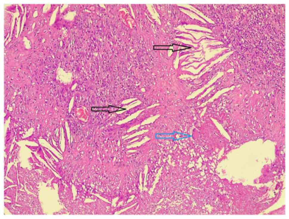
Cholesterol granulomas (ChGs) are benign fibro‑granulomatous lesions that develop following trauma and inflammation. The most common sites of presentation are the middle ear, paranasal sinuses and petrous apex; however, they can present in any area of the body where cholesterol deposition can occur. The present study describes the case of a 62‑year‑old male who presented with a painless right scrotal mass that had been there for the past 6 years. Upon a physical examination, a hard non‑tender mass at the lower pole of the right testis with normal overlying scrotal skin was detected. Blood analyses revealed normal levels of tumor markers (β‑human chorionic gonadotropin, alpha‑fetoprotein and lactate dehydrogenase) and a normal lipid profile. A right scrotal color Doppler ultrasound revealed a well‑defined hypoechoic 13x14 mm lower pole testicular mass. Scrotal magnetic resonance imaging revealed a well‑defined nodule (13x11x12 mm) at the lower pole of the right testis near the epididymis. Under spinal anesthesia, via inguinal incision, a right partial orchiectomy was performed. The post‑operative period was uneventful. The results of the histopathological examination were consistent with testicular ChG. ChG of the testis is extremely rare, and only one other case has been recorded in the literature to date, at least to the best of our knowledge. Differentiating between ChGs of the testis and testicular tumors before surgery can be relatively challenging. This should be regarded as a differential diagnosis in cases of testicular masses.