Introduction
Epidermal inclusion cysts (EICs), also termed
epidermoid or infundibular cysts, are benign, keratin-filled
nodules often misidentified as sebaceous cysts containing sebum
rather than keratin (1). These
lesions result from the entrapment of epidermal cells. Although the
terms ‘EIC’ and ‘sebaceous cyst’ are frequently used
interchangeably in clinical practice, the two are histologically
distinct: True sebaceous cysts arise from sebaceous glands and
contain sebum, whereas EICs are lined by stratified squamous
epithelium and contain laminated keratin (1). Recognizing this distinction is
clinically relevant, as nipple EICs are exceedingly uncommon and
are readily mistaken for other benign or malignant breast lesions
(2). Although EICs can occur at
any site on the body, they are most commonly found on the face,
trunk, neck, extremities and scalp, while their occurrence in the
breast, particularly in the nipple area, is exceedingly rare
(2,3). These cysts can develop at any age,
but are more frequently observed in adulthood, typically during the
third and fourth decades of life, and are more prevalent in males,
with a male-to-female ratio of 2:1 (3,4).
Although the precise basis for this male predominance has not yet
been fully established, it has been attributed to the
androgen-driven stimulation of the pilosebaceous apparatus and
sebaceous gland activity, together with the higher frequency of
acne, folliculitis and cutaneous trauma in men, all of which favor
the follicular occlusion and epidermal implantation that underlie
cyst formation (3,4).
The pathogenesis of EICs is closely related to the
pilosebaceous unit, as these lesions are generally believed to
originate from the infundibulum of the hair follicle. In the
majority of cases, they arise spontaneously when the follicular
orifice becomes plugged, resulting in the progressive retention and
lamellated accumulation of keratin within an epithelial-lined
cavity that may communicate with the skin surface through a central
punctum (3). Alternatively, true
inclusion cysts develop when fragments of the surface epidermis are
displaced into the dermis, where the implanted epithelium continues
to proliferate and keratinize; the designation epidermal inclusion
cyst is often reserved for these acquired, implantation-related
lesions (3,5).
A number of factors have been implicated in the
development of EICs. The disruption or occlusion of the follicular
unit is central to their formation, and individuals with acne
vulgaris are consequently predisposed, as retained comedones may
give rise to multiple cysts (3).
Local trauma is a well-recognized precipitant, since penetrating or
blunt injury, surgical incisions, needle biopsy and, in the breast,
procedures such as reduction mammoplasty can implant epidermal
elements into the dermis (2,5).
Other contributory factors reported in the literature include
chronic inflammation, human papillomavirus infection and prolonged
ultraviolet exposure, whereas multiple or atypically distributed
cysts may occasionally indicate an underlying genetic disorder,
such as Gardner syndrome (3).
Cutaneous cysts are broadly categorized into true
cysts and pseudocysts based on their morphology and differentiation
patterns. True cysts include those lined with a stratified squamous
epithelium and those lined with a non-stratified squamous
epithelium, whereas pseudocysts lack an epithelial lining.
Epidermoid cysts belong to the category of true cysts (1). The literature demonstrates that
epidermal cysts in uncommon sites, such as the breast, axillary
lymph node and nipple are rare (1,2,4). The
majority of cases are effectively managed through excision and show
no recurrence (3).
The present case report describes a rare case of
EICs located in the nipple of a male patient. In addition, a review
of the literature is performed to identify similar published
cases.
Case report
Patient information
A 29-year-old male patient presented to Smart Health
Tower, Sulaymaniyah, Iraq, on September, 2024, with a 3-year
history of swelling in his left nipple associated with pain.
Additionally, the patient reported recurrent episodes of pain,
tenderness and yellowish discharge from the nipple. An
investigation of his past medical, surgical and family history did
not reveal any notable findings. He was an active smoker and
consumed alcohol occasionally.
Clinical findings
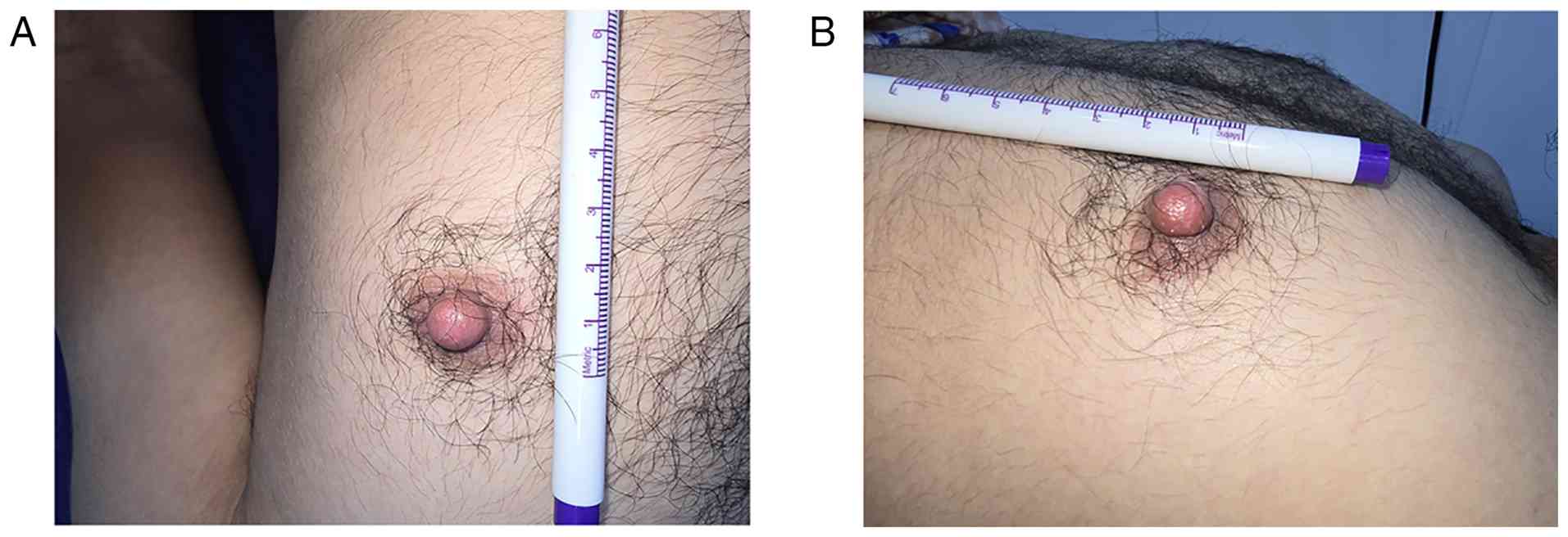
A clinical examination revealed a small, tender,
soft-tissue growth on the left nipple of the patient. It was a
centrally located, freely movable cyst with a smooth surface and
well-defined margins, measuring ~10 mm in diameter (Fig. 1).
Diagnostic assessment and therapeutic
intervention
Laboratory tests and radiological imaging were not
conducted. As the lesion presented as a small, superficial,
clinically typical cutaneous cyst confined to the nipple skin, and
the patient elected to proceed directly to excision, an
ultrasonography was not performed. Based on the clinical findings,
the physician suspected an EIC in the nipple. The patient underwent
surgical excision of the nipple using an elliptical incision under
local anesthesia, and the excised tissue was sent for a
histopathological examination. The tissue was fixed in 10%
neutral-buffered formalin at room temperature (20-25˚C) for ~24 h
before routine tissue processing and paraffin embedding. Sections
of 4 µm thickness were prepared and stained with hematoxylin and
eosin (H&E; Merck KGaA) according to the manufacturer's
standard protocol. Staining was performed at room temperature, with
hematoxylin for ~5 min, followed by differentiation and bluing, and
eosin for ~1-2 min. The stained sections were examined using a
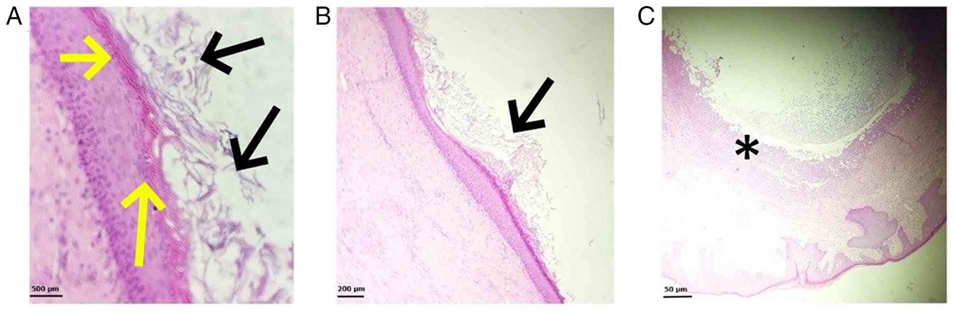
light microscope (BX43; Olympus Corporation). Histopathological
analysis confirmed an infected keratinous cyst consistent with an
EIC of the nipple, with no evidence of malignancy (Fig. 2).
Follow-up
The post-operative period was uneventful, and no
recurrences were observed at the 4-month follow-up. The cosmetic
outcome was satisfactory, with preservation of the nipple contour
and no evidence of distortion or deformity of the nipple
anatomy.
Discussion
EICs, also referred to as epidermal cysts,
infundibular cysts, or sebaceous cysts, are benign, keratin-filled
sacs that arise from the overgrowth or localized proliferation of
epithelial cells within the dermal or subcutaneous layers (6).
These cysts can develop in various body parts;
however, their occurrence in the nipple is rare. They typically
appear as nodules beneath the skin and have a noticeable central
punctum. In contrast to sebaceous glands, they are not formed from
these glands and primarily contain keratin instead of sebum.
Therefore, they differ from sebaceous cysts. Despite these
differences, the terms ‘sebaceous cysts’ and ‘epidermoid cysts’ are
frequently used interchangeably in clinical practice (1). They may be clinically and
radiologically misidentified as other benign or malignant lesions,
making an accurate preoperative diagnosis challenging (7). EICs vary in size, ranging from 4 mm,
as documented by Dilek et al (8), to 3.4 cm, as described by Ak et
al (9). The cyst in the case
described herein measured ~10 mm in diameter, placing it within the
moderate range of reported sizes.
The formation of EICs typically arises from factors,
such as trauma, epithelial proliferation and minimal inflammation.
These cysts may develop in the breast through various mechanisms
that damage the epidermis and lead to its implantation within the
breast tissue. Such mechanisms include congenital cysts caused by
obstructed hair follicles or pores, trauma, reduction mammoplasty,
or needle biopsy (5). Jain et
al (10) documented a case
involving a 15-month-old girl who developed an EIC following blunt
trauma inflicted by her mother, who had squeezed the child's breast
during the neonatal period to express ‘witch's milk’. Conversely,
in the case report by Dilek et al (8), which described the case of a
27-year-old woman, no history of trauma was reported. Similarly, in
the present case report, the patient reported no history of trauma
or surgical interventions in the nipple area where the cyst was
identified.
Although a history of trauma or surgical
intervention is a well-recognized cause of EICs, a considerable
proportion of these lesions, including the present case, arise in
its absence, reflecting the several non-traumatic mechanisms by
which they may form. The majority of spontaneous cysts are
considered to originate from the pilosebaceous unit, developing
when the follicular infundibulum becomes occluded and desquamated
keratin is progressively retained within an epithelial-lined
cavity; this process may be promoted by conditions that favor
follicular plugging, such as acne vulgaris and its associated
comedones (3). In the breast and
nipple, EICs may additionally result from squamous metaplasia of
the columnar cells lining dilated ducts, a change reported in
association with fibrocystic disease, fibroadenoma and phyllodes
tumors, as well as from the congenital sequestration of ectodermal
rests displaced during embryonic development (2). The nipple-areolar complex is
particularly rich in pilosebaceous structures and Montgomery
glands, providing an anatomical substrate for spontaneous
follicular or ductal obstruction, whereas human papillomavirus
infection and chronic ultraviolet exposure have been implicated as
contributory factors in a subset of cases (3). Collectively, these mechanisms explain
how an EIC, such as that observed in the patient described herein,
may develop in the nipple despite the absence of any preceding
trauma or surgical manipulation.
In addition, herein, a review of the published
literature was conducted using the PubMed, Google Scholar and
Scopus databases. The search employed combinations of the key words
‘epidermal inclusion cyst’, ‘epidermoid cyst’, ‘nipple’ and
‘breast’. Articles published in the English language up to the time
of manuscript preparation were screened, and relevant case reports
were included for analysis. The review identified six reported
cases of EICs involving the nipple, highlighting the rarity of this
condition (Table I) (1,2,4,8-10).
These lesions are typically characterized by slow growth and an
indolent clinical course and are often asymptomatic, which may
explain why patients do not readily associate their development
with a history of prior trauma or local injury. The majority of
reported cases presented as painless, well-circumscribed nodules,
consistent with the generally benign nature of EICs. For example,
the cases described by Marchesi et al (4) and Jain et al (10) were entirely painless, whereas Dilek
et al (8) reported a
symptomatic lesion associated with pain. The present case report
shares similarities with the latter report, as the patient
experienced localized pain in addition to nipple swelling. Notably,
the lesion was also associated with intermittent discharge and
clinical signs of infection, features that are uncommon among
previously documented nipple EICs. These findings suggest a more
complex clinical presentation compared with the majority of
reported cases, which were largely asymptomatic and detected
because of progressive enlargement or cosmetic concerns.
 | Table IReview of cases of epidermal inclusion
cysts of the breast and nipple reported in the literature. |
Table I
Review of cases of epidermal inclusion
cysts of the breast and nipple reported in the literature.
| Authors/year of
publication | Age | Sex | Study design | Size | Location | Imaging tool | Intervention | Follow-up | (Refs.) |
|---|
| Abdullah et
al, 2024 | 55 Years | Female | Case report | 13 mm | Axillary lymph
node | Ultrasound | Mass removal | No recurrence | (1) |
| Dilek et al,
2014 | 27 Years | Female | Case report | 4 mm | Nipple | Ultrasound | Excisional
biopsy | No recurrence | (8) |
| Ak et al,
2022 | 51 Years | Male | Case report | 34 mm | Breast
(subareolar) | Ultrasound | Surgical
excision | No recurrence | (9) |
| Paliotta et
al, 2016 | 70 Years | Female | Case report | 31x30 mm | Breast (lower outer
quadrant) | Ultrasound | Excisional
biopsy | No recurrence | (2) |
| Jain et al,
2012 | 15 Months | Female | Case report | 8x8 mm | Nipple | N/A | Excisional
biopsy | N/A | (10) |
| Marchesi et
al, 2014 | 39 Years | Male | Case report | 13 mm | Nipple | Ultrasound | Excisional
biopsy | No recurrence | (4) |
An additional noteworthy aspect of the case
described herein is the sex of the patient. The overwhelming
majority of nipple EICs reported in the literature have occurred in
female patients. To the best of current knowledge, only one
previous case involving a male nipple has been documented.
Furthermore, the symptomatic nature of the lesion, including pain,
intermittent discharge and secondary infection, distinguishes it
from most previously reported cases and more closely resembles the
uncommon painful presentation described by Dilek et al
(8). Given the scarcity of
reported male nipple EICs, important aspects of their clinical
behavior, diagnostic evaluation, and optimal management remain
insufficiently characterized (8).
The detailed documentation of such cases is therefore essential to
improve recognition, facilitate accurate diagnosis, and expand the
existing evidence base regarding this rare clinical entity.
A variety of conditions are included in the
differential diagnosis of male breast masses, ranging from benign
lesions, such as gynecomastia, fibrocystic changes, fibroadenomas,
hematomas, abscesses, and lipomas to malignant conditions like
ductal carcinoma, metastases and lymphoma (9).
Among the available diagnostic modalities,
ultrasonography is the primary and preferred technique for nipple
EICs, as it confirms the cystic nature of the lesion and rules out
any association with the mammary gland or other tumors (2,4). A
physical examination alone is unreliable, since these lesions
present as smooth, round nodules that are difficult to assess by
palpation. Mammography has a reported diagnostic accuracy of
approximately 79%, whereas magnetic resonance imaging documented in
only a few cases, has also shown consistent accuracy in detecting
breast EICs (2). According to
studies, fine-needle aspiration cytology and fine-needle aspiration
biopsy may support the diagnosis, but are less reliable than
ultrasonography (2,11). In the present case report,
ultrasonography was not performed; the diagnosis was instead
confirmed by excisional biopsy and histopathological
examination.
The complications of EIC include rupture,
inflammation and abscess formation (5). Malignant transformation into squamous
cell carcinoma within the cyst wall has been reported in rare
cases, occurring in ~2% of patients (6). Solid nodules are detected within the
cyst wall, which should raise suspicion for malignant degeneration
(1). In the present case report,
while clinical signs of infection were observed, no evidence of
malignant transformation was identified.
Small, uncomplicated cysts generally do not require
treatment. However, in the event that removal is preferred, it can
be performed through a simple surgical excision, ensuring the cyst
and its wall are completely and instantly removed (1,3). All
surgically excised epidermoid cysts should undergo pathological
examination to confirm complete removal, rule out misdiagnosis,
prevent recurrence, and reduce the risk of malignant transformation
(2,3). According to the literature, the
post-operative period and follow-up are uneventful, with no
reported recurrence of the nipple EIC (4,8,10).
The present case report has a number of limitations
that should be mentioned. First, pre-operative imaging, in
particular ultrasonography, was not performed; thus, the imaging
characteristics of the lesion could not be documented and
associated with the histopathological findings. Second, the
post-operative follow-up was relatively short (4 months); although
no recurrence was observed, a longer period of surveillance would
more reliably exclude late recurrence. Finally, as a single case
report, these observations cannot be generalized, and larger series
are warranted to better define the presentation and management of
male nipple EICs.
In conclusion, the present case report adds to the
very limited literature available on EICs of the male nipple,
representing one of the small number of documented cases. It
reinforces that EICs should be included in the differential
diagnosis of a male nipple swelling. Clinically, ultrasonography is
the preferred modality for pre-operative characterization, whereas
complete surgical excision with histopathological confirmation
remains both diagnostic and curative; this approach ensures a
favorable prognosis with a minimal risk of recurrence and allows
malignant transformation to be excluded.
Acknowledgements
Not applicable.
Funding
Funding: No funding was received.
Availability of data and materials
The data generated in the present study may be
requested from the corresponding author.
Authors' contributions
ZDH and FHK were major contributors to the
conception of the study, as well as to the literature search for
related studies. SMA and SHM contributed to the literature search,
data acquisition and interpretation, and manuscript preparation.
ROB and KKM contributed to the design of the study, literature
review, critical revision of the manuscript, and the processing of
the table. HOB and RSA assisted in the diagnosis and management of
the patient and participated in manuscript review. AMA and RMA were
the pathologists who performed the diagnoses. FHK and SMA confirm
the authenticity of all the raw data. All authors have read and
approved the final manuscript.
Ethics approval and consent to
participate
Written informed consent was obtained from the
patient for participation in the present study.
Patient consent for publication
Written informed consent was obtained from the
patient for the publication of the present case report and any
accompanying images.
Competing interests
The authors declare that they have no competing
interests.
References
|
1
|
Abdullah AM, Omar SS, Qadir AA, Salih AM,
Pshtiwan LR, Ahmed RS, Ali RM, Baba HO, Mohammed RO, Ali HO and
Kakamad FH: Epidermal inclusion cyst in an axillary lymph node with
breast cancer: A case report. Mol Clin Oncol. 21(72)2024.PubMed/NCBI View Article : Google Scholar
|
|
2
|
Paliotta A, Sapienza P, D'ermo G, Cerone
G, Pedullà G, Crocetti D, DE Gori A and DE Toma G: Epidermal
inclusion cyst of the breast: A literature review. Oncol Lett.
11:657–660. 2016.PubMed/NCBI View Article : Google Scholar
|
|
3
|
Hoang VT, Trinh CT, Nguyen CH, Chansomphou
V, Chansomphou V and Tran TT: Overview of epidermoid cyst. Eur J
Radiol Open. 6:291–301. 2019.PubMed/NCBI View Article : Google Scholar
|
|
4
|
Marchesi A, Parodi PC, Palitta G, Marchesi
M and Vaienti L: A ‘Polypoid’ nipple: An unusual case of epidermoid
cyst. J Plast Reconstr Aesthet Surg. 67:e27–e28. 2014.PubMed/NCBI View Article : Google Scholar
|
|
5
|
Kini YK, Kharkar VR, Rudagi BM and
Kalburge JV: An unusual occurrence of epidermoid cyst in the buccal
mucosa: A case report with review of literature. J Maxillofac Oral
Surg. 12:90–93. 2013.PubMed/NCBI View Article : Google Scholar
|
|
6
|
Lee JY: Giant epidermal inclusion cyst of
the axilla: A case report with diagnostic ultrasound imaging
features. Radiol Case Rep. 17:64–67. 2022.PubMed/NCBI View Article : Google Scholar
|
|
7
|
Abu-Mandeel E, Saadeh A, Rababaa H and
Al-Share MS: Epidermal inclusion cyst of the axilla with
calcifications. Radiol Case Rep. 18:784–787. 2023.PubMed/NCBI View Article : Google Scholar
|
|
8
|
Dilek N, Dilek AR, Saral Y and Şehitoğlu
I: Epidermoid cyst on the nipple: A rare location. Breast J.
20:203–204. 2014.PubMed/NCBI View Article : Google Scholar
|
|
9
|
Ak M, Yurtsever C, Cakir OF and Yurtsever
N: Epidermal inclusion cyst in male breast: How to differentiate
from other male breast lesions. Radiol Case Rep. 17:3919–3922.
2022.PubMed/NCBI View Article : Google Scholar
|
|
10
|
Jain S, Sarkar R, Garg VK and Khurana N:
Epidermal inclusion cyst or giant milium of the nipple. Indian J
Dermatol Venereol Leprol. 78(103)2012.PubMed/NCBI View Article : Google Scholar
|
|
11
|
Singh M, Maheshwari B, Khurana N and Jain
S: Epidermal inclusion cyst in breast: Is it so rare? J Cytol.
29:169–172. 2012.PubMed/NCBI View Article : Google Scholar
|