Introduction
MicroRNAs (miRNAs), which were first identified in
1993 (1), are non-coding RNAs
consisting of 21–23 nucleotides. They constitute a recently
emerging class of endogenous negative regulators of gene expression
which possess a noteworthy evolutionary conservation (2,3). miRNA
products are single-stranded RNAs of 19–22 nucleotides cleaved from
70- and 100-nucleotide hairpin pre-miRNA precursors (1,4).
miRNAs are thought to modulate gene expression at the
post-transcriptional level (5,6). These
small molecules exert their regulatory effects by base-pairing to
partially complementary mRNAs and function by two mechanisms:
degrading target mRNA transcripts or inhibiting mRNA translation
(6,7). miRNAs are also associated with the
main phenotypes of many cancer cells (including pancreatic cancer),
such as proliferation, invasion and apoptosis (8–10).
Therefore, study of the functions and mechanisms of miRNAs may lead
to new approaches for the classification, diagnosis and treatment
of human cancers. The growing interest in these regulatory miRNAs
has led to the continued exploration of miRNA expression in cancer
samples and the identification of new miRNAs that may act as
oncogenes and tumor suppressors (11). Accumulating studies have shown the
dysregulation of miRNA expression in various tumor types, including
esophageal squamous cell carcinoma (12), lung cancer (13), breast cancer (14), pancreatic adenocarcinoma (15), hepatocellular carcinoma (16), colon cancer (17) and gastric cancer (18). Furthermore, it has been reported
that miRNA genes are frequently located at fragile sites and
genomic regions involved in cancers, suggesting that aberrant miRNA
expression plays an important role in cancer pathogenesis (19).
Pancreatic cancer (PC) is an aggressive malignancy
with one of the worst mortality rates. It is the sixth leading
cause of death from malignant disease in China and the fourth
leading cause of cancer-related death in the United States
(20–22). The estimated mortality is almost the
same as the estimated incidence, with an overall 5-year survival
rate of <5% (22). Therefore,
new associated factors and novel therapeutic targets for pancreatic
cancer remains to be identified. Although the etiology of
pancreatic cancer is attributed to numerous serious factors, the
accumulation of genetic and epigenetic changes remains the
fundamental mechanism of tumorigenesis.
Previously, the aberrant expression of several
miRNAs was identified in pancreatic carcinoma. miR-375 is one of
the most consistently downregulated miRNAs in pancreatic cancer
(23,24). The effects of miR-375 may be
cell-type specific (23). However,
a limited number of studies on pancreatic cancer have focused on
the targeting, clinical and prognostic significance of miR-375.
In the present study, we examined miR-375 expression
in 44 pancreatic cancer tissues and four pancreatic cancer cell
lines and found that miR-375 was frequently down-regulated in
pancreatic carcinomas. Additionally, the low expression of miR-375
in 44 pancreatic cancer tissues was relative to the matched
adjacent non-tumor tissues by real-time PCR, suggesting that it was
significantly associated with pT stage, lymph node metastases and
pTNM stage.
Materials and methods
Tissues samples
Pancreatic cancer and matched adjacent non-tumor
tissues from 44 patients were obtained post-operatively in 2009
from the Department of General Surgery, The First Affiliated
Hospital of Soochow University (Suzhou, China). The patients
provided signed, informed consent for their tissues to be used for
scientific research. Ethical approval for the study was obtained
from the Department of General Surgery, The First Affiliated
Hospital of Soochow University (Suzhou, China). Diagnoses were
based on pathological and/or cytological evidence. Histological
features of the specimens were evaluated by two senior pathologists
according to classification criteria from the WHO (World Health
Organization) (1990). Cancers were classified using the TNM staging
system of the American Joint Committee on Cancer (AJCC; 2010) and
the International Union against Cancer (UICC). Tissues were
obtained from patients prior to chemotherapy or radiation therapy.
Specimens were immediately frozen and stored at −80°C prior to
microarray and real-time PCR analyses. One section of each sample
was stained with hematoxylin and eosin.
Cell lines and culture conditions
The human pancreatic cancer cell lines (Panc-1,
SW1990, BxPC3 and Patu8988) were maintained in DMEM supplemented
with 10% fetal bovine serum (FBS). Cells were cultured in a 37°C
incubator with 5% CO2. The human Panc-1, SW1990, BxPC3
and Patu8988 pancreatic cancer cell lines were obtained from
Shanghai Genechem Co., Ltd. (Genechem, Shanghai, China). Panc-1
cells were cultured in DMEM supplemented with 10% FBS, while
SW1990, BxPC3 and Patu8988 were cultured in RMPI-1640 supplemented
with 10% FBS. The normal pancreatic tissues (3 matched adjacent
non-tumor tissues) were randomly selected from the 44 cases of
pancreatic cancer as the controls. All of the cell lines were
cultured at 37°C and 5% CO2.
Extraction, polyadenylation and reverse
transcriptase reaction
Total RNA was extracted from patients or cell line
samples using Trizol (Invitrogen) according to the manufacturer’s
protocol. The concentration and purity of RNA were controlled by UV
spectrophotometry using a NanoPhotometer UV/Vis spectrophotometer
(Implen, Schatzbogen, Munich, Germany).
The reverse transcription was using Taqman assay
kits (Applied Biosystems, Foster City, CA, USA) with U6 small
nuclear RNA as an internal normalized reference. A 15 μl
reverse transcriptase reaction mixture containing 5 μl of
the RNA sample, 3 μl RT-primer, 1.5 μl 10X Reverse
Transcription Buffer, 1 μl MultiScribe™ Reverse
Transcriptase, 0.15 μl 100 mmol/l dNTPs (with dTTP), 0.19
μl RNase inhibitor and 4.16 μl DEPC-treated water was
incubated for 5 min at 65°C. Subsequent to the addition of 1
μl RNase H to the mixture, the total reaction mixture was
incubated in a 96-well plate of a GeneAmp PCR 9700 Thermocycler
(Applied Biosystems, Hayward, CA, USA) for 30 min at 16°C, 30 min
at 42°C, 5 min at 85°C, and maintained at 4°C.
Real-time PCR
Real-time PCR was performed using Taqman assay kits
(Applied Biosystems, Foster City, CA, USA) according to the
manufacturer’s instructions, with a PRISM 7900 real-time PCR
machine (Applied Biosystems).
The 20-μl mixture of PCR consisted of 10
μl Taqman 2X Universal PCR Master Mix, 1 μl Taqman
MicroRNA assay (20X), 5 μl reverse-transcribed product, and
4 μl DEPC. Threshold cycle data were determined by setting a
default threshold. The reactive condition was 40 amplification
cycles of 95°C for 10 min, 90°C for 15 sec and 60°C for 1 min in a
96-well optical plate using a 7500 Fast Real-Time PCR System
(Applied Biosystems). The U6 RNA was adopted as an endogenous
reference compared to the expression levels of miR-375, and the
2−ΔΔCt method was used to calculate the relative
expression levels of miR-375 in cancerous samples compared to their
non-tumor counterparts. Samples were performed in triplicate. The
products of real-time PCR were confirmed by TA cloning and a
sequencing assay.
Statistical analysis
The expression levels in pancreatic cancer tissues
relative to the non-tumor controls were analyzed using the
2−ΔΔCt method. Briefly, the threshold cycle (Ct) of
fluorescence for each sample was determined. ΔCt indicated the
difference in expression levels with the Ct value between miR-375
and U6 (ΔCt = CtmiR-375 − CtU6), and ΔΔCt
indicated the difference in the ΔCt value between cancer tissue and
the matched control (ΔΔCt = ΔCtcancer −
ΔCtcontrol). The 2−ΔΔCt value (fold value)
was also calculated. When the fold value was <1, there was a low
expression of miR-375 in the cancer tissues and cancer cell lines
compared to their non-tumorous counterparts.
The statistical differences in miR-375 expression in
cancer tissues and cell lines relative to the matched adjacent
non-tumor tissues were analyzed by a paired t-test. Moreover, the
association between miR-375 expression and clinicopathological
parameters was analyzed by a non-parametric test (Mann-Whitney U
test between 2 groups and Kruskal-Wallis H test for ≥3 groups).
P<0.05 was considered to indicate a statistically significant
difference. Statistical analysis was performed using the
Statistical Program for Social Sciences (SPSS) software 17.0 (SPSS
Inc., Chicago, IL, USA).
Results
Expression of miR-375 is frequently
downregulated in pancreatic cancer tissues
Quantitative real-time reverse
transcription-polymerase chain reaction (qRT-PCR) analysis of
miR-375 was performed in 44 pairs of pancreatic cancer tumor
tissues and matched adjacent non-tumor tissues. The results showed
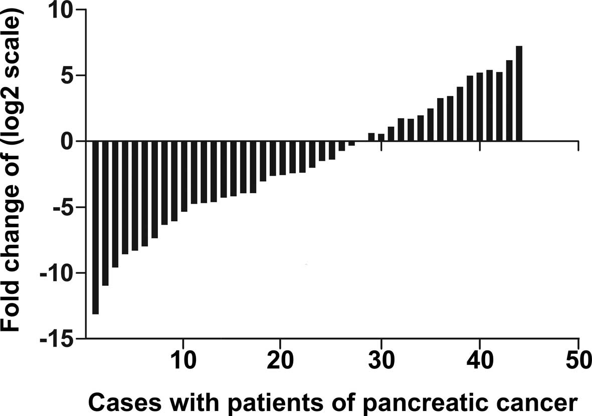
that miR-375 was significantly down-regulated in pancreatic cancer
tumor tissues. The value of ΔCt (mean ± SD) was −0.208±1.529 in
pancreatic cancer tissues and −1.992±3.447 in their matching
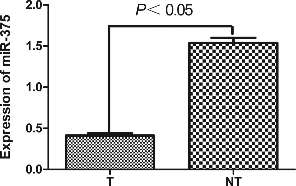
adjacent non-tumor tissues (P=0.021, paired t-test; Fig. 1). miR-375 was 0.413±0.026 in 44
cases of pancreatic cancer tissues and 1.538±0.061 in their
matching adjacent non-tumor tissues. miR-375 was significantly
downregulated in pancreatic cancer tissues with a median 3.4-fold
reduction relative to their matched adjacent non-tumor tissues
(Fig. 2). Among 44 pancreatic
cancer patients, 32 (72.7%) cases revealed a >50% reduction in
the expression levels of miR-375 compared to their matched adjacent
non-tumor tissues.
Expression of miR-375 is significantly
downregulated in pancreatic cancer cell lines
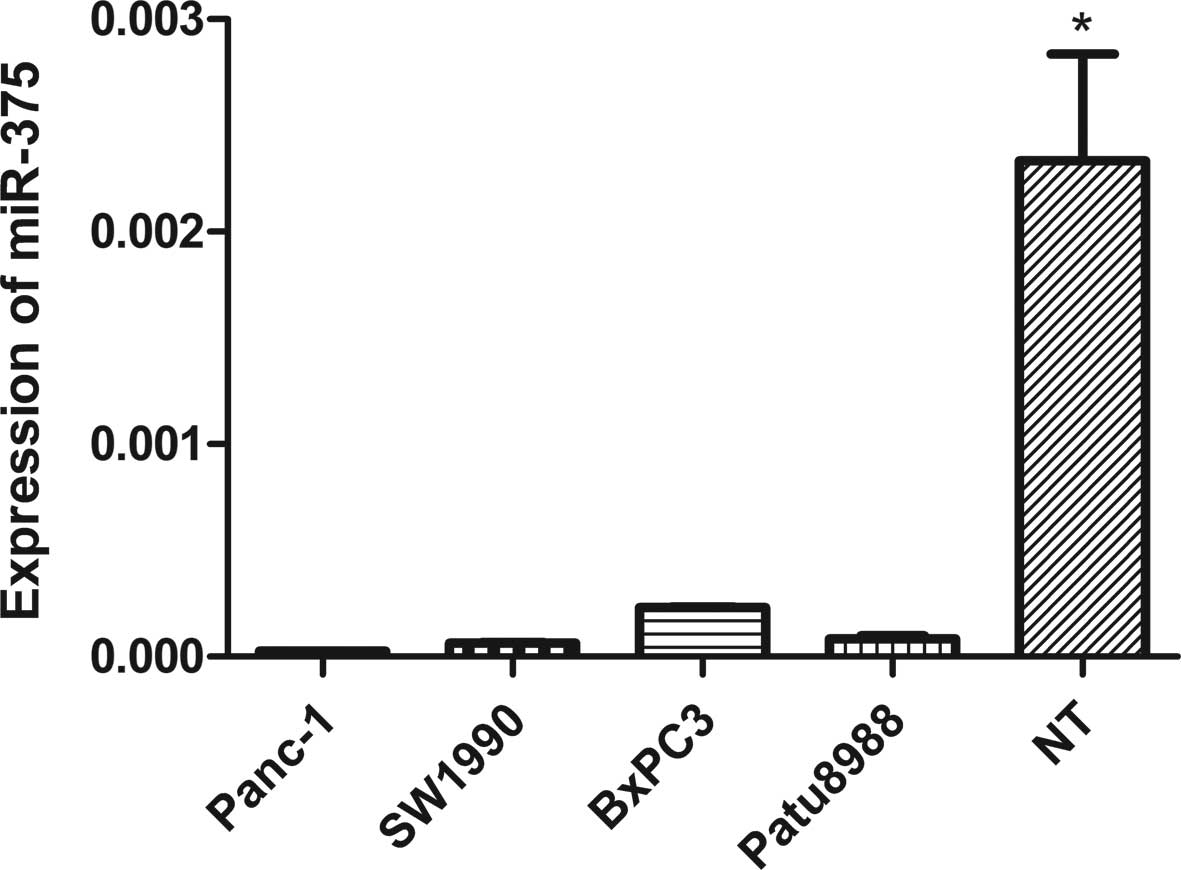
To confirm the association between miR-375
expression and pancreatic cancer, we measured miR-375 expression in
three pancreatic cancer cell lines using qRT-PCR. We found a
significantly low expression of miR-375 in Panc-1 (P=0.016), SW1990
(P=0.016), BxPC3 (P=0.018) and Patu8988 cells (P=0.017) relative to
three matching adjacent non-tumor tissues randomly selected from
the patients (Fig. 3).
Correlation between miR-375 expression
levels and clinicopathological characteristics in pancreatic cancer
patients
The non-parametric test between the relative
expression levels of miR-375 in pancreatic cancer cases and its
clinicopathological characteristics showed that there was no
significant correlation between the low expression of miR-375 and
parameters such as gender, age, tumor size, tumor location and
histological grade (P>0.05). However, the low expression of
miR-375 was correlated with pT stage, lymph node metastases and
pTNM stage (P<0.05) (Table
I).
 | Table IAssociation between the expression of
miR-375 with clinicopathological characteristics in patients with
pancreatic cancer. |
Table I
Association between the expression of
miR-375 with clinicopathological characteristics in patients with
pancreatic cancer.
| Parameters | No. | miR-375a | P-value |
|---|
| Total | 44 | | |
| Age (years) | | | 0.659 |
| ≤60 | 20 | 0.00175
(0.000075–0.011896) | |
| >60 | 24 | 0.00180
(0.000071–0.011022) | |
| Gender | | | 0.124 |
| Male | 30 | 0.0022
(0.000058–0.003811) | |
| Female | 14 | 0.0015
(0.000076–0.013231) | |
| Tumor size
(cm) | | | 0.090 |
| ≤2 | 1 | 0.1826 | |
| >2 | 43 | 0.0017
(0.000074–0.011266) | |
| pT stage | | | 0.002b |
| T1 | 1 | 0.1826 | |
| T2 | 22 | 0.0090
(0.00078–0.013505) | |
| T3 | 21 | 0.0012
(0.000049–0.001853) | |
| T4 | 0 | - | |
| pN stage | | | 0.018b |
| N0 | 28 | 0.0066
(0.001811–0.013365) | |
| N1 | 16 | 0.000067
(0.000028–0.000077) | |
| pTNM stage | | | 0.001b |
| I | 16 | 0.0644 | |
| IA | 1 | 0.3533 | |
| IB | 15 | 0.0664
(0.041764–0.071184) | |
| II | 28 | 0.0018 | |
| IIA | 12 | 0.0178
(0.011330–0.026199) | |
| IIB | 16 | 0.0014
(0.000670–0.001585) | |
| III | 0 | - | |
| IV | 0 | - | |
| Histological
grade | | | 0.097 |
| Poorly
differentiated | 9 | 0.0009
(0.00025–0.006525) | |
| Moderately
differentiated | 34 | 0.0018
(0.00007–0.012628) | |
| Well
differentiated | 1 | 0.0259 | |
Discussion
Due to the asymptomatic onset of pancreatic cancer,
most patients are in advanced or metastatic condition at the time
of diagnosis, resulting in a poor prognosis. The majority of
patients diagnosed with have pancreatic cancer succumb to the
disease within 12 months, and few survive 5 years after diagnosis.
The poor prognosis of these patients is due to its late clinical
presentation with symptoms, early and aggressive local invasion,
and high metastatic potential (25).
miRNA alterations have been shown to play an
important role in different steps of tumor formation and
progression (26). miRNA expression
has been studied by cloning and sequencing, northern blotting,
in situ hybridization, microarrays, real-time PCR and other
techniques. A number of studies have analyzed the global expression
pattern of miRNAs in pancreatic carcinoma (23,24,27,28).
However, real-time PCR has an advantage in that it is a more
quantitative and more sensitive method compared to high-throughput
assays.
Szafranska et al(23) have performed the first comprehensive
miRNA expression profile study in tissues from normal pancreas
(n=7), chronic pancreatitis (n=7), pancreatic cancer (n=10) and 33
human tissues of different non-pancreatic origin, to identify miRNA
candidates with a potential for future clinical application from a
pool of 377 known and novel miRNAs. Their findings demonstrated
that miR-375 may be used to classify normal, chronic pancreatitis
and cancerous tissues, and discriminate between neoplastic and
non-neoplastic processes in pancreatic cancer. miR-375 expression
was high in normal pancreas but was significantly lower in both
diseased tissues and absent in cell lines. miR-375 has been
previously described to be expressed in mouse pancreatic islet
cells suppressing glucose-induced insulin secretion and miR-375 was
recently identified as a pancreatic islet-specific miRNA regulating
insulin secretion (29). Thus, it
is likely, that the lower content of miR-375 in chronic
pancreatitis and pancreatic cancer tissues reflects the reduced
number of islet cells present in these tissues.
Therefore, we used real-time PCR to assess the
expression levels of miR-375 in a large number of cases and
clarified the correlation between miR-375 and clinicopathological
characteristics in pancreatic carcinoma.
The non-parametric test between the relative
expression levels of miR-375 in pancreatic cancer cases and its
clinicopathological characteristics showed that there was no
significant correlation between the low expression of miR-375 and
parameters such as gender, age, tumor size, tumor location and
histological grade (P>0.05). However, the low expression of
miR-375 was correlated with pT stage, lymph node metastases and
pTNM stage (P<0.05).
To the best of our knowledge, we performed the
largest study to date that assesses the expression levels of
miR-375 in pancreatic cancers by real-time PCR. The results showed
that miR-375 was frequently downregulated in 44 cancer and matching
adjacent non-tumor tissue pairs. The significantly reduced
expression of miR-375 was found in four pancreatic cancer cell
lines. Our results were consistent with previous studies on the
global expression pattern of miRNAs in pancreatic carcinoma
(25,28). Therefore, as the low expression of
miR-375 is frequently observed in pancreatic cancers, it may be
crucial in the process of carcinogenesis.
This study demonstrated that miR-375 was
significantly downregulated in pancreatic cancer. Due to the
resected tissues samples being obtained, according to the TNM
staging system of the American Joint Committee on Cancer (AJCC;
2010) and the International Union against Cancer (UICC), there were
no T4 stage patients, and only one case of T1 stage. miR-375 was
mainly associated with stages T2 and T3, thus miR-375 was
associated with whether the pancreatic cancer invaded the adjacent
organs or vessels. miR-375 expression was correlated with pT stage,
suggesting that miR-375 is involved in the carcinogenesis,
development and metastasis of pancreatic carcinoma. miR-375 was
also correlated with pN or lymph node metastases, thus miR-375 is
crucial in the metastasis of pancreatic cancer. There was a
decrease in the expression level of miR-375, but an increase in
pTNM stage suggesting that miR-375 is associated with the
progression and metastasis of pancreantic cancer. Findings of this
study therefore suggest that miR-375 is involved in the
carcinogenesis, development and metastasis of pancreatic
carcinoma.
miRNA expression can be reduced by a number of
factors, including transcriptional factors, mutations, deletions
and methylation. However, miR-375 plays an anti-oncogene role.
Results of bioinformatic algorithms, such as Target
Scan 4.2, demonstrated that 3-phosphoinositide-dependent protein
kinase-1 (PDK1) is a potential target gene of miR-375. Previous
studies have confirmed this role in pancreatic (30), gastric (31) and esophageal cancer (32). Various growth factors activate the
phosphatidylinositol 3-kinase (PI3K) pathway, which in turn
phosphorylates phosphatidylinositol-4,5-biphosphate (PIP2) to
generate phosphatidylinositol-3,4,5-triphosphate (PIP3). One of the
most studied signaling events controlled by PIP3 is the activation
of a group of AGC family protein kinases, including isoforms of
protein kinase B (Pkb/Akt) and the ribosomal S6 kinase (S6K), which
play crucial roles in regulating physiological processes relevant
to metabolism, cell growth, proliferation and survival (33,34).
PDK1 is a PH domain-containing protein that is activated following
PI3K activity, which in turn phosphorylates Akt1 at threonine 308
(or cognate locations on other isoforms) together with a large
variety of other AGC kinase substrates. Although this kinase is
important in PI3K-Akt-mTOR signaling, activating mutations of the
gene encoding PDK1 have not been described. Findings of a recent
study (35) suggest that PDK1
expression levels control the growth, proliferation and survival of
developing cells in pancreatic cancer, however, this has yet to be
adequately investigated. PDK1 activation is dependent primarily on
cytoplasmic membrane localization, and is considered to be
constitutively active. Thus, while it is unlikely that activating
mutations in the kinase domain occur, it is possible that
membrane-targeting PDK1 mutations may result in pathway activation
(36).
PDK1 is a gene that has been identified as a direct
target of miR-375 (30). PDK-1 is a
key component in Akt signaling, a well-documented pathway that
regulates cancer cell survival and proliferation.
The reduced expression of miR-375 has been reported
in pancreatic cancer (27),
hepatocellular carcinoma (37) as
well as head or neck squamous cell carcinoma (38). Apart from its role in cancer,
miR-375 is an important regulator in mammalian pancreatic
islet-cell development and regulation of insulin secretion
(39), indicating its diverse role
in normal physiology. The target genes of miR-375 may function
cooperatively through different cell mechanisms. PDK1 as a target
of miR-375 provides new insights into the molecular networks of
miR-375. However, further studies are required to investigate the
targets of miR-375 that may favor the process of tumorigenesis. The
introduction of a single miRNA that modulates complex downstream
signals and, in turn, retards the process of tumorigenesis would be
useful.
At present, the yes-associated protein (YAP)
(40), JAK2 (18) and 14-3-3 ζ (30) are other genes that have been
identified as a direct target of miR-375.
In conclusion, the results of this study suggest
that miR-375 interferes with the PI3K-Akt-mTOR signaling.
Therefore, miR-375 is a potential therapeutic target against the
PI3K-Akt-mTOR signaling axis for the prevention of pancreatic
cancer development and progression.
Acknowledgements
This study was supported by the Young
Scientist Fund of the National Natural Science Foundation of China
(no. 81201905)
References
|
1
|
Lee RC, Feinbaum RL and Ambros V: The
C. elegansheterochronic gene lin-4 encodes small RNAs with
antisense complementarity to lin-14. Cell. 75:843–854. 1993.
|
|
2
|
Bushati N and Cohen SM: microRNA
functions. Annu Rev Cell Dev Biol. 23:175–205. 2007. View Article : Google Scholar
|
|
3
|
Carthew RW and Sontheimer EJ: Origins and
mechanisms of miRNAs and siRNAs. Cell. 136:642–655. 2009.
View Article : Google Scholar : PubMed/NCBI
|
|
4
|
Zeng Y: Principles of micro-RNA production
and maturation. Oncogene. 25:6156–6162. 2006. View Article : Google Scholar : PubMed/NCBI
|
|
5
|
Ambros V: The functions of animal
microRNAs. Nature. 431:350–355. 2004. View Article : Google Scholar : PubMed/NCBI
|
|
6
|
Bartel DP: MicroRNAs: genomics,
biogenesis, mechanism, and function. Cell. 116:281–297. 2004.
View Article : Google Scholar : PubMed/NCBI
|
|
7
|
He L and Hannon GJ: MicroRNAs: small RNAs
with a big role in gene regulation. Nat Rev Genet. 5:522–531. 2004.
View Article : Google Scholar : PubMed/NCBI
|
|
8
|
Zaman MS, Chen Y, Deng G, et al: The
functional significance of microRNA-145 in prostate cancer. Br J
Cancer. 103:256–264. 2010. View Article : Google Scholar : PubMed/NCBI
|
|
9
|
Yu S, Lu Z, Liu C, et al: miRNA-96
suppresses KRAS and functions as a tumor suppressor gene in
pancreatic cancer. Cancer Res. 70:6015–6025. 2010. View Article : Google Scholar : PubMed/NCBI
|
|
10
|
Schickel R, Park SM, Murmann AE and Peter
ME: miR-200c regulates induction of apoptosis through CD95 by
targeting FAP-1. Mol Cell. 38:908–915. 2010. View Article : Google Scholar : PubMed/NCBI
|
|
11
|
Esquela-Kerscher A and Slack FJ: Oncomirs
- microRNAs with a role in cancer. Nat Rev Cancer. 6:259–269. 2006.
View Article : Google Scholar
|
|
12
|
Mori Y, Ishiguro H, Kuwabara Y, et al:
MicroRNA-21 induces cell proliferation and invasion in esophageal
squamous cell carcinoma. Mol Med Rep. 2:235–239. 2009.PubMed/NCBI
|
|
13
|
Takamizawa J, Konishi H, Yanagisawa K, et
al: Reduced expression of the let-7 microRNAs in human lung cancers
in association with shortened postoperative survival. Cancer Res.
64:3753–3756. 2004. View Article : Google Scholar : PubMed/NCBI
|
|
14
|
Iorio MV, Ferracin M, Liu CG, et al:
MicroRNA gene expression deregulation in human breast cancer.
Cancer Res. 65:7065–7070. 2005. View Article : Google Scholar : PubMed/NCBI
|
|
15
|
Ikenaga N, Ohuchida K, Mizumoto K, et al:
MicroRNA-203 expression as a new prognostic marker of pancreatic
adenocarcinoma. Ann Surg Oncol. 17:3120–3128. 2010. View Article : Google Scholar : PubMed/NCBI
|
|
16
|
Murakami Y, Yasuda T, Saigo K, et al:
Comprehensive analysis of microRNA expression patterns in
hepatocellular carcinoma and non-tumorous tissues. Oncogene.
25:2537–2545. 2006. View Article : Google Scholar : PubMed/NCBI
|
|
17
|
Dai X, Chiang Y, Wang Z, et al: Expression
levels of microRNA-375 in colorectal carcinoma. Mol Med Rep.
5:1299–1304. 2012.PubMed/NCBI
|
|
18
|
Ding L, Xu Y, Zhang W, et al: MiR-375
frequently downregulated in gastric cancer inhibits cell
proliferation by targeting JAK2. Cell Res. 20:784–793. 2010.
View Article : Google Scholar : PubMed/NCBI
|
|
19
|
Calin GA, Sevignani C, Dumitru CD, et al:
Human microRNA genes are frequently located at fragile sites and
genomic regions involved in cancers. Proc Natl Acad Sci USA.
101:2999–3004. 2004. View Article : Google Scholar
|
|
20
|
Li D, Xie K, Wolff R and Abbruzzese JL:
Pancreatic cancer. Lancet. 363:1049–1057. 2004. View Article : Google Scholar
|
|
21
|
Guo X and Cui Z: Current diagnosis and
treatment of pancreatic cancer in China. Pancreas. 31:13–22. 2005.
View Article : Google Scholar : PubMed/NCBI
|
|
22
|
Siegel R, Naishadham D and Jemal A: Cancer
statistics, 2012. CA Cancer J Clin. 62:10–29. 2012. View Article : Google Scholar
|
|
23
|
Szafranska AE, Davison TS, John J, et al:
MicroRNA expression alterations are linked to tumorigenesis and
non-neoplastic processes in pancreatic ductal adenocarcinoma.
Oncogene. 26:4442–4452. 2007. View Article : Google Scholar
|
|
24
|
Bloomston M, Frankel WL, Petrocca F, et
al: MicroRNA expression patterns to differentiate pancreatic
adenocarcinoma from normal pancreas and chronic pancreatitis. JAMA.
297:1901–1908. 2007. View Article : Google Scholar
|
|
25
|
Park JY, Helm J, Coppola D, Kim D, Malafa
M and Kim SJ: MicroRNAs in pancreatic ductal adenocarcinoma. World
J Gastroenterol. 17:817–827. 2011. View Article : Google Scholar : PubMed/NCBI
|
|
26
|
Calin GA and Croce CM: MicroRNA signatures
in human cancers. Nat Rev Cancer. 6:857–866. 2006. View Article : Google Scholar : PubMed/NCBI
|
|
27
|
Lee EJ, Gusev Y, Jiang J, et al:
Expression profiling identifies microRNA signature in pancreatic
cancer. Int J Cancer. 120:1046–1054. 2007. View Article : Google Scholar : PubMed/NCBI
|
|
28
|
Zhang Y, Li M, Wang H, et al: Profiling of
95 microRNAs in pancreatic cancer cell lines and surgical specimens
by real-time PCR analysis. World J Surg. 33:698–709. 2009.
View Article : Google Scholar : PubMed/NCBI
|
|
29
|
Poy MN, Hausser J, Trajkovski M, et al:
miR-375 maintains normal pancreatic alpha- and beta-cell mass. Proc
Natl Acad Sci USA. 106:5813–5818. 2009. View Article : Google Scholar : PubMed/NCBI
|
|
30
|
El Ouaamari A, Baroukh N, Martens GA,
Lebrun P, Pipeleers D and van Obberghen E: miR-375 targets
3′-phosphoinositide-dependent protein kinase-1 and regulates
glucose-induced biological responses in pancreatic beta-cells.
Diabetes. 57:2708–2717. 2008.
|
|
31
|
Tsukamoto Y, Nakada C, Noguchi T, et al:
MicroRNA-375 is downregulated in gastric carcinomas and regulates
cell survival by targeting PDK1 and 14-3-3zeta. Cancer Res.
70:2339–2349. 2010. View Article : Google Scholar : PubMed/NCBI
|
|
32
|
Li X, Lin R and Li J: Epigenetic silencing
of microRNA-375 regulates PDK1 expression in esophageal cancer. Dig
Dis Sci. 56:2849–2856. 2011. View Article : Google Scholar : PubMed/NCBI
|
|
33
|
Kozma SC and Thomas G: Regulation of cell
size in growth, development and human disease: PI3K, PKB and S6K.
Bioessays. 24:65–71. 2002. View Article : Google Scholar : PubMed/NCBI
|
|
34
|
Mora A, Komander D, van Aalten DM and
Alessi DR: PDK1, the master regulator of AGC kinase signal
transduction. Semin Cell Dev Biol. 15:161–170. 2004. View Article : Google Scholar : PubMed/NCBI
|
|
35
|
Westmoreland JJ, Wang Q, Bouzaffour M,
Baker SJ and Sosa-Pineda B: Pdk1 activity controls proliferation,
survival, and growth of developing pancreatic cells. Dev Biol.
334:285–298. 2009. View Article : Google Scholar : PubMed/NCBI
|
|
36
|
Storz P and Toker A:
3′-phosphoinositide-dependent kinase-1 (PDK-1) in PI 3-kinase
signaling. Front Biosci. 7:d886–d902. 2002.
|
|
37
|
Ladeiro Y, Couchy G, Balabaud C, et al:
MicroRNA profiling in hepatocellular tumors is associated with
clinical features and oncogene/tumor suppressor gene mutations.
Hepatology. 47:1955–1963. 2008. View Article : Google Scholar : PubMed/NCBI
|
|
38
|
Avissar M, Christensen BC, Kelsey KT and
Marsit CJ: MicroRNA expression ratio is predictive of head and neck
squamous cell carcinoma. Clin Cancer Res. 15:2850–2855. 2009.
View Article : Google Scholar : PubMed/NCBI
|
|
39
|
Poy MN, Eliasson L, Krutzfeldt J, et al: A
pancreatic islet-specific microRNA regulates insulin secretion.
Nature. 432:226–230. 2004. View Article : Google Scholar : PubMed/NCBI
|
|
40
|
Liu AM, Poon RT and Luk JM: MicroRNA-375
targets Hippo-signaling effector YAP in liver cancer and inhibits
tumor properties. Biochem Biophys Res Commun. 394:623–627. 2010.
View Article : Google Scholar : PubMed/NCBI
|