Introduction
Benexate hydrochloride betadex (BHB) is used
clinically as an antiulcer agent. In the gastric mucosa, BHB
promotes prostaglandin synthesis, inhibits acid secretion and
increases mucosal blood flow (1).
The effects of BHB are proposed to be mediated by nitric oxide
(NO), through inhibition of cyclooxygenases (COXs) and inflammatory
cytokines. However, the mechanism by which BHB promotes mucosal
angiogenesis and ulcer healing remains unclear.
NO is involved in numerous physiological functions,
including healing processes (2). NO
acts as a local vasodilator in the gastric mucosal
microvasculature, and stimulates mucus and bicarbonate secretion in
the stomach. Previous studies have suggested that the inhibition of
NO synthase (NOS) significantly delays ulcer healing by reducing
gastric blood flow around the ulcer (3). NO inhibitors, including
L-NG-nitroarginine methyl ester (L-NAME), have an inhibitory
effect on gastric ulcer healing.
COX may also be involved in gastrointestinal mucosal
integrity. COX is expressed in inflammatory cells and fibroblasts
of the gastric mucosa; in a previous study, COX mRNA expression
increased rapidly in injured mucosa, and the level of expression
was correlated with the severity of the gastric mucosal injuries
(4). mRNA expression levels of
certain inflammatory cytokines, including interleukin-1β (IL-1β)
and tumor necrosis factor-α (TNF-α), are also significantly
increased in gastric ulcers (5).
Despite numerous studies investigating anti-gastric
ulcer agents, the molecular mechanism of BHB is not fully
understood. In the present study, the anti-gastric ulcer effects of
BHB with respect to the modulation of NOS, COX and inflammatory
cytokine expression have been investigated.
Materials and methods
Materials
BHB was purchased from Ildong Pharmaceutical Co.,
Ltd. (Seoul, Korea). Protein expression was detected using the
following primary antibodies from Santa Cruz Biotechnology, Inc.
(Dallas, TX, USA): Rabbit polyclonal IL-1β (dilution, 1:500; cat.
no. sc-7884); goat polyclonal IL-6 immunoglobulin G (IgG)
(dilution, 1:500; cat. no. sc-1265); goat polyclonal TNF-α IgG
(dilution, 1:500; cat. no. sc-1351); rabbit polyclonal
cyclooxygenase-1 (COX-1; dilution, 1:1,000; cat. no. sc-7950); and
goat polyclonal cyclooxygenase-2 IgG (COX-2; dilution, 1:1,000;
cat. no. sc-1745) (all purchased from Santa Cruz Biotechnology,
Inc.). The following primary antibodies were also used: Rabbit
polyclonal inducible NOS (iNOS; dilution, 1:1,000; cat. no.
ab15323); rabbit monoclonal neuronal NOS (nNOS; dilution, 1:1,000;
cat. no. ab76067) (both purchased from Abcam, Cambridge, UK); and
rabbit monoclonal endothelial NOS (eNOS; dilution, 1:1,000; cat.
no. 32027; Cell Signaling Technology, Inc., Beverly, MA, USA).
Horseradish peroxidase (HRP-conjugated goat anti-mouse IgG
(dilution, 1:2,000; cat. no. sc-2005), HRP-conjugated goat anti-rat
IgG (dilution, 1:2,000; cat. no. sc-2006) and HRP-conjugated donkey
anti-goat IgG (dilution, 1:2,000; cat. no. sc-2020) were used (all
purchased from Santa Cruz Biotechnology, Inc.).
Animals
A total of 30 male Sprague-Dawley rats, weighing
between 240–250 g, were purchased from Samtako Laboratory Animal
Company (Osan, Korea) and housed for 1 week in the animal facility
for acclimation. Constant environmental conditions were maintained
with a temperature of 23±1°C, humidity of 55% and a 12-h light/dark
cycle. Following acclimation, the rats underwent a fast for the 24
h prior to the experiments. The present study was approved by the
Institutional Animal Care & Use Committee of Korea University
(approval number, KUIACUC-2013-181).
Study methods
Gastric ulcers were induced in all rats by direct
injection of acetic acid. The rats were anesthetized using
tiletamine/zolazepam (10 mg/kg; intramuscular injection; Zoetis,
Inc., Florham Park, NJ, USA) and xylazine (5 mg/kg; intraperitoneal
injection; Bayer AG, Leverkusen, Germany). A longitudinal incision
of 2 cm was made in the upper abdomen. The stomach was then exposed
and directly injected with 2 cm3 of 60% acetic acid
solution, as described by Okabe et al (6). After 45 sec, the gastric contents were
aspirated by syringe. The abdomen was sutured and oral intake of
food and water was permitted following closure. Following gastric
ulcer induction, the rats were randomly divided into 6 groups, with
5 rats per group. The groups were organized as follows: Control, no
BHB treatment; BHB 100; 300; or 1,000 mg/kg treatment; L-NAME
(Sigma-Aldrich, St. Louis, MO, USA) 70 mg/kg treatment; and L-NAME
70 mg/kg treatment with BHB at 1,000 mg/kg. The drugs were
dissolved in 2 ml 5% dextrose water (DW; JW Pharmaceutical
Corporation, Seoul, Korea) and administered orally once per day for
5 days. The rats in the control group were administered 2 ml 5% DW
without BHB.
Assessment of the gastric lesions
A total of 5 days after the induction of gastric
ulcers, the rats were sacrificed using CO2. The stomachs
were dissected, gently incised along the longer curvature, opened
and rinsed with phosphate-buffered saline (PBS) to remove the
gastric contents. The gastric mucosa lesions were macroscopically
examined with a magnifier using a metric measurement scale. The
areas of the ulcerous lesions were measured in mm2 using
the lesion index (7).
Western blot analysis
The expression levels of COXs (COX-1 and COX-2),
cytokines (IL-1β, IL-6 and TNF-α) and NOS (nNOS, eNOS and iNOS)
were measured using western blot analysis. The gastric tissues were
frozen using liquid nitrogen and stored at −80°C. Samples were
pulverized by a mortar and pestle, then mixed with
radioimmunoprecipitation assay buffer (a lysis buffer) and
centrifuged at 14,200 × g for 15 min. The supernatants were
collected and the protein content was determined using a Bio-Rad
Protein Assay Dye Reagent Concentrate (Bio-Rad Laboratories, Inc.,
Hercules, CA, USA). A similar mass of total protein was loaded from
each sample onto a 5–12% sodium dodecyl sulfate gel and transferred
to polyvinylidene fluoride membranes using electrophoresis. The
membranes were blocked with a blocking buffer (5% skimmed milk in
PBS) for 1 h at room temperature, then incubated with the primary
antibody. Following several washes with PBS-Tween 20 over 30 min,
the membranes were incubated with the secondary antibody specific
to the primary antibody for 1 h at room temperature. Following
several additional washes with PBS-Tween 20 over 30 min, detection
was performed using an enhanced chemiluminescence kit (Pierce ECL
Western Blotting Substrate; Thermo Fisher Scientific, Inc.,
Waltham, MA, USA) and the images were analyzed using ImageJ
software (National Institutes of Health, Bethesda, MD, USA). The
intensity of each band was compared with that of the internal
control, β-actin.
Statistical analysis
Data were processed and analyzed using SPSS, version
20.0 (IBM SPSS, Armonk, NY, USA). Statistical comparisons were
performed using a Student's t-test. P≤0.05 was considered to
indicate a statistically significant difference.
Results
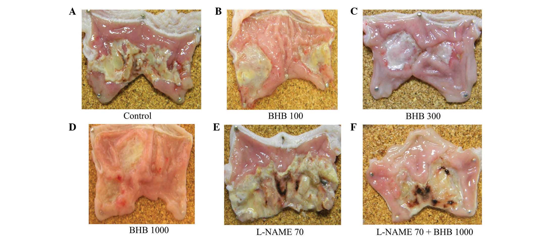
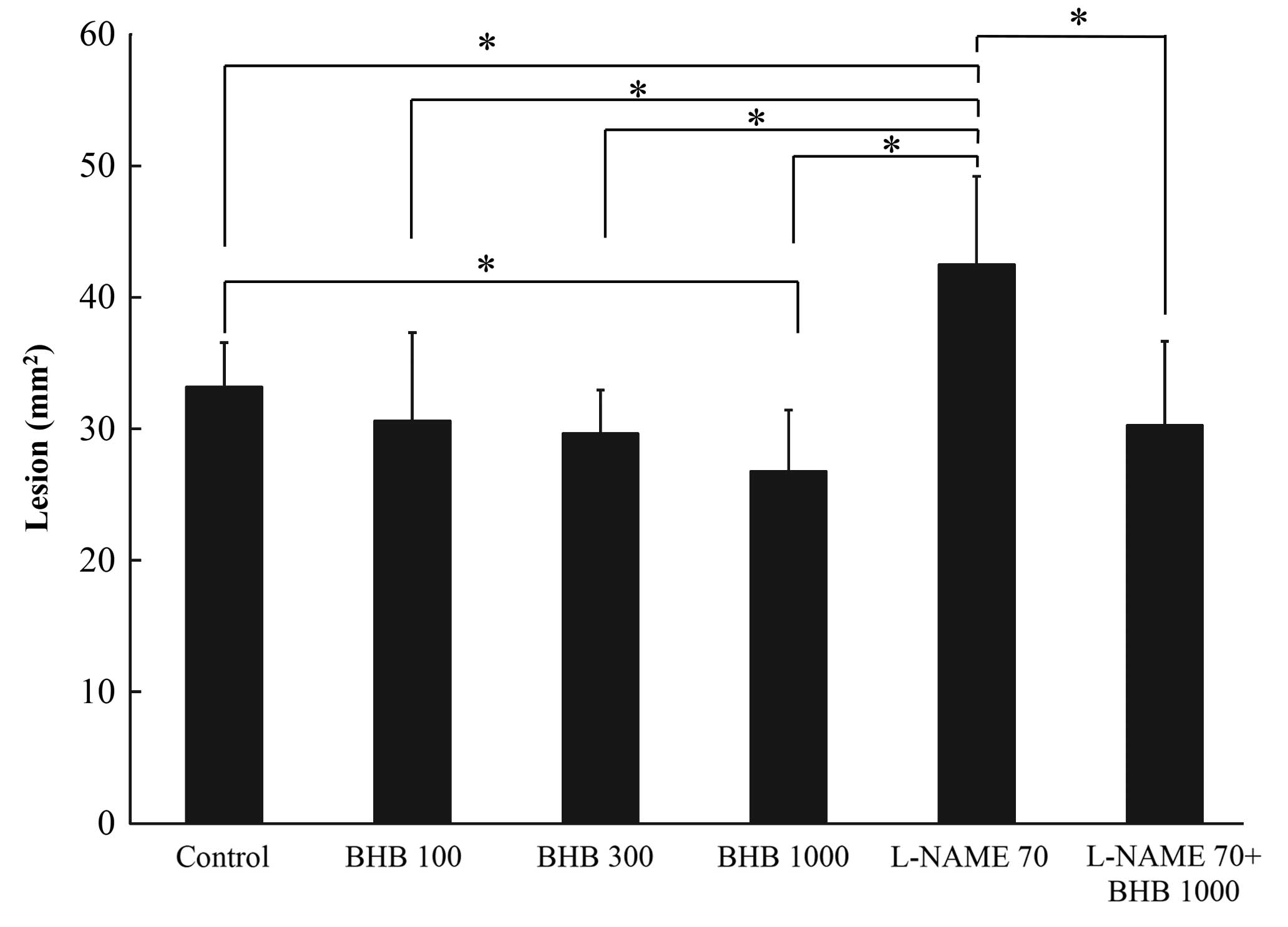
Gastric ulcers
The control group developed ulcerous lesions
(Fig 1). Rats that received BHB at
doses of 100, 300 and 1,000 mg/kg demonstrated reductions in
mucosal injury of 7.8, 10.7 and 19.3%, respectively, compared with
the control (Fig. 2); the area of
ulcerous lesions significantly decreased in the group treated with
1,000 mg/kg BHB (Fig. 2). L-NAME
aggravated the acetic acid-induced ulcerous lesions, observed
macroscopically (Fig. 1E), but the
effect of L-NAME was somewhat reversed when it was administered
with 1,000 mg/kg BHB (Fig. 1F). The
L-NAME + BHB group exhibited significantly reduced lesion area
compared with the L-NAME group (P<0.05; Fig. 2).
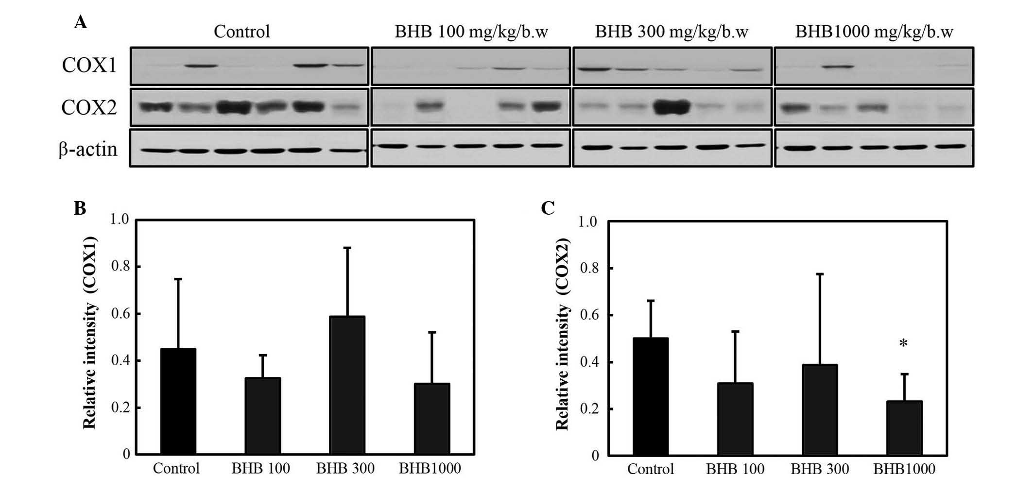
COX and cytokine levels of gastric
ulcers
The expression levels of COX-1 and −2 are reported
in Fig. 3. COX-2 expression was
decreased in the BHB groups compared with the control group;
however, a significant difference was only observed at 1,000 mg/kg
(P<0.05; Fig. 3C). No significant
difference in COX-1 expression was observed between the BHB
treatment and control groups.
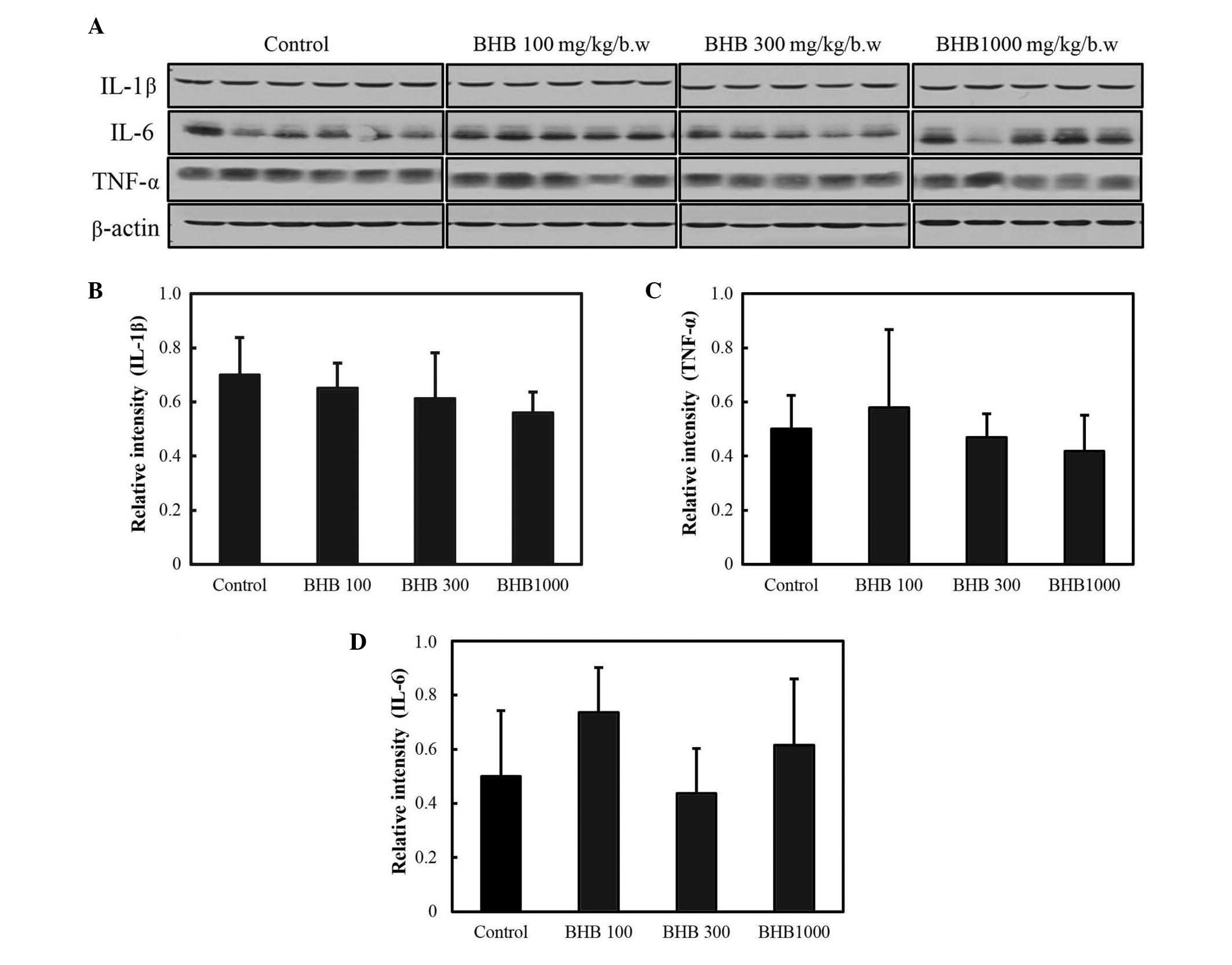
The expression levels of pro-inflammatory cytokines
are reported in Fig. 4; and the
expression levels of IL-1β, IL-6 and TNF-α were not observed to be
significantly different following BHB treatment.
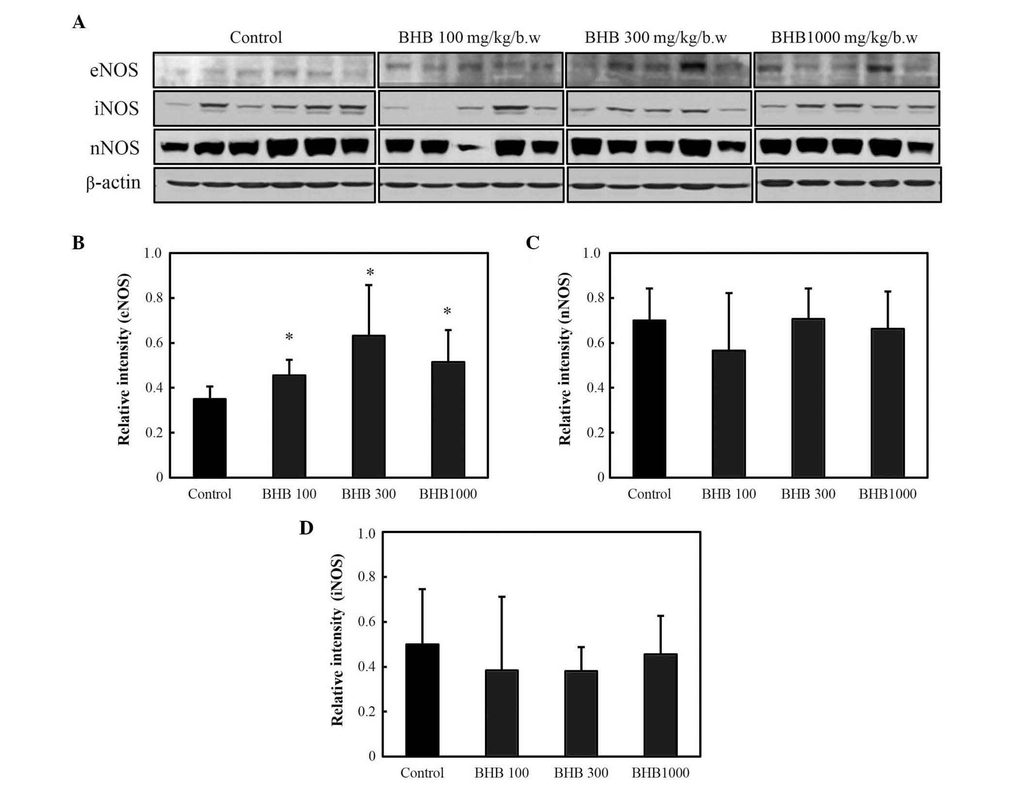
NOS levels in the gastric ulcers
The effect of BHB on eNOS, iNOS and nNOS protein
expression was also assessed by western blot (Fig. 5A). All BHB treatments significantly
increased eNOS expression compared with the control group
(P<0.05; Fig. 5B), but no
significant differences were identified in the expression levels of
nNOS or iNOS between the groups (Fig. 5C
and D).
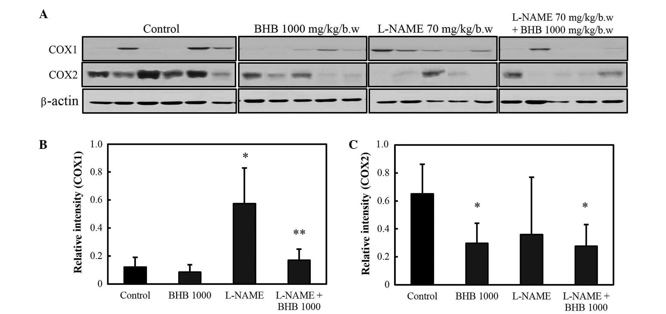
COX and inflammatory cytokine levels
of gastric ulcers in the presence of an NO inhibitor
As presented in Fig.
6, COX-1 protein expression levels increased in rats
administered L-NAME compared with the control group (P<0.05),
and the co-treatment with BHB significantly decreased the
expression of COX-1 compared with the L-NAME group (P<0.05).
COX-2 protein expression levels increased in rats in the control
group, and the administration of BHB significantly decreased COX-2
protein expression in both groups, with or without L-NAME
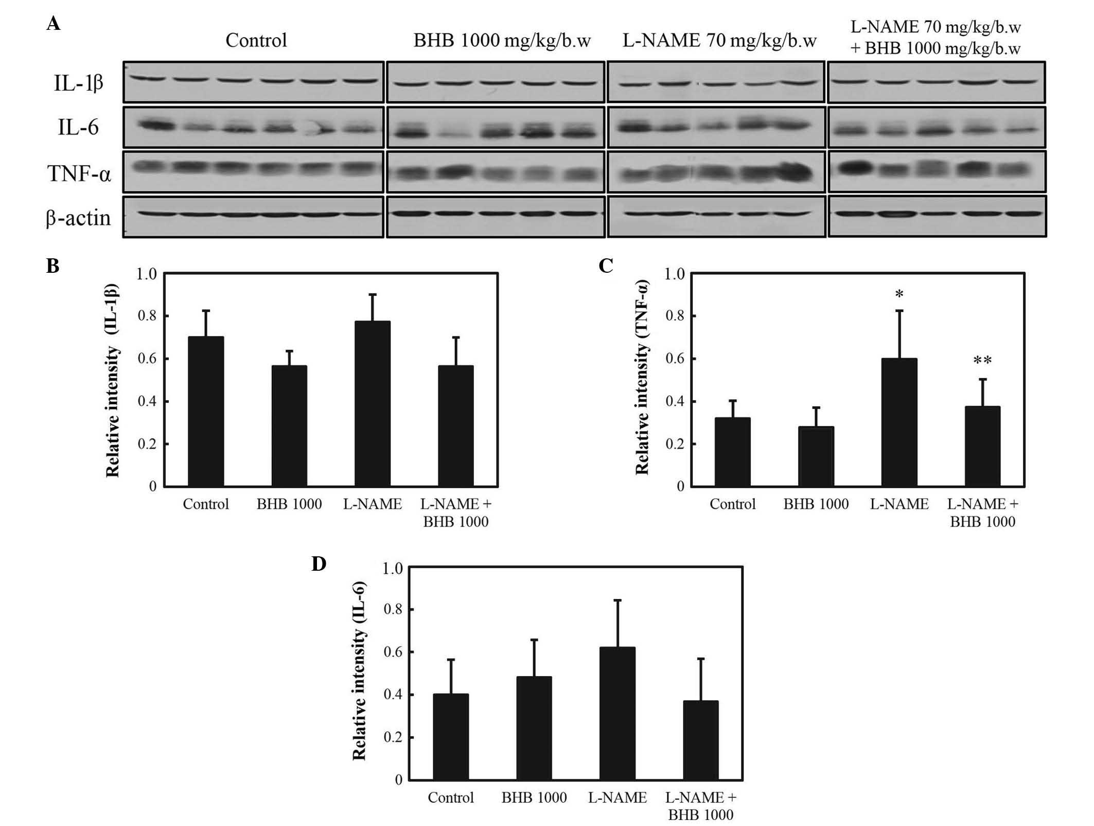
(P<0.05). As presented in Fig. 7,
L-NAME administration significantly increased the expression of the
inflammatory cytokine TNF-α (P<0.05 vs. control group), whereas
combined treatment suppressed its expression (P<0.05 vs. L-NAME
group). No significant difference was observed in the expression of
IL-1β and IL-6 with either drug.
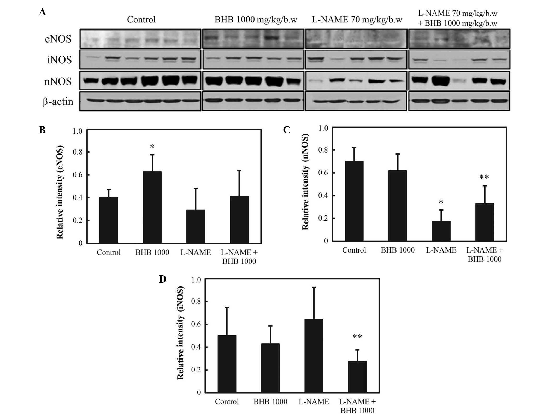
NOS levels of gastric ulcers in the
presence of an NO inhibitor
In the presence of L-NAME, eNOS levels did not
differ between those groups that were or were not administered BHB
(P<0.05; Fig. 8A and B). The
expression levels of nNOS were decreased in the L-NAME group
compared with levels in the control group; and compared with the
L-NAME group, these levels were significantly increased in the
L-NAME + BHB group (P<0.05; Fig. 8A
and C). By contrast, the expression levels of iNOS were
suppressed by BHB + L-NAME co-treatment compared with L-NAME
treatment alone (P<0.05; Fig. 8A and
D).
Discussion
The gastric microcirculation has an essential role
in healing gastric ulcers. Local vasodilators, including NO, are
important in the maintenance of mucosal integrity and in mucosal
defense mechanisms (2). Previous
studies have reported that increased levels of NO significantly
enhance ulcer healing by the maintenance of gastric blood flow; NO
also promotes angiogenesis and endothelial cell proliferation and
migration (8–10). It has previously been proposed that
the anti-ulcer effects of NO result from gastric mucosal NOS
activation (3). The
calcium-dependent isoforms of NOS, eNOS and nNOS are constitutively
expressed in the endothelium of blood vessels and in the brain,
respectively. However, expression of iNOS, the calcium-independent
isoform, is induced by pro-inflammatory agents (11,12).
eNOS is known to promote ulcer healing through angiogenesis,
enhancing gastric blood flow and stimulating mucus and bicarbonate
secretion (13–15). iNOS is associated with acute and
chronic inflammation, and it has previously been reported that
changes in iNOS expression and activity levels are correlated with
the severity of tissue inflammation (16).
Iwasaki and Matsunaga (17) reported that the vasorelaxant effects
of BHB, which were blocked by an NO inhibitor in defective
endothelium, were associated with NOS activation. By contrast,
Arimoto et al (18)
demonstrated that the in vitro activity of NOS was
suppressed by BHB. The current study, therefore, hypothesized that
a specific NOS may be activated through BHB administration, leading
to gastric mucosal healing. In the present study, BHB significantly
increased eNOS expression in the rat gastric ulcer model,
suggesting that the protective effect of BHB against gastric ulcers
may involve an increase in eNOS. iNOS and nNOS were not
significantly increased following BHB administration. These results
are partially in agreement with the hypothesis that the anti-ulcer
effects of BHB are associated with increased blood flow through the
activation of the specific NOS isoform, eNOS. However, BHB did not
increase eNOS in the presence of L-NAME, which aggravated the
ulcerous lesion. Additional study is therefore required in order to
establish the mechanisms of the BHB-induced increased blood flow in
gastric ulcers.
An increase in COX-2 expression has previously been
demonstrated to be a major contributor to inflammation (10). Previous studies have reported that
COX-1 and COX-2 levels are positively correlated with the severity
of gastric mucosal damage (19–21).
COX-2 mRNA expression is induced during the acute stage of gastric
ulceration (22); the early phase of
inflammation is primarily mediated by constitutive COX-1
expression, whilst the late and acute phases are mediated by COX-1
and COX-2. COX-2 expression is typically observed 1–2 h after
stimulation (4,23). In the present study, BHB
administration at a dose of 1,000 mg/kg significantly decreased
COX-2 levels, which may indicate that the protective effect of BHB
is associated with modulation of COX-2 expression.
Inflammatory cytokines mediate the upregulation of
COX-2 expression and inhibit growth factors that are responsible
for recovery from mucosal damage (24,25). The
current results suggest that BHB enhances ulcer healing through a
reduction of gastric inflammation and of the inflammatory cytokine
TNF-α; however, no association was observed between BHB and
interleukin levels.
There are a number of differences between the
mechanism of action of BHB and of other mucoprotective agents,
including rebamipide. Rebamipide reportedly suppresses COX-2
expression and NF-κB activation (22,26). In
the present study, however, BHB only decreased COX expression
whilst upregulating eNOS. Together, the current results suggest
that the anti-ulcer effects of BHB are, in part, associated with
decreased COX-2 and pro-inflammatory cytokine levels and with eNOS
activation. In conclusion, BHB administration ameliorates acetic
acid-induced mucosal injury in a rat model of gastric ulcers by
BHB-induced decreases to TNF-α and increases to eNOS expression
levels.
Acknowledgements
This study was supported by the Industrial Core
Technology Development Program (grant no. 10060251; Development of
the diagnostic device for functional dyspepsia based on
Korean-Western medicine fusion abdominal diagnosis) and the Basic
Science Research Program (grant no. NRF2012R1A2A2A01016829) funded
By the Ministry of Trade, Industry and Energy (MI, Korea) and the
Ministry of Science, ICT and Future Planning.
References
|
1
|
Hori Y, Odaguchi K, Jyoyama H, Yasui K and
Mizui T: Differential effect of benexate hydrochloride betadex on
prostaglandin levels in stomach and inflammatory site in rats. Jpn
J Pharmacol. 72:183–190. 1996. View Article : Google Scholar : PubMed/NCBI
|
|
2
|
Pique JM, Whittle BJ and Esplugues JV: The
vasodilator role of endogenous nitric oxide in the rat gastric
microcirculation. Eur J Pharmacol. 174:293–296. 1989. View Article : Google Scholar : PubMed/NCBI
|
|
3
|
Ma L and Wallace JL: Endothelial nitric
oxide synthase modulates gastric ulcer healing in rats: Am J
Physiol Gastrointest. Liver Physiol. 279:G341–G346. 2000.
|
|
4
|
Brzozowski T, Konturek PC, Sliwowski Z,
Pajdo R, Drozdowicz D, Kwiecien S, Burnat G, Konturek SJ and Pawlik
WW: Prostaglandin/cyclooxygenase pathway in ghrelin-induced
gastroprotection against ischemia-reperfusion injury. J Pharmacol
Exp Ther. 319:477–487. 2006. View Article : Google Scholar : PubMed/NCBI
|
|
5
|
Dembiński A, Warzecha Z, Ceranowicz P,
Cieszkowski J, Dembiński M, Ptak-Belowska A, Kuwahara A and Kato I:
Administration of obestatin accelerates the healing of chronic
gastric ulcers in rats. Med Sci Monit. 17:BR196–BR200. 2011.
View Article : Google Scholar : PubMed/NCBI
|
|
6
|
Okabe S, Roth JL and Pfeiffer CJ: A method
for experimental, penetrating gastric and duodenal ulcers in rats.
Observations on normal healing. Am J Dig Dis. 16:277–284. 1971.
View Article : Google Scholar : PubMed/NCBI
|
|
7
|
Morini G and Grandi D: Methods to measure
gastric mucosal lesions in the rat. Curr Protoc Toxicol. 21:Unit
21.2. 2010. View Article : Google Scholar : PubMed/NCBI
|
|
8
|
Kato S, Abe Y, Konishi M, Kuroda N and
Takeuchi K: Mechanism of gastric hyperemic response during acid
secretion in rats: Relation to nitric oxide, prostaglandins, and
sensory neurons. J Clin Gastroenterol. 25(Suppl 1): S48–S55. 1997.
View Article : Google Scholar : PubMed/NCBI
|
|
9
|
Moncada S, Palmer RMJ and Higgs EA: Nitric
oxide: Physiology, pathophysiology and pharmacology. Pharmacol Rev.
43:109–142. 1991.PubMed/NCBI
|
|
10
|
Konturek SK and Konturek PC: Role of
nitric oxide in the digestive system. Digestion. 56:1–13. 1995.
View Article : Google Scholar : PubMed/NCBI
|
|
11
|
Konturek PC, Brzozowski T, Meixner H, Ptak
A, Hahn EG and Konturek SJ: Central and peripheral neural aspects
of gastroprotective and ulcer healing effects of
lipopolysaccharides. J Physiol Pharmacol. 52:611–623.
2001.PubMed/NCBI
|
|
12
|
Brzozowska I, Konturek PC, Brzozowski T,
Konturek SJ, Kwiecien S, Pajdo R, Drozdowicz D, Pawlik M, Ptak A
and Hahn EG: Role of prostaglandins, nitric oxide, sensory nerves
and gastrin in acceleration of ulcer healing by melatonin and its
precursor, L-tryptophan. J Pineal Res. 32:149–162. 2002. View Article : Google Scholar : PubMed/NCBI
|
|
13
|
Guo JS, Cheng CL and Koo MW: Inhibitory
effects of Centella asiatica water extract and asiaticoside on
inducible nitric oxide synthase during gastric ulcer healing in
rats. Planta Med. 70:1150–1154. 2004. View Article : Google Scholar : PubMed/NCBI
|
|
14
|
Cho SO, Lim JW, Kim KH and Kim H:
Involvement of Ras and AP-1 in Helicobacter pylori-induced
expression of COX-2 and iNOS in gastric epithelial AGS cells. Dig
Dis Sci. 55:988–996. 2010. View Article : Google Scholar : PubMed/NCBI
|
|
15
|
Li Y, Wang WP, Wang HY and Cho CH:
Intragastric administration of heparin enhances gastric ulcer
healing through a nitric oxide-dependent mechanism in rats. Eur J
Pharmacol. 399:205–214. 2000. View Article : Google Scholar : PubMed/NCBI
|
|
16
|
McCafferty DM, Mudgett JS, Swain MG and
Kubes P: Inducible nitric oxide synthase plays a critical role in
resolving intestinal inflammation. Gastroenterology. 112:1022–1027.
1997. View Article : Google Scholar : PubMed/NCBI
|
|
17
|
Iwasaki T and Matsunaga K: Nitric
oxide-associated vasorelaxing effect of an anti-ulcer agent,
benexate hydrochloride betadex. Drug Dev Res. 36:13–19. 1995.
View Article : Google Scholar
|
|
18
|
Arimoto T, Yoshikawa T, Komori Y and
Kumagai Y: Inhibition of constitutive nitric oxide synthase by
benexate. Life Sci. 59:953–959. 1996. View Article : Google Scholar : PubMed/NCBI
|
|
19
|
Wallace JL, McKnight W, Reuter BK and
Vergnolle N: NSAID-induced gastric damage in rats: Requirement for
inhibition of both cyclooxygenase 1 and 2. Gastroenterology.
119:706–714. 2000. View Article : Google Scholar : PubMed/NCBI
|
|
20
|
Tanaka A, Araki H, Hase S, Komoike Y and
Takeuchi K: Up-regulation of COX-2 by inhibition of COX-1 in the
rat: A key to NSAID-induced gastric injury. Aliment Pharmacol Ther.
16(Suppl 2): 90–101. 2002. View Article : Google Scholar : PubMed/NCBI
|
|
21
|
Tanaka A, Araki H, Komoike Y, Hase S and
Takeuchi K: Inhibition of both COX-1 and COX-2 is required for
development of gastric damage in response to nonsteroidal
antiinflammatory drugs. J Physiol Paris. 95:21–27. 2001. View Article : Google Scholar : PubMed/NCBI
|
|
22
|
Murata H, Yabe Y, Tsuji S, Tsujii M, Fu
HY, Asahi K, Eguchi H, Kawano S and Hayashi N: Gastroprotective
agent rebamipide induces cyclooxygenease-2 (COX-2) in gastric
epithelial cells. Dig Dis Sci. 50:S70–S75. 2005. View Article : Google Scholar : PubMed/NCBI
|
|
23
|
Gudis K and Sakamoto C: The role of
cyclooxygenase in gastric mucosal protection. Dig Dis Sci. 50(Suppl
1): S16–S23. 2005. View Article : Google Scholar : PubMed/NCBI
|
|
24
|
Brzozowska I, Targosz A, Sliwowski Z,
Kwiecien S, Drozdowicz D, Pajdo R, Konturek PC, Brzozowski T,
Pawlik M, Konturek SJ, et al: Healing of chronic gastric ulcers in
diabetic rats treated with native aspirin, nitric oxide
(NO)-derivative of aspirin and cyclooxygenase (COX)-2 inhibitor. J
Physiol Pharmacol. 55:773–790. 2004.PubMed/NCBI
|
|
25
|
Motilva V, de la Alarcón Lastra C,
Bruseghini L, Manuel Herrerias J and Sánchez-Fidalgo S: COX
expression and PGE(2) and PGD(2) production in experimental acute
and chronic gastric lesions. Int Immunopharmacol. 5:369–379. 2005.
View Article : Google Scholar : PubMed/NCBI
|
|
26
|
Qi Z, Jie L, Haixia C and Xiaoying Z:
Effect of rebamipide on quality of peptic ulcer healing in rat. Dig
Dis Sci. 54:1876–1883. 2009. View Article : Google Scholar : PubMed/NCBI
|