Introduction
Preeclampsia is a severe complication of gestational
hypertension and an important source of perinatal mortality for
mothers as well as infants. Impaired cardio-pulmonary function due
to proteinuria and severe hypoalbuminemia during treatment are
important indications for the termination of pregnancy (1). Vascular endothelial injury hypothesis
is a widely accepted hypothesis explaining the pathogenesis
mechanisms of preeclampsia relating to endothelial injury,
oxidative stress, inflammation, proliferation, apoptosis and immune
disorders. It was considered the center piece for the pathogenesis
of preeclampsia (2). Previous
studies revealed a correlation between abnormal lipid metabolism
and abnormal endothelial function (3). The characteristics of preeclampsia have
been described using the uteroplacental ischemia model, chronic
nitric oxide synthase inhibition model, adriamycin nephropathy
model and chronic high insulin model (3,4). In that
study, the role of abnormal lipid metabolism and vascular
endothelial cell damage in the pathogenesis of preeclampsia was
analyzed by establishing an ApoE gene knockout mouse model.
However, to the best of our knowledge, few studies
are available in which the preeclampsia model was established based
on abnormal lipid metabolism.
Materials and methods
Experimental animals
ApoE−/− mice and wild-type (WT) mice
(B6.129) were purchased from the Animal Center of Nanjing
University and bred in SPF room. These mice were weaned 20 days
after birth and fed with regular feed (4% fat and 0.07%
cholesterol). The mice were kept in cages in a quiescent
environment with a 12-h light/dark cycle, temperature at (30+0.5),
and relative humidity of (55+0.5)%, and had free access to water.
ApoE+/− mice were produced by mating ApoE−/−
mice and ApoE+/− mice of the same genotype, the progeny
mice were assigned into 3 groups (6 mice in each group), i.e.,
ApoE−/− group, ApoE+/− group and WT group,
based on the genotype.
ApoE genotyping
ApoE−/− mice were produced by replacing a
part of exon 3 and intron 3 in ApoE gene by neo gene, thus, ApoE
gene was inactivated. WT specific DNA fragment (155 bp) was
amplified using sense primer P2 and antisense primer P3. DNA
fragment of homozygous allele (255 bp) of ApoE−/− was
amplified with sense primer P1 and antisense primer P3. Primers
used in this part were: P1, 5-GCCTAGCCGAGGGAGAGCCG-3′; P2,
5′-TGTGACTTGGGAGCTCTGCAGC-3′; P3, 5′-GCCGCCCCGACTGCATCT-3′.
PCR reaction system used: ddH2O 11.55 µl,
buffer 1.5 µl, dNTPs (10 µM) 0.5 µl, P1 (180) (10 µM) 0.25 µl, P2
(671) (10 µM) 0.25 µl, P3 (672) (10 µM) 0.25 µl; Taq enzyme (5
U/µl): 0.2 µl, gDNA (1 µg/µl) 0.5 µl (4). PCR conditions were: 94°C, for 3 min;
94°C, for 20 sec, 68°C, for 40 sec, 72°C, for 2 min, 35 cycles;
72°C, for 10 min. PCR product was characterized using 2% agarose
gel electrophoresis, with loading volume of 8 µl. The
electrophoresis set-up was 90 V for 30 min.
Specimen preparation
Twelve-week mice of the same genotype (male:female
=1:1) were housed in a cage. Vaginal plug indicated successful
mating. Ten days after vaginal plug, non-pregnant mice were
re-housed in the cage. The day after successful mating was recorded
as day 1 of gestation. Blood pressure was measured every 4 days
starting from day 0, mouse urine was collected on day 4, 8, 12 and
16 in metabolic cages. On day 19, mice were anaesthetized using
intraperitoneal injection of pentobarbital sodium (3 g/l, 30 mg/kg)
after 12-h fasting, and orbital blood was collected. The placenta
was removed after cesarean section, placed in an ice bath and
stored at −80°C until use. The number and weight of fetal mice were
measured and recorded.
Observational measurements and
analysis methods
Using ELISA and an automatic biochemical analyzer,
total cholesterol (TC), triglyceride (TG), low-density lipoprotein
(LDL) and high-density lipoprotein (HDL) levels measured at the end
of gestation. The systolic blood pressure in caudal artery was
measured using the non-invasive tail-cuff method with a CODA
non-invasive mouse-tail blood pressure gauge (Kent Scientific
Corporation, Torrington, CT, USA). The mouse was placed in the CODA
fixation device and tail was exposed and immersed in 40°C water for
30 min, after which the tail was soft enough and dilated
adequately, and fixed at the tail root. The caudal artery was
closely contacted to the pulse sensor of CODA non-invasive
mouse-tail blood pressure gauge. The resting blood pressure was
measured 3 times and the mean blood pressure was calculated.
Urinary protein was measured with using bicinchoninic acid (BCA)
protein kit (Thermo Fisher Scientific, Inc., Waltham, MA, USA) and
urinary creatinine was measred with a creatinine kit (ab65340;
Abcam, Cambridge, MA, USA). The pathological changes of glomerular
filtration membrane and macroscopic/microscopic morphological
changes of placenta were evaluated using hematoxylin and eosin
(H&E) staining and TEM. Cardiac perfusion of PBS was performed
for 5 min, and then 2% paraformaldehyde for 2 min. One kidney was
removed and dissected longitudinally and fixed in 2%
paraformaldehyde for 4 h, dehydrated in 30% sucrose/PBS at 4°C for
24 h, embedded in cool embedding medium (OCT compound), cut into
sections and stained using H&E staining. The morphological
changes in glomerular slices were observed under a microscope. For
TEM observation, the sections were fixed in 2% glutaraldehyde for 2
h, washed 3 times with 0.1 mol/l phosphate buffer, fixed in 1%
osmium tetroxide, dehydrated in ethanol and acetone subsequently,
replaced with oxypropylene, embedded in epoxy resin (Epon 812), cut
into ultrathin sections with an LKB-III microtome, dual-stained by
uranyl acetate and lead citrate, and observed with JME-1200-EX
transmission electron microscope (Olympus, Tokyo, Japan).
The observational measurements included the
morphology of capillary endothelial cells, thickening or thinning
of basal membrane, precipitation of electron dense material, the
location, number and shape of the deposits, the changes of
podocytes and foot process, the proliferation of mesangial cells
and mesangial matrix in mesangial region. Slices of placenta were
prepared and observed in the same way.
The expression levels of toll-like receptor 4 (TLR4)
and soluble fms-like tyrosine kinase-1 (sFlt-1) at the end of
gestation were measured with an ELISA kit (MVR100; R&D Systems,
Inc., Minneapolis, MN, USA).
Statistical analysis
SPSS 20.0 software (Chicago, IL, USA) was used for
statistical analyses. Quantitative data were presented as mean ±
SD. One-way ANOVA was used for the comparison among multiple
groups. Qualitative data are presented as number and percentage.
The Chi-square test was used for inter-group comparison. P<0.05
was considered as statistically significant.
Results
Serum lipids
As shown in Table I,
serum lipid levels in the ApoE−/−, ApoE+/−
and WT groups were not significantly different (P>0.05).
 | Table I.Serum lipids (mmol/l). |
Table I.
Serum lipids (mmol/l).
| Groups | TC | TG | LDL | HDL |
|---|
|
ApoE−/− | 4.6±1.2 | 1.2±0.6 | 2.7±0.8 | 0.6±0.2 |
|
ApoE+/− | 4.5±1.3 | 1.3±0.7 | 2.6±0.9 | 0.7±0.3 |
| WT | 4.4±1.2 | 1.2±0.5 | 2.5±0.7 | 0.6±0.3 |
| F-value | 0.526 | 0.427 | 0.326 | 0.528 |
| P-value | 0.411 | 0.325 | 0.241 | 0.427 |
Blood pressure
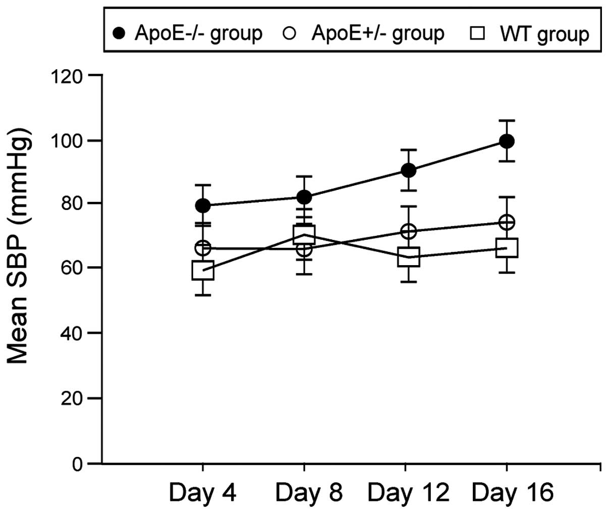
As shown in Fig. 1,
mean systolic blood pressures in ApoE−/− group on day 12
and 16 were significantly higher than the ApoE+/− and WT
groups (P<0.05).
Urinary proteins and creatinine
As shown in Table
II, urinary proteins and creatinine levels in
ApoE−/− group on day 12 and 16 were significantly higher
than the ApoE+/− and WT groups (P<0.05).
| Table II.Urinary proteins and creatinine. |
Table II.
Urinary proteins and creatinine.
| Groups | Urinary proteins
(mg/l) | Urninary creatinine
(µmol/l) |
|---|
|
|
|
|
|---|
|
| 0 day | 4 days | 8 days | 12 days | 16 days | 0 day | 4 days | 8 days | 12 days | 16 days |
|---|
|
ApoE−/− | 50.2±13.6 | 65.6±17.2 | 73.2±16.6 | 104.7±23.6 | 232.5±46.5 | 45.2±13.2 | 56.5±14.6 | 66.8±18.2 | 154.7±36.4 | 246.7±52.7 |
|
ApoE+/− | 53.3±14.5 | 64.7±13.6 | 75.3±12.3 | 82.0±15.8 | 94.5±26.5 | 44.3±12.6 | 53.2±15.4 | 65.9±13.7 | 83.2±23.1 | 96.3±26.5 |
| WT | 51.7±15.2 | 63.2±14.8 | 72.4±14.9 | 81.6±11.3 | 96.5±24.7 | 43.7±13.6 | 52.7±16.5 | 64.2±18.9 | 80.5±24.7 | 94.5±26.5 |
| F-value | 0.526 | 0.432 | 0.857 | 4.847 | 9.655 | 0.541 | 0.329 | 0.502 | 5.302 | 8.657 |
| P-value | 0.214 | 0.303 | 0.768 | 0.034 | 0.000 | 0.236 | 0.214 | 0.548 | 0.031 | 0.000 |
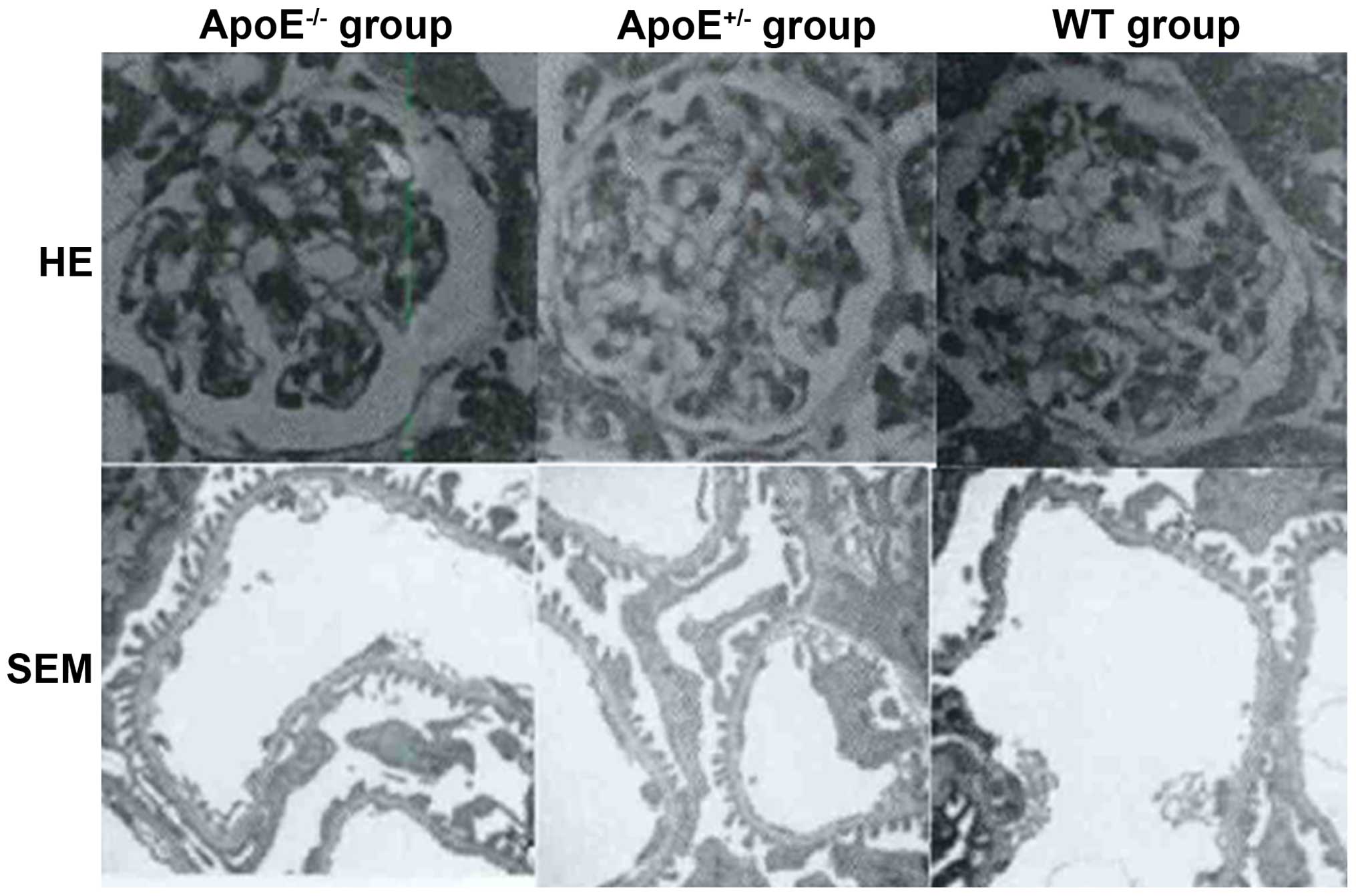
Structural changes of glomerular
filtration membrane and placenta
As shown in Fig. 2,
thickening and edema in the glomerular filtration membrane as well
as capillary thrombosis was evident in the ApoE−/−
group, while no significant changes were detected in the
ApoE+/− or WT group.
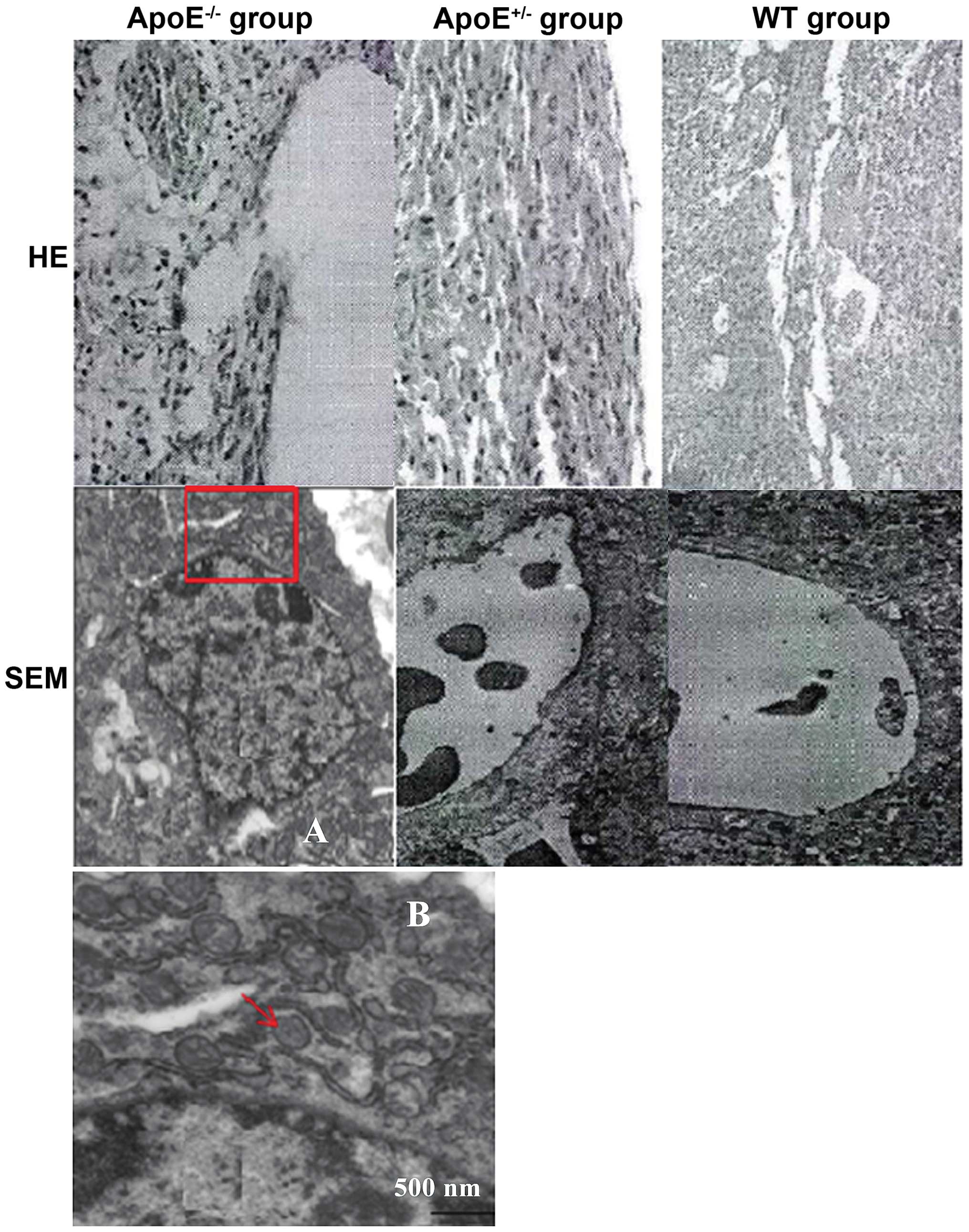
Significant edema and necrosis of placental villous
stroma, irregular nuclear morphology, degeneration of cytoplasmic
membrane structures, fat deposition in placenta and mitochondrial
swelling and deformation were observed in in the ApoE−/−
group (Fig. 3), while no significant
changes were detected in the ApoE+/− or WT group.
The levels of serum TLR4 and
sFlt-1
TLR4 and sFlt-1 levels in ApoE−/− group
were significantly higher than the ApoE+/− and WT groups
(P<0.05) (Table III).
| Table III.Levels of serum TLR4 and sFlt-1
(ng/ml). |
Table III.
Levels of serum TLR4 and sFlt-1
(ng/ml).
| Groups | TLR-4 | sFlt-1 |
|---|
|
ApoE−/− | 45.7±4.7 | 32.4±5.6 |
|
ApoE+/− | 5.3±1.2 | 4.2±1.3 |
| WT | 5.2±1.3 | 3.6±1.4 |
| F-value | 10.524 | 12.645 |
| P-value | 0.000 | 0.000 |
Discussion
The theory of vascular endothelial injury during
gestational hypertension suggested that vascular endothelial injury
increased the synthesis and release of vasoconstrictor factors,
decreased the synthesis and release of endothelium-derived relaxing
factor, leading to the disturbance of vasoactive factors and
subsequent vasoconstriction, impaired connection of vascular
endothelial cells, increased vascular permeability, extravasation
of intravascular proteins and fluid, platelet aggregation and
activation of coagulation system (5). Previous findings showed that lipid
peroxidation and inflammatory response due to abnormal lipid
metabolism played a major role in inducing vascular endothelial
dysfunction (6). The possible
mechanism may be that the activation of neutrophils and adherence
of neutrophils to vascular endothelium, led to vascular injury
(7). Placenta could secrete various
cytokines, leading to vascular endothelial injury, thus it could
mediate various pathology and play important roles in the
pathogenesis of gestational hypertension (8).
The present study established the ApoE−/−
mouse model of preeclampsia and found the following symptoms in
these mice: i) thickening and edema in glomerular filtration
membrane; ii) capillary thrombosis; iii) significant edema and
necrosis of placental villous stroma; iv) irregular nuclear
morphology; v) degeneration of cytoplasmic membrane structures; and
vi) fat deposition in placenta and mitochondrial swelling and
deformation. This model could simulate the pathological process of
preeclampsia and indicated that dyslipidemia is important in
preeclampsia pathogenesis. In the ApoE−/− group, we
observed no changes in serum lipids, which was inconsistent with
previous studies (9,10). Those studies showed that preeclampsia
occurred in parallel with dyslipidemia. On the other hand, our
results were consistent with the results of the study by Belo et
al (11). Their results revealed
that ApoE gene polymorphism was not a risk factor for preeclampsia
(11,12).
The results of this study showed that both TLR4 and
sFlt-1 expression levels in the ApoE−/− group were
considerably higher than those of the ApoE+/− and WT
groups. Results from previous studies reported that TLR4 was
involved in ischemic and hypoxic pulmonary hypertension, pulmonary
edema and cerebral edema due to barrier dysfunciton of endothelial
cells, and proteinuria due to barrier dysfunction of glomerular
filtration membrane. TLR4 recognized the pathogen-associated
molecular pattern and endogenous ligands, induced intracellular
signal transduction, and led to an inflammatory response (13). TLR4 was overexpressed in immune
cells, renal podocytes, trophoblast cells, and vascular endothelial
cells (14). In addition,
overexpression of sFlt-1 was also associated with the pathogenesis
of preeclampsia. It was shown that sFlt-1 bound to VEGF, reduced
the expression of glomerular slit diaphragm protein, promoted
endothelial isolation and hypertrophy, and led to proteinuria
(15,16). The mean systolic blood pressures in
the ApoE−/− group on day 12 and 16 were significantly
higher than those of the ApoE+/− and WT groups,
indicating the probability of preeclampsia in the middle and late
phase of gestation. This was consistent with natural disease
course. A previous study established TLR4-knockout pregnant mouse,
in which the proteinuria in preeclampsia mice were reversible, and
the hypertention in preeclampsia mice was also relieved, indicating
that TLR4 may be involved in the pathological process of
preeclampsia due to sFlt-1 (15).
In conclusion, ApoE-knockout mouse simulated the
pathologic process of preeclampsia, while the change of serum
lipids was not significant, thus the pathogenesis of preeclampsia
may be mediated by TLF-4 and sFlt-1.
Acknowledgements
The present study was supported by grant no.
2013GGE27035.
References
|
1
|
Gong YH, Jia J, Lü DH, Dai L, Bai Y and
Zhou R: Outcome and risk factors of early onset severe
preeclampsia. Chin Med J (Engl). 125:2623–2627. 2012.PubMed/NCBI
|
|
2
|
Yang X, Wang F, Lau WB, Zhang S, Zhang S,
Liu H and Ma XL: Autoantibodies isolated from preeclamptic patients
induce endothelial dysfunction via interaction with the angiotensin
II AT1 receptor. Cardiovasc Toxicol. 14:21–29. 2014. View Article : Google Scholar : PubMed/NCBI
|
|
3
|
Mannarino E and Pirro M: Endothelial
injury and repair: a novel theory for atherosclerosis. Angiology.
59 Suppl 2:69S–72S. 2008. View Article : Google Scholar : PubMed/NCBI
|
|
4
|
Podjarny E, Baylis C and Losonczy G:
Animal models of preeclampsia. Semin Perinatol. 23:2–13. 1999.
View Article : Google Scholar : PubMed/NCBI
|
|
5
|
Sircar M, Thadhani R and Karumanchi SA:
Pathogenesis of preeclampsia. Curr Opin Nephrol Hypertens.
24:131–138. 2015. View Article : Google Scholar : PubMed/NCBI
|
|
6
|
Redman CW, Sacks GP and Sargent IL:
Preeclampsia: an excessive maternal inflammatory response to
pregnancy. Am J Obstet Gynecol. 180:499–506. 1999. View Article : Google Scholar : PubMed/NCBI
|
|
7
|
Bayhan G, Koçyigit Y, Atamer A, Atamer Y
and Akkus Z: Potential atherogenic roles of lipids, lipoprotein(a)
and lipid peroxidation in preeclampsia. Gynecol Endocrinol. 21:1–6.
2005. View Article : Google Scholar : PubMed/NCBI
|
|
8
|
Matsuo K, Kooshesh S, Dinc M, Sun CC,
Kimura T and Baschat AA: Late postpartum eclampsia: report of two
cases managed by uterine curettage and review of the literature. Am
J Perinatol. 24:257–266. 2007. View Article : Google Scholar : PubMed/NCBI
|
|
9
|
Cekmen MB, Erbagci AB, Balat A, Duman C,
Maral H, Ergen K, Ozden M, Balat O and Kuskay S: Plasma lipid and
lipoprotein concentrations in pregnancy induced hypertension. Clin
Biochem. 36:575–578. 2003. View Article : Google Scholar : PubMed/NCBI
|
|
10
|
Francoual J, Audibert F, Trioche P, Chalas
J, Capel L, Lindenbaum A, Labrune P and Frydman R: Is a
polymorphism of the apolipoprotein E gene associated with
preeclampsia? Hypertens Pregnancy. 21:127–133. 2002. View Article : Google Scholar : PubMed/NCBI
|
|
11
|
Belo L, Gaffney D, Caslake M, Santos-Silva
A, Pereira-Leite L, Quintanilha A and Rebelo I: Apolipoprotein E
and cholesteryl ester transfer protein polymorphisms in normal and
preeclamptic pregnancies. Eur J Obstet Gynecol Reprod Biol.
112:9–15. 2004. View Article : Google Scholar : PubMed/NCBI
|
|
12
|
Kobayashi K, Oyama S, Numata A, Rahman MM
and Kumura H: Lipopolysaccharide disrupts the milk-blood barrier by
modulating claudins in mammary alveolar tight junctions. PLoS One.
8:e621872013. View Article : Google Scholar : PubMed/NCBI
|
|
13
|
Lee KM and Seong SY: Partial role of TLR4
as a receptor responding to damage-associated molecular pattern.
Immunol Lett. 125:31–39. 2009. View Article : Google Scholar : PubMed/NCBI
|
|
14
|
Banas MC, Banas B, Hudkins KL, Wietecha
TA, Iyoda M, Bock E, Hauser P, Pippin JW, Shankland SJ, Smith KD,
et al: TLR4 links podocytes with the innate immune system to
mediate glomerular injury. J Am Soc Nephrol. 19:704–713. 2008.
View Article : Google Scholar : PubMed/NCBI
|
|
15
|
Suzuki H, Ohkuchi A, Matsubara S, Takei Y,
Murakami M, Shibuya M, Suzuki M and Sato Y: Effect of recombinant
placental growth factor 2 on hypertension induced by full-length
mouse soluble fms-like tyrosine kinase 1 adenoviral vector in
pregnant mice. Hypertension. 54:1129–1135. 2009. View Article : Google Scholar : PubMed/NCBI
|
|
16
|
Lin M, Yiu WH, Li RX, Wu HJ, Wong DW, Chan
LY, Leung JC, Lai KN and Tang SC: The TLR4 antagonist CRX-526
protects against advanced diabetic nephropathy. Kidney Int.
83:887–900. 2013. View Article : Google Scholar : PubMed/NCBI
|