Introduction
Soft tissue sarcoma (STS) is a rare malignancy with
an incidence rate of <1% of all adult cancers (1). Among all STS cases, the 10–15% that
originate in the retroperitoneum are known as retroperitoneal soft
tissue sarcomas (RSTS) (2). The
prognosis of RSTS is poor, with a 5-year overall survival (OS) of
20–60% due to the low efficacy of local control and the high
incidence of metastasis after resection (2–5). Local
reoccurrence is the primary reason for relapse in retroperitoneal
sarcoma in up to 90% of relapsing patients, while distant
metastasis is the primary cause of tumor-associated mortality in
sarcoma (4). Furthermore, local
recurrence is common, with a consistent relapse rate of ~5% per
year between 60 and 120 months following primary treatment
(4). Therefore, it remains a
challenge to manage RSTS and long-term local tumor control of RSTS
remains a key obstacle (1,6).
Surgery is the most effective treatment for RSTS
(7). However, as RSTS are often
diagnosed at very advanced stages with anatomic localization and
frequent invasion of retroperitoneal adjacent structures, surgery
is not suitable for numerous patients (8,9). When
surgical en-bloc resection is used as the sole-treatment,
the outcomes are poor, with side effects due to the excision of
neighboring structures accompanied by the occurrence of positive
resection margins despite the aggressive surgical approach
(5). A high rate of recurrence
typically occurs following surgery (9). Therefore, external radiotherapy in
combination with chemotherapy (ERBT) is the current strategy used
to reduce the rate of local tumor reoccurrence in these patients.
However, an adequate therapeutic dose of ERBT in RSTS may damage
the adjacent tissues and organs (10). The adjacent structures, including the
small bowel, kidney and stomach, are often radiosensitive and have
a low radiation tolerance (10).
Additionally, no consistent evidence of a disease-free survival
benefit has been shown for neoadjuvant/adjuvant chemotherapy for
the majority of histological subtypes, although there may be
certain situations where it is advantageous (11). Therefore, treatment options for
patients with unresectable RSTS are limited, particularly where the
aim is the relief of pain and local RSTS control.
To overcome these problems, different
radiotherapeutic techniques have been developed to create a local
boost of irradiation that is restricted to the tumor site (9). Brachytherapy (BRT) can deliver to the
target tumor a large total radiotherapy dose, relieve pain and
decrease complications, and has shown potential for improving local
control and pain relief for RSTS (5,11).
Accumulating evidence has shown that BRT could be performed in
patients with unresectable RSTS as a monotherapy (12). However, the published descriptions of
brachytherapy via 125I implantation have been limited to
case reports (12–14). Thus, the available literature cannot
serve as a guide for the widespread clinical application of
brachytherapy using 125I. Additionally, the feasibility,
efficacy and safety of 125I seed implantation in
patients with unresectable RSTS has not yet been evaluated on a
large scale. Herein, we present a brachytherapy 125I
treatment technique that was performed in our hospital for
unresectable RSTS and the effect of this treatment technique on
patient outcomes.
Patients and methods
Patients
Between January 2009 and August 2013, 23 patients
with primary, localized recurrent or metastasized, histologically
confirmed and unresectable RSTS at the Department of Abdominal
Oncology, West China Hospital (Sichuan, China) were recruited into
the present study. The patients were reevaluated for eligibility
for BRT and were required to be in good general condition,
including a normal blood pressure or hypertension controlled by
drugs, adequate liver and renal function and adequate hematological
function (white blood cell count >3,000/l, platelet count
>80,000/l and hemoglobin level >9.5 gm/dl). In addition, the
patients were excluded if there was any evidence of cardiac disease
(congestive heart failure or history of myocardial infarction
within the previous 3 months) and if the patient had a history of
acute tumor rupture with hemoperitoneum. Certain patients who would
otherwise have been excluded due to poor general condition, but who
had no other contraindications, were included when their general
status improved and delayed BRT was performed. Pediatric and
gynecological sarcomas were excluded due to the uncertain risk
posed by the potential displacement of radioactive seeds after
intervention therapy. The present reviewed the pathological
characteristics of recurrent cases and conducted biopsies for all
primary tumors prior to brachytherapy. All suitable patients
received the 125I seed implantation therapy at the Department of
Abdominal Oncology, West China Hospital, Sichuan University
(Chengdu, China). The present study was approved by the Ethics
Committee of Sichuan University. Written informed consent was
obtained from all patients.
Implant preparation
Before protocol enrollment, patients were
reevaluated with basic history and pathological examination of the
tumor/s, physical examination and laboratory tests. Chest/abdomen
axial computed tomography (CT) scan of 3-mm slice thickness was
performed using a SOMATOM Emotion CT scanner (Siemens Healthcare,
Erlangen, Germany) to assess tumor number, location, size,
association with adjacent organs and tissues, and any accompanying
metastasis. Oncologists and radiologists with >10 years of
experience assessed areas at risk for subclinical disease, the
optimal puncture route, and the number and distribution of seeds.
Any discrepancy in assessment was solved by discussion. For lesions
that did not respond to treatment as expected, a post-treatment
plan was designed to enhance the radiation dose while considering
patient safety and the limitations of this operation, which
generally requires a number of sessions.
Operation
For brachytherapy, a metal strip was placed in the
body as a surface marker to better visualize the location and
orientation of the target tumor. Then the patient was placed in the
prone or supine position and a local anesthetic (0.5% lidocaine;
Shanghai Zhpharma, Co., Ltd., Shanghai, China) was administered.
The physician (usually a radiologist) guided 18-gauge needles
(Hakko Trading, Co., Ltd., Shanghai, China) into the predetermined
locations under CT imaging. Using a real-time technique, the needle
was inserted into the tumor, avoiding important issues like the
aortaventralis, inferior vena cava and nerves. Needle tip location
in relation to the tumor and surrounding structures was confirmed
by CT imaging. A Mick applicator (Mick Radio-Nuclear Instruments,
Inc., Mt. Vernon, NY, USA) was used to deposit the radioactive
seeds (t1/2, 59.6 days; energy activity
range, 0.6–0.8 mCi; mean, 0.78 mCi; Atom-Hitech, Co., Ltd.,
Beijing, China). The space between permanently implanted seeds was
1.0 cm within rows and the rows were ~1.0 cm apart. Following the
operation, a reevaluation for errant seeds was performed via a CT
scan of the whole abdomen.
Severe post-procedural pain was controlled with
additional moderate lidocaine subcutaneous injections at the
procedure location. Hemorrhaging during the operation was
controlled by blocking the catheter (Hakko Trading, Co., Ltd.).
After the completion of the brachytherapy, the patient remained in
the observation room for 2 h to be monitored for any unexpected
complications.
Follow-up and assessment indices
Following the 125I implantation, pain remission and
local control were considered to be primary outcome indices;
complete response and overall survival were deemed secondary
outcomes. RECIST guidelines (version 1.1) were used to assess the
efficiency, as follows: Complete response (CR), disappearance of
all target lesions; Partial response (PR), at least a 30% reduction
in the sum of diameters of target lesions; stable disease (SD),
neither sufficient shrinkage to qualify for PR nor sufficient
increase to qualify for progressive disease (PD); and PD, at least
a 20% increase in the sum of diameters of target lesions (15). Duration of overall response (OR) was
calculated as CR/PR. As 125I has an effective dose following
implantation of six months, local control was defined as patients
being free from the disease in the original location after
brachytherapy for six months following the procedure. Overall
survival was defined as the percentage of patients surviving at the
conclusion of the follow-up period. All patients rated their pain
status on a visual analog scale (VAS) before and after the
procedure: 0 indicated no pain and a score of 10 represented
maximal pain. This rating was repeated 24 h after the operation and
at 1, 3, 6, 12, 24 and 36 months after operation. Regular CT scans
of the abdominal and lesion regions were performed every three
months on patients to assess local control and complete response
after treatment until August 2014, or until patient mortality or
loss to follow-up. A researcher was trained to conduct clinical
interviews in person, via phone call or via email to determine
overall survival. Interviews collected information about treatment
response, primarily in terms of degree of pain, local control, OR
and OS. All adverse effects of the procedure for each patient were
recorded in this series, including nerve damage, liver or renal
damage, drifting seed, stent-tract bleeding, infection or
mortality.
Statistical analysis
Data are presented as percentages of patients or as
the mean ± standard deviation with ranges. Survival curves were
generated using the Kaplan-Meier method. Results of pain relief,
renal function and liver function were calculated by the paired
t-test. P<0.05 was considered to indicate a statistically
significant difference. All data were calculated using SPSS
software, version 16.0 (SPSS, Inc., Chicago, IL, USA).
Results
Patient demographics
A total of 23 patients with unresectable RSTS were
recruited for this study from the Department of Abdominal Oncology.
Patient demographics are listed in Table
I. The median age of patients at the time of diagnosis was
50.17±14.57 years (range, 19–78 years). Among the 23 patients, 9
were male (39%) and 14 were female (61%). The diameters of tumors
ranged between 2.70 and 19.90 cm. The median tumor size was
6.78±3.85 cm. The histologic grades (intermediate and high grade)
and the histological types were as follows: Liposarcomas, 6 (26%);
leiomyosarcomas, 6 (26%); small round cell liposarcoma, 2 (9%);
epithelioid sarcoma, 3 (13%); rhabdomyosarcoma, 2 (9%); malignant
fibrous histiocytoma, 1 (4%); synovial sarcoma, 1 (4%);
extraskeletal chondrosarcoma, 1 (4%); and extraskeletal
osteosarcoma, 1 (4%). Patients received pretreatment with a mean of
1.43±0.99 surgical operations and 0.70±0.97 courses of
interventional therapies, such as transarterial chemoembolization
(TACE).
 | Table I.Pathological characteristics and
distribution of events in the 23 RSTS patients in this study. |
Table I.
Pathological characteristics and
distribution of events in the 23 RSTS patients in this study.
| Variable | Data |
|---|
| Age (years) |
50.17±14.57 |
| Gender
(male/female) | 9/14
(39/61) |
| Histological
subtype |
|
|
Liposarcoma | 6
(26) |
|
Leiomyosarcoma | 6
(26) |
| Small
round cell liposarcoma | 2 (9) |
|
Epithelioid sarcoma | 3
(13) |
| Other
typesa | 6
(26) |
| Location of
lesions |
|
|
Pararenal space | 6
(26) |
|
Lumbosacral anterior area | 2 (9) |
|
Paravertebral area | 3
(13) |
|
Posterior pancreatic area | 2 (9) |
| Para
aortic region | 9
(39) |
| Portal
vein adjacent area | 1 (4) |
| Initial
presentation |
|
|
Primary | 2 (9) |
|
Recurrent | 15 (65) |
|
Metastasis | 6
(26) |
| Tumor size
(cm) |
6.78±3.85 |
|
<5 | 4
(17) |
|
5–10 | 14 (61) |
|
>10 | 5
(22) |
| No. prior
intervention operations |
0.70±0.97 |
| No. prior
surgeries |
1.43±0.99 |
| 0 | 3
(13) |
| 1 | 11 (48) |
|
>1 | 9
(39) |
| No. of
sessions |
2.57±1.43 |
| 1 | 7
(30) |
|
>1 | 16 (70) |
| No. of seeds |
|
| First
time procedure |
70.87±52.28 |
|
Subsequent procedures |
46.32±30.73 |
| Energy
activity (mean, mCi) | 0.78
(0.6–0.8) |
| Follow-up
(months) |
20.87±13.22 |
| Coagulation
function (s) |
|
| PT | 11.56±2.44 |
|
APTT | 28.13±7.38 |
| TT | 19.15±1.81 |
CT-guided 125I implantation is a
feasible, safe and effective treatment for RSTS
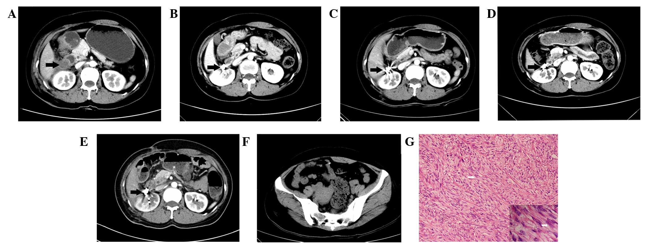
We successfully implanted CT-guided 125I seeds in 23
RSTS patients (Fig. 1). A mean of
70.87±52.28 seeds were implanted in RSTS (range, 10–210) in the
first session and an average number of 46.32±30.73 seeds were used
during post-treatment sessions (Table
I). No patients were recalled to collect data specifically for
this study. All data was obtained from medical records and imaging.
All of the patients were treated, with satisfactorily outcomes. The
P-values of alpha-fetoprotein and TBil were 0.023 and 0.015
(Table II), which were P<0.05
and were considered statistically significant. However, there was
no clinical significance as the pre-operation and post-operation
concentrations of alpha-fetoprotein and TBil were in the normal
range. Thus, no clinically significant damage to renal and liver
functions was detected, indicating that CT-guided 125I implantation
is a safe treatment for RSTS.
| Table II.Pre-operative and one month
post-operative blood test. |
Table II.
Pre-operative and one month
post-operative blood test.
| Parameter | Pre-operative | Post-operative | P-value |
|---|
| Liver function |
|
|
|
| TBil
(µmol/l) | 13.11±5.57 | 9.80±3.24 | 0.015 |
| ALT
(IU/l) | 22.78±18.26 | 22.63±14.07 | 0.278 |
| AST
IU/l) | 24.30±7.06 | 22.42±7.85 | 0.122 |
| Renal function |
|
|
|
| Urea
(mmol/l) | 4.75±1.44 | 4.02±1.40 | 0.389 |
|
Creatinine (µmol/l) | 71.91±16.10 | 62.97±22.82 | 0.192 |
| Uric
acid (µmol/l) | 307.87±86.91 | 276.28±106.65 | 0.652 |
| Tumor markers |
|
|
|
|
Alpha-fetoprotein (ng/ml) | 3.41±2.22 | 3.58±2.03 | 0.023 |
|
Carcino-embryonic antigen
(ng/ml) | 1.76±1.38 | 1.55±1.25 | 0.445 |
|
Carbohydrate antigen 1–25
(U/ml) | 24.11±29.38 | 28.98±29.03 | 0.139 |
|
Carbohydrate antigen 199
(U/ml) | 10.01±6.54 | 12.89±7.39 | 0.601 |
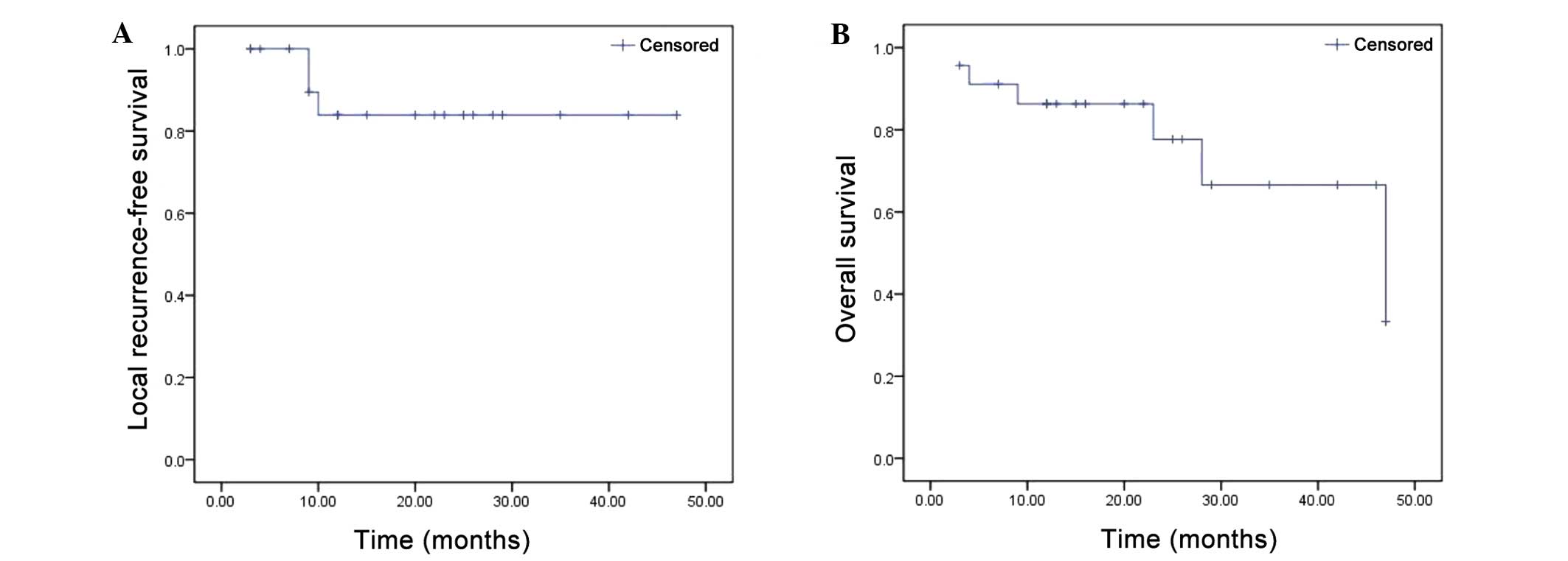
Local recurrence was detected in two patients at 9
months and one patient at 10 months after the operation. The early
OR of brachytherapy, evaluated at 90 days from the second cycle of
completed treatment, was observed in all 23 patients (100%). Two
patients did not complete the follow-up period of the study. Local
recurrence was detected in three patients during the follow-up
period (20.87±13.22 months). Therefore, local control by
125I seed implantation was 87.0% (Fig. 2A). The median overall survival was
21.56±14.16 months (Fig. 2B). The
VAS scores were increased from 7.4±3.2 preoperatively to 7.6±3.0 by
24 h after the operation, but remained at low levels throughout the
follow-up period: 2.3±2.6 at one month, 2.0±2.7 at 3 months,
1.2±1.3 at 6 months, 1.5±1.3 at 12 months, 1.4±1.0 at 24 months and
2.5±0.8 at 36 months. Therefore, the mean VAS scores differed
significantly from the preoperative baseline at each postoperative
time point and all P-values were <0.05 (Table III), demonstrating that patients
received significant pain relief one month after brachytherapy.
| Table III.VAS scores of patients pre-and
post-operatively. |
Table III.
VAS scores of patients pre-and
post-operatively.
|
|
|
| Post-op time
(months) |
|---|
|
|
|
|
|
|---|
| Parameter | Pre-op | Post-op 24 h | 1 | 3 | 6 | 12 | 24 | 36 |
|---|
| Patients (n) | 24 | 24 | 24 | 24 | 20 | 16 | 8 | 4 |
| VAS score | 7.4±3.2 | 7.6±3.0 | 2.0±2.6 | 2.0±2.7 | 1.2±1.3 | 1.5±1.3 | 1.4±1.0 | 2.5±0.8 |
| P-value | – | P=0.26 | P<0.001 | P<0.001 | P<0.001 | P<0.001 | P<0.001 | P=0.006 |
Adverse events
Four asymptomatic seed drafts were observed in three
patients. One seed was drafted to the liver and the remainder were
drafted to the pelvic cavity (Fig.
1). However, no serious complications were detected. None of
these patients exhibited clinical symptoms associated with the
procedure. One patient with light radioactive intestinal bleeding
was observed three months after the operation and received further
treatment in the form of a rectal diversion operation. Some other
complications were observed in some of the 23 patients, including
fever in 4 patients and nausea in 2 patients (Table IV). These complications were solved
following symptomatic treatments.
| Table IV.Complications detected among 23
patients during follow-up. |
Table IV.
Complications detected among 23
patients during follow-up.
| Complication | Patients (n) |
|---|
| Seed draft | 3 |
| Stent-tract
bleeding | 1 |
| Fever | 4 |
| Enterobrosis | 0 |
| Radioactive
intestines, bleeding | 1 |
| Vascular
perforation | 0 |
| Nerve damage | 0 |
| Loss of
appetite | 2 |
| Diarrhea | 1 |
| Ventosity | 2 |
| Nausea | 2 |
Discussion
In the present study, 23 patients were successfully
treated with CT-guided 125I seeds implanted via
different approaches relative to their RSTS tumors. Among this
patient group, only one severe complication was observed during the
follow-up period. Four drafting seeds were detected in 3/23
patients. Good local control (87.0%), OR (100%) and median overall
survival (21.56±14.16 months) values were achieved by brachytherapy
in all recruited patients. The present findings demonstrated a
significant improvement in the management of patients with
inoperable RSTS by CT-guided 125I seed implantation.
The average number of seeds we used (mean=70.87) in
the present study was more than that used by Li et al, who
used 30 seeds in the first session in one case (13), though less than Kumar and Good, who
used 229 seeds (12). Unlike the
present study, these studies were case reports without any
statistical significance. The number of sessions (n=2.57) in the
present study was similar to Chen et al (14) (n=2.6) and Li et al (13) (n=2). During follow-up, it was found
that the mortality rate in the present study was 21.7% (5/23), and
the number of adverse events was 16/23, which was more than
previous similar studies (12,16).
Notably, one patient had radioactive intestinal bleeding, which was
not observed in previous studies (12–14).
This may have been due to the patients having had several previous
external-beam radiation therapy (EBRT) treatments, and the
relatively high activity of the seeds that were used in this study.
Based on the results in this patient, we speculate that, for
safety, lower activity seeds should be applied in cases where the
target tumor is close to the aorta or intestines. The discrepancy
in overall survival and other complications may be due in part to
the smaller number of cases in the study by Chen et al
(14). Furthermore, previous studies
were limited to case reports and did not have as large a sample
size (12,16). Lastly, the follow-up period in the
present study was longer compared with the previous studies. VAS
scores were elevated 24 h after the operation, while they remained
low during follow-up periods from 1 to 24 months, suggesting that
the duration of radiation treatment may be too short to relieve
cancer pain and that the puncture might increase pain in the local
region. Notably, pain was found to be elevated at 36 months,
supporting the observation that 125I seeds have ~180
days of effective radiation cover following implantation, and that
the residual sarcoma tissue may progress after the duration of the
effective radiation has elapsed.
Collectively, the present data suggest that
CT-guided 125I seed implantation is feasible, safe and
effective for patients with unresectable RSTS. In the following
sections, we report the operative procedure and thereby provide
clinical guidelines for patients with retroperitoneal sarcoma.
Surgical resection remains the standard primary
treatment for patients with RSTS, and may improve overall survival.
By contrast, the primary outcome aims of en-bloc resection
for patients with obstinate RSTS are to achieve pain relief and
local control. Considering the palliative aims, surgery may not be
the optimal treatment option, particularly in patients with
multiple unresectable metastases, an unfavorable overall prognosis
or poor performance status. It has been well-documented that
surgical resection of tumors with sizes >5 cm (5,7) and
high-grade histology (17) is
accompanied by a high probability of loco-regional recurrence and
distant metastases within the first two years (13). This is due to the difficulty in
surgical resection of RSTS of achieving complete excision; there is
often a positive postoperative margin, owing to adjacent structures
like the gastrointestinal tract (3).
Unless there is further treatment, 90% of patients will succumb to
recurrent tumors (18,19). Bonvalot et al (20) once pointed out that no long-term
overall survival benefit has been demonstrated in patients that
have undergone resection of uninvolved organs. Furthermore,
Mullinax et al (19) reported
that 13 patients (4%) succumbed in the perioperative setting, while
3 succumbed intraoperatively. These numbers are high (though
reportedly acceptable, per the authors) and should be taken into
consideration when evaluating this approach for patients with
retroperitoneal sarcomas. In addition, poor tissue healing further
limits the role of repeated surgery as a therapeutic option for
this disease. Lastly, though common surgical oncological principles
prevail, every operation will be different. Therefore, an
experienced surgical team should plan each operation after careful
study, as soft-tissue sarcoma may occur at any site. Thus, for
certain patients, less invasive therapies may be more effective for
managing local disease recurrence.
Chemotherapy is widely used for treating advanced
cancer. Although particular subtypes of soft-tissue sarcoma are
sensitive to chemotherapeutic agents, neoadjuvant/adjuvant
chemotherapy has not yet been shown to confer a disease-free
survival benefit for the majority of histological subtypes
(11). Palliative systemic
chemotherapy is the cornerstone therapy; however, the response rate
is 20–30% and the median overall survival is generally lower than
12 months (21). Furthermore, since
the use of adjuvant chemotherapy remains controversial, there are
no standard guidelines for systemic chemotherapy in patients with
RSTS (19). A meta-analysis of
adjuvant chemotherapy did not demonstrate an overall survival
advantage, although progression-free survival was improved
(22), suggesting that RSTS may be
insensitive to adjuvant chemotherapy. Consistently, a previous
study showed that the outcome of therapeutic chemotherapy for RSTS
was unsatisfactory in terms of overall survival (23). In addition, chemotherapy usually
results in complications such as vomiting, diarrhea and decreased
platelet count. Collectively, these previous results indicate that
chemotherapy is not a good choice for patients with RSTS.
Radiotherapy is another well-established modality in
the management of RSTS and radiation treatment is generally
considered beneficial. Preoperative EBRT is able to facilitate
marginally negative resection and postoperative radiation treatment
can diminish local recurrence and may improve survival (18,24).
However, the role of ERBT in primary retroperitoneal sarcoma has,
to date, been only poorly defined due to a lack of randomized
clinical series (3,9,10).
Furthermore, the role of radiotherapy in relieving pain is limited.
Since the 125I provides a source of continuous low dose
radiation, it may be more effective than daily pulsed high dose
irradiation in treating the hypoxic portion of large, slow-growing
necrotic tumors (12).
Unfortunately, in the retroperitoneum and spinal region, the
required curative doses exceed 45–50 Gy, which is neither easily
nor safely delivered without a high risk of radiation-induced
gastrointestinal, genitourinary or spinal cord complications
(7). EBRT was excluded as it might
cause skin injury (13). The higher
doses required for local control and the inherent
normal-tissue-tolerance limitations of external beam radiation
therapy may explain the failure of this modality to adequately
control retroperitoneal soft-tissue sarcomas (11,12,23).
Therefore, radiotherapy, as a monotherapy, may not be suitable for
patients with RSTS.
There are other options for patients with
unresectable RSTS, including different interventional radiology
therapies. These interventional radiology approaches have common
advantages; for example, they are simple, cheap, have high safety,
good efficacy, low invasiveness and few complications (25). However, there are also a number of
drawbacks associated with them.
Percutaneous ethanol injection has been clinically
applied for several decades and has proven to be a safe technique,
but its effects have been limited by alcohol tolerance and local
blood flow (26). Furthermore,
multiple sessions may be required, leading to a prolonged treatment
time.
Transcatheter embolization (TAE) or TACE has shown
varying degrees of efficacy. A tumor may have a number of feeding
arteries other than the main feeders, and the vascularly-rich
bottom portion of the tumor does not respond effectively to TAE
(26). Nevertheless, TAE has been
shown to be effective for the treatment of rapidly growing tumors
(26). Unfortunately, RSTS is not an
ideal target for TAE due to its slow growth (12,26).
TACE has increased intratumoral chemotherapeutic concentration,
reduced systemic toxicity and increased local effects, and thus has
improved therapeutic results when compared with systemic
chemotherapy (27). With TACE,
embolization of the tumor feeding vessels slows blood flow, creates
ischemia and increases the contact time between the
chemotherapeutic agent and the tumor cells (27). Unfortunately, RSTS are less vascular
compared with numerous other tumor types, reducing the efficacy of
the therapy while leaving the patient just as vulnerable to the
most common complications associated with the toxicity of the
chemotherapeutic agents, including nausea, neutropenia,
myelosuppression and bacteremia (28). Furthermore, for osteosarcoma, the
intraarterial infusion of cisplatin did not improve the local tumor
response (29).
Radiofrequency ablation (RFA) has been widely used
for two decades in the treatment of various neoplasms, including
renal cancer located in the retroperitoneum (30,31).
Successful treatment of retroperitoneal lymph nodes has also been
reported. However, the application of RFA requires particular
caution be observed because of the high risk of thermal damage to
neighboring organs, such as the bowel, nerves and nearby vessels
(32). Therefore, considering the
uncertain effectiveness and difficulty in avoiding complications,
these intervention therapies may not be a good choice for RSTS.
Traditionally, interstitial implants were performed
with 226Ra needles (31).
Due to radiation safety considerations, however, 226Ra
has largely been replaced by other radionuclides (33). Currently, the majority of
interstitial brachytherapy treatments are delivered using different
radioactive sources, such as 192Ir, 103Pd and
125I (33).
192Ir is ideal for temporary
brachytherapy. It decays with a half-life of 73.83 days and emits
gamma rays with an average energy of ~370 keV. 192Ir is
most commonly used in the form of a wire as a transient
brachytherapy without any ‘fixicity’ problems when compared with
permanent implantation seeds like 125I and
103Pd (34).
125I decays with a half-life of 59.4 days and emits
photons with an average energy of 27.4–35.5 keV (35). 125I is commercially
available in the form of small ‘seed’ sources for interstitial
permanent implants. If rectal toxicity is a concern, the very low
dose rate of 125I should favor 125I implants
(36). Furthermore, the dose
homogeneity inside the target volume is very high with
125I (36). Crucially,
125I is an ideal isotope to use for large volume
irradiation of retroperitoneal tumors close to the spinal cord due
to its low gamma photon energy of 35.5 keV (37), which results in a rapidly decreasing
radiation dose outside of the implanted volume (12).
103Pd is ideal for use as a permanent
interstitial source, similar to 125I. 103Pd
decays with a half-life of 17.0 days and emits photons with an
average energy of 21–30 keV. A 103Pd source is similar
in size and encapsulation to 125I sources (33). Although it also offers the practical
advantage of low energy, reducing the dose to surrounding organs
and minimizing shielding requirements, the difference of the
half-lives between 103Pd (17 days) and 125I
(61 days) is marked (33). In
addition, 125I has been in practical use for longer than
103Pd (33).
Brachytherapy is an established method of safely
providing local adjuvant radiotherapy that may be used alone or in
combination with resection for prostate cancer, breast cancer,
cervical cancer and soft tissue sarcomas. Compared with surgery,
125I has fewer complications, a larger application field
and requires fewer procedures to be conducted under sedation or a
short general anesthetic (38).
Compared with chemotherapy, 125I implantation has fewer
toxic complications (16). Compared
with ERBT, 125I implantation has an advantage as
brachytherapy inflicts less radiation damage to adjacent structures
such as the bowel and genitourinary tract (10). For certain tumors, consecutive
radiotherapy can be performed by repeatedly implanting
125I seeds, and the curative effects are better than
external radiotherapy (13).
Compared with vascular intervention therapies, 125I is
not limited by the size of the cancer or the distribution of the
vessels (39). In comparison with
RFA, 125I has the advantage of avoiding damage to
critical structures, such as blood vessels and nerves (39). Compared with other radioactivity
sources, 125I has a long half-life, a low level of
radiant energy that is steadily released over 200 days following
implantation and is suitable for targeting slowly growing tumors
such as RSTS (34).
There were several limitations in the present study.
The first limitation is a shortage of a treatment planning system,
which limits the user's ability to manipulate the isodose lines
manually to ensure adequate target coverage and spare critical
structures (14). Secondly, due to
the difficulties in maintaining optimum implant geometry in the
irregular anatomy of the retroperitoneal space, bones, arteries and
veins, the ability to deliver an optimum dose to the tumor is often
limited. This difficulty has also been observed in the results of
previous studies (34). Although
peripheral nerves are generally tolerant of radiation, the high
doses of radiation adjacent to the sources may be injurious
(14). By adjusting implant geometry
to avoid this complication, the likelihood of hot or cold spots
occurring within the tumor bed may be increased (14). Third, patients with large and closely
applied dorsal veins and vessels are at particular risk for seed
migration to the lung. Fourth, certain patients succumbed during
the follow-up period. Fifth, the present patients were recruited
from a single center, and the sample size was relatively small due
to the rarity of RSTS. Sixth, we did not perform an arm-to-arm
study, comparing CT-guided 125I implantation with any
other treatments. Seven, to the best of our knowledge, there has
been no economic assessment of 125I implantation for
RSTS. Finally, seed migration may cause cold or hot spots, move to
important structures like the bladder and urethra, or move into a
vascular structure (34).
Several methods to remedy the drawbacks outlined
above were implemented. First, we implanted seeds with a 1-cm
interval seed-to-seed (0.8 cm raw-to-raw) to produce an array with
the best possible coverage. Second, in order maximize patient
retention in the study we facilitated a close patient relationship,
improved patient communication and maximum kindness of care by not
only maintaining contact with patients via phone and/or e-mail but
also maintaining contact with their family. Third,
multidisciplinary clinical teams with a substantial knowledge and
experience in the management of sarcomas worked together on this
project. The team included surgeons, oncologists, radiologists and
pathologists. Fourth, to decrease the ‘cold spots’, we usually
performed a post-therapy plan by supplementing 125I
seeds as necessary in the target location to enhance the radiation
dose.
Although 125I implantation does not
represent a cure for RSTS, the principal goals of 125I
implantation of significant pain relief and good local control have
been achieved. The present results suggest that CT-guided
125I implantation is safe, feasible and effective for
the treatment of patients with obstinate RSTS. However, familiarity
with local anatomy, experience and skill are prerequisites for
success. The present findings may aid physicians who are
determining the appropriate management of their patients.
Additional large-scale and multicenter studies are required.
Acknowledgements
The present study was supported by the National
Nature Science Foundation of China (grant no. 81470141) and the
Science & Technology Department of Sichuan Province (grant no.
2014SZ0002-8).
References
|
1
|
Lewis JJ and Brennan MF: Soft tissue
sarcomas. Curr Probl Surg. 33:817–872. 1996. View Article : Google Scholar : PubMed/NCBI
|
|
2
|
Karakousis CP, Gerstenbluth R, Kontzoglou
K and Driscoll DL: Retroperitoneal sarcomas and their management.
Arch Surg. 130:1104–1109. 1995. View Article : Google Scholar : PubMed/NCBI
|
|
3
|
Windham TC and Pisters PW: Retroperitoneal
sarcomas. Cancer Control. 12:36–43. 2005.PubMed/NCBI
|
|
4
|
Heslin MJ, Lewis JJ, Nadler E, Newman E,
Woodruff JM, Casper ES, Leung D and Brennan MF: Prognostic factors
associated with long-term survival for retroperitoneal sarcoma:
Implications for management. J Clin Oncol. 15:2832–2839.
1997.PubMed/NCBI
|
|
5
|
Jenkins MP, Alvaranga JC and Thomas JM:
The management of retroperitoneal soft tissue sarcomas. Eur J
Cancer 32A. 622–626. 1996. View Article : Google Scholar
|
|
6
|
Jaques DP, Coit DG, Hajdu SI and Brennan
MF: Management of primary and recurrent soft-tissue sarcoma of the
retroperitoneum. Ann Surg. 212:51–59. 1990. View Article : Google Scholar : PubMed/NCBI
|
|
7
|
Lewis JJ, Leung D, Woodruff JM and Brennan
MF: Retroperitoneal soft-tissue sarcoma: Analysis of 500 patients
treated and followed at a single institution. Ann Surg.
228:355–365. 1998. View Article : Google Scholar : PubMed/NCBI
|
|
8
|
McGrath PC: Retroperitoneal sarcomas.
Semin Surg Oncol. 10:364–368. 1994. View Article : Google Scholar : PubMed/NCBI
|
|
9
|
Dziewirski W, Rutkowski P, Nowecki ZI,
Salamacha M, Morysiński T, Kulik A, Kawczyńska M, Kasprowicz A,
Lyczek J and Ruka W: Surgery combined with intraoperative
brachytherapy in the treatment of retroperitoneal sarcomas. Ann
Surg Oncol. 13:245–252. 2006. View Article : Google Scholar : PubMed/NCBI
|
|
10
|
Classen J, Hehr T, Lamprecht U, Zumbrägel
A, Bamberg M and Budach W: Hyperfractionated 192Ir brachytherapy
for recurrent retroperitoneal sarcoma: A technique for delivery of
local tumor boost dose. Strahlenther Onkol. 179:118–122. 2003.
View Article : Google Scholar : PubMed/NCBI
|
|
11
|
Strauss DC, Hayes AJ and Thomas JM:
Retroperitoneal tumours: Review of management. Ann R Coll Surg
Engl. 93:275–280. 2011. View Article : Google Scholar : PubMed/NCBI
|
|
12
|
Kumar PP and Good RR: Interstitial 125I
implantation in the retreatment of retroperitoneal soft tissue
sarcoma. Report of a case. Acta Radiol Oncol. 25:37–39. 1986.
View Article : Google Scholar : PubMed/NCBI
|
|
13
|
Li Y, Wang Y, Liu B, Li Z and Wang W:
(125)I brachytherapy seeds implantation for inoperable low-grade
leiomyosarcoma of inferior vena cava. Korean J Radiol. 14:278–282.
2013. View Article : Google Scholar : PubMed/NCBI
|
|
14
|
Chen ME, Zhang B and Li HP: PET/CT-Guided
radioactive 125I seeds implanted in retroperitoneal sarcoma. Guang
Dong Yi Xue. 33:32012.(In Chinese).
|
|
15
|
Eisenhauer E, Therasse P, Bogaerts J,
Schwartz LH, Sargent D, Ford R, Dancey J, Arbuck S, Gwyther S,
Mooney M, et al: New response evaluation criteria in solid tumours:
Revised RECIST guideline (version 1.1). Eur J Cancer. 45:228–247.
2009. View Article : Google Scholar : PubMed/NCBI
|
|
16
|
Li Y, Wang Y, Liu B, Li Z and Wang W:
125I Brachytherapy Seeds Implantation for Inoperable
Low-Grade Leiomyosarcoma of Inferior Vena Cava. Korean J Radiol.
14:278–282. 2013. View Article : Google Scholar : PubMed/NCBI
|
|
17
|
Neuhaus SJ, Barry P, Clark MA, Hayes AJ,
Fisher C and Thomas JM: Surgical management of primary and
recurrent retroperitoneal liposarcoma. Br J Surg. 92:246–252. 2005.
View Article : Google Scholar : PubMed/NCBI
|
|
18
|
Clark MA, Fisher C, Judson I and Thomas
JM: Soft-tissue sarcomas in adults. N Engl J Med. 353:701–711.
2005. View Article : Google Scholar : PubMed/NCBI
|
|
19
|
Mullinax JE, Zager JS and Gonzalez RJ:
Current diagnosis and management of retroperitoneal sarcoma. Cancer
Control. 18:177–187. 2011.PubMed/NCBI
|
|
20
|
Bonvalot S, Rivoire M, Castaing M,
Stoeckle E, Le Cesne A, Blay JY and Laplanche A: Primary
retroperitoneal sarcomas: A multivariate analysis of surgical
factors associated with local control. J Clin Oncol. 27:31–37.
2009. View Article : Google Scholar : PubMed/NCBI
|
|
21
|
D'Adamo DR: Appraising the current role of
chemotherapy for the treatment of sarcoma. Semin Oncol. 38:(Suppl
3). S19–S29. 2011. View Article : Google Scholar : PubMed/NCBI
|
|
22
|
Adjuvant chemotherapy for localised
resectable soft-tissue sarcoma of adults, . Meta-analysis of
individual data. Sarcoma Meta-analysis Collaboration. Lancet.
350:1647–1654. 1997.PubMed/NCBI
|
|
23
|
Lewis JJ and Benedetti F: Adjuvant therapy
for soft tissue sarcomas. Surg Oncol Clin N Am. 6:847–862.
1997.PubMed/NCBI
|
|
24
|
Hines OJ, Nelson S, Quinones-Baldrich WJ
and Eilber FR: Leiomyosarcoma of the inferior vena cava: Prognosis
and comparison with leiomyosarcoma of other anatomic sites. Cancer.
85:1077–1083. 1999. View Article : Google Scholar : PubMed/NCBI
|
|
25
|
Ryder SD: British Society of
Gastroenterology: Guidelines for the diagnosis and treatment of
hepatocellular carcinoma (HCC) in adults. Gut. 52:(Suppl 3).
iii1–iii8. 2003. View Article : Google Scholar : PubMed/NCBI
|
|
26
|
Imai Y, Habe K, Imada M, Hakamada A, Isoda
KI, Yamanishi K, Uchida A and Mizutani H: A case of a large
dermatofibrosarcoma protuberans successfully treated with
radiofrequency ablation and transcatheter arterial embolization. J
Dermatol. 31:42–46. 2004. View Article : Google Scholar : PubMed/NCBI
|
|
27
|
Chu JP, Chen W, Li JP, Zhuang WQ, Huang
YH, Huang ZM and Yang JY: Clinicopathologic features and results of
transcatheter arterial chemoembolization for osteosarcoma.
Cardiovasc Intervent Radiol. 30:201–206. 2007. View Article : Google Scholar : PubMed/NCBI
|
|
28
|
Avritscher R and Javadi S: Transcatheter
intra-arterial limb infusion for extremity osteosarcoma: Technical
considerations and outcomes. Tech Vasc Interv Radiol. 14:124–128.
2011. View Article : Google Scholar : PubMed/NCBI
|
|
29
|
Winkler K, Bielack S, Delling G,
Salzer-Kuntschik M, Kotz R, Greenshaw C, Jürgens H, Ritter J,
Kusnierz-Glaz C and Erttmann R: Effect of intraarterial versus
intravenous cisplatin in addition to systemic doxorubicin,
high-dose methotrexate, and ifosfamide on histologic tumor response
in osteosarcoma (study COSS-86). Cancer. 66:1703–1710. 1990.
View Article : Google Scholar : PubMed/NCBI
|
|
30
|
Zhao M, Li X, Wang J, Li W and Huang Z:
Retroperitoneal schwannoma treated with percutaneous computed
tomography-guided radiofrequency ablation. J Neurosurg Spine.
17:173–176. 2012. View Article : Google Scholar : PubMed/NCBI
|
|
31
|
Shariat SF, Raptidis G, Masatoschi M,
Bergamaschi F and Slawin KM: Pilot study of radiofrequency
interstitial tumor ablation (RITA) for the treatment of
radio-recurrent prostate cancer. Prostate. 65:260–267. 2005.
View Article : Google Scholar : PubMed/NCBI
|
|
32
|
Keil S, Bruners P, Brehmer B and Mahnken
AH: Percutaneous radiofrequency ablation for treatment of recurrent
retroperitoneal liposarcoma. Cardiovasc Intervent Radiol. 31:(Suppl
2). S213–S216. 2008. View Article : Google Scholar : PubMed/NCBI
|
|
33
|
Nath R: Response to “Comments on
‘Dosimetry of interstitial brachytherapy sources: Recommendations
of the AAPM Radiation Therapy Committee Task Group No. 43’”. [Med.
Phys. 22, 209–234 (1995)]. Med Phys. 22:209–234. 1995. View Article : Google Scholar : PubMed/NCBI
|
|
34
|
Stone NN and Stock RG: Complications
following permanent prostate brachytherapy. Eur Urol. 41:427–433.
2002. View Article : Google Scholar : PubMed/NCBI
|
|
35
|
Sloboda RS and Menon GV: Experimental
determination of the anisotropy function and anisotropy factor for
model 6711 I-125 seeds. Med Phys. 27:1789–1799. 2000. View Article : Google Scholar : PubMed/NCBI
|
|
36
|
Nickers P, Thissen B, Jansen N and
Deneufbourg JM: 192Ir or 125I prostate brachytherapy as a boost to
external beam radiotherapy in locally advanced prostatic cancer: A
dosimetric point of view. Radiother Oncol. 78:47–52. 2006.
View Article : Google Scholar : PubMed/NCBI
|
|
37
|
Dutreix A and Wambersie A: Letter:
Specification of gamma-ray brachytherapy sources. Br J Radiol.
48:1034–1035. 1975. View Article : Google Scholar : PubMed/NCBI
|
|
38
|
Zhang P and Li HP: Application of
radioactive 125I seeds in abdominal malignant solid
tumors. Yingxiang Zhenduan Yu Jieru Fangshexue. 20:313–316.
2011.(In Chinese).
|
|
39
|
Holloway CL, Delaney TF, Alektiar KM,
Devlin PM, O'Farrell DA and Demanes DJ: American Brachytherapy
Society (ABS) consensus statement for sarcoma brachytherapy.
Brachytherapy. 12:179–190. 2013. View Article : Google Scholar : PubMed/NCBI
|