Introduction
Congestive heart failure (CHF) is a condition in
which the heart does not pump as well as it should. CHF can be
considered the final stage of a variety of organic diseases of the
heart, with higher mortality. The occurrence rate of CHF is higher
after acute myocardial infarction (AMI) and the prognosis is poorer
(1). The main mechanism of
AMI-induced CHF is the ventricular remodeling, in which the
vascular endothelial cells may play an important role (2). Previous findings revealed that the
endothelial cells as the body's largest endocrine organ with
abnormally active functions could, not only sense the hemodynamic
changes and blood transfer signal, but also synthesize and secrete
a variety of vasoactive substances (3). These vasoactive substances included NO,
cell adhesion molecule-1 (PGI2), endothelin-1 (ET-1), angiotensin
II (Ang II), platelet activating factor, plasminogen activator (PA)
[such as tissue type PA (tPA)] and intercellular adhesion
molecule-1 (ICAM-1) (4–7). They fulfil several functions in human
body: i) Maintaining the dynamic equilibrium between vascular
relaxing factor and shrinkage factor (8); ii) maintaining the dynamic equilibrium
between the coagulation and fibrinolytic system (9); iii) inhibiting the proliferation of
vascular smooth muscle cells; iv) involving in the inflammation and
immune response (10); and v)
regulating the lipid oxidation and vascular permeability (11).
ATP-sensitive potassium (K+) channel
(KATP) is the link between the cell electrophysiology
and the metabolism. There are a few scholars who have proposed
(12) that the selective activation
of endothelial cell SUR2B/Kir6.1 KATP could correct the
endothelial dysfunction and restore endothelial normal function,
which was taken as an important new target for treatment of
cardiovascular disease. Based on this idea, in this study, the
action mechanism of natakalim, a novel KATP channel
opener, was analyzed in ameliorating the post-infarction CHF.
Materials and methods
Animals
Twenty-five healthy Wistar male rats (age, 10 weeks;
average weight, 300 g) were selected under regular feeding. The
rats were randomly selected and divided into the sham operation
group, the model group and the groups of 1, 3 and 9 mg/kg/day
natakalims (n=5). The gastric infusion was given to rats in the
model group and each of the natakalim groups from the third day and
after. Natakalim was dissolved in the normal saline, with the
perfusion volume of 0.5 ml/100 g, the model group received an equal
volume of distilled water for 8 weeks continuously, once a day.
The study was approved by the Animal Ethics
Committee of Xiangyang No. 1 People's Hospital.
CHF models after AMI
AMI were conducted by ligation of left anterior
descending coronary artery. The main steps were the following: i)
Rats were under anesthesia by intraperitoneal injection of
pentobarbitalum natricum (40 mg/kg); ii) they were in supine
position and fixed on the operating table; iii) they underwent
tracheostomy and were connected to small animal respirators, with
the respiratory rate of 60/min and the inspiratory to expiratory
ratio of 1:1.5; and iv) electrocardiogram (ECG) electrode needle
was buried under subcutaneous area in limbs and normal ECG was
recorded. The hair in the operation area was cleared, disinfected
by iodophor and the skin was cut along the left sternum. Sternum
was cut between third and fourth ribs on the left and the
intercostal muscle was bluntly dissected layer by layer to expose
the heart. Hemostat was used to carefully distract the intercostal
incision and the ophthalmic forceps was used to strip the
pericardium. Cotton swab was used to gently pick up the left atrial
appendage. The left anterior descending coronary artery was passed
through by 7–0 atraumatic needle at the site, 2 mm from the left
anterior descending coronary artery starting site between the
pulmonary cone and left aurcle. Then it was under ligation together
with a small bundle of myocardia. ECG showed that lead II was
elevated at ST segment, while it was clearly seen that the ligation
region became white and pulse was weakened, proving that the
ligation was successful. In the sham operation group, the left
anterior descending coronary artery site was only passed through by
the needle between the left aurcle left edge and pulmonary arterial
cone without ligation, while the rest of the procedure was the same
as the model group. A thin plastic hose was placed in before
closing thoracic cavity to absorb gas after the closure and to
drain the oozy blood in order to recover intrathoracic negative
pressure. Subsequently, the thoracic cavity was closed layer by
layer. After the spontaneous breathing was established in rats, the
thoracic cavity was unplugged and the trachea and neck skin were
sutured. After operation, 200,000 µ/day penicillin was administered
for three days to prevent infection.
Observation indicators and test
methods
After 8 weeks, rats were sacrificed and left
ventricular end-diastolic diameter (LVEDD), left ventricular
ejection fraction (LVEF), N-terminal prohormone of brain
natriuretic peptide (NT-proBNP), left ventricular mass index,
myocardial cell cross-sectional area, myocardial collagen content,
plasma ET-1 and endothelial nitric oxide synthase (eNOS) levels
were compared among groups. LVEDD and LVEF were measured by
medium-sized animal dedicated cardiac ultrasound instrument
(Beijing Liuyi Instrument Factory, Beijing, China). Tail vein blood
(5 ml) was collected and centrifuged at 3,000 rpm for 20 min and
stored at −80°C. NT-proBNP and eNOS were tested by ELISA kits
(Sigma-Aldrich, St. Louis, MO, USA) and ET-1 was tested by
radioimmunoassay kits (Beijing Institute of East Asian RIA,
Beijing, China). Rat hearts were taken out and quickly placed in
ice-cold saline at −4°C and then rinsed and dried by filter paper.
An electronic scale was used to accurately weigh the hearts, the
left and right ventricles were divided along the ventricle and
their weights were measured separately. The ratio of the left
ventricular weight to body weight was calculated. The paraffin
tissue sections were prepared and stained by hematoxylin and eosin.
Medical image analysis system was used to measure the individual
cell cross-sectional area and the mean values were recorded.
Masson's staining was performed and the myocardial collagen fibers
were stained in green, while the myocardial cells were stained in
red. Image-Pro Plus 6.0 image analysis software was used to measure
the myocardial interstitial collagen volume fraction.
Statistical analysis
SPSS 19.0 statistical software (Chicago, IL, USA)
was used for statistical analysis. Quantitative data were expressed
as mean ± standard deviation and the comparison between the two
groups was conducted using one-way ANOVA. Qualitative data were
expressed as number of cases or percentage (%) and the comparison
between the two groups was done using χ2 test method.
P<0.05 was considered to indicate a statistically significant
difference.
Results
Comparison of LVEDD, LVEF and
NT-proBNP
Before the experiment, when LVEDD, LVEF and
NT-proBNP were compared among the groups, the differences were not
statistically significant (P>0.05). After the experiment LVEDD
and NT-proBNP values in the model group were the highest compared
to other groups followed by the 1, 9 and 3 mg/kg/day groups. LVEF
decreased in all groups, but the lowest value was recorded for the
model group, followed by the 1 and 9 mg/kg/day groups. The highest
value for LVEF was in the 3 mg/kg/day group. Differences were
statistically significant (P<0.05) (Table I).
 | Table I.Comparison of LVEDD, LVEF and
NT-proBNP. |
Table I.
Comparison of LVEDD, LVEF and
NT-proBNP.
|
| LVEDD (mm) | LVEF (%) | NT-proBNP
(pg/ml) |
|---|
|
|
|
|
|
|---|
| Groups | Before
experiment | After experiment | Before
experiment | After experiment | Before
experiment | After experiment |
|---|
| Sham operation | 23.4±2.8 | 23.6±2.5 | 62.4±3.5 | 62.3±3.3 | 52.6±10.3 | 53.3±11.1 |
| Model | 23.5±2.7 | 35.5±2.9 | 62.5±3.6 | 42.7±3.2 | 55.3±11.2 | 432.7±34.5 |
| 1 mg/kg/day | 23.2±2.5 | 34.3±2.7 | 62.3±3.7 | 45.5±3.2 | 50.7±11.9 | 405.3±36.6 |
| 3 mg/kg/day | 22.8±2.6 | 33.6±2.8 | 62.4±3.6 | 47.6±3.4 | 52.4±12.2 | 362.2±38.2 |
| 9 mg/kg/day | 22.9±2.8 | 34.0±2.9 | 62.6±3.8 | 45.9±3.4 | 51.9±10.6 | 396.4±39.7 |
| F-value | 0.462 | 7.529 | 0.629 | 8.636 | 0.935 | 23.524 |
| P-value | 0.328 | 0.008 | 0.744 | <0.001 | 0.766 | <0.001 |
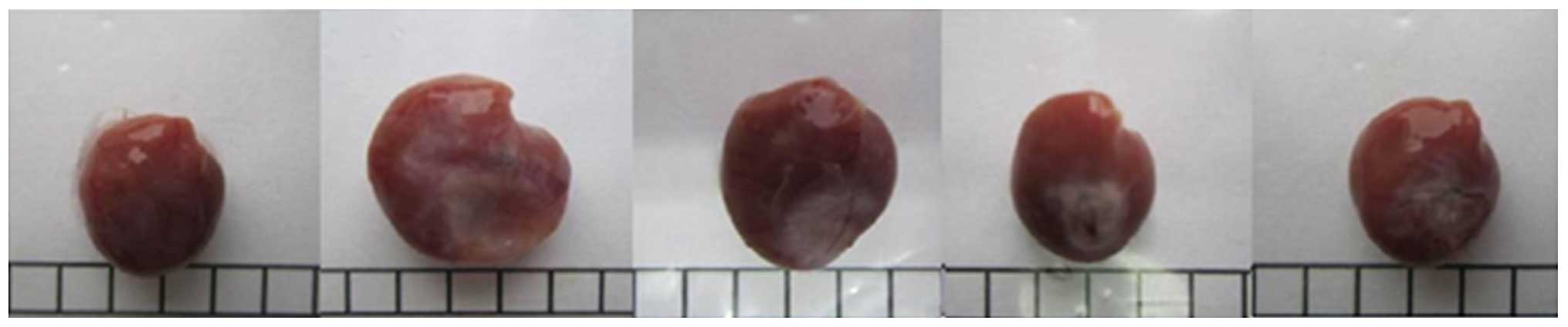
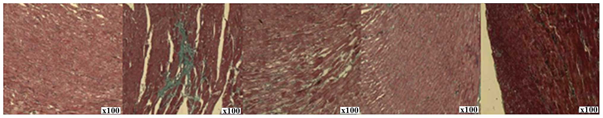
Comparison of left ventricular mass
index, myocardial cell cross-sectional area and collagen
content
Compared with the sham operation group, the left
ventricular mass index, myocardial cell cross-sectional area and
collagen levels were higher in all other groups. The highest value
was recorded in the model group, followed by the 1 and 9 mg/kg/day
groups. The lowest values were observed in the 3 mg/kg/day group.
All differences were statistically significant (P<0.05)
(Table II and Figs. 1–3).
| Table II.Comparison of left ventricular mass
index, myocardial cell cross-sectional area and collagen content
(%). |
Table II.
Comparison of left ventricular mass
index, myocardial cell cross-sectional area and collagen content
(%).
| Groups | Left ventricular mass
index | Cell cross-sectional
area | Collagen |
|---|
| Model | 8.5±1.3 | 178.6±23.4 | 6.4±1.5 |
| 1 mg/kg/day | 8.2±1.4 | 152.3±21.2 | 6.0±1.6 |
| 3 mg/kg/day | 6.9±1.2 | 120.5±20.7 | 4.7±1.3 |
| 9 mg/kg/day | 8.0±1.5 | 146.6±22.3 | 5.5±1.4 |
| F-value | 6.458 | 6.935 | 6.625 |
| P-value | 0.026 | 0.021 | 0.023 |
In the model group, the myocardial fiber arrangement
was thickened, disordered and even ruptured. Myocardial
hypertrophy, myocardial interstitial significant hyperplasia,
inflammatory cell infiltration and significant increase in
myocardial cell cross-sectional area occurred. Compared with the
model group, the cell cross-sectional area in each natakalim group
was reduced, and the reduction in the 3 mg/kg/day group was the
most obvious.
Comparison of ET-1 and eNOS
levels
Prior to the experiment, differences between the
ET-1 and eNOS levels in different groups did not reveal any
statistically significant differences (P>0.05). In addition to
the sham operation, after the experiment ET-1 and eNOS levels were
considerably elevated in all the groups. The highest values were
recorded in the model group, followed by the 1 and 9 mg/kg/day
groups. The lowest values were observed in the 3 mg/kg/day group.
Differences were statistically significant (P<0.05) (Table III).
| Table III.Comparison of ET-1 and eNOS
levels. |
Table III.
Comparison of ET-1 and eNOS
levels.
|
| ET-1 (pg/ml) | eNOS (µg/ml) |
|---|
|
|
|
|
|---|
| Groups | Before
experiment | After experiment | Before
experiment | After experiment |
|---|
| Sham operation | 213.4±42.6 | 217.5±43.2 | 6.6±2.2 | 6.5±2.3 |
| Model | 206.3±43.3 | 356.8±55.6 | 6.5±2.3 | 17.2±2.2 |
| 1 mg/kg/day | 215.5±41.4 | 321.5±51.8 | 6.3±2.4 | 14.3±2.6 |
| 3 mg/kg/day | 208.7±43.5 | 277.9±48.2 | 6.4±2.5 | 8.8±2.4 |
| 9 mg/kg/day | 210.2±42.7 | 303.6±56.4 | 6.6±2.3 | 11.5±2.6 |
| F-value | 0.526 | 7.012 | 0.864 | 7.714 |
| P-value | 0.649 | 0.019 | 0.963 | 0.005 |
Discussion
Previous findings revealed that natakalim was
capable of protecting the aortic endothelial cells with hypoxia and
homocysteine injury, reversing the ventricular remodeling in animal
models with abdominal aorta coarctation-induced overloading
pressure and preventing the development and progression of
ventricular remodeling to heart failure (13). The mechanism was related to the
correction of endothelial dysfunction and protection of endothelial
function. Our results showed that compared with the sham operation
group, the LVEDD and NT-proBNP in the model group and each of the
natakalim groups were elevated. LVEF decreased, the left
ventricular mass index, myocardial cell cross-sectional area,
myocardial collagen content, plasma ET-1 and eNOS levels increased.
Natakalim positively affected all of the above-mentioned indexes.
This effect was more significant in the 3 mg/kg/day group. LVEDD,
LVEF, left ventricular mass index and microcosmic myocardial cell
cross-sectional area and myocardial collagen content, NT-proBNP
reflected the neuroendocrine regulation. ET-1 and eNOS are two
important cytokines secreted by endothelial cells and play
important roles in ventricular remodeling.
Under myocardial ischemia and hypoxia, a large
number of ATP molecules is consumed and K+ concentration
inside cells is significantly reduced. This can open the
KATP, shorten the myocardial action potential duration,
reduce Ca2+ influx, which is an important
self-protection mechanism (14)
against myocardial ischemia and hypoxia. Watanuki et al
showed that after being pre-incubated for 60 min with 5 mg/l
pertussis toxin in the isolated guinea pig ventricular myocytes,
ET-1-induced K+ influx was significantly eliminated
(15). ET-1 and its receptor
influenced the metabolic state of the cells and inhibited the
openness of the KATP by intracellular signal
transduction.
The mechanism of natakalim against the endothelin
system may be explained by the fact that inhibiting ET-1 synthesis
and its release, could reduce the ET-1 levels in the circulation
(16) leading to the release of NO
and PGI2. Inhibition of the vasoconstriction effect of ET-1,
improved the hemodynamics of high-load status (17) and ameliorated the cardiac systolic
and diastolic functions (18). It
was manifested as the reversal of cardiac remodeling, restoration
of reduced heart function and prevention of the development and
progression of heart failure in animal models.
NO generated by catalysis of eNOS can relax the
blood vessels, reduce the cardiac preload and afterload which lead
to myocardial protection (19). It
can also directly enhance the dilatation effect of ventricular
muscle, maintain cardiac output through the left ventricle
Frank-Starling regulation mechanism and be beneficial for the heart
pump function under physiological and pathological states (20). Scherrer-Crosbie et al found
out that deletion of eNOS gene in the mouse model with myocardial
infarction, significantly reduced eNOS levels. Compared to
wild-type mice, these mice developed a more severe myocardial
infarction and ventricular remodeling (21). Natakalim could promote the eNOS
protein expression, raise the level of endothelium-derived NO,
increase eNOS-NO pathway activity (22), reduce inducible NOS (iNOS) protein
expression and reduce the generation of a large number of
iNOS-induced NOs and inhibit iNOS-NO pathway (23).
We concluded that natakalim can improve the
ventricular remodeling of CHF after AMI, and 3 mg/kg/day was the
most effective dose. This may be related to the inhibition effect
of ET-1 and promotion of NO and eNOS activities.
References
|
1
|
McMurray JJ, Adamopoulos S, Anker SD,
Auricchio A, Böhm M, Dickstein K, Falk V, Filippatos G, Fonseca C,
Gomez-Sanchez MA, et al: Task Force for the Diagnosis and Treatment
of Acute and Chronic Heart Failure 2012 of the European Society of
Cardiology; ESC Committee for Practice Guidelines: ESC guidelines
for the diagnosis and treatment of acute and chronic heart failure
2012: The Task Force for the Diagnosis and Treatment of Acute and
Chronic Heart Failure 2012 of the European Society of Cardiology.
Developed in collaboration with the Heart Failure Association (HFA)
of the ESC. Eur J Heart Fail. 14:803–869. 2012. View Article : Google Scholar : PubMed/NCBI
|
|
2
|
Gao S, Long CL, Wang RH and Wang H: K(ATP)
activation prevents progression of cardiac hypertrophy to failure
induced by pressure overload via protecting endothelial function.
Cardiovasc Res. 83:444–456. 2009. View Article : Google Scholar : PubMed/NCBI
|
|
3
|
Khazaei M, Moien-Afshari F and Laher I:
Vascular endothelial function in health and diseases.
Pathophysiology. 15:49–67. 2008. View Article : Google Scholar : PubMed/NCBI
|
|
4
|
Otsuka F, Finn AV, Yazdani SK, Nakano M,
Kolodgie FD and Virmani R: The importance of the endothelium in
atherothrombosis and coronary stenting. Nat Rev Cardiol. 9:439–453.
2012. View Article : Google Scholar : PubMed/NCBI
|
|
5
|
Pan X, Wang J, Pu Y, Yao J and Wang H:
Effect of puerarin on expression of ICAM-1 and TNF-alpha in kidneys
of diabetic rats. Med Sci Monit. 21:2134–2140. 2015. View Article : Google Scholar : PubMed/NCBI
|
|
6
|
Scalera F, Schlembach D and Beinder E:
Production of vasoactive substances by human umbilical vein
endothelial cells after incubation with serum from preeclamptic
patients. Eur J Obstet Gynecol Reprod Biol. 99:172–178. 2001.
View Article : Google Scholar : PubMed/NCBI
|
|
7
|
Tsikouris JP, Simoni J, Suarez JA,
Sutthiwan P, Ziska M and Meyerrose GE: Comparison of effects of
quinapril versus enalapril on vasoactive substances following acute
myocardial infarction. Am J Cardiol. 94:641–643. 2004. View Article : Google Scholar : PubMed/NCBI
|
|
8
|
Vanhoutte PM, Shimokawa H, Feletou M and
Tang EH: Endothelial dysfunction and vascular disease - a
thirthieth anniversary update. Acta Physiol (Oxf). 26:123–124.
2015.
|
|
9
|
Butta NV, Fernández-Bello I, López-Longo
FJ and Jiménez-Yuste V: Endothelial dysfunction and altered
coagulation as mediators of thromboembolism in Behçet disease.
Semin Thromb Hemost. 41:621–628. 2015. View Article : Google Scholar : PubMed/NCBI
|
|
10
|
Mudau M, Genis A, Lochner A and Strijdom
H: Endothelial dysfunction: the early predictor of atherosclerosis.
Cardiovasc J Afr. 23:222–231. 2012. View Article : Google Scholar : PubMed/NCBI
|
|
11
|
Kadowaki D, Anraku M, Sakaya M, Hirata S,
Maruyama T and Otagiri M: Olmesartan protects endothelial cells
against oxidative stress-mediated cellular injury. Clin Exp
Nephrol. 19:1007–1014. 2015. View Article : Google Scholar : PubMed/NCBI
|
|
12
|
Zingman LV, Alekseev AE, Hodgson-Zingman
DM and Terzic A: ATP-sensitive potassium channels: metabolic
sensing and cardioprotection. J Appl Physiol 1985. 103:1888–1893.
2007. View Article : Google Scholar : PubMed/NCBI
|
|
13
|
Chen X, Han W, Zhang Y, Cui W, Pan Z, Jin
X, Long C and Wang H: The molecular pathway of ATP-sensitive
potassium channel in endothelial cells for mediating arteriole
relaxation. Life Sci. 137:164–169. 2015. View Article : Google Scholar : PubMed/NCBI
|
|
14
|
Seino S and Miki T: Physiological and
pathophysiological roles of ATP-sensitive K+ channels.
Prog Biophys Mol Biol. 81:133–176. 2003. View Article : Google Scholar : PubMed/NCBI
|
|
15
|
Watanuki M, Horie M, Tsuchiya K, Obayashi
K and Sasayama S: Endothelin-1 inhibition of cardiac ATP-sensitive
K+ channels via pertussis-toxin-sensitive G-proteins.
Cardiovasc Res. 33:123–130. 1997. View Article : Google Scholar : PubMed/NCBI
|
|
16
|
Vita JA and Keaney JF Jr: Endothelial
function: a barometer for cardiovascular risk? Circulation.
106:640–642. 2002. View Article : Google Scholar : PubMed/NCBI
|
|
17
|
Marti CN, Gheorghiade M, Kalogeropoulos
AP, Georgiopoulou VV, Quyyumi AA and Butler J: Endothelial
dysfunction, arterial stiffness, and heart failure. J Am Coll
Cardiol. 60:1455–1469. 2012. View Article : Google Scholar : PubMed/NCBI
|
|
18
|
Jandeleit-Dahm KA and Watson AM: The
endothelin system and endothelin receptor antagonists. Curr Opin
Nephrol Hypertens. 21:66–71. 2012. View Article : Google Scholar : PubMed/NCBI
|
|
19
|
Sun Y, Ye L, Jiang C, Jiang J, Hong H and
Qiu L: Over-expression of HSPA12B protects mice against myocardium
ischemic/reperfusion injury through a PPARγ-dependent PI3K/Akt/eNOS
pathway. Am J Transl Res. 7:2724–2737. 2015.PubMed/NCBI
|
|
20
|
Liu YH, Carretero OA, Cingolani OH, Liao
TD, Sun Y, Xu J, Li LY, Pagano PJ, Yang JJ and Yang XP: Role of
inducible nitric oxide synthase in cardiac function and remodeling
in mice with heart failure due to myocardial infarction. Am J
Physiol Heart Circ Physiol. 289:H2616–H2623. 2005. View Article : Google Scholar : PubMed/NCBI
|
|
21
|
Scherrer-Crosbie M, Ullrich R, Bloch KD,
Nakajima H, Nasseri B, Aretz HT, Lindsey ML, Vançon AC, Huang PL,
Lee RT, et al: Endothelial nitric oxide synthase limits left
ventricular remodeling after myocardial infarction in mice.
Circulation. 104:1286–1291. 2001. View Article : Google Scholar : PubMed/NCBI
|
|
22
|
Starling RC: Inducible nitric oxide
synthase in severe human heart failure: impact of mechanical
unloading. J Am Coll Cardiol. 45:1425–1427. 2005. View Article : Google Scholar : PubMed/NCBI
|
|
23
|
Jones SP, Greer JJ, van Haperen R, Duncker
DJ, de Crom R and Lefer DJ: Endothelial nitric oxide synthase
overexpression attenuates congestive heart failure in mice. Proc
Natl Acad Sci USA. 100:4891–4896. 2003. View Article : Google Scholar : PubMed/NCBI
|