Introduction
Hypertension is one of the major risk factors for
cardiovascular accidents (1). Its
main complications include stroke, myocardial infarction, heart
failure and chronic kidney disease (2–4).
Hypertension is a serious threat to human health, and is one of the
most actively researched areas in the biomedical field. Blood
pressure is maintained by the regulation of vascular tone, which
can be affected by many factors. For example, nitric oxide (NO) has
been shown to be an effective vasodilator (5). Furthermore, the NO synthase inhibitor
NG-nitro-L-arginine methyl ester (L-NAME) is known to
induce sustained blood pressure elevation and left ventricular
hypertrophy (6). Soluble guanylyl
cyclase (sGC) is an important effector of NO (7). It acts by increasing intracellular
cyclic GMP (cGMP) levels to mediate numerous biological functions
(8). The compound
1H-[1,2,4]oxadiazolo[4,3,-a]quinoxalin-1-one (ODQ) has been
identified as a selective inhibitor of this enzyme; ODQ treatment
is able to increase contractile tone and inhibit relaxation in
response to authentic NO (8).
Indomethacin (INDO), a known cyclooxygenase (COX) inhibitor has
been reported to significantly increase mean arterial pressure
without altering other hemodynamic parameters through the
inhibition of vasodilation (9).
Antihypertensive drugs exert their actions through a
variety of pathways that regulate blood pressure. The major effects
of these drugs include: Modulation of the sympathetic branch of the
peripheral nervous system and of the renin-angiotensin system
(RAS); blockade of calcium channels; improvement of endothelial
function; regulation of cardiac blood flow; and inhibition of
vascular remodeling and increased urination (10). Antihypertensive drugs include:
Diuretics, calcium channel blockers (CCBs), angiotensin-converting
enzyme inhibitors (ACEIs), angiotensin II (ATII) receptor
antagonists (ARBs), α1 receptor blockers, β-blockers, renin
inhibitors, central hypotensive agents, ganglion blockers and
vasodilators (11). Despite their
important therapeutic effects, these drugs all have potential side
effects. For example, the use of diuretic antihypertensive drugs
can lead to hypokalemia, hyperglycemia, hypercholesterolemia,
hypertriglyceridemia, and accumulation of uric acid in the blood;
β-blockers can cause bronchospasm, peripheral circulation
disorders, and insulin insensitivity; and ACEIs can give rise to a
dry persistent cough, for example (12).
Panax notoginseng is a species of the genus
Panax which is a traditional Chinese herbal medicine
(13). The main bioactive ingredient
of this species is Panax notoginseng saponins (PNS), which
is a phytoestrogenic composition (14). It is known that PNS exerts extensive
effects on the cardiovascular system, including inhibition of
platelet aggregation, augmentation of the coronary blood flow,
improvement of left ventricular diastolic function in hypertensive
patients, and myocardial ischemia remodeling protection (15–18). PNS
also reduces myocardial oxygen consumption and is endowed with
antiarrhythmic effects (19–23).
PNS is a chemical mixture containing >50
different saponins, and the five major components of PNS are
ginsenosides Rg1, Rb1, Re and Rd, and notoginsenoside R1 (24–29). PNS
saponins are classified into two main groups: Namely the
20(S)-protopanaxatriol saponins (PTS) such as ginsenoside Rg1 and
ginsenoside Rd; and the 20(S)-protopanaxadiol saponins (PDS) such
as ginsenoside Rb1 and Re, and notoginsenoside R1 (30,31).
In the present study, the aim was to assess the role
of PNS and its main components in vascular tone, and thereby
explain the mechanism by which they benefit cardiovascular
function. The study was conducted using in vitro aortic
vascular rings. The endothelium-derived relaxing factors and
pathways were examined to elucidate the vasodilation effects of PNS
and its major components. This should provide an experimental basis
for and improve the clinical application of PNS and its major
components.
Materials and methods
Drugs and reagents
PNS, ginsenoside Rg1, ginsenoside Rb1,
notoginsenoside R1, ginsenoside Re and ginsenoside Rd were provided
by Zhongxin Pharmaceutical Group Corporation, Ltd. (Tianjin,
China). Norepinephrine (NE), acetylcholine chloride (ACh), dimethyl
sulfoxide (DMSO), L-NAME, INDO, sodium chloride (NaCl), ODQ,
potassium chloride (KCl), potassium dihydrogen phosphate
(KH2PO4), magnesium sulfate heptahydrate
(MgSO4.7H2O), sodium bicarbonate
(NaHCO3), glucose and calcium chloride
(CaCl2) were purchased from Sigma-Aldrich (Merck
Millipore, Darmstadt, Germany).
Animals
Adult male Wistar rats (weight, 250–300 g), were
purchased from the Experimental Animal Center, Institute of
Radiation Medicine, Chinese Academy of Medical Sciences, Tianjin,
China [permit: SCXK200F (JING) 0004]. Rats (8–10 weeks) were housed
at 22 ± 2°C and a relative humidity of 40 ± 10% under a 12-h
light/dark cycle and given standard laboratory diet and water. Rats
were fasted for 12 h before experiments but allowed to access water
freely. All experimental procedures that involved animals were
submitted to and approved by the Animal Ethics Committee of Tianjin
University of Traditional Chinese Medicine Medical Center (permit:
LAEC2013005).
Preparation of aortic rings
Male Wistar rats were sacrificed by cervical
dislocation and their thoracic aortas were carefully dissected and
removed. Fat and other non-vascular tissues were dissected, and the
thoracic aortas were sliced into 3-4-mm ring segments after placing
in Krebs-Henseleit (K-H) solution at 4°C (in mM: NaCl, 118; KCl,
4.7; NaHCO3, 25; KH2PO4, 1.2;
MgSO4, 1.2; CaCl2, 1.3; glucose, 10)
(32). The vessels were not
stretched and the endothelium was protected during handling. For
some of the rings, the endothelium was removed gently by rubbing
the ring with a glass rod.
The aorta rings were mounted onto two
stainless-steel stirrups immersed in a 10-ml organ chamber,
containing K-H buffer that was continuously bubbled with 95%
O2 and 5% CO2, and maintained at 37°C.
Isometric tension change was measured with the force-displacement
transducer and recorded using a PowerLab 8/30 bio-signal recording
system (AD Instruments Pty Ltd., Bella Vista, Australia). The aorta
rings were stretched progressively to a basal tension of 2.0 g and
equilibrated for 90 min; during this period the bath solution was
replaced with K-H buffer every 15 min. After stabilization the
rings were repeatedly contracted with KCl (60 mmol/l) three times
until the muscle tension returned to the basal level.
The aortic rings were pre-treated with NE
(1×10⁻⁶ mol/l) to achieve the plateau phase, and ACh
(1×10⁻⁵ mol/l) was then added to induce vasodilation.
Compared with the maximum contraction extent induced by NE, if the
relaxation extent achieved was >60%, the endothelium was
regarded as intact and functional, while if it was <10%, the
aorta rings would be regarded as completely denuded
endothelium.
Measurement of vascular
relaxation
Once a sustained contraction plateau in response to
NE (1×10⁻⁶ mol/l) was achieved, 10 µl H2O or
DMSO; PNS (0.2, 0.4, 0.6 or 0.8 mg/ml); or Rg1, Rb1, Re, R1 and Rd
(1×10⁻⁸, 1×10⁻⁷, 1×10⁻⁶ or
1×10⁻⁵ mol/l) was cumulatively added with an interval of
8 min to the organ bath containing the aortic rings with or without
endothelium. H2O was used for PNS. DMSO was used for
Rg1, Rb1, R1, Re and Rd.
In order to investigate the involvement of the
endothelial NO pathway and cyclooxygenase (COX) pathway in
vasorelaxation to PNS and its main five components (ginsenoside
Rg1, Re, Rb1 and Rd, and notoginsenoside R1), the rings were
exposed to 0.1 mmol/l L-NAME, 0.01 mmol/l ODQ and 0.01 mmol/l INDO
for 20 min prior to application of NE to blunt the endothelial
function by inhibiting NO and COX synthesis following repeated
washout and subsequent equilibration for 45 min.
Statistical analysis
Results are shown as mean ± standard deviation
values. Statistical comparisons were carried out by analysis of
variance followed by a Dunnett's multiple comparison test.
P<0.05 was considered to indicate a statistically significant
difference. Each data point represents the mean of a minimum of 10
aortic rings from different animals unless otherwise noted.
Results
Effects of solvents (H2O
and DMSO) on aortic rings
It was first verified that the solvents
H2O and DMSO each had no effect on the NE-induced
vasoconstriction of the rat aortic rings. Mean values of the
contraction at four different time points for each solvent are
presented in Table I. As detailed in
Materials and methods, the test substances (H2O or DMSO)
were delivered every 8 min and no wash-out was performed during the
experiment. Statistical comparison demonstrated that the curves for
the two solvents were not significantly different (P>0.05).
 | Table I.Effects of solvent on the
vasoconstriction of aortic rings (%; mean ± standard
deviation). |
Table I.
Effects of solvent on the
vasoconstriction of aortic rings (%; mean ± standard
deviation).
| Time (min) | H2O
(n=15) | DMSO (n=15) |
|---|
| 8 | 104.13±3.83 | 104.04±2.75 |
| 16 | 105.22±6.09 | 107.39±4.93 |
| 24 | 102.68±4.95 | 106.82±5.97 |
| 32 | 95.31±5.06 | 99.98±7.69 |
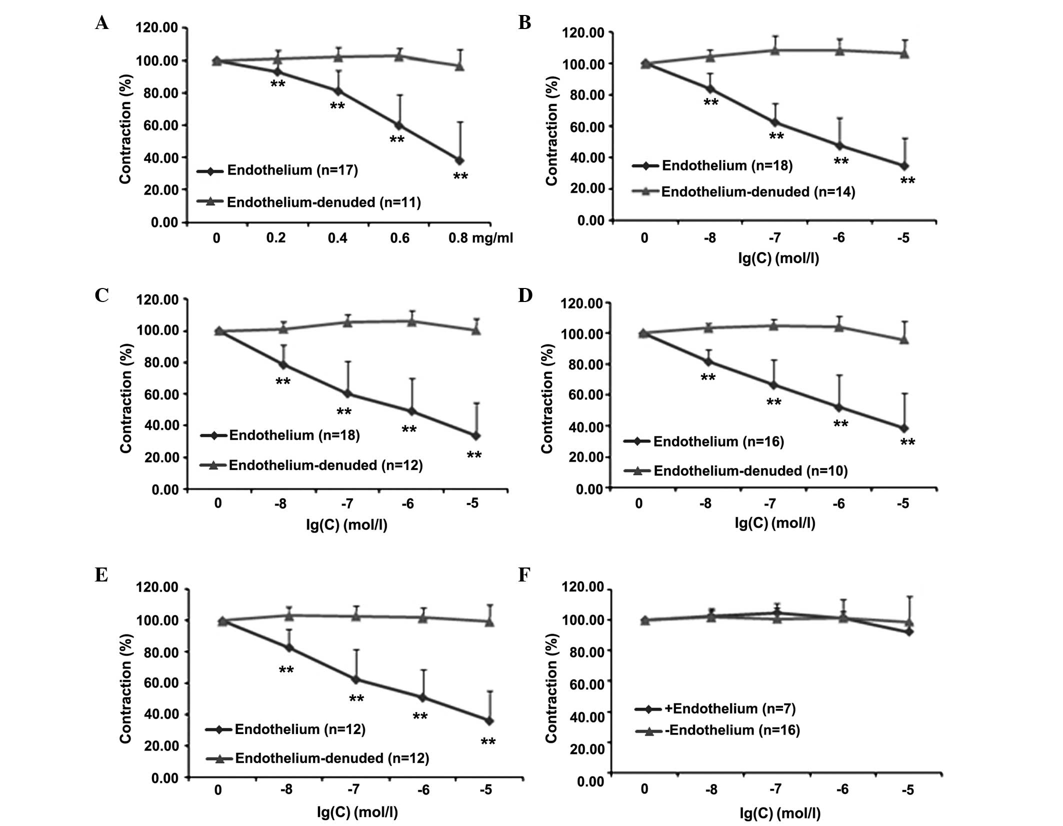
Effects of PNS and its main components
on aortic rings with or without endothelium
Whether the endothelium plays a major role in
mediating the effect of PNS and its five main components (Rg1, Re,
Rb1, R1 and Rd) was then investigated. In Fig. 1, dose-response curves for these
compounds are shown; each curve represents the ability of the
compound to reduce the tonic contraction induced by NE in the
presence and absence of the endothelium. Results clearly indicate
that when the endothelium lining was intact, PNS, Rg1, Re, Rb1 and
R1 (Fig. 1A-E, respectively)
significantly reduced the tonic contraction (% of NE) at all doses
investigated. The effect was completely lost when the endothelium
was absent. Notably, the administration of Rd was not able to
induce any response in either the presence or the absence of the
endothelium. Detailed statistical comparisons of the data are
presented in Table II.
| Table II.Effects of PNS and five main
components on aortic rings with or without endothelium. |
Table II.
Effects of PNS and five main
components on aortic rings with or without endothelium.
|
|
| Contraction (% of
NE) |
|---|
|
|
|
|
|---|
| Drugs | Concentration | Endothelium
(n=10–18) | Endothelium-denuded
(n=10–18) |
|---|
| PNS (mg/ml) | 0.2 |
93.02±9.11a |
100.91±5.29 |
|
| 0.4 |
80.37±13.44a |
102.43±5.37 |
|
| 0.6 |
60.62±20.71a |
102.73±4.96 |
|
| 0.8 |
37.19±25.23a |
96.69±10.37 |
| Rg1 (mol/l) |
1×10⁻⁸ |
83.67±10.41a |
104.28±4.50 |
|
|
1×10⁻⁷ |
62.47±12.07a |
108.68±8.96 |
|
|
1×10⁻⁶ |
47.61±17.61a |
108.68±7.67 |
|
|
1×10⁻⁵ |
34.75±17.88a |
106.68±8.61 |
| Re (mol/l) |
1×10⁻⁸ |
78.50±12.82a |
101.30±4.79 |
|
|
1×10⁻⁷ |
60.44±20.44a |
105.36±5.20 |
|
|
1×10⁻⁶ |
49.26±20.63a |
105.97±7.28 |
|
|
1×10⁻⁵ |
33.63±20.74a |
100.39±7.35 |
| Rb1 (mol/l) |
1×10⁻⁸ |
81.77±7.78a |
103.24±3.06 |
|
|
1×10⁻⁷ |
66.69±16.36a |
104.67±4.12 |
|
|
1×10⁻⁶ |
52.13±20.94a |
103.83±7.21 |
|
|
1×10⁻⁵ |
38.69±22.79a |
95.90±11.76 |
| R1 (mol/l) |
1×10⁻⁸ |
83.06±11.58a |
103.21±5.73 |
|
|
1×10⁻⁷ |
62.55±19.06a |
103.13±6.19 |
|
|
1×10⁻⁶ |
51.01±17.96a |
101.90±6.66 |
|
|
1×10⁻⁵ |
36.04±19.23a |
99.57±10.87 |
| Rd (mol/l) |
1×10⁻⁸ |
101.87±5.57 |
102.78±3.29 |
|
|
1×10⁻⁷ |
101.06±10.06 |
104.93±3.55 |
|
|
1×10⁻⁶ |
101.52±12.46 |
101.73±3.95 |
|
|
1×10⁻⁵ |
99.06±16.80 |
92.43±5.90 |
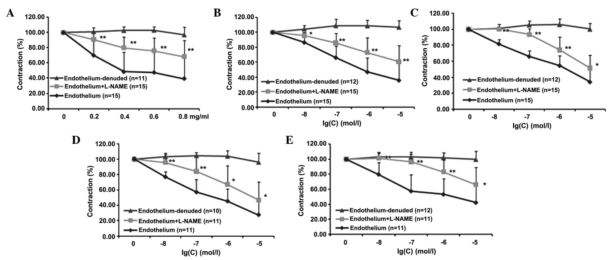
Effects of PNS and its main components
on the endothelial NO pathway
Effects of PNS and its main components are
partially conserved by L-NAME
In order to evaluate whether endothelial NO plays a
significant role in the mediation of the previously observed
effects of PNS and four of its main components (Rg1, Re, Rb1 and
R1), experiments were carried out with L-NAME, which is a compound
known to selectively block NO synthase and therefore the pathways
downstream of NO production. The results shown in Fig. 2 reveal that pre-incubation of the
aortic rings with intact endothelium using L-NAME significantly
reduced the effects of the drugs at all concentrations tested
(P<0.01). Detailed statistical comparisons of the data are
presented in Table III.
| Table III.Effects of PNS and four main
components on aortic rings in the presence of L-NAME. |
Table III.
Effects of PNS and four main
components on aortic rings in the presence of L-NAME.
|
|
| Contraction (% of
NE) |
|---|
|
|
|
|
|---|
| Drugs | Concentration | Endothelium
(n=11–15) | Endothelium +
L-NAME (n=11–15) | Endothelium-denuded
(n=10–12) |
|---|
| PNS (mg/ml) | 0.2 |
70.03±21.71 |
90.59±7.80a |
100.91±5.29 |
|
| 0.4 |
48.39±25.60 |
79.81±14.41a |
102.43±5.37 |
|
| 0.6 |
47.41±26.50 |
75.94±16.68a |
102.73±4.96 |
|
| 0.8 |
39.30±27.01 |
68.23±21.04a |
96.69±10.37 |
| Rg1 (mol/l) |
1×10⁻⁸ |
86.58±13.21 |
95.48±4.42b |
104.28±4.50 |
|
|
1×10⁻⁷ |
66.23±15.37 |
85.71±12.67a |
108.68±8.96 |
|
|
1×10⁻⁶ |
46.96±18.98 |
72.85±19.64a |
108.68±7.67 |
|
|
1×10⁻⁵ |
35.72±22.03 |
60.54±21.56a |
106.68±8.61 |
| Re (mol/l) |
1×10⁻⁸ |
81.72±5.53 |
100.40±5.53a |
101.30±4.79 |
|
|
1×10⁻⁷ |
65.98±7.06 |
93.49±7.46a |
105.36±5.20 |
|
|
1×10⁻⁶ |
54.59±12.43 |
74.19±16.16a |
105.97±7.28 |
|
|
1×10⁻⁵ |
34.14±19.26 |
51.36±16.36b |
100.39±7.35 |
| Rb1 (mol/l) |
1×10⁻⁸ |
76.76±6.63 |
95.61±12.15a |
103.24±3.06 |
|
|
1×10⁻⁷ |
56.99±16.34 |
83.97±19.92a |
104.67±4.12 |
|
|
1×10⁻⁶ |
45.25±16.09 |
66.92±24.55b |
103.83±7.21 |
|
|
1×10⁻⁵ |
27.40±16.61 |
46.92±23.21b |
95.90±11.76 |
| R1 (mol/l) |
1×10⁻⁸ |
79.83±15.76 |
101.40±6.26a |
103.21±5.73 |
|
|
1×10⁻⁷ |
57.67±21.30 |
96.26±10.68a |
103.13±6.19 |
|
|
1×10⁻⁶ |
53.34±20.68 |
83.03±15.84a |
101.90±6.66 |
|
|
1×10⁻⁵ |
42.40±23.16 |
66.02±23.26b |
99.57±10.87 |
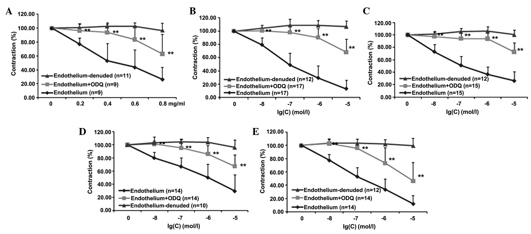
Effects of PNS and its main components are
partially conserved by ODQ
It was then evaluated whether the endothelial
NO-mediated pathway requires the functional integrity of guanylyl
cyclase and, therefore, cGMP production. Thus, whether
pre-treatment of aortic rings with intact endothelium with ODQ, a
selective, irreversible, heme-site inhibitor of soluble guanylyl
cyclase and competitive inhibitor of NO, was able to inhibit the
effects of PNS, Rg1, Re, Rb1 and R1 was tested. As shown in
Fig. 3, pre-incubation of the aortic
rings with intact endothelium using ODQ significantly reduced the
effects of PNS and four of its main components (Rg1, Re, Rb1 and
R1) at all concentrations tested (P<0.05). Detailed statistical
comparisons of the data are presented in Table IV.
| Table IV.Effects of PNS and four main
components on aortic rings in the presence of ODQ. |
Table IV.
Effects of PNS and four main
components on aortic rings in the presence of ODQ.
|
|
| Contraction (% of
NE) |
|---|
|
|
|
|
|---|
| Drug | Concentration | Endothelium
(n=9–17) | Endothelium + ODQ
(n=9–17) | Endothelium-denuded
(n=11–14) |
|---|
| PNS (mg/ml) | 0.2 |
77.25±8.74 |
96.249±10.18a |
100.91±5.29 |
|
| 0.4 |
53.15±24.64 |
93.82±12.36a |
102.43±5.37 |
|
| 0.6 |
43.75±25.06 |
83.74±19.50a |
102.73±4.96 |
|
| 0.8 |
26.31±17.15 |
62.98±28.98a |
96.69±10.37 |
| Rg1 (mol/l) |
1×10⁻⁸ |
79.48±10.38 |
100.59±4.64a |
104.28±4.50 |
|
|
1×10⁻⁷ |
49.00±17.82 |
98.15±8.38a |
108.68±8.96 |
|
|
1×10⁻⁶ |
29.48±18.62 |
90.36±13.49a |
108.68±7.67 |
|
|
1×10⁻⁵ |
13.23±13.16 |
68.28±19.82a |
106.68±8.61 |
| Re (mol/l) |
1×10⁻⁸ |
73.04±11.74 |
97.52±6.79a |
101.30±4.79 |
|
|
1×10⁻⁷ |
51.18±14.45 |
94.06±11.81a |
105.36±5.20 |
|
|
1×10⁻⁶ |
36.87±11.64 |
93.87±9.34a |
105.97±7.28 |
|
|
1×10⁻⁵ |
26.38±14.52 |
72.73±14.95a |
100.39±7.35 |
| Rb1 (mol/l) |
1×10⁻⁸ |
80.01±8.93 |
101.39±10.77a |
103.24±3.06 |
|
|
1×10⁻⁷ |
66.97±13.66 |
95.26±12.94a |
104.67±4.12 |
|
|
1×10⁻⁶ |
50.44±20.56 |
86.12±12.67a |
103.83±7.21 |
|
|
1×10⁻⁵ |
29.81±24.59 |
67.31±17.27a |
95.90±11.76 |
| R1 (mol/l) |
1×10⁻⁸ |
77.64±9.05 |
102.81±6.96a |
103.21±5.73 |
|
|
1×10⁻⁷ |
53.26±13.45 |
95.89±13.65a |
103.13±6.19 |
|
|
1×10⁻⁶ |
33.62±15.63 |
73.17±23.19a |
101.90±6.66 |
|
|
1×10⁻⁵ |
12.14±12.43 |
46.44±28.21a |
99.57±10.87 |
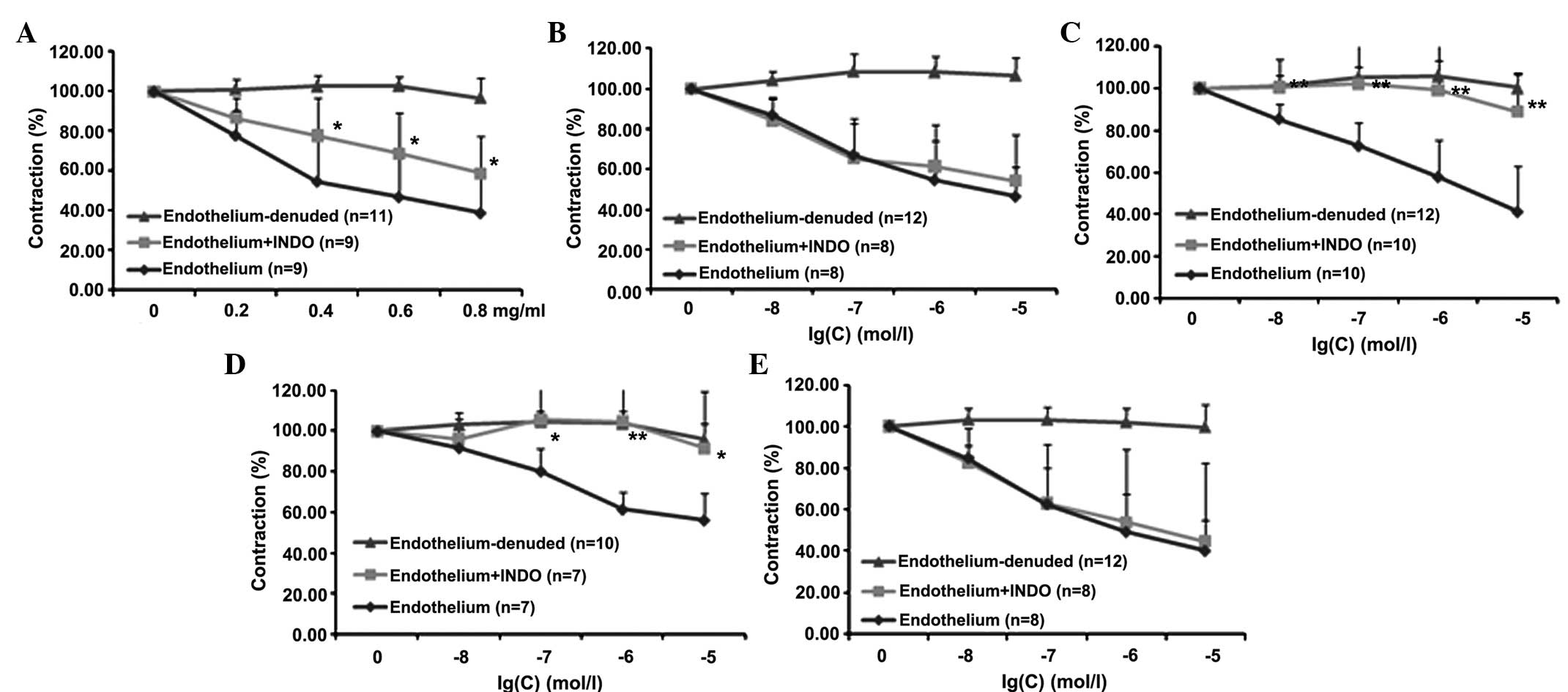
Effects of PNS and its main components on the COX
pathway
In this set of experiments, the involvement of the
COX pathway in mediating the effects of PNS, Rg1, Re, Rb1 and R1
was investigated. As shown in Fig.
4, pre-incubation of the rat aortic rings with intact
endothelium using INDO, a known COX inhibitor, significantly
reduced the effects of PNS, Re and Rb1 (P<0.05); by contrast,
Rg1 and R1 did not elicit any effects. Detailed statistical
comparisons of the data are provided in Table V.
| Table V.Effects of PNS and four main
components on the cyclooxygenase pathway in aortic rings. |
Table V.
Effects of PNS and four main
components on the cyclooxygenase pathway in aortic rings.
|
|
| Contraction (% of
NE) |
|---|
|
|
|
|
|---|
| Drugs | Concentration | Endothelium
(n=7–10) | Endothelium + INDO
(n=7–10) | Endothelium-denuded
(n=10–12) |
|---|
| PNS (mg/ml) | 0.2 |
77.96±13.39 |
86.70±10.23 |
100.91±5.29 |
|
| 0.4 |
54.72±21.15 |
77.67±19.08a |
102.43±5.37 |
|
| 0.6 |
46.86±21.48 |
68.71±20.56a |
102.73±4.96 |
|
| 0.8 |
38.79±18.11 |
58.69±18.68a |
96.69±10.37 |
| Rg1 (mol/l) |
1×10⁻⁸ |
87.06±7.99 |
84.24±11.55 |
104.28±4.50 |
|
|
1×10⁻⁷ |
67.11±18.11 |
65.32±17.48 |
108.68±8.96 |
|
|
1×10⁻⁶ |
54.66±19.60 |
61.45±120.90 |
108.68±7.67 |
|
|
1×10⁻⁵ |
46.46±14.63 |
54.25±22.84 |
106.68±8.61 |
| Re (mol/l) |
1×10⁻⁸ |
85.41±7.41 |
100.67±13.15b |
101.30±4.79 |
|
|
1×10⁻⁷ |
72.67±11.16 |
102.29±21.72b |
105.36±5.20 |
|
|
1×10⁻⁶ |
57.98±17.28 |
99.28±21.33b |
105.97±7.28 |
|
|
1×10⁻⁵ |
41.47±21.54 |
89.04±17.89b |
100.39±7.35 |
| Rb1 (mol/l) |
1×10⁻⁸ |
91.80±5.28 |
95.88±13.18 |
103.24±3.06 |
|
|
1×10⁻⁷ |
80.37±10.72 |
105.62±24.91a |
104.67±4.12 |
|
|
1×10⁻⁶ |
61.59±8.39 |
104.70±25.69b |
103.83±7.21 |
|
|
1×10⁻⁵ |
56.32±13.22 |
91.48±28.07a |
95.90±11.76 |
| R1 (mol/l) |
1×10⁻⁸ |
84.62±6.04 |
82.64±16.32 |
103.21±5.73 |
|
|
1×10⁻⁷ |
62.56±17.43 |
62.95±28.20 |
103.13±6.19 |
|
|
1×10⁻⁶ |
49.19±18.10 |
53.80±35.32 |
101.90±6.66 |
|
|
1×10⁻⁵ |
40.13±14.70 |
44.55±37.98 |
99.57±10.87 |
Discussion
PNS is a traditional Chinese herbal medicine that
has protective effects on heart function; particularly, it
significantly ameliorates myocardial ischemia-reperfusion injury,
reduces myocardial damage, decreases the incidence of irreversible
ventricular fibrillation, and protects against myocardial ischemia
(33). In addition, PNS has a
protective effect on blood vessels; it inhibits the proliferation
of vascular smooth muscle, protects the vascular endothelium,
lowers blood pressure, and exhibits anti-thrombosis,
anti-atherosclerosis and anti-platelet aggregation effects
(34). Due to these multiple
beneficial effects, PNS is frequently used in Chinese medicine for
the treatment of cardiovascular diseases.
In the present study, the effects and mechanism of
action of PNS and it main five components (Rg1, Re, Rb1, R1 and Rd)
on rat aorta rings re-contracted with NE were evaluated.
Ginsenosides Rg1, Re, Rb1 and Rd and notoginsenoside R1 are all
found in the root and rhizome of Panax notoginseng, and they
are main components of PNS. Ginsenosides Rg1 and Rb1 present
anti-amnestic and anti-aging effects (35). Rb1 also plays a role in neurogenesis
and the cardiovascular system (36,37).
Ginsenoside Re mainly functions in the cardiovascular system, where
its effects include changing cardiac electrophysiological
properties, which may account for its antiarrhythmic effect
(38). Finally, ginsenoside Rd, a
dammarane-type steroid glycoside, presents a neuroprotective effect
on ischemic brain (39). R1
protected the rat heart from I/R-induced structure and function
injury, suggesting R1 as a potential adjuvant therapy for patients
presenting with acute myocardial infarction (40). Under the experimental conditions used
in the present study, PNS, Rg1, Re, Rb1 and R1 induced a
significant concentration-dependent relaxation, while Rd was not
effective at any of the concentrations investigated (Fig. 1).
NE induces vasoconstriction through increasing
intracellular Ca2+ concentration (41,42). It
stimulates α1 adrenergic receptors located on vascular
smooth muscle causing Ca2+ to move into cells through
receptor-operated Ca2+ channels as well as
Ca2+ release from internal stores (43,44).
Increasing intracellular Ca2+ concentration is an
important condition for the production of vascular endothelial
relaxing factor and the regulation of vascular tone.
The endothelium in blood vessels has been identified
to be a critical regulator of vascular tone (45). Endothelial dependence has since been
reported for a number of other vasoactive substances (46,47). In
particular, it is now known that the vascular endothelium plays a
critical role in vascular tone regulation due to its ability to
produce both vasoconstrictors (endothelin-1, ATII and thromboxane
A2) and vasodilation (NO and prostacyclin) factors
(48,49). In the present study, it was
demonstrated that PNS and four of its main components (Rg1, Re, Rb1
and R1) had vasodilation effects in aortic rings with intact
endothelium. By contrast, these vasodilation effects were not
present when the endothelium was removed from the aortic rings.
Endothelial cells release various vasodilators to
exert their diastolic activity upon being stimulated. Endothelial
cells release vasodilators which include the following three main
categories: NO, prostaglandin I2 (PGI2) and
endothelium-derived hyperpolarizing factor (EDHF). NO and
PGI2 appear to be important vasodilators as they share a
redundancy interaction and they are both activated by multiple
compounds and mechanical signals (50). NO is the strongest vasodilator in
vascular endothelial cells, and is synthesized by L-arginine via a
reaction that is catalytically synthesized by endothelial NO
synthase (51). NO is able to
activate sGC to produce cGMP which in turn activates protein kinase
G (PKG, also known as cGMP-dependent protein kinase). PKG acts to
prevent calcium influx and increase the opening of ATP channels,
thus decreasing intracellular Ca2+ levels and ultimately
causing vasodilation (52).
L-NAME, an NO synthase inhibitor, reduces the
formation of NO and inhibits vasodilation (53). The key enzyme of the NO-cGMP pathway
is guanylate cyclase; if guanylate cyclase is inhibited, the
NO-cGMP pathway is subsequently blocked and vasodilation is
inhibited (54). ODQ, a guanylate
cyclase inhibitor, is able to prevent the generation of cGMP and
the activation of PKG, thus leading to inhibition of the
vasodilation (55). When endothelial
cells are stimulated, COX oxidizes arachidonic acid to generate
unstable prostaglandin G2 (PGG2) and
prostaglandin H2 (PGH2). Through the action
of prostacyclin synthase, PGG2 and PGH2
generate PGI2 (56).
PGI2 plays a role in vascular smooth muscle cells by
promoting the generation of intracellular cAMP for vasodilation
(57). COX is thus a key enzyme for
the synthesis of PGI2. INDO is an inhibitor of COX and,
through the reduction of this enzyme, reduces the generation of
PGI2, thereby interfering with vasodilation.
In the present study, the NO synthase inhibitor
L-NAME and the guanylate cyclase inhibitor ODQ were shown to reduce
the diastolic effects of PNS and four of its main components (Rg1,
Re, Rb1 and R1) in aortic rings with intact endothelium (Figs. 2 and 3). It thus may be concluded that these
substances cause vasodilation by increasing the production of NO in
blood vessels and, furthermore, the endothelium-dependent
vasodilation effects of PNS and four of its main components (Rg1,
Re, Rb1 and R1) are exerted upon the guanylate cyclase pathway.
INDO, which is a COX inhibitor, is able to block the vasodilatory
effects exerted by PNS and ginsenosides Re and Rb1 (Fig. 4). This indicates that PNS, Re and Rb1
may stimulate the release of PGI2 to dilate blood
vessels. However, INDO was not found to block the vasodilatory
effects of Rg1 and R1, which indicates that the vasodilation
effects of Rg1 and R1 are not associated with the release of
PGI2.
The present study has certain limitations, which are
that it was only performed in normal rat aorta in vitro and
so it does not best represent hypertensive circumstances or the
situation in vivo. Therefore, the conclusions reached in
this study require further clarification in future studies.
References
|
1
|
Kim SJ, Lee J, Jee SH, Nam CH, Chun KH,
Park IS and Lee SY: Cardiovascular Risk Factors for Incident
Hypertension in the Prehypertensive Population. Epidemiol Health.
32:e20100032010. View Article : Google Scholar : PubMed/NCBI
|
|
2
|
Vasan RS, Larson MG, Leip EP, Evans JC,
O'Donnell CJ, Kannel WB and Levy D: Impact of high-normal blood
pressure on the risk of cardiovascular disease. N Engl J Med.
345:1291–1297. 2001. View Article : Google Scholar : PubMed/NCBI
|
|
3
|
Chobanian AV, Bakris GL, Black HR, Cushman
WC, Green LA, Izzo JL Jr, Jones DW, Materson BJ, Oparil S, Wright
JT Jr and Roccella EJ: National Heart, Lung, and Blood Institute
Joint National Committee on Prevention, Detection, Evaluation, and
Treatment of High Blood Pressure; National High Blood Pressure
Education Program Coordinating Committee: The Seventh Report of the
Joint National Committee on Prevention, Detection, Evaluation, and
Treatment of High Blood Pressure: the JNC 7 report. JAMA.
289:2560–2572. 2010. View Article : Google Scholar
|
|
4
|
Erdine S, Ari O, Zanchetti A, Cifkova R,
Fagard R, Kjeldsen S, Mancia G, Poulter N, Rahn KH, Rodicio JL, et
al: ESH-ESC guideline for the management of hypertension. Herz.
31:331–338. 2006. View Article : Google Scholar : PubMed/NCBI
|
|
5
|
Toda N, Nakanishi S and Tanabe S:
Aldosterone affects blood flow and vascular tone regulated by
endothelium-derived NO: Therapeutic implications. Br J Pharmacol.
168:519–533. 2013. View Article : Google Scholar : PubMed/NCBI
|
|
6
|
Kopincova J, Puzserova A and Bernatova I:
Chronic low-dose L-NAME treatment effect on cardiovascular system
of borderline hypertensive rats: Feedback regulation? Neuro
Endocrinol Lett. 29:784–789. 2008.PubMed/NCBI
|
|
7
|
Tsai AL, Berka V, Sharina I and Martin E:
Dynamic ligand exchange in soluble guanylyl cyclase (sGC):
Implications for sGC regulation and desensitization. J Biol Chem.
286:43182–43192. 2011. View Article : Google Scholar : PubMed/NCBI
|
|
8
|
Feelisch M, Kotsonis P, Siebe J, Clement B
and Schmidt HH: The soluble guanylyl cyclase inhibitor
1H-[1,2,4]oxadiazolo[4,3,-a] quinoxalin-1-one is a nonselective
heme protein inhibitor of nitric oxide synthase and other
cytochrome P-450 enzymes involved in nitric oxide donor
bioactivation. Mol Pharmacol. 56:243–253. 1999.PubMed/NCBI
|
|
9
|
Kasznicki J and Wiktorowsha-Owczarek A:
Effects of indomethacin on hymodynamic parameters after intravenous
administration of propranolol and enalaprilat in rabbits. Pol J
Pharmacol. 53:487–493. 2001.PubMed/NCBI
|
|
10
|
Zhang XY, Guo SL and Lv GY: Research
progress of the mechanism of antihypertensive Traditional Chinese
Medicine. Zhejiang Zhong Yi Yao Da Xue Xue Bao. 29:84–86. 2005.(In
Chinese).
|
|
11
|
Wang HQ, Yang HJ and Li ZH: Research
progress of antihypertension drug. Jibing Jiance Yu Kongzhi.
1:35–38. 2013.(In Chinese).
|
|
12
|
Bai Y: Evaluation and rational application
of antihypertensive drugs. Chifeng Xue Yuan Xue Bao. 88–89.
2007.(In Chinese).
|
|
13
|
Guo HB, Cui XM, An N and Cai GP: Sanchi
ginseng (Panax notoginseng (Burkill) F. H. Chen) in China:
Distribution, cultivation and variations. Genet Resour Crop Evol.
57:453–460. 2010. View Article : Google Scholar
|
|
14
|
Ng TB: Pharmacological activity of sanchi
ginseng (Panax notoginseng). J Pharm Pharmacol. 58:1007–1719. 2006.
View Article : Google Scholar : PubMed/NCBI
|
|
15
|
He L, Chen X, Zhou M, Zhang D, Yang J,
Yang M and Zhou D: Radix/rhizoma notoginseng extract
(Sanchitongtshu) for ischemic stroke: A randomized controlled
study. Phytomedicine. 18:437–442. 2011. View Article : Google Scholar : PubMed/NCBI
|
|
16
|
Zheng X, Deng YH, Feng Y, Liu Y, Yang L,
Huang Y, Sun J, Liang W and Guan Y: Pharmacokinetics and safety of
ginsenoside Rd following a single or multiple intravenous dose in
healthy Chinese volunteers. J Clin Pharmacol. 50:285–292. 2010.
View Article : Google Scholar : PubMed/NCBI
|
|
17
|
Liu J, Wang Y, Qin L, Yu Y and Wang C:
Saponins of Panax notoginseng: Chemistry, cellular targets and
therapeutic opportunities in cardiovascular diseases. Expert Opin
Investig Drugs. 23:523–539. 2014. View Article : Google Scholar : PubMed/NCBI
|
|
18
|
Uzayisenga R, Ayeka PA and Wang Y:
Anti-diabetic potential of Panax notoginseng saponins (PNS): A
review. Phytother Res. 28:510–516. 2014. View Article : Google Scholar : PubMed/NCBI
|
|
19
|
Cicero AF, Bandieri E and Arletti R:
Orally administered Panax notoginseng influence on rat spontaneous
behaviour. J Ethnopharmacol. 73:387–391. 2000. View Article : Google Scholar : PubMed/NCBI
|
|
20
|
Huang YS, Yang ZC, Yan BG, Hu XC, Li AN
and Crowther RS: Improvement of early postburn cardiac function by
use of Panax notoginseng and immediate total eschar excision in one
operation. Burns. 25:35–41. 1999. View Article : Google Scholar : PubMed/NCBI
|
|
21
|
Wang XJ, Ichikawa H and Konishi T:
Antioxidant potential of qizhu tang, a Chinese herbal medicine, and
the effect on cerebral oxidative damage after ischemia reperfusion
in rats. Biol Pharm Bull. 24:558–563. 2001. View Article : Google Scholar : PubMed/NCBI
|
|
22
|
Li XH and Li SH: Effects of total saponins
of Sanchi (Panax pseudo-ginseng var. notoginseng) on TNF, NO and
its mechanisms. Zhong Cao Yao. 307:514–515. 1999.(In Chinese).
|
|
23
|
Liu S and Chen JX: Study on effects of raw
and cooked Panax notoginseng on blood lipids. Acta Pharmacology
Sinica. 5:100–103. 1984.
|
|
24
|
Chen W, Dang Y and Zhu C: Simultaneous
determination of three major bioactive saponins of Panax
notoginseng using liquid chromatography-tandem mass spectrometry
and a pharmacokinetic study. Chin Med. 5:122010. View Article : Google Scholar : PubMed/NCBI
|
|
25
|
Wang JR, Yao LF, Gao WN, Liu Y, Yick PW,
Liu L and Jiang ZH: Quantitative comparison and metabolite
profiling of saponins in different parts of the root of Panax
notoginseng. J Agric Food Chem. 62:9024–9034. 2014. View Article : Google Scholar : PubMed/NCBI
|
|
26
|
Wang L, Li Z, Zhao X, Liu W, Liu Y, Yang
J, Li X, Fan X and Cheng Y: A network study of Chinese medicine
xuesaitong injection to elucidate a complex mode of action with
multicompound, multitarget and multipathway. Evid Based Complement
Alternat Med. 2013:6523732013.PubMed/NCBI
|
|
27
|
Qin F, Yu SL, Zheng Y and Gao YH:
Simultaneous determination of ginsenoside Rg1 and ginsenoside Re in
total saponins of fibrous roots of Panax notoginseng by HPLC-MS/MS.
Zhongguo Yao Fang. 21:2922–2924. 2010.(In Chinese).
|
|
28
|
Zhang C, Bao J, Li X and Zheng Y: HPLC
determination of the amount of ginsenosides in different parts of
Panax ginseng C.A.Mey. and P. quiquefolius L. and P. notoginseng
(Burk) F.H.Chen. Yao Wu Fen Xi Za Zhi. 25:1190–1194. 2005.(In
Chinese).
|
|
29
|
Jiang YQ, Wang Q, Ma SP and Danf XD:
Quantitative determination of saponins in the root of Panax
pseudoginseng var.notoginseng by HPLC-ELSD and UV
spectrophotometry. Zhong Cao Yao. 31:737–739. 2000.(In
Chinese).
|
|
30
|
Wan JB, Wang YT and Li SP: Sanqi (Panax
notoginseng)Pharmacological Activity-Based Quality Control of
Chinese Herbs. Li SP and Wang YT: Nova Science Publishers; New
York: pp. 179–203. 2008
|
|
31
|
Wan JB, Zhang QW, Ye WC and Wang YT:
Quantification and separation of protopanaxatriol and
protopanaxadiol type saponins from Panax notoginseng with
macroporous resins. Sep Pur Tech. 60:198–205. 2008. View Article : Google Scholar
|
|
32
|
Xu SY: Methodology of Pharmacological
Experiments. 2nd. People's Health Publishing House; Beijing: pp.
886–887. 1994, (In Chinese).
|
|
33
|
Qiao CL, Ding YF and Yang CR:
Pharmacologic research progress of notoginseng total saponins.
Zhongguo Xian Dai Zhong Yao. 11:25–30. 2012.(In Chinese).
|
|
34
|
Guo WJ, Yang M, Zhu JG, et al: New
progress on pharmacological study of Panax notoginsenoside on the
cardiovascular effects. Xian Dai Shi Pin Yu Yao Pin Za Zhi. 2:1–4.
2007.(In Chinese).
|
|
35
|
Cheng Y, Shen LH and Zhang JT:
Anti-amnestic and anti-aging effects of ginsenoside Rg1 and Rb1 and
its mechanism of action. Acta Pharmacol Sin. 26:143–149. 2005.
View Article : Google Scholar : PubMed/NCBI
|
|
36
|
Zhang JT: Nootropic mechanisms of
ginsenoside Rg1-influence on neuronal plasticity and neurogenesis.
Yao Xue Xue Bao. 40:385–388. 2005.(In Chinese). PubMed/NCBI
|
|
37
|
Zhong ZD, Wang CM, Wang W, Shen L and Chen
ZH: Major hypoglycemic ingredients of Panax notoginseng saponins
for treating diabetes. Sichuan Da Xue Xue Bao Yi Xue Ban.
45:235–239. 2014.(In Chinese). PubMed/NCBI
|
|
38
|
Peng L, Sun S, Xie LH, Wicks SM and Xie
JT: Ginsenoside Re: Pharmacological effects on cardiovascular
system. Cardiovasc Ther. 30:e183–1e88. 2012. View Article : Google Scholar : PubMed/NCBI
|
|
39
|
Ye R, Zhao G and Liu X: Ginsenoside Rd for
acute ischemic stroke: Translating from bench to bedside. Expert
Rev Neurother. 13:603–613. 2013. View Article : Google Scholar : PubMed/NCBI
|
|
40
|
He K, Yan L, Pan CS, Liu YY, Cui YC, Hu
BH, Chang X, Li Q, Sun K, Mao XW, et al: ROCK-dependent ATP5D
modulation contributes to the protection of notoginsenoside NR1
against ischemia-reperfusion-induced myocardial injury. Am J
Physiol Heart Circ Physiol. 307:H1764–H1776. 2014. View Article : Google Scholar : PubMed/NCBI
|
|
41
|
Wang H, Qu JT, Zhao X, Guo Y and Mao HP:
Vasodilator effect of oroxylin A on thoracic aorta isolated from
rats. Zhong Xi Yi Jie He Xue Bao. 10:880–885. 2012.(In Chinese).
View Article : Google Scholar : PubMed/NCBI
|
|
42
|
Martinsen A, Baccelli C, Navarro I, Abad
A, Quetin-Leclercq J and Morel N: Vascular activity of a natural
diterpene isolated from Croton zambesicus and of a structurally
similar synthetic trachylobane. Vascul Pharmacol. 52:63–69. 2010.
View Article : Google Scholar : PubMed/NCBI
|
|
43
|
McFadzean I and Gibson A: The developing
relationship between receptor-operated and store-operated calcium
channels in smooth muscle. Br J Pharmacol. 135:1–13. 2002.
View Article : Google Scholar : PubMed/NCBI
|
|
44
|
Slish DF, Arvigo R and Balick MJ: Alseis
yucatanensis: A natural product from Belize that exhibits multiple
mechanisms of vasorelaxation. J Ethnopharmacol. 92:297–302. 2004.
View Article : Google Scholar : PubMed/NCBI
|
|
45
|
Stone DJ and Johns RA:
Endothelium-dependent effects of halothane, enflurane and
isoflurane on isolated rat aortic vascular rings. Anesthesiology.
71:126–132. 1989. View Article : Google Scholar : PubMed/NCBI
|
|
46
|
Furchgott RF: The role of endothelium in
the responses of vascular smooth muscle to drugs. Annu Rev
Pharmacol Toxicol. 24:175–194. 1984. View Article : Google Scholar : PubMed/NCBI
|
|
47
|
Vanhoutte PM, Rubanyi GM, Miller VM and
Houston DS: Modulation of vascular smooth muscle contraction by the
endothelium. Annu Rev Physiol. 48:307–320. 1986. View Article : Google Scholar : PubMed/NCBI
|
|
48
|
Corvol P, Alhenc-Gelas F and Soubrier F:
The vascular endothelium, a site of production and metabolism of
vasoactive peptides. Med Sci (Paris). 9:1050–1060. 1993.(In
French). View Article : Google Scholar
|
|
49
|
Negro R: Endothelial effects of
antihypertensive treatment: Focus on irbesartan. Vasc Health Risk
Manag. 4:89–101. 2008. View Article : Google Scholar : PubMed/NCBI
|
|
50
|
Hellsten Y, Nyberg M, Jensen LG and
Mortensen SP: Vasodilator interactions in skeletal muscle blood
flow regulation. J Physiol. 590:6279–6305. 2012. View Article : Google Scholar
|
|
51
|
Wang GZ, Luo XF, Sun B, Hou YL, Li LP,
Zheng DD and Qiao GF: Effects and mechanism of the flavonoid
glycoside of Polygonum aviculare L. on vascular endothelium
relaxation. Haerbin Yi Ke Da Xue Xue Bao. 4:315–318. 2010.(In
Chinese).
|
|
52
|
Li YJ, Zhou JW and Bin K: The diastolic
mechanism of Gualou Xiebai Banxia Decoction. Zhong Yao Yao Li Yu
Lin Chuang. 4:5–7. 2010.(In Chinese).
|
|
53
|
Li XL, Zou XM, Nie G, Song ML and Li G:
Roles of neuronal nitric oxide synthase and inducible nitric oxide
synthase in intestinal transplantation of rats. Transplant Proc.
45:2497–2501. 2013. View Article : Google Scholar : PubMed/NCBI
|
|
54
|
Denninger JW and Marletta MA: Guanylate
cyclase and the NO/cGMP signaling pathway. Biochim Biophys Acta.
1411:334–350. 1999. View Article : Google Scholar : PubMed/NCBI
|
|
55
|
Marinko M, Novakovic A, Nenezic D,
Stojanovic I, Milojevic P, Jovic M, Ugresic N, Kanjuh V, Yang Q and
He GW: Nicorandil directly and cyclic GMP-dependently opens K+
channels in human bypass grafts. J Pharmacol Sci. 128:59–64. 2015.
View Article : Google Scholar : PubMed/NCBI
|
|
56
|
Fang WT, Li HJ, Zhou LS and Su LQ:
Prostacyclin signal pathway: Research advances. Guo Ji Yao Xue Yan
Jiu Za Zhi. 4:276–278. 2010.(In Chinese).
|
|
57
|
Zhou P, Wang HP and Jiang HD: The Extracts
of Cortex Eucammiae induces both directly and endothelium-dependent
relaxation in rat thoracic aorta. Zhongguo Xian Dai Ying Yong Yao
Xue. 3:182–185. 2007.(In Chinese).
|