Introduction
Intravenous indwelling needles are widely used in
clinical practice due to their advantages. Meanwhile, the resulting
various types of complications become one of the difficulties in
clinical care. Research indicates that as the most common
complication of indwelling needles phlebitis has an incidence rate
of 77.5% (1). Phlebitis clinically
presents as, local tissue pain, pyrexia, and swelling at the sites
of puncture and transfusion, and even formation of a hard strip
along the direction of the vein. The phlebitis caused by
intravenous indwelling needles presents with various symptoms after
the indwelling needles stimulate the inner wall of the blood
vessels and produce a series of pathological and physiological
responses (2,3). Mechanical phlebitis is most common and
an acute aseptic inflammation (4).
The phlebitis caused by intravenous indwelling needle severely
affects patient's comfort. Reducing the time for use of indwelling
needles would increase patient's pain in repeated puncture and the
treatment cost. Thus, the prevention and nursing of the phlebitis
caused by intravenous indwelling needles become the focus and
difficulty in indwelling needle nursing. Magnesium sulfate wet
compression is currently a widely used effective method for
treatment of phlebitis in clinical practice but it is not
acceptable in clinical practice and patients due to tis poor
operability. Research (5) has
reported that use of Comfeel hydrocolloid dressings can effectively
prevent occurrence of the phlebitis caused by intravenous
indwelling needles but its high cost increases the economic burden
on patients. Therefore, seeking a simple and cost-effective method
for prevention and treatment of phlebitis has become a concern of
the researchers.
External application of Chinese medicine is a method
using the traditional Chinese medical theory. The medicine is
absorbed into the lesions via skin for promoting circulation and
removing stasis, clearing heat and removing toxicity, and
subsidence of swelling. Mirabilite is a type of crystalline solid
made from the sulfate mineral of the mirabilite family, mainly
comprising hydrous sodium sulfate (Na2SO4
10H2O). In the theory of traditional Chinese medicine,
it is believed that mirabilite is bitter (6). Pharmacopeia of the People's Republic of
China (7), edition 2010 states that
mirabilite is mainly used to treat abdominal distension, dry stool.
The research by Ke and Wang (8)
demonstrates that external application of mirabilite can diminish
inflammation, relieve pain, and prevent infections. In some
clinical reports, mirabilite is used to treat phlebitis of various
causes (9–11). However, the majority of these reports
are clinical observations, which lack support of the experimental
data.
At present, it is believed that the major cause of
phlebitis is the mechanical stimulation of the venous vascular wall
by the indwelling needle catheter. In terms of the pathological
mechanism, the stimulation by the indwelling needle catheter and
the drug can cause mechanical damage to the vascular intima,
promote inflammatory reactions to occur in the venous wall and
release inflammatory factors thus activating the inflammatory
reaction chain (12). IL-1 and IL-6,
and tumour necrosis factor-α (TNF-α) in the inflammatory factors
are important proinflammatory factors that mediate the acute
inflammatory responses (13). In the
case of trauma and inflammation, the expression of IL-1, IL-6, and
TNF-α increases. Particularly, the increase is more significant in
inflammatory responses (14). The
research by Li et al (15)
demonstrated that the inflammatory factors promote occurrence of
vascular inflammatory responses and increase vascular damage.
In the research, we observe the effect of external
application of mirabilite on the pathological changes in the
vascular tissue with phlebitis caused by indwelling needles at
different points in time for indwelling of the intravenous
indwelling needle using the animal experiment and hematoxylin and
eosin (H&E) staining. We use the immunofluorescence method to
detect the changes in the expression of IL-1, IL-6, and TNF-α in
the vascular tissue and investigate their role in preventing
mechanical phlebitis caused by intravenous indwelling needle thus
providing experimental data for clinical prevention of phlebitis
caused by indwelling needles.
Materials and methods
Grouping of the experimental animals
and the treatment methods
A total of 57 healthy big-eared New Zealand rabbits
weighing 2.5–3.0 kg, male or female, were divided into three groups
using the random number table; blank control group, raised
conventionally (Jinzhou Medical University, Jinzhou, China) without
any treatment; indwelling needle group, conventionally punctured
with indwelling needles; group with external application of
mirabilite (Beijing Sanyao Science & Technology Development
Co., Ltd., Beijing, China), punctured with intravenous indwelling
needles followed by external application of mirabilite for
intervention. The study was approved by the Ethics Committee of
Jinzhou Medical University.
Operating procedures for intravenous
indwelling needles
A total of 24 G positive pressure needle-free
connecting type indwelling needles were used. Thick and straight
venous blood vessels of rabbit ears were selected. The skin was
disinfected with iodophor cotton ball according to the principles
of sterile operation. The needle was directly inserted into the
vein at an angle of 10–15°C relative to the rabbit ear vein after
the skin surface became dry. The puncture degree was decreased
after blood returned. The rabbit ear was propped with the left
hand. The catheter and the needle tip were pushed into the vein
with the right hand with the supporting effect of the needle. The
catheter was completely pushed into the vein while extracting the
needle. It was then fixed with sterile adhesive tape.
Method for external application of
mirabilite
A piece of sterile gauze was immersed in 10%
mirabilite solution before intravenous infusion each day. The gauze
was wrung until it did not drip. The gauze was applied in front of
the punctured vein. The site was covered by preservative film and
fixed by adhesive tape. The skin was exposed to the medicine for 6
h. A total of 20 ml of 0.9% sodium chloride injection was
intravenously infused at a fixed time in sequence each day. A 3 ml
pre-charged catheter (Beijing Beifang Pasture Biotechnology
Research Institute, Beijing, China) irrigator was used for sealing
the catheter under positive pressure. It was then properly fixed by
bandage.
Collection of the tissue
specimens
Three rabbits were randomly selected from the blank
control group when the indwelling time was reached. Eight rabbits
were randomly selected from the indwelling needle group and the
group with external application of mirabilite, and the indwelling
needles were extracted, respectively. They were subjected to
intravenous anesthesia with 10% chloral hydrate and 4%
paraformaldehyde perfusion. The rabbit ear vein was isolated. A
segment of 1 cm-long ear vein was taken with the puncture point as
the center and fixed in 4% paraformaldehyde solution.
H&E staining
The ear vein was routinely dehydrated and
paraffined. The paraffined blocks were sectioned into 5 m-thick
slices. After being baked for 15 min, the sections were dewaxed for
15 min with xylene (I) and xylene (II), respectively washed for 5
min with ethanol of different concentrations (100, 95, 90, 85, 80,
75 and 70%), washed 3 times for 3 min each with phosphate-buffered
saline (PBS), immersed in hematoxylin for 5 min for staining,
washed for 20 min with tap water, washed 3 times for 3 min each
with PBS, washed for 1–3 sec with 1% acidic alcohol, washed for 3
sec with water, washed 3 times for 3 min each with PBS, stained for
1–3 min with 0.5% eosin solution, washed 3 times for 3 min each
with PBS, immersed in: 100% alcohol for 10 min, 100% for 10 min,
95% for 5 min, 90% for 5 min, 85% for 5 min, 80% for 2 min, 75% for
2 min, 70% for 2 min, immersed in xylene (I) and xylene (II) for 5
min, respectively mounted with neutral gum, and observed under a
microscope (Olympus Corp., Tokyo, Japan). All reagents were
purchased from Beijing Chemical Reagents Co., Ltd. (Beijing,
China).
Immunofluorescence
The steps for preparation of paraffin sections were
the same as mentioned above. The sections were baked for 60 min,
dewaxed for 15 min with xylene (I) and xylene (II), respectively
washed 3 times for 5 min each with 1× PBS, and blocked for 30 min
with the antibody diluent. The sections were blocked with the goat
serum. Primary antibodies of IL-1, IL-6, and TNF-α (purchased from
Abcam, Cambridge, MA, USA) were added. Dilution was performed with
the antibody diluent according to the dilution ratio. It was added
to the tissue sections. The sections were incubated overnight at
4°C, washed 3 times for 5 min each with 1× PBS. The fluorescence
marked second antibody (Wuhan Boster Biological Technology Ltd.,
Wuhan, China) was added to the goat serum. Dilution was performed
with the antibody diluent according to the dilution ratio. It was
added to the tissue sections. The sections were incubated for 2 h
at room temperature, washed 3 times for 5 min each with 1× PBS. The
nucleus was 4′, 6-diamidino-2-phenylindole (DAPI) stained for 15
min. The sections were washed 3 times for 5 min each with 1× PBS.
The sections were mounted with glycerol, preserved away from light,
and observed and photographed under the fluorescence microscope
(Olympus Corp.).
Statistical analysis
The ImageJ software (Tree Star, Inc., Ashland, OR,
USA) was used to count the tissue nuclei and positive cells of each
section. The percentage of the count of the positive cells in the
total number of all cells was computed as a comparison indicator.
The SPSS 19.0 statistical software package (SPSS, Inc., Chicago,
IL, USA) was used for statistical analysis of the results. The
one-way analysis of variance was used for comparison at different
points in time within each group. The t-test was used for
comparison of different groups at the same point in time. A
P<0.05 was considered to indicate a significant analysis.
Results
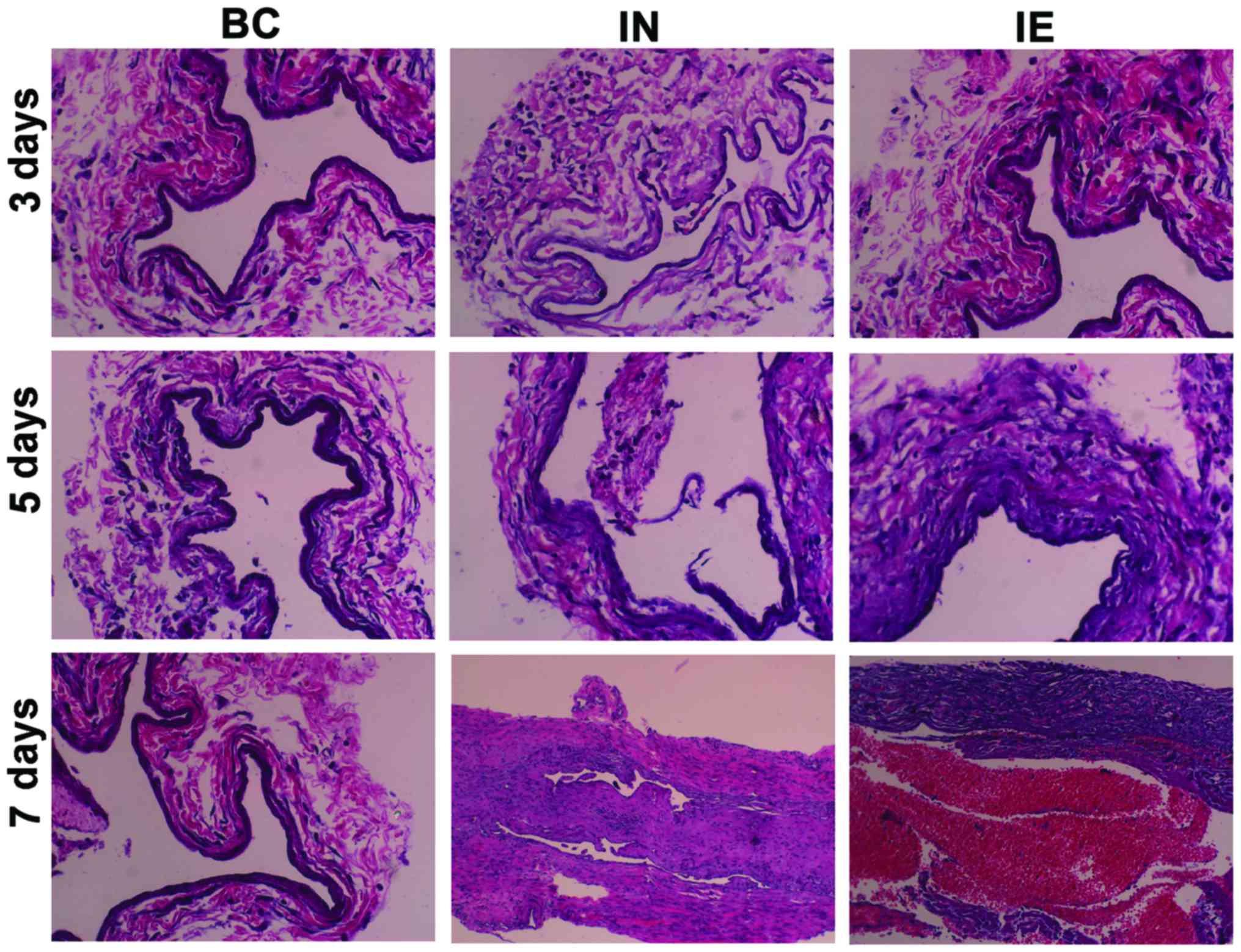
Microscopic examination results of
H&E staining in various experimental groups
In the blank group, no inflammatory cellular
infiltration occurred in the vascular wall, and the no
proliferation of fibrous tissue occurred around the vascular wall.
A little inflammatory cellular infiltration occurred in both the
indwelling needle group and the group with external application of
mirabilite, and mild proliferation of fibrous tissue occurred
around the vascular wall at 3 days. In both the indwelling needle
and the mirabilite group, we could observe substantial inflammatory
cellular infiltration in the vascular wall, proliferation of
fibrous tissue in the vascular wall, and a little white thrombosis
in the blood vessels at 5 days. In the indwelling needle group, we
observed substantial inflammatory cellular infiltration around the
vascular wall, significant fibrous tissue hyperplasia, and
substantial thrombosis in the blood vessels at 7 days. Compared
with the indwelling needle group, the group with external
application of mirabilite had slight inflammatory cellular
infiltration and thrombosis (Fig.
1).
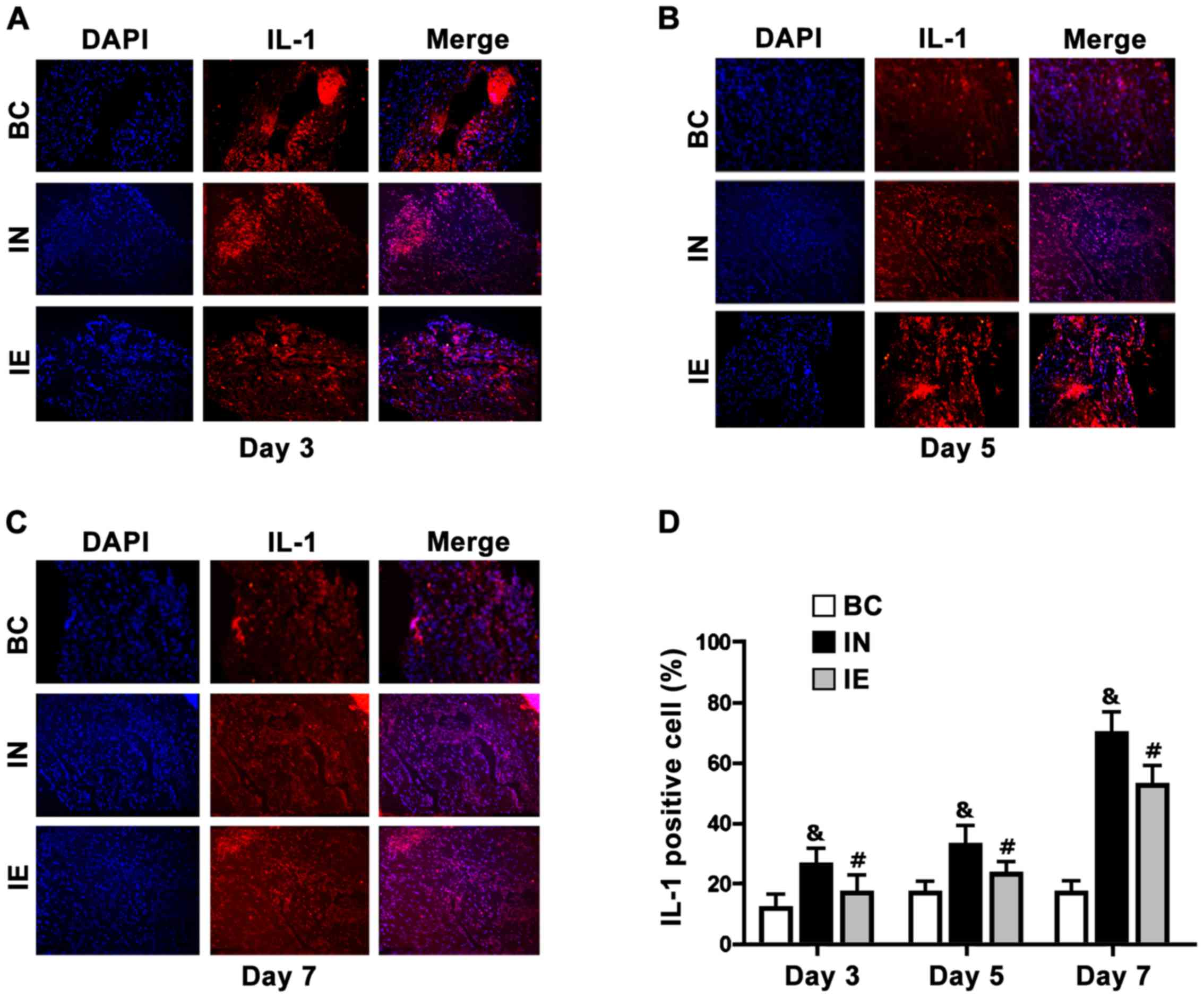
Expression levels of IL-1 in the
rabbit ear veins in various experimental groups
The detection results showed that with the increase
of the indwelling time of the intravenous indwelling needles, the
expression levels of IL-1 in the indwelling needle group and the
group with external application of mirabilite increased. The
expression levels in the indwelling needle and the group with
external application of mirabilite were higher than that in the
blank control group (P<0.05). The expression levels of IL-1 in
the group with external application of mirabilite were lower than
that in the indwelling needle group at 3, 5, and 7 days. The
differences were statistically significant (Fig. 2 and Table
I).
 | Table I.Percentages of IL-1 positive cells in
various experimental groups (mean ± SD). |
Table I.
Percentages of IL-1 positive cells in
various experimental groups (mean ± SD).
|
| n=24 |
|---|
|
|
|
|---|
| Groups | 3 days | 5 days | 7 days |
|---|
| Blank | 11.65±1.71 | 11.65±1.71 | 11.65±1.71 |
| Indwelling
needle |
18.22±2.56a,b |
33.68±1.62a,b |
73.49±7.73a,b |
| Mirabilite
intervention |
13.78±1.87a,b |
23.52±2.12a,b |
56.8±5.54a,b |
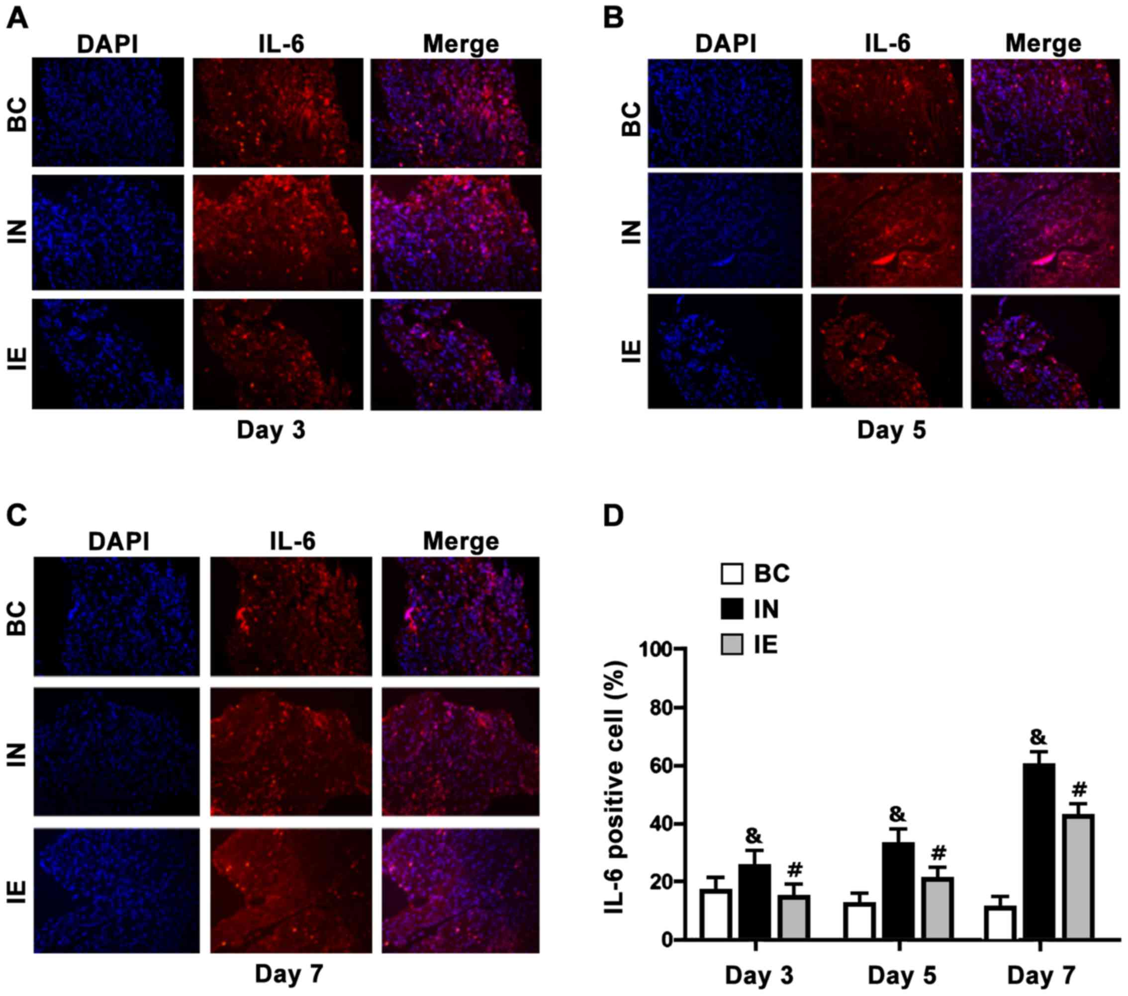
Expression of IL-6 in the rabbit ear
veins in various experimental groups
The detection results showed that with the increase
of the indwelling time of the intravenous indwelling needles, the
expression levels of IL-6 in both the indwelling needle group and
the group with external application of mirabilite increased. The
expression levels in both the indwelling needle group and the group
with external application of mirabilite were higher than that in
the blank control group at 3, 5, and 7 days. The expression of IL-6
in the group with external application of mirabilite was lower than
that in the indwelling needle group. The differences were
statistically significant (P<0.05) (Fig. 3 and Table
II).
| Table II.Percentages of IL-6 positive cells in
various experimental groups (mean ± SD) n=24. |
Table II.
Percentages of IL-6 positive cells in
various experimental groups (mean ± SD) n=24.
| Groups | 3 days | 5 days | 7 days |
|---|
| Blank control | 10.44±1.72 | 10.44±1.72 | 10.44±1.72 |
| Indwelling
needle |
13.87±1.13a,b |
31.17±1.61a,b | 80.1±2.7a,b |
| With external
application of mirabilite |
12.26±1.7a,b |
25.16±2.38a,b |
69.4±2.77a,b |
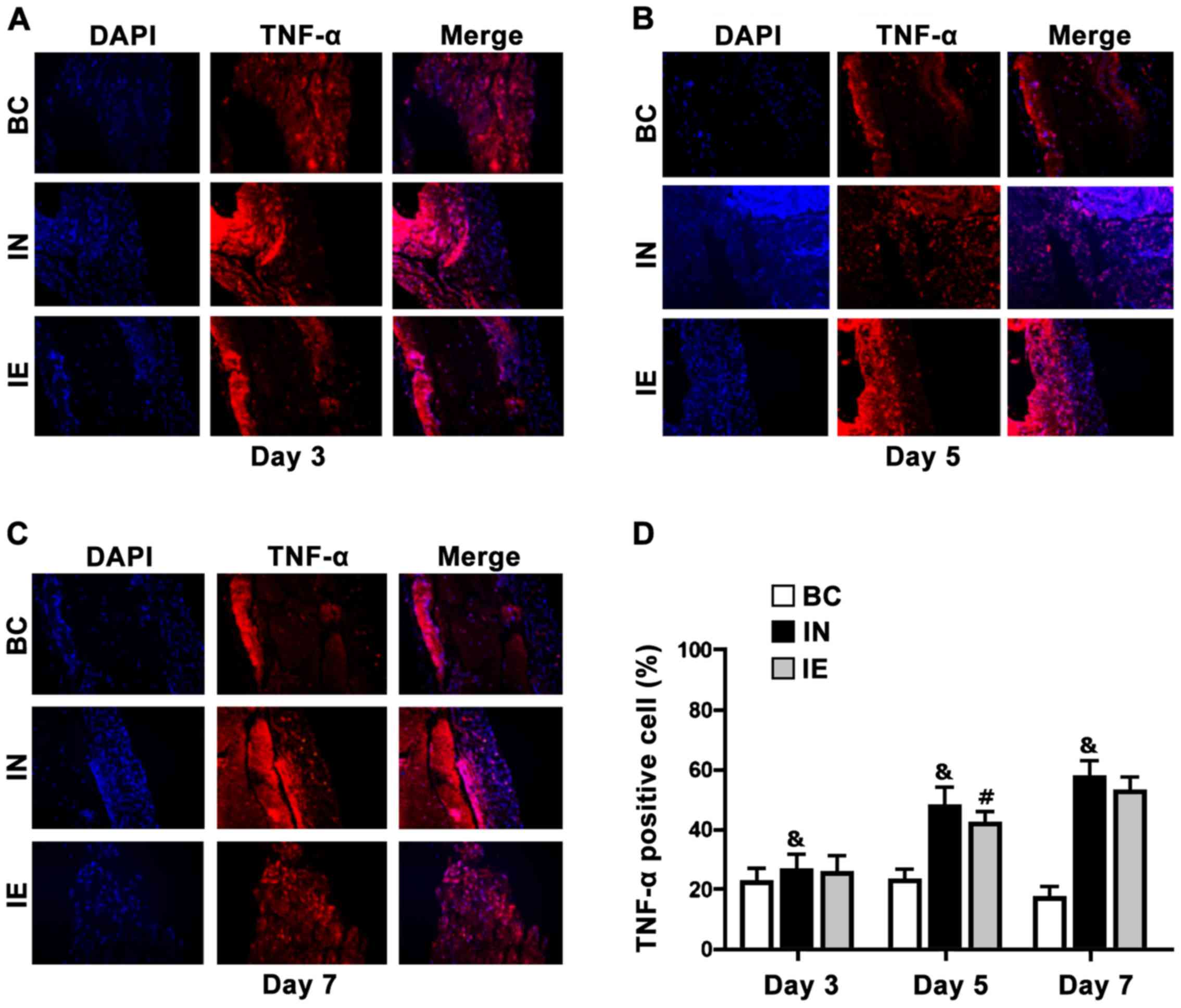
Expression of TNF-α in the rabbit ear
veins in various experimental groups
The detection results indicated that with the
increase of the indwelling time of the intravenous indwelling
needles, the expression levels of TNF-α in both the indwelling
needle group and the group with external application of mirabilite
increased. The expression levels in the indwelling needle group and
the group with external application of mirabilite were higher than
that in the blank control group (P<0.05). The expression of
TNF-α in the group with external application of mirabilite was
lower than that in the indwelling needle group. The differences
were statistically significant (P<0.05) (Fig. 4 and Table III).
| Table III.Percentages of the TNF-α positive
cells in various experimental groups (mean ± SD) n=24. |
Table III.
Percentages of the TNF-α positive
cells in various experimental groups (mean ± SD) n=24.
| Groups | 3 days | 5 days | 7 days |
|---|
| Blank control | 15.5±1.2 | 15.5±1.2 | 15.5±1.2 |
| Indwelling
needle |
19.28±1.41a |
37.22±1.78a |
57.14±3.53a |
| With external
application of mirabilite |
17.67±1.57a,b |
31.58±3.57a,b |
46.0±4.53a,b |
Discussion
Venous transfusion is an important approach to
clinical treatment. Intravenous indwelling needles have been
substituted for steel needles and become an important tool for
clinical intravenous therapy due to their advantages. With the
extensive application of indwelling needles in clinical treatment,
the occurrence of various complications severely perplexed the
clinical nursing personnel (16). In
the complications caused by indwelling needles, phlebitis is the
most common complication. Based on the clinical statistical data,
the incidence of phlebitis is as high as 80% (17). Phlebitis can be divided into
mechanical, chemical, and infectious by cause (18). The majority of the phlebitis cases
caused by intravenous indwelling needles are mechanical phlebitis
and acute aseptic inflammation. Mechanical phlebitis is an
inflammatory response of the venous wall irritated by vasospasm and
intimal injury arising from stimulation and friction of the
vascular wall by the indwelling needle catheter (19). According to relevant literature
(20–22), no phlebitis can be observed by naked
eye if the indwelling time of the intravenous indwelling needles
exceeds 3 days. Actually, inflammatory cellular infiltration occurs
on the inner vascular wall. Thus, the indwelling time of the
intravenous indwelling needles is considered as one of the
important factors that influence the occurrence of phlebitis. In
the experiment, significant inflammatory cellular infiltration
could be observed in the venous tissue at 3 days after indwelling
in the indwelling needle group, which is consistent with the
relevant research result. With the increase of the indwelling time,
the severity of phlebitis of the rabbit ear vein increases
significantly, suggesting that the prevention of the mechanical
phlebitis caused by indwelling needles should start as early as
possible.
With the in-depth research on the pathological
mechanism of phlebitis, it has been found that inflammatory
responses play an important role in occurrence and progression of
phlebitis (23). The mechanical
stimulation of the vascular wall by intravenous indwelling needles
can directly damage the vascular endothelial cells. The damaged
vascular endothelial cells would increase the vascular
permeability. The inflammatory cells effuse from the vascular wall
thus leading to inflammatory responses under the action of
chemotactic factors. Anti-inflammatory cytokines and
pro-inflammatory factors are mutually conditioning thus forming a
balance when no inflammatory responses occur in a normal organism
(24). Various causes lead to an
increase in pro-inflammatory factors. The balance would be damaged
thus leading to inflammatory responses when the anti-inflammatory
cytokines are insufficient to constrain the pro-inflammatory
factors. In the experiment, the detection indexes of IL-1, IL-6,
and TNF-α are the pro-inflammatory factors. The expression levels
of the pro-inflammatory factors of IL-1, IL-6, and TNF-α increase
abnormally when the indwelling time of the indwelling needles
exceeds 7 days. Under the action of the chemotaxis of the
pro-inflammatory factors, the inflammatory cells effuse to the
extravascular tissue via the damaged vascular wall thus leading to
inflammatory responses in the extravascular tissue.
IL-1 primarily comprises the inflammatory factors
secreted by such phagocytes as macrophages, and monocytes. It has
extensive biological activities. The biological activities mainly
include: Promoting proliferation of lymphocytes, hematopoietic
cells, and fibroblasts, promoting wound healing. In addition, IL-1
can promote adherence and infiltration of the leukocytes by
inducing the endothelial cells to express adhesion molecules. The
increase of the expression of IL-1 promotes the leukocytes to
migrate to the vascular endothelial cells after adherence to wall.
The leukocytes regularly release plasmin thus making the blood
vessel endothelium shift from anticoagulation to coagulation
accelerating and leading to development of phlebitis (25). In the experiment, the expression of
IL-1 increases significantly when the indwelling time of indwelling
needles exceeds 3 days. With the further increase of the indwelling
time, the expression level also increases.
IL-6 is a glycosylated protein comprising 183 amino
acids. IL-6 is produced by many types of cells including
macrophages, T cells and B cells. It can regulate the growth and
differentiation of many types of cells. It plays roles in
regulation of immune responses, acute phase response, and
hematopoiesis. It also plays an important role in anti-inflammation
immune responses. IL-6 plays a vital role in occurrence of
inflammation. In the experiment, the expression of IL-6 starts to
increase when the indwelling time of indwelling needles exceeds 3
days. The expression at 7 days is significantly higher than that at
3 days.
TNF-α is synthesized by activated macrophages,
monocytes, some T cells, and NK cells. It participates in such
pathological processes as inflammatory reactions, immune responses,
antitumor activity, endotoxic shock, arteriosclerosis, venous
thrombosis, and vasculitis. It also plays an important role in the
physiological processes of inflammation and immune responses. In
the experiment, the expression of TNF-α starts to increase at 3
days of indwelling of the indwelling needles. It reaches the peak
at 7 days.
Research demonstrates that inflammatory cells play
roles in triggering and strengthening thrombosis. IL-1 and TNF-α
can inhibit fibrinolysis. TNF-α can also inhibit the expression of
the thrombomodulin of endothelial cells thus making the endothelial
cells shift from an anticoagulation state to a pro-coagulation
state. Therefore, the increased expression levels of IL-1 and TNF-α
can not only promote progression of inflammations but also promote
thrombosis.
Mirabilite is a white granular mineral medicine
primarily hydrous sodium sulfate. In the traditional Chinese
medicine, mirabilite has many functions. Modern research indicates
external application of mirabilite plays roles in moisture
absorption, subsidence of swelling, clearing heat and removing
toxicity, and anti-inflammatory action. It is primarily used to
treat pancreatitis, haemorrhoids and phlebitis.
The research results indicate that IL-1, IL-6, and
TNF-α were highly expressed in the venous blood vessels of the
rabbit ears at 3 days after the indwelling needles were indwelt.
The expression increased with time. The expression increased
significantly at 7 days, suggesting that infiltration of
inflammatory cells occurred in the vascular tissue and the blood
vessel endothelium released IL-1, IL-6, and TNF-α thus leading to
local inflammatory responses under stimulation. The increase of the
expression of IL-1 and TNF-α can induce the normal cells to produce
IL-6. The increase of the expression of IL-6 further aggravates
inflammatory responses. Continuous stimulation from the indwelling
needle catheter makes the inflammatory factors to interact mutually
thus aggravating phlebitis. Some research has indicated that the
increase of inflammatory factors can lead to vasoconstriction and
spasm, cause inflammatory responses to the blood vessels, and give
rise to blood damage (15), which is
consistent with the above result.
Moreover, the research also demonstrates that
external application of mirabilite can effectively decrease the
expression of IL-1, IL-6, and TNF-α in the rabbit ear veins and
decrease the severity of inflammatory responses, which is
consistent with the result reported by Thao et al (26). Mirabilite may make the local blood
supply sufficient by stimulating the nervous reflex, improve the
blood circulation of the local tissue, accelerate local lymph
circulation, strengthen the phagocytic functions of the
reticuloendothelial cells, reduce local endothelial cell
infiltration, strengthen anti-inflammatory function, recover the
vascular functions, and alleviate phlebitis.
In conclusion, early application of mirabilite for
prevention of mechanical phlebitis arising from intravenous
indwelling needles is effective. Its specific action mechanism is
expected to be further studied.
Acknowledgements
This study was financially supported by The Nature
Science Foundation of Liaoning Provience (no. 201602283, for Yanyan
Lu).
References
|
1
|
Kovalenko OM, Mal'tsev DV, Kazmirchuk VIe
and Kovalenko AO: Studying of cytokine dynamics in injured persons
with severe burns for estimation of severity and prognosis. Klin
Khir. 2:49–53. 2014.(In Ukrainian).
|
|
2
|
Ray-Barruel G, Polit DF, Murfield JE and
Rickard CM: Infusion phlebitis assessment measures: A systematic
review. J Eval Clin Pract. 20:191–202. 2014. View Article : Google Scholar : PubMed/NCBI
|
|
3
|
Saunders H: Translating knowledge into
best practice care bundles: A pragmatic strategy for EBP
implementation via moving postprocedural pain management nursing
guidelines into clinical practice. J Clin Nurs. 24:2035–2051. 2015.
View Article : Google Scholar : PubMed/NCBI
|
|
4
|
Powell J, Tarnow KG and Perucca R: The
relationship between peripheral intravenous catheter indwell time
and the incidence of phlebitis. J Infus Nurs. 31:39–45. 2008.
View Article : Google Scholar : PubMed/NCBI
|
|
5
|
Grange-Prunier A, Couilliet D, Grange F
and Guillaume JC: Allergic contact dermatitis to the Comfeel
hydrocolloid dressing. Ann Dermatol Venereol. 129:725–727. 2002.(In
French). PubMed/NCBI
|
|
6
|
Kourbeti IS, Vakis AF, Ziakas P,
Karabetsos D, Potolidis E, Christou S and Samonis G: Infections in
patients undergoing craniotomy: Risk factors associated with
post-craniotomy meningitis. J Neurosurg. 122:1113–1119. 2015.
View Article : Google Scholar : PubMed/NCBI
|
|
7
|
China Pharmacopoeia Committee:
Pharmacopoeia of the People's Republic of China. China Medical
Science Press; Beijing: pp. 118–119. 2010
|
|
8
|
Wang Y, Zhang X and Li C: Applying hot
compresses with Rhubarb and mirabilite to reduce pancreatic leakage
occurrence in the treatment of severe acute pancreatitis. Iran
JPublic Health. 46:136–138. 2017.
|
|
9
|
Milutinović D, Simin D and Zec D: Risk
factor for phlebitis: A questionnaire study of nurses' perception.
Rev Lat Am Enfermagem. 23:677–684. 2015. View Article : Google Scholar : PubMed/NCBI
|
|
10
|
Abolfotouh MA, Salam M, Bani-Mustafa A,
White D and Balkhy HH: Prospective study of incidence and
predictors of peripheral intravenous catheter-induced
complications. Ther Clin Risk Manag. 10:993–1001. 2014. View Article : Google Scholar : PubMed/NCBI
|
|
11
|
Hegerova LT, Leal AD, Grendahl DC, Seisler
DK, Sorgatz KM, Anderson KJ, Hilger CR and Loprinzi CL: An analysis
of fosaprepitant-induced venous toxicity in patients receiving
highly emetogenic chemotherapy. Support Care Cancer. 23:55–59.
2015. View Article : Google Scholar : PubMed/NCBI
|
|
12
|
Mermel LA, Allon M, Bouza E, Craven DE,
Flynn P, O'Grady NP, Raad II, Rijnders BJ, Sherertz RJ and Warren
DK: Clinical practice guidelines for the diagnosis and management
of intravascular catheter-related infection: Update by the
Infectious Diseases Society of America. Clin Infect Dis. 49:1–45.
2009. View
Article : Google Scholar : PubMed/NCBI
|
|
13
|
Ward-Kavanagh LK, Lin WW, Šedý JR and Ware
CF: The TNF receptor superfamily in co-stimulating and
co-inhibitory responses. Immunity. 44:1005–1019. 2016. View Article : Google Scholar : PubMed/NCBI
|
|
14
|
Ng PC, Li K, Leung TF, Wong RP, Li G, Chui
KM, Wong E, Cheng FW and Fok TF: Early prediction of sepsis-induced
disseminated intravascular coagulation with interleukin-10,
interleukin-6, and RANTES in preterm infants. Clin Chem.
52:1181–1189. 2006. View Article : Google Scholar : PubMed/NCBI
|
|
15
|
Li WJ, Liu Y, Wang JJ, Zhang YL, Lai S,
Xia YL, Wang HX and Li HH: ‘Angiotensin II memory’ contributes to
the development of hypertension and vascular injury via activation
of NADPH oxidase. Life Sci. 149:18–24. 2016. View Article : Google Scholar : PubMed/NCBI
|
|
16
|
Rickard CM, McCann D, Munnings J and
McGrail MR: Routine resite of peripheral intravenous devices every
3 days did not reduce complications compared with clinically
indicated resite: A randomised controlled trial. BMC Med. 8:532010.
View Article : Google Scholar : PubMed/NCBI
|
|
17
|
Zingg W and Pittet D: Peripheral venous
catheters: An under-evaluated problem. Int J Antimicrob Agents.
34:38–42. 2009. View Article : Google Scholar : PubMed/NCBI
|
|
18
|
Karamanoglu A, Yumuk PF, Gumus M, Ekenel
M, Aliustaoglu M, Selimen D, Sengoz M and Turhal NS: Port needles:
Do they need to be removed as frequently in infusional
chemotherapy? J Infus Nurs. 26:239–242. 2003. View Article : Google Scholar : PubMed/NCBI
|
|
19
|
Falkowski A: Improving the PICC insertion
process. Nursing. 36:26–27. 2006. View Article : Google Scholar : PubMed/NCBI
|
|
20
|
Pandey D, Mehta S, Grover A and Goel N:
Indwelling catheterization in Caesarean section: Time to retire it!
J Clin Diagn Res. 9:01–04. 2015.
|
|
21
|
Mo XR, Luo XJ, Li CP, Pan XF and Zhou LL:
Effect ofmannitol injection by intravenous catheter on ear vein
endothelial cell apoptosis and venous thrombus in rabbits. Eur Rev
Med Pharmacol Sci. 19:491–497. 2015.PubMed/NCBI
|
|
22
|
Watanabe D, Fukunaga M and Yamamoto H:
Verification of the injection pressure reduction effect using the
novel indwelling needle for contrast-enhanced CT. Nihon Hoshasen
Gijutsu Gakkai Zasshi. 73:267–272. 2017. View Article : Google Scholar : PubMed/NCBI
|
|
23
|
dos Reis PE, Silveira RC, Vasques CI and
de Carvalho EC: Pharmacological interventionsto treat phlebitis:
Systematic review. J Infus Nurs. 32:74–79. 2009. View Article : Google Scholar : PubMed/NCBI
|
|
24
|
Duan F, Yu Y, Guan R, Xu Z, Liang H and
Hong L: Vitamin K2 induces mitochondria related apoptosis in human
bladder cancer cells via ROS and JNK/p38 MAPK signal pathways. PLoS
One. 11:e01618862016. View Article : Google Scholar : PubMed/NCBI
|
|
25
|
Park KH and Park WJ: Endothelial
dysfunction: Clinical implications in cardiovascular disease and
therapeutic approaches. J Korean Med Sci. 30:1213–1225. 2015.
View Article : Google Scholar : PubMed/NCBI
|
|
26
|
Thao NP, Luyen BT, Koo JE, Kim S, Koh YS,
Thanh NV, Cuong NX, Kiem PV, Minh CV and Kim YH: In vitro
anti-inflammatory components isolated from the carnivorous plant
Nepenthes mirabilis ELour. F Rafarin. Pharm Biol.
54:588–594. 2016. View Article : Google Scholar : PubMed/NCBI
|