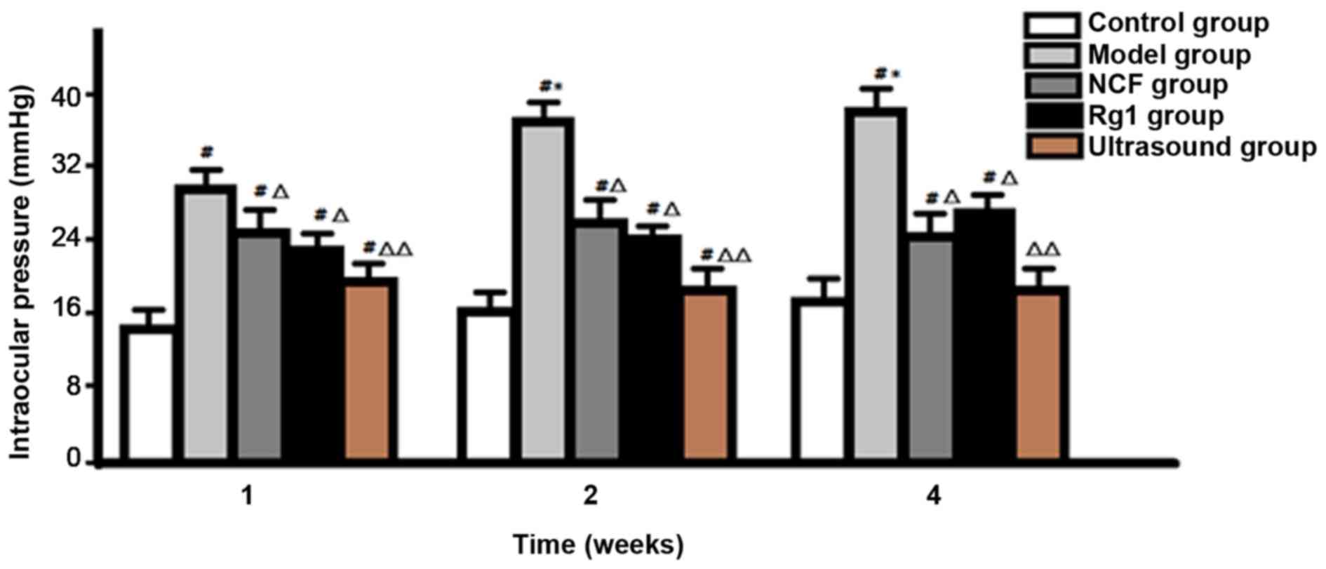
|
1
|
Quigley HA: Understanding Glaucomatous
Optic Neuropathy: The synergy between clinical observation and
investigation. Annu Rev Vis Sci. 2:235–254. 2016. View Article : Google Scholar : PubMed/NCBI
|
|
2
|
Chen X and Zhao Y: Diagnostic performance
of isolated-check visual evoked potential versus retinal ganglion
cell-inner plexiform layer analysis in early primary open-angle
glaucoma. BMC Ophthalmol. 17:772017. View Article : Google Scholar : PubMed/NCBI
|
|
3
|
Oddone F, Roberti G, Micera A, Busanello
A, Bonini S, Quaranta L, Agnifili L and Manni G: Exploring serum
levels of brain derived neurotrophic factor and nerve growth factor
across glaucoma stages. PLoS One. 12:e01685652017. View Article : Google Scholar : PubMed/NCBI
|
|
4
|
Mesentier-Louro LA, De Nicolò S, Rosso P,
De Vitis LA, Castoldi V, Leocani L, Mendez-Otero R, Santiago MF,
Tirassa P, Rama P, et al: Time-dependent nerve growth factor
signaling changes in the rat retina during optic nerve
crush-induced degeneration of retinal ganglion cells. Int J Mol
Sci. 18:E982017. View Article : Google Scholar : PubMed/NCBI
|
|
5
|
Hussein F, Antonescu C and Karshafian R:
Ultrasound and microbubble induced release from intracellular
compartments. BMC Biotechnol. 17:452017. View Article : Google Scholar : PubMed/NCBI
|
|
6
|
Luo H, Huang WX, Yang C, Zhao JQ, Liu S,
Xu YS and Liu CW: Therapeutic efficacy and mechanism of action of
ginsenoside Rg1 in treating acute hepatic failure in mice. Zhonghua
Gan Zang Bing Za Zhi. 25:217–222. 2017.(In Chinese). PubMed/NCBI
|
|
7
|
Garcia TB, Hollborn M and Bringmann A:
Expression and signaling of NGF in the healthy and injured retina.
Cytokine Growth Factor Rev. 34:43–57. 2017. View Article : Google Scholar : PubMed/NCBI
|
|
8
|
Kimura A, Namekata K, Guo X, Harada C and
Harada T: Neuroprotection, growth factors and BDNF-TrkB signalling
in retinal degeneration. Int J Mol Sci. 17:E15842016. View Article : Google Scholar : PubMed/NCBI
|
|
9
|
Sun ZG, Chen LP, Wang FW, Xu CY and Geng
M: Protective effects of ginsenoside Rg1 against hydrogen
peroxide-induced injury in human neuroblastoma cells. Neural Regen
Res. 11:1159–1164. 2016. View Article : Google Scholar : PubMed/NCBI
|
|
10
|
Huang L, Liu LF, Liu J, Dou L, Wang GY,
Liu XQ and Yuan QL: Ginsenoside Rg1 protects against
neurodegeneration by inducing neurite outgrowth in cultured
hippocampal neurons. Neural Regen Res. 11:319–325. 2016. View Article : Google Scholar : PubMed/NCBI
|
|
11
|
Huo DS, Zhang M, Cai ZP, Dong CX, Wang H
and Yang ZJ: The role of nerve growth factor in ginsenoside
Rg1-induced regeneration of injured rat sciatic nerve. J Toxicol
Environ Health A. 78:1328–1337. 2015. View Article : Google Scholar : PubMed/NCBI
|
|
12
|
Wang B, He L, Cui B and Lv H: Protection
of ginsenoside Rg1 on central nerve cell damage and the influence
on neuron apoptosis. Pak J Pharm Sci. 27 Suppl 6:2035–2040.
2014.PubMed/NCBI
|
|
13
|
Li YB, Wang Y, Tang JP, Chen D and Wang
SL: Neuroprotective effects of ginsenoside Rg1-induced neural stem
cell transplantation on hypoxic-ischemic encephalopathy. Neural
Regen Res. 10:753–759. 2015. View Article : Google Scholar : PubMed/NCBI
|
|
14
|
Guo X, Guo S, Pan L, Ruan L, Gu Y and Lai
J: Anti-microRNA-21/221 and microRNA-199a transfected by ultrasound
microbubbles induces the apoptosis of human hepatoma HepG2 cells.
Oncol Lett. 13:3669–3675. 2017.PubMed/NCBI
|
|
15
|
Luo J, Zhou X, Diao L and Wang Z:
Experimental research on wild-type p53 plasmid transfected into
retinoblastoma cells and tissues using an ultrasound microbubble
intensifier. J Int Med Res. 38:1005–1015. 2010. View Article : Google Scholar : PubMed/NCBI
|
|
16
|
Rokicki W, Zalejska-Fiolka J,
Pojda-Wilczek D, Hampel A, Majewski W, Ogultekin S and
Mrukwa-Kominek E: Differences in serum oxidative status between
glaucomatous and nonglaucomatous cataract patients. BMC Ophthalmol.
17:132017. View Article : Google Scholar : PubMed/NCBI
|
|
17
|
Panchal SS, Patidar RK, Jha AB, Allam AA,
Ajarem J and Butani SB: Anti-inflammatory and antioxidative stress
effects of oryzanol in glaucomatous rabbits. J Ophthalmol.
2017:14687162017. View Article : Google Scholar : PubMed/NCBI
|
|
18
|
Mumcu UY, Kocer I, Ates O and Alp HH:
Decreased paraoxonase1 activity and increased malondialdehyde and
oxidative DNA damage levels in primary open angle glaucoma. Int J
Ophthalmol. 9:1518–1520. 2016.PubMed/NCBI
|