Introduction
Cholangiocarcinoma is a malignant neoplasm that
originates from the epithelium of the biliary tree (1). Hilar cholangiocarcinoma (HCC) is the
most common type of cholangiocarcinoma, accounting for about 50–60%
(2,3). HCC arises from the right and left
hepatic ducts, the confluence of the right and left hepatic bile
ducts, and above cystic duct openings. At present, surgical
resection is still the only effective way to cure HCC (4). However, because of its location, prone
to local einvasion, adjacent structure infiltration and
metastatisation, surgical operation is very difficult and quite
often not radical. In addition, presenting with insidious onset and
lacking specific symptoms, most patients are diagnosed in advanced
stage and lost the opportunity of surgical treatment. Consequently,
the radical (R0) resection rate is only 10–20% (5). The survival rates of five years after
complete surgery range from 11 to 41% (6). So as a safe and effective palliative
therapy, the biliary stent placed by endoscopic retrograde
cholangiopancreatography (ERCP) may be the best choice for patients
with unresectable HCC (7). However,
violated by the tumor, the left and right hepatic bile ducts are
not communicating with each other, which leads to the difficulty of
superselective catheterization and the failure of ERCP. Correct and
reasonable selection of the target bile duct is the key to
superselective catheterization of bile duct successfully. At
present, the choice of target bile duct is mainly based on
preoperative CT, magnetic resonance cholangiopancreatography (MRCP)
and intraoperative cholangiography. But for complex HCC, it is
difficult to determine the target bile duct. So the ERCP is still
full of challenge.
Since the early 2000s, three-dimensional printing
(3DP) technologies have begun to be applied in medical domain
(8). In recent years, the
application of 3DP technologies is more and more extensive, such as
personalized surgical planning and guidance, medical research and
premedical education (8–11). Compared with CT, magnetic resonance
imaging (MRI) and other two-dimensional images, three-dimensional
visualization (3DV) and 3DP models are more intuitive and more
accurate in displaying anatomical and spatial structure of the
organs and have made great achievements. Their application value on
surgical treatment of HCC has been affirmed (12,13). But
the application of 3DV and 3DP technologies on ERCP is still in the
early stage and less reported in the literature.
The objective of this study was to assess the role
of ERCP for HCC based on 3DV and 3DP technologies at a center in
China.
Materials and methods
Patients
The present study was in accordance with the ethical
standards of the responsible committee on human experimentation
(institutional or regional) and with the Helsinki Declaration of
1975, as revised in 2000. The Institutional Ethics Committee of our
hospital approved this study, and written informed consent was
obtained from each participant before the study. 15 patients with
HCC who underwent ERCP were enrolled from January 2015 to January
2017, including 9 males and 6 females. CT and MRCP examination were
acquired and 3DV models were constructed. The real
three-dimensional models of the tumor and surrounding bile ducts
were created by the 3DP technologies. Before the operation, all
patients were informed of the indication and details of ERCP and
the risks and complications of the operation.
Instruments
CT: The data of 1 mm slice thickness were acquired
from the 64-MDCT provided by American GE Company. MRCP: The 1.5 T
Signa HDxt provided by GE Healthcare Bio-Sciences (Pittsburgh, PA,
USA) was utilized to obtain the 1.8 mm slice thickness.
3DV and 3DP technologies
Data of CT and MRCP were imported into 3D
visualization workstation that called Mimics Innovation Suite v17.0
software in DICOM format, where 3DV models of liver tumor and bile
ducts were reconstructed. The reconstructed models were exported as
Standard Template Library files. The topological correction,
decimation, Laplacian smoothing, and local smoothing were needed to
create a 3D model for 3DP. ProJet® 4500 3D printer (3D
Systems Corp., Circle Rock Hill, SC, USA) was used to manufacture
the models and the printing material was Visijet C4 Spectrum Core.
Bismuth-Corlette (BC) type was performed and diameter of the
dilated bile ducts was calculated. The bile duct with the largest
diameter and the widest drainage area was selected as the target
bile duct and compared with the actually selected ones in ERCP.
ERCP
Now it is still controversial whether unilateral or
bilateral drainage is better in the treatment of HCC. Unilateral
stenting appears to be adequate for relieving jaundice in most
unresectable cases. And it is technically easier and less expensive
than bilateral stenting, with reintervention for stent dysfunction
also being considerably easier (14). At the same time, in consideration of
the tolerance of surgery and postoperative complications, so
unilateral stenting was performed for all patients in our study. At
first, the drainage area and target bile duct were selected
preliminarily on the basis of preoperative abdominal CT, MRCP image
and intraoperative cholangiography. The bile duct was selectively
cannulated using a guide wire. If encountered difficulties, the
sphincterotomy was performed. The bile was extracted by a syringe
after successful selective intrahepatic bile duct cannulation as
much as possible to lower the pressure of the bile duct. The
dilating bougie was used to dilate the stenosis along the guide
wire. According to the patients' medical history, BC type, patients
and their families' wishes, the biliary stents were chosen
rationally. Finally, the metal or plastic stent was placed
successfully and nasobiliary duct was inserted and fixed
smoothly.
Results
3DV models were successfully manufactured for all
patients that clearly displayed all the relevant structures,
including intrahepatic bile ducts; size, shape, location of the
tumor; anatomical structure between them; location and degree of
the biliary stricture. Moreover, 3DV models could be amplified,
rotated, and hyalinized to clarify the anatomic character of these
structures with omnidirectional, multiple-angle, and multilevel
views. The models can localize correctly the tumor and surrounding
bile ducts, identify the extent of biliary stenosis and
cholangiectasis, contribute to determining the target bile duct and
make target bile duct be selected intuitively, concretely and
objectively. The location and direction of target bile duct can be
used to direct selective intrahepatic bile duct cannulation in
ERCP. Furthermore, 3DV models can avoid the influence of subjective
factors such as the operator's own experience, the level of imaging
ability to ERCP, and the success rate is higher. Demographics and
research variables are shown in the Table I. The target bile duct screened by
the models had a high concordance rate of 86.7% with that in ERCP.
The results were different in two patients. The target bile duct
identified by 3DV and 3DP models was the right posterior lobe bile
duct. However, selective cannulation of the right posterior lobe
bile duct failed, we had to try the left lateral lobe bile duct and
succeed. The diagnostic accuracy of BC type results by 3DV and 3DP
models was 93.3%.
 | Table I.Demographics and research
variables. |
Table I.
Demographics and research
variables.
| Variable | No. of patients
(n=15) |
|---|
| Female/Male | 6/9 |
| Age, years | 65.4±14.9 |
| BC type
(II/IIIA/IIIB/IV) |
|
|
Model | 2/4/4/5 |
| ERCP | 2/5/4/4 |
|
Concordance rate | 14/15 (93.3%) |
| Target bile duct |
|
|
Model | R, R, L, L, R, R, L,
R, R, R, L, R, R, L, L |
| ERCP | R, R, L, L, L, R, L,
R, R, L, L, R, R, L, L |
|
Concordance rate | 13/15 (86.7%) |
ERCP was performed successfully for all patients.
After ERCP, the malignant obstructive jaundice symptoms of 13
patients were alleviated or disappeared. Preoperative and
postoperative laboratory features including white blood cell (WBC),
neutrophil ratio (Neu%), alanine transaminase (ALT), aspartate
transaminase (AST), total bilirubin (TBIL) and direct bilirubin
(DBIL), are shown in Table II. The
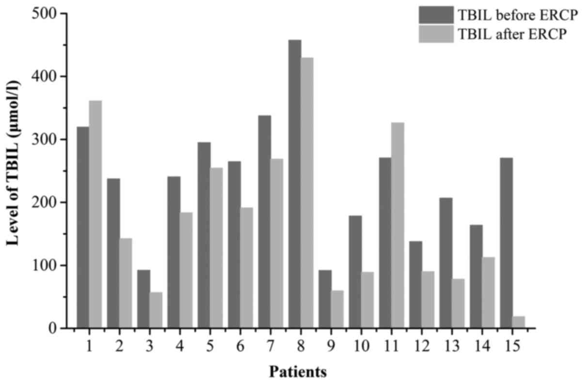
TBIL level of 2 cases did not decrease, or even increased. BC type
for the two patients was both Type IV and the cancer metastasize to
the liver and portal vein. Besides, cholangitis was induced by
ERCP. So their prognosis was poor, just like the findings of Vienne
and his colleagues. They thought complete resolution of jaundice
might not occur for patients with Bismuth IV after ERCP because it
was now thought that at least 50% of normal liver needs to be
drained to achieve complete resolution of jaundice (15). The change of TBIL for all patients
before and 3 days after operation was listed in Fig. 1. Cholangitis, postoperative
pancreatitis and hyperamylasemia occurred in 2,1 and 4 cases
respectively. And the patients were discharged in 6–20 days. All
patients were followed up for three months to one year and three
cases were died.
| Table II.Preoperative and postoperative
laboratory features. |
Table II.
Preoperative and postoperative
laboratory features.
| Variable | Preoperative | Postoperative day
1 | Postoperative day
3 |
|---|
| WBC | 5.8±2.0 | 9.8±2.0 | 7.1±2.8 |
| Neu% | 71.0±11.6 | 82.5±10.7 | 77.6±9.6 |
| ALT | 156.1±148.3 | 92.6±89.1 | 81.3 ±91.2 |
| AST | 135.3±96.5 | 95.3±51.1 | 78.6±82.5 |
| TBIL | 237.5±97.6 | 226.6±102.8 | 186.2±112.7 |
| DBIL | 196.6±82.0 | 194.1±88.8 | 158.0±104.6 |
The specific diagnosis and treatment process of a
typical case were reported as follows: A 75-year-old male was
admitted for yellow skin and mucosa for one week. And he had a
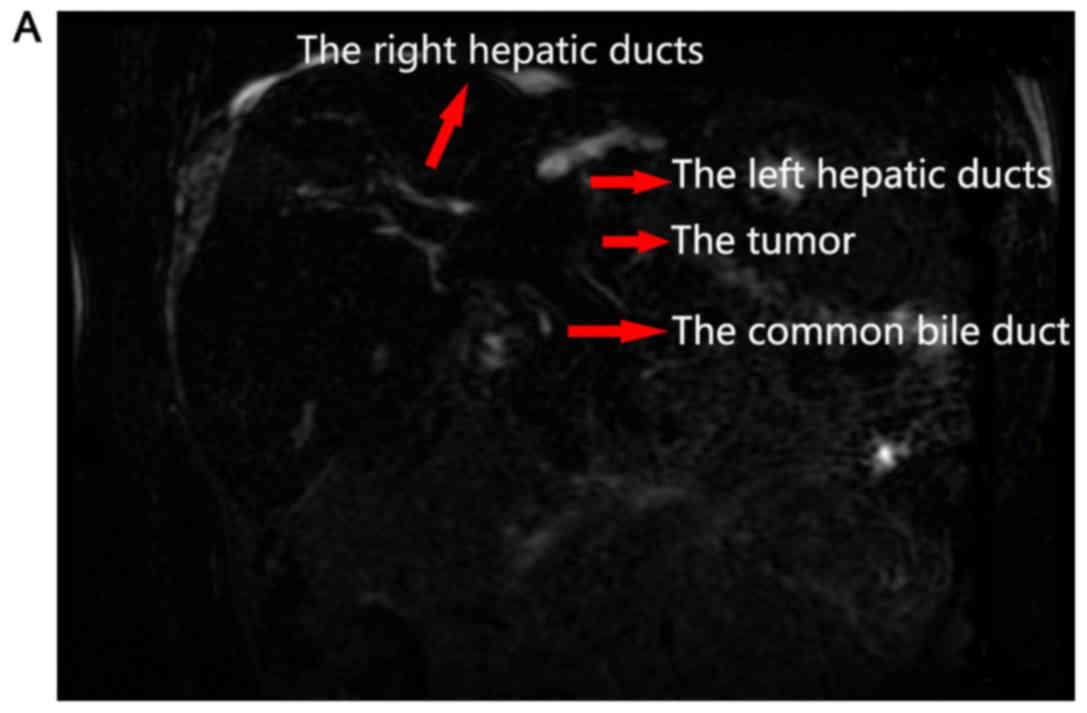
medical history of hepatitis B cirrhosis. CT scan and MRCP showed a
mass in hepatic portal and dilatation of intrahepatic bile ducts,
and the BC type was type II (Fig.
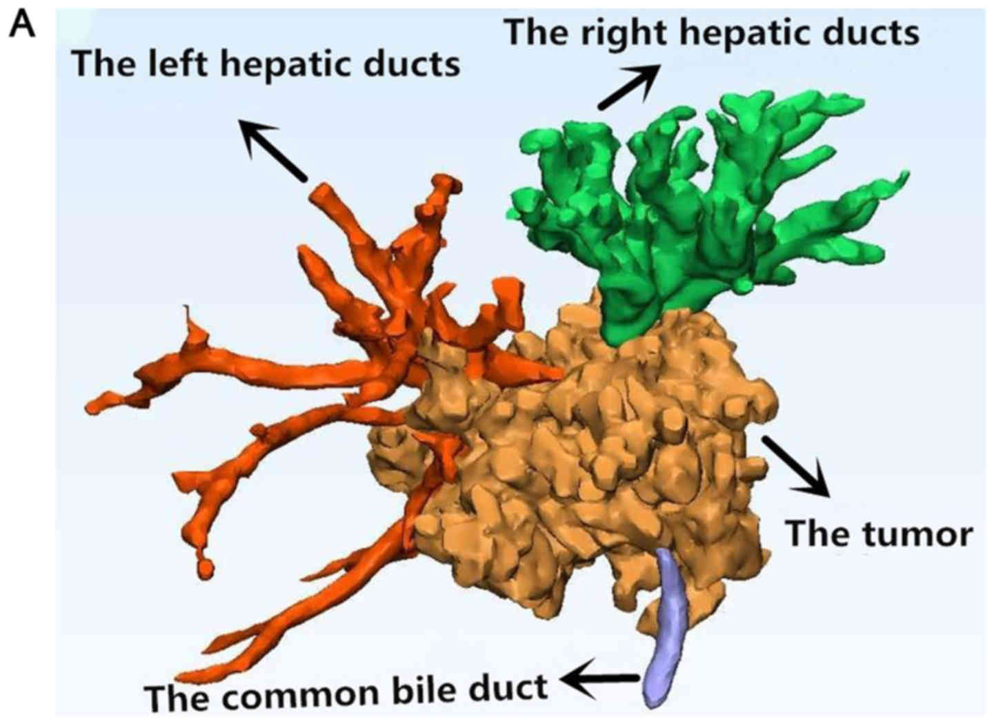
2); The 3DV and 3DP model were constructed successfully and the
BC type was type II (Fig. 3).
However, in this model of the patient, because of severe
dilatation, most of the left bile ducts fused together. But some
bile ducts can still be distinguished. The diameters of the dilated
bile ducts were calculated by Mimics Innovation Suite v17.0
software (Materialise, Leuven, Belgium),. The method was averaging
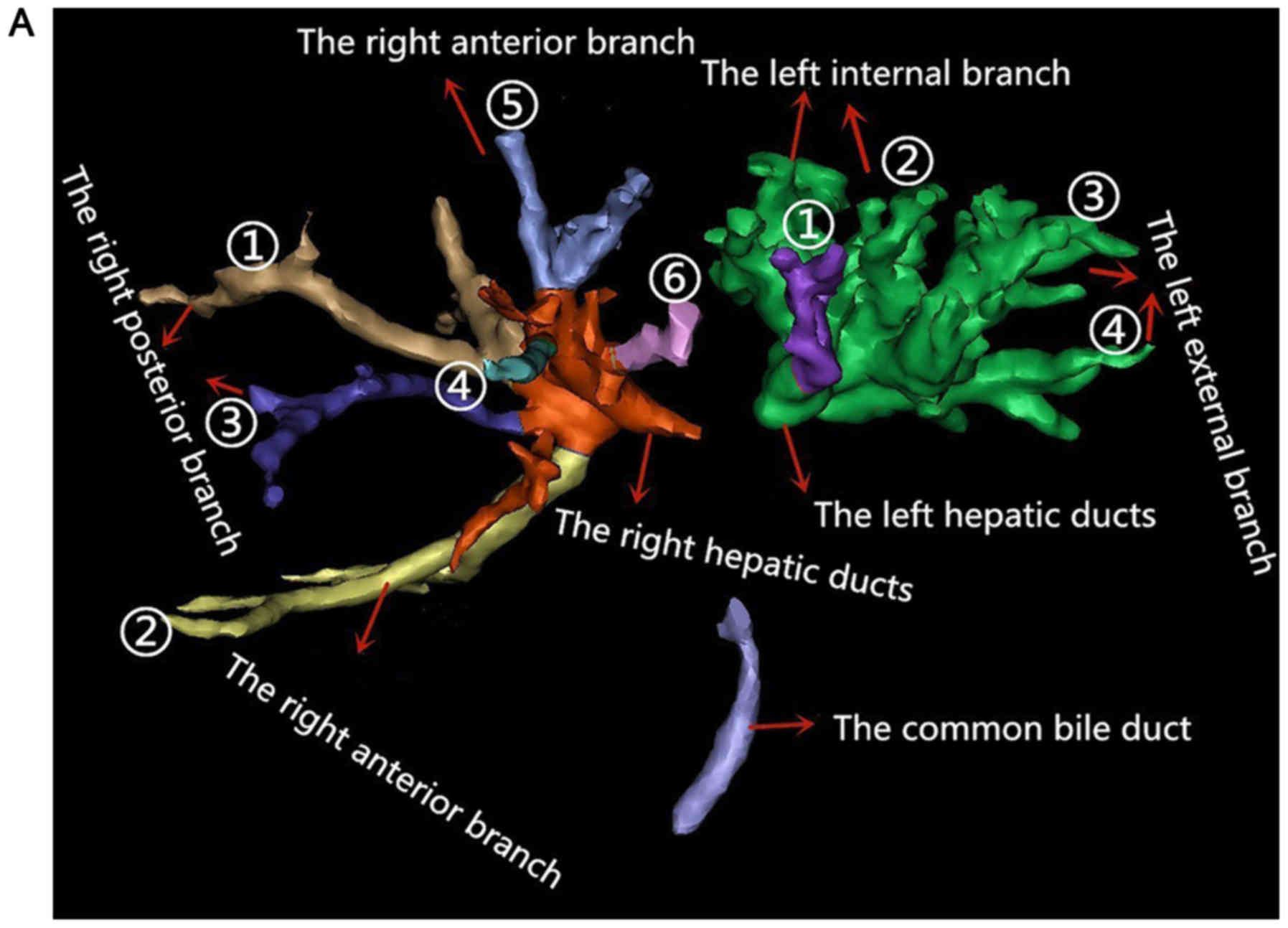
the testing values of repeated measurements. The left and right
hepatic bile ducts were marked with different colors and
corresponding sequence numbers (Fig.
4A). The diameters of hepatic bile ducts were as follows
(Table III).
| Table III.Diameter of bile ducts with different
colors and the corresponding sequence numbers. |
Table III.
Diameter of bile ducts with different
colors and the corresponding sequence numbers.
| A, Right hepatic duct
system |
|---|
|
|---|
| No. | Color | Diameter of bile duct
(mm) |
|---|
| 1 | Brown | 7.28
(8.45+5.95+6.74+7.97/4) |
| 2 | Yellow | 5.68
(6.09+5.49+5.44+5.69/4) |
| 3 | Blue | 3.77
(4.06+3.30+4.40+3.37/4) |
| 4 | Light blue | 3.72
(3.00+4.49+3.74+3.65/4) |
| 5 | Lilac | 6.77
(7.56+6.31+7.01+6.19/4) |
| 6 | Pink | 4.78
(4.93+4.47+4.50+5.24/4) |
|
| B, Left hepatic
duct system |
|
| No. | Color | Diameter of bile
duct (mm) |
|
| 1 | Purple | 7.01
(7.74+5.91+8.12+6.27/4) |
| 2 | Green | 9.12
(8.59+9.54+10.2+8.1/4) |
| 3 | Green | 13.57
(15.54+12.61+13.52+16.4/4) |
| 4 | Green | 12.00
(12.78+11.52+10.8+12.72/4) |
The final right target bile duct was the right brown
bile duct and the corresponding serial number was ①, namely, the
right posterior branch bile duct. And the final left target bile
duct was ①, namely the left lateral branch. In ERCP, the bile duct
of the right lobe (Fig. 4B) was the
actual selected bile duct. The double-wire guided technique was
applied. Along the guide wire, the nasobiliary duct was placed to
the right lobe of the liver. The 3DV model was consistent with
intraoperative condition in ERCP, and the selected bile duct was
the same. After operation, the patient's symptoms were
significantly relieved without complications and he was discharged
on postoperative day 14, without any complications.
Discussion
HCC is a malignant tumour with high mortality. BC
type can accurately delineate the anatomic location of tumor and
the degree of invasion and is the most widely used preoperative
reference system for predicting resectability and defining surgical
strategy. And it also contributes to choose reasonable drainage way
in endoscopic therapy and evaluating therapeutic effect. Since
proposed by Bismuth and Corlette in 1975 (16), the method is still widely used today.
The majority of patients with advanced HCC are inappropriate for
surgical resection because of the tumor location in the hepatic
hilum and adjacent areas, advanced tumor stage, or comorbidities.
Additionally, some people are aged, bad general condition and seem
hardly to tolerate surgical operation. Therefore, these patients
often have a poor prognosis in terms of survival and quality of
life. So as a nonsurgical and palliative treatment, biliary stent
placed by ERCP may be the best choice for these people to relieve
malignant obstructive jaundice and improve hepatic function, raise
living quality and prolong survival time. This kind of treatment
corresponds to the normal physiological characteristics. And
compared with surgery, it is less invasive and has fewer
postoperative complications.
Since 3DV and 3DP technologies are applied to the
clinical medicine, they have been widely used in oral and
maxillofacial surgery and orthopedics (17–19). 3D
visualization and printing can successfully show the anatomic
relationship between tissue organs and surrounding structures. 3DV
models could be amplified, rotated, and hyalinized to clarify the
anatomic character of tissue and organ structure with
omnidirectional, multiple-angle, and multilevel views. And 3DV
models can also be employed to accurately calculate the size and
volume of a liver tumor, the diameter and length of blood vessels
and bile ducts and the residual liver volume. These functions are
also of important clinical significance for preoperative planning
and guiding the actual surgical operations. However, compared with
3DP models, there is a certain sense of spatial distance in 3DV
models. 3DP models are constructed based on 3DV images and can
foresee anatomic structure at surgery truly from multiple
dimensions before operation. In addition, 3DP models can be brought
into the operation room, provide real-time navigation intuitively
for key steps in operation and identify and locate key parts
quickly. At present, the research and application of these
technologies have also made significant achievements in liver
surgery, such as liver transplantation, laparoscopic liver
resection, hepatectomy for hepatolithiasis, hepatic hydatid disease
(20–27). There also are researches on surgical
treatment of HCC. Zeng and colleagues (12) applied 3DV and 3DP technologies to the
surgery of HCC and found they played an important role in the
accurate preoperative diagnosis, the establishment of the operation
plan, precise operation in surgery and improvement of the success
rate of surgery. With the help of these technologies, Oshiro Y and
Ohkohchi N performed successfully left hepatic lobectomy and
extrahepatic bile duct resection for a HCC patient (28).
But for the clinical application of 3DV and 3DP
techniques for ERCP in patients with HCC, there were no reports and
literature of the relevant studies. In our study, we discover that
3DV and 3DP technologies play a significant role in the successful
implementation of ERCP in patients with HCC. This article mainly
carries on the elaboration from the following three aspects.
The role of 3DV and 3DP technologies in the
selection of target bile duct: Correct and reasonable selection of
the target bile duct are important to the successful selective
intrahepatic bile duct cannulation in ERCP. There are two factors
that determine the target bile duct: The dilatation of bile duct is
distinct and the drainage range is extensive. In the present study,
15 patients had a high consistency rate of 86.7% on target bile
duct selected by the 3DV models and ERCP respectively. Preoperative
3D reconstruction can clearly show the three-dimensional anatomy of
the tumor, dilated bile duct and its anatomical relationship with
the tumor (Fig. 3) (29,30).
With the help of Mimics Innovation Suite v17.0 software
(Materialise, Leuven, Belgium), the diameter of the proximal
dilated bile ducts could be calculated directly and accurately, and
the bile duct with the largest diameter and the most extensive
drainage was selected as the target bile duct. The target bile duct
is ordinarily selected based on CT, MRCP and intraoperative
cholangiography and other two-dimensional images, which can not
exhibit the spatial structure of the bile duct, causing the
specific direction of the guide wire in superselective
catheterization of bile duct is not completely clear, and even a
blind insertion. So the success rate of operation is closely
related to the operator's experience and the ability of
interpreting the images. However, compared to 2D images, 3DV and
3DP models can show the biliary tree more intuitively, the width
and direction of target bile duct more clearly, and can rotate by
360 degree and avoid the influence of subjective factors on
ERCP.
The role of 3DV and 3DP technologies in guiding the
selection and placement of a biliary stent: The location of
drainage in patients with HCC can be selected as the left hepatic
duct system, the right hepatic duct system or both. The key
determinant is the site of the bile duct stricture. BC type is very
important for the rational selection of the drainage site and the
judgement of curative effect in endoscopic stent placement. Our
study discovers that BC classification classified by the 3DV and
3DP models is more accurate, the 3DV and 3DP models can demonstrate
the location of bile duct invaded by the tumor more accurately than
CT and MRCP and can be used to guide the selection and placement of
a biliary stent.
3DV and 3DP technologies can reduce the use of
contrast media: Contrast agents are often needed to outline the
stricture in ERCP. However, contrast media injection into
intrahepatic ducts without adequate drainage easily leads to
uncontrolled cholangitis and poor prognosis. The rate of
cholangitis after ERCP was 1–5% (31–33). And
what's worse, invaded by the tumor, intrahepatic biliary drainage
is poor in HCC patients. To decrease the incidence of this
complication, some studies have explored the usefulness of CT or MR
cholangiography (MRC) prior to stenting (34,35).
Once the bile duct tree is delineated by CT or MRC, selective
guidewire cannulation and stenting of the targeted bile duct can be
accomplished without contrast injection. Compared with CT/MRI, 3DV
and 3DP models are more intuitive and more accurate (36). Consequently, they can reduce the use
of contrast media. And we can speculate that 3DV and 3DP models can
decrease the incidence of cholangitis. But further case control
studies are needed to verify the hypothesis.
In conclusion, compared with 2D images, 3DV and 3DP
models be amplified, rotated, and hyalinized to clarify the
anatomic character of the tumor and its relationship with the
surrounding bile ducts with omnidirectional, multiple-angle, and
multilevel views. 3DV and 3DP technologies can help to determine
the target bile duct, assist in selective cannulation, guide the
selection and placement of biliary stent in ERCP, and reduce the
use of contrast media. But our findings require further
reverification, because the number of cases in this study is too
small. More case studies are needed to further validate the
conclusion.
Acknowledgements
The present study was supported by the scientific
and technological project of Hubei Province (grant no. 2013BKB013),
and Natural Science Foundation of Hubei Province in China (grant
no, 2011CHB025).
References
|
1
|
Razumilava N and Gores GJ:
Cholangiocarcinoma. Lancet. 383:2168–2179. 2014. View Article : Google Scholar : PubMed/NCBI
|
|
2
|
Blechacz B, Komuta M, Roskams T and Gores
GJ: Clinical diagnosis and staging of cholangiocarcinoma. Nat Rev
Gastroenterol Hepatol. 8:512–522. 2011. View Article : Google Scholar : PubMed/NCBI
|
|
3
|
Rizvi S and Gores GJ: Pathogenesis,
diagnosis, and management of cholangiocarcinoma. Gastroenterology.
145:1215–1229. 2013. View Article : Google Scholar : PubMed/NCBI
|
|
4
|
Paik WH, Loganathan N and Hwang JH:
Preoperative biliary drainage in hilar cholangiocarcinoma: When and
how. World J Gastrointest Endosc. 6:68–73. 2014. View Article : Google Scholar : PubMed/NCBI
|
|
5
|
Li H, Qin Y, Cui Y, Chen H, Hao X and Li
Q: Analysis of the surgical outcome and prognostic factors for
hilar cholangiocarcinoma: A Chinese experience. Dig Surg.
28:226–231. 2011. View Article : Google Scholar : PubMed/NCBI
|
|
6
|
Nagorney DM and Kendrick ML: Hepatic
resection in the treatment of hilar cholangiocarcinoma. Adv Surg.
40:159–171. 2006. View Article : Google Scholar : PubMed/NCBI
|
|
7
|
Gerges C, Schumacher B, Terheggen G and
Neuhaus H: Expandable metal stents for malignant hilar biliary
obstruction. Gastrointest Endosc Clin N Am. 21:481–497. 2011.
View Article : Google Scholar : PubMed/NCBI
|
|
8
|
Rengier F, Mehndiratta A, von
Tengg-Kobligk H, Zechmann CM, Unterhinninghfen R, Kauczor HU and
Giesel FL: 3D printing based on imaging data: Review of medical
applications. Int J Comput Assist Radiol Surg. 5:335–341. 2010.
View Article : Google Scholar : PubMed/NCBI
|
|
9
|
Gross BC, Erkal JL, Lockwood SY, Chen C
and Spence DM: Evaluation of 3D printing and its potential impact
on biotechnology and the chemical sciences. Anal Chem.
86:3240–3253. 2014. View Article : Google Scholar : PubMed/NCBI
|
|
10
|
Michalski MH and Ross JS: The shape of
things to come: 3D printing in medicine. JAMA. 312:2213–2214. 2014.
View Article : Google Scholar : PubMed/NCBI
|
|
11
|
Murphy SV and Atala A: 3D bioprinting of
tissues and organs. Nat Biotechnol. 32:773–785. 2014. View Article : Google Scholar : PubMed/NCBI
|
|
12
|
Zeng N, Fang CH, Fan YF, Yang J, Xiang N,
Zhu W, Liu J, Cai W and Mo ZK: The construction of
three-dimensional visualization platform and its application in
diagnosis and treatment for hilar cholangiocarcinoma. Zhonghua Wai
Ke Za Zhi. 54:680–685. 2016.(In Chinese). PubMed/NCBI
|
|
13
|
Ji GW, Zhu FP, Wang K, Jiao CY, Shao ZC
and Li XC: Clinical implications of biliary confluence pattern for
bismuth-corlette type IV hilar cholangiocarcinoma applied to
hemihepatectomy. J Gastrointest Surg. 21:666–675. 2017. View Article : Google Scholar : PubMed/NCBI
|
|
14
|
Yasuda I, Mukai T and Moriwaki H:
Unilateral versus bilateral endoscopic biliary stenting for
malignant hilar biliary strictures. Dig Endosc. 25 Suppl 2:S81–S85.
2013. View Article : Google Scholar
|
|
15
|
Vienne A, Hobeika E, Gouya H, Lapidus N,
Fritsch J, Choury AD, Chryssostalis A, Gaudric M, Pelletier G,
Buffet C, et al: Prediction of drainage effectiveness during
endoscopic stenting of malignant hilar strictures: The role of
liver volume assessment. Gastrointest endosc. 72:728–735. 2010.
View Article : Google Scholar : PubMed/NCBI
|
|
16
|
Bismuth H and Corlette MB: Intrahepatic
cholangioenteric anastomosis in carcinoma of the hilus of the
liver. Surg Gynecol Obstet. 140:170–178. 1975.PubMed/NCBI
|
|
17
|
Lee KY, Cho JW, Chang NY, Chae JM, Kang
KH, Kim SC and Cho JH: Accuracy of three-dimensional printing for
manufacturing replica teeth. Korean J Orthod. 45:217–225. 2015.
View Article : Google Scholar : PubMed/NCBI
|
|
18
|
Obregon F, Vaquette C, Ivanovski S,
Hutmache DW and Bertassoni LE: Three-dimensional bioprinting for
regenerative dentistry and craniofacial tissue engineering. J Dent
Res. 94 9 Suppl:143S–152S. 2015. View Article : Google Scholar : PubMed/NCBI
|
|
19
|
Wu XB, Wang JQ, Zhao CP, Sun X, Shi Y,
Zhang ZA, Li YN and Wang MY: Printed three-dimensional anatomic
templates for virtual preoperative planning before reconstruction
of old pelvic injuries: Initial results. Chin Med J (Engl).
128:477–482. 2015. View Article : Google Scholar : PubMed/NCBI
|
|
20
|
Ikegami T and Maehara Y: Transplantation:
3D printing of the liver in living donor liver transplantation. Nat
Rev Gastroenterol Hepatol. 10:697–698. 2013. View Article : Google Scholar : PubMed/NCBI
|
|
21
|
Zein NN, Hanouneh IA, Bishop PD, Samaan M,
Eghtesad B, Quintini C, Miller C, Yerian L and Klatte R:
Three-dimensional print of a liver for preoperative planning in
living donor liver transplantation. Liver Transpl. 19:1304–1310.
2013. View
Article : Google Scholar : PubMed/NCBI
|
|
22
|
Baimakhanov Z, Soyama A, Takatsuki M,
Hidaka M, Hirayama T, Kinoshita A, Natsuda K, Kuroki T and Eguchi
S: Preoperative simulation with a 3-dimensional printed solid model
for one-step reconstruction of multiple hepatic veins during living
donor liver transplantation. Liver Transpl. 21:266–268. 2015.
View Article : Google Scholar : PubMed/NCBI
|
|
23
|
Velayutham V, Fuks D, Nomi T, Kawaguchi Y
and Gayet B: 3D visualization reduces operating time when compared
to high-definition 2D in laparoscopic liver resection: A
case-matched study. Surg Endosc. 30:147–153. 2016. View Article : Google Scholar : PubMed/NCBI
|
|
24
|
Xie A, Fang C, Huang Y, Fan Y, Pan J and
Peng F: Application of three-dimensional reconstruction and visible
simulation technique in reoperation of hepatolithiasis. J
Gastroenterol Hepatol. 28:248–254. 2013. View Article : Google Scholar : PubMed/NCBI
|
|
25
|
He YB, Bai L, Aji T, Jiang Y, Zhao JM,
Zhang JH, Shao YM, Liu WY and Wen H: Application of 3D
reconstruction for surgical treatment of hepatic alveolar
echinococcosis. World J Gastroenterol. 21:10200–10207. 2015.
View Article : Google Scholar : PubMed/NCBI
|
|
26
|
Fang C: Minimally invasive digital
technology: A new edge tool for the diagnosis and treatment of
hepatolithiasis. Dig Med. 2:1–5. 2016. View Article : Google Scholar
|
|
27
|
Koganemaru M, Horiuchi H and Abe T:
Treatment of hypohepatia after transplantation of liver from a
living donor liver by transcatheter embolization, using a simulated
3-dimensional printing vascular model. Gastroenterology.
151:e11–e13. 2016. View Article : Google Scholar : PubMed/NCBI
|
|
28
|
Oshiro Y and Ohkohchi N: Three-dimensional
liver surgery simulation: Computer-assisted surgical planning with
three-dimensional simulation software and three-dimensional
printing. Tissue Eng Part A. 23:474–480. 2017. View Article : Google Scholar : PubMed/NCBI
|
|
29
|
Fang CH, Zhu W, Wang H, Xiang N, Fan Y,
Yang J, Wang X and Zhong Sz: A new approach for evaluating the
resectability of pancreatic and periampullary neoplasms.
Pancreatology. 12:364–371. 2012. View Article : Google Scholar : PubMed/NCBI
|
|
30
|
Fang CH, Liu J, Fan YF, Yang J, Xiang N
and Zeng N: Outcomes of hepatectomy for hepatolithiasis based on
3-dimensional reconstruction technique. J Am Coll Surg.
217:280–288. 2013. View Article : Google Scholar : PubMed/NCBI
|
|
31
|
Katsinelos P, Lazaraki G, Chatzimavroudis
G, Gkagkalis S, Vasiliadis I, Papaeuthimiou A, Terzoudis S,
Pilpilidis I, Zavos C and Kountouras J: Risk factors for
therapeutic ERCP-related complications: An analysis of 2,715 cases
performed by a single endoscopist. Ann Gastroenterol. 27:65–72.
2014.PubMed/NCBI
|
|
32
|
Iorgulescu A, Sandu I, Turcu F and
Iordache N: Post-ERCP acute pancreatitis and its risk factors. J
Med Life. 6:109–113. 2013.PubMed/NCBI
|
|
33
|
Kostrzewska M, Baniukiewicz A, Wroblewski
E, Laszewicz W, Swidnicka-Siergiejko A, Piotrowska-Staworko G,
Dlugosz JW and Dabrowski A: Complications of endoscopic retrograde
cholangiopancreatography (ERCP) and their risk factors. Adv Med
Sci. 56:6–12. 2011. View Article : Google Scholar : PubMed/NCBI
|
|
34
|
Hintze RE, Abou-Rebyeh H, Adler A,
Veltzke-Schlieker W, Felix R and Wiedenmann B: Magnetic resonance
cholangiopancreatography-guided unilateral endoscopic stent
placement for Klatskin tumors. Gastrointest Endosc. 53:40–46. 2001.
View Article : Google Scholar : PubMed/NCBI
|
|
35
|
Freeman ML and Overby C: Selective MRCP
and CT-targeted drainage of malignant hilar biliary obstruction
with self-expanding metallic stents. Gastrointest Endosc. 58:41–49.
2003. View Article : Google Scholar : PubMed/NCBI
|
|
36
|
Fang CH, Tao HS, Yang J, Fang ZS, Cai W,
Liu J and Fan YF: Impact of three-dimensional reconstruction
technique in the operation planning of centrally located
hepatocellular carcinoma. J Am Coll Surg. 220:28–37. 2015.
View Article : Google Scholar : PubMed/NCBI
|