Introduction
Neonatal necrotizing enterocolitis (NEC) is a severe
acquired disease that causes mucosal damage and necrosis in the
bowel. NEC most frequently occurs in premature and sick infants,
with an incidence of 1.5–7.2%, and its development is influenced by
many different factors (1). It has
been demonstrated that ischemic hypoxia leads to localized necrosis
or necrosis of the entire intestine and colon (2). Abdominal distension and hematochezia
are the primary symptoms of neonatal NEC, which is characterized by
necrosis of the intestinal mucosa and may also lead to systemic
sepsis and multi-organ failure (3).
Necrosis of the intestinal tract significantly compromises the
function of the digestive system and results in a loss of appetite
and weight loss (4). Additionally,
previous studies have indicated that inflammation and apoptosis are
associated with disease progression in patients with neonatal NEC
(5,6). Therefore, attenuating inflammation and
inhibiting the apoptosis of epithelial cells in the small intestine
are potential methods of treating neonatal NEC.
Inflammation is one of the most common
characteristics of NEC and serves a key role in the progression of
necrotic diseases (7). It has been
demonstrated that inflammation is a potential pathophysiological
mechanism that contributes to the development of neonatal NEC, as
it induces the excessive production of pro-inflammatory cytokines
(8). Chan et al (9) proposed that blocking LPS activation and
leukocyte infiltration may be a potential method of treating
lipopolysaccharide (LPS)/cluster of differentiation 14/toll like
receptor 4 (TLR4)-mediated inflammation. A previous review
systematically elaborated on the pathogenesis and therapeutic
implications of bacterial infection, intestinal barrier function
and inflammatory responses of immature enterocytes (2). Furthermore, a number of studies have
demonstrated that inhibiting intestinal inflammation contributes to
the improvement of neonatal NEC and have analyzed the potential of
factors involved in inflammatory signaling pathways as therapeutic
targets (10,11). It has previously been demonstrated
that berberine attenuates LPS-induced inflammation and
extracellular matrix accumulation by regulating the nuclear factor
(NF)-κB signaling pathway (12).
Therefore, the present study hypothesized that berberine may be
beneficial in the treatment of neonatal NEC and the
anti-inflammatory effect of berberine in a mouse model was
investigated.
Apoptosis of epithelial cells in the small intestine
is the most common characteristic observed in patients with NEC
(13). It has been reported that
intestinal epithelial apoptosis initiates gross bowel necrosis and
leads to anabrosis in an experimental rat model of NEC (14). In an experimental model of NEC,
epidermal growth factor (EGF) and the apoptosis regulator B-cell
lymphoma (Bcl)-2 reduced the apoptosis of intestinal epithelial
cells and protected the intestinal epithelium against NEC injury
(15). In addition, it has been
demonstrated that decreasing intestinal apoptosis improves neonatal
NEC in rats via the heparin-binding EGF-like growth factor
(16). Furthermore, berberine
attenuated ischemia/reperfusion-induced myocardial apoptosis by
activating 5′ adenosine monophosphate-activated protein kinase and
the phosphoinositide 3-kinase (PI3K)/protein kinase B (AKT)
signaling pathways in a diabetic rat model (17).
In the present study, the anti-inflammatory and
anti-apoptotic effects of berberine in the intestinal epithelium of
a neonatal NEC mouse model were investigated. The PI3K/AKT
signaling pathway in epithelial cells was investigated as the
potential mechanism by which berberine alleviates neonatal NEC. The
results revealed that treatment with berberine increased EGF
expression, the activity of epithelial cells and decreased the area
of infarction within the small intestine. These results suggest
that berberine may be a potential therapeutic agent for the
treatment of patients with NEC.
Materials and methods
Ethics statement
The present study was conducted in accordance with
the Guide for the Care and Use of Laboratory Animals by Renmin
Hospital of Wuhan University (Hubei, China) (18). All surgeries and experiments were
performed under anesthetic to minimize any pain caused to the
animal. The present study was approved by the Ethics Committee of
Renmin Hospital of Wuhan University.
Animals
A neonatal NEC mouse model was established following
a previously described protocol (19). A total of 40 postnatal 4-day-old male
C57BL/6JCnc mice (weight, 5–10 g; Beijing Vital River Laboratory
Animal Technology, Beijing, China) were used. All mice were housed
in a temperature and humidity controlled room at 25±1°C and 55±5%,
respectively, with an artificial 12 h light/dark cycle and they had
free access to food and water. To establish a neonatal NEC model,
mice were administered 150 mmol/l acetic acid by enema following
induction of anesthesia with sodium pentobarbital (40 mg/kg; China
National Pharmaceutical Group Corporation, Beijing, China) by
intraperitoneal injection. Mice were then separated into two groups
(15 mice/group) and received oral treatment of berberine (5 mg/ml)
or PBS (5 mg/ml) once a day for 10 days. The body weights and food
intake of experimental mice were measured prior to each injection.
All mice were sacrificed on day 10 for histological analysis.
Cell culture
Epithelial cells from the small intestine were
isolated from the 10 experimental mice, and cultured in Dulbecco's
modified Eagle's medium with 10% fetal bovine serum (both from
Gibco; Thermo Fisher Scientific, Inc., Waltham, MA, USA).
Epithelial cells were cultured in a 5% CO2 humidified
atmosphere at 37°C, following the attachment of the cells, passage
1 epithelial cells were treated with PI3K (5 mg/ml) (Promega
Corporation, Madison, WI, USA) and/or berberine (Xi'an Le Sen
Bio-Technology Co., Ltd., Xian, China) (5 mg/ml) for 24 h to
analyze the effects of PI3K on apoptosis and the expression of
apoptosis-related proteins.
ELISA assay
ELISA kits were used to determine serum levels of
myeloid differentiation protein-2 (MD-2; cat. no. MBS261609;
MyBioSource, Inc., San Diego, CA, USA), NF-κB (cat. no. ab176647),
tumor necrosis factor (TNF)-α (cat. no. ab100747), interleukin
(IL)-6 (cat. no. ab100712), chemokine (C-X-C motif) ligand 1
(Cxcl-1) (cat. no. ab216951) (all from Abcam, Cambridge, MA, USA)
and TLR-4 (cat. no. ABIN424269; antibodies-online Inc., Atlanta,
GA, USA). Procedures were conducted following the manufacturer's
protocol and the final results were measured at 450 nm using an
ELISA plate reader.
Western blot analysis
Epithelial cells were isolated from
berberine-treated mice, homogenized in a RIPA buffer (cat. no.
R0278; Sigma-Aldrich; Merck KGaA, Darmstadt, Germany) containing
protease-inhibitor and centrifuged at 3,000 × g at 4°C for 10 min,
and the protein concentration was measured by a BCA assay kit (cat.
no. 23225; Thermo Fisher Scientific, Inc.). Proteins (20 µg/lane)
were loaded for SDS-PAGE: The stacking gel concentration was 5% and
separating gel was 12%. The proteins were then transferred to a
PVDF membrane (EMD Millipore, Billerica, MA, USA). All the primary
antibodies except for anti-EGF were purchased from Cell Signaling
Technology, Inc. (Danvers, MA, USA), including caspase3 (cat. no.
9662), caspase9 (cat. no. 9502), survivin (cat. no. 2808), Cyto
c (cat. no. 11940), caspase8 (cat. no. 4790), FADD (cat. no.
2782), c-Myc (cat. no. 13987), P53 (cat. no. 2524), IFN-γ (cat. no.
8455), PI3K (cat. no. 4249), AKT (cat. no. 4691), pAKT (cat. no.
4060), pERK (cat. no. 3192), Bcl-2 (cat. no. 3498) (all 1:1,000)
and GAPDH (cat. no. 5174; 1:2,000). Primary antibodies directed
against EGF (cat. no. ab9695) was purchased from Abcam. Primary
antibodies were incubated with the membranes at 4°C for overnight
then blocked with 5% skimmed milk for 1 h at 37°C. Subsequently,
proteins were and incubated with corresponding horseradish
peroxidase (HRP)-conjugated secondary anti-rabbit (cat. no. 7071)
and anti-mouse (cat. no. 4410) antibodies (all 1:5,000; Cell
Signaling Technology, Inc.) for 30 min at room temperature.
Proteins were visualized using an enhanced chemiluminescence
detection system (Pierce ECL Plus Western Blotting Substrate;
Thermo Fisher Scientific, Inc.) and analyzed by ImageJ software
(version 1.47; National Institutes of Health, Bethesda, MD,
USA).
Flow cytometry
Apoptosis rates of the epithelial cells were
evaluated using an Annexin V-fluorescein isothiocyanate (FITC) and
propidium iodide (PI) apoptosis detection kit (BD Biosciences, San
Jose, CA, USA). Epithelial cells were isolated from berberine- or
PBS-treated mice and suspended with Annexin V-FITC and PI,
following the manufacturer's instructions. Fluorescence was
measured using a fluorescence activated cell sorting flow
cytometer.
Cell viability assay
Epithelial cells (2×10³ cells/well) were
isolated from berberine-treated mice and seeded in a 96 well
microplate; the epithelial cells were cultured until they reached
90% confluence. The culture medium was then removed, epithelial
cells were washed three times and subsequently incubated with 1%
TritonCleanCSS_6 X-100 (cat. no. 9002-93-1;
Sigma-Aldrich; Merck KGaA) for 30 min in room temperature. Lactate
dehydrogenase activity in the lysates was measured using a CytoTox
96® assay kit (Promega Corporation) and a microplate
reader at a wavelength of 490 nm. All procedures were conducted
following the manufacturer's instructions.
Analysis of physical activity by
indirect calorimetry
Indirect calorimetry was performed to evaluate the
physical activity of mice with neonatal NEC using the comprehensive
laboratory animal monitoring system (Columbus Instruments
International, Columbus, OH, USA). On day 10 after treatment with
berberine, the physical activity of mice was monitored every 30 min
for 24 h.
Immunohistochemistry
Fluorescein conjugated staining was performed to
analyze the apoptosis of small intestine cells and was conducted
using the in situ Cell Death Detection kit (Roche
Diagnostics, Basel, Switzerland), following the manufacturer's
instruction. Paraffin-embedded epithelial tissue sections
(4–5-µm-thick) were fixed with 3.7% formalin at room temperature
for 18–24 h and then the epitope retrieval was performed. The
slides were incubated in 10X Antigen Retrieval Reagent-Basic
(R&D Systems, Inc., Minneapolis, MN, USA) at 92–95°C for 2–10
min, cool in room temperature, rinsed with ddH2O and
washed with PBS. Paraffin sections were exposed to hydrogen
peroxide (3%) for 10–15 min in room temperature and were then
blocked using a regular blocking solution [StartingBlock (PBS)
Blocking Buffer; Thermo Fisher Scientific, Inc.] for 10–15 min at
37°C. In addition, sections were incubated with rabbit anti-mouse
primary antibodies directed against EGF (cat. no. ab9695) or Bcl-2
(cat. no. ab59348) (both 1:200; Abcam) for 2 h at 37°C and washed
three times with PBS. Subsequently, sections were incubated with
HRP-conjugated goat anti-rabbit secondary antibodies (cat. no.
ab6721, 1:1,000; Abcam) for 1 h at 37°C. The TUNEL assay was
performed using the TUNEL apoptosis detection kit (cat. no. L00297;
GenScript, Piscataway Township, NJ, USA) according to a
manufacturers instructions. Fluorescence Mounting medium was
purchased from Trevigen, Inc. (Gaithersburg, MD, USA) and images
were captured using nine fields of view. Finally, sections were
stained with hematoxylin and eosin stain at 4°C for 12 h. A Nikon
Eclipse TE2000-U Inverted Microscope (Nikon Corporation, Tokyo,
Japan) combined with Nikon optics was used to photograph all
sections.
Statistical analysis
All data are presented as the mean ± standard error
of the mean. Unpaired data was analyzed using a Student's t test.
Comparisons of data among multiple groups were analyzed by a
two-way analysis of variance followed by Tukey's post hoc test.
P<0.05 was considered to indicate a statistically significant
difference.
Results
Berberine decreases levels of
inflammatory cytokines in the peripheral blood of mice with
neonatal NEC
The anti-inflammatory effects of berberine in mice
with neonatal NEC were investigated. Levels of TLR-4 (Fig. 1A), MD-2 (Fig. 1B), TNF-α (Fig. 1C), NF-κB (Fig. 1D), IL-6 (Fig. 1E) and Cxcl-1 (Fig. 1F) were all significantly decreased in
the peripheral blood of mice treated with berberine compared with
those treated with PBS. These results indicate that berberine
significantly decreases inflammatory cytokine levels in the
peripheral blood of mice with neonatal NEC.
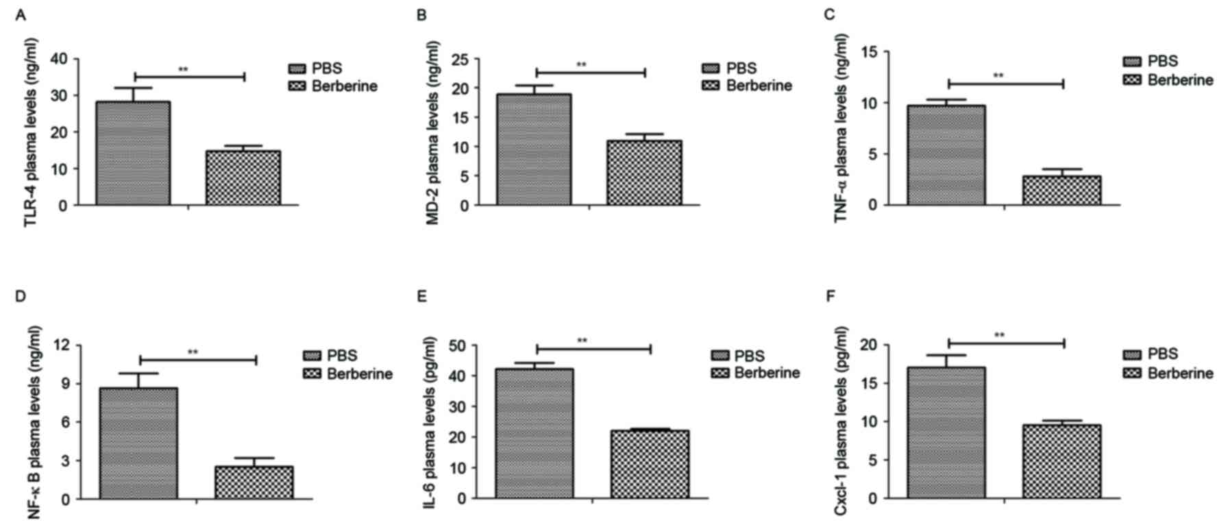 | Figure 1.Berberine significantly reduces
inflammatory cytokine levels in the peripheral blood of mice with
neonatal NEC. Mice with neonatal NEC were treated with either
berberine or PBS (as a control) and levels of (A) TLR-4, (B) MD-2,
(C) TNF-α, (D) NF-κB, (E) IL-6 and (F) Cxcl-1 in the peripheral
blood was determined by ELISA. **P<0.01. NEC, necrotizing
enterocolitis; TLR, toll like receptor; MD, myeloid differentiation
protein; TNF, tumor necrosis factor; NF nuclear factor; IL,
interleukin; Cxcl, (C-X-C motif) ligand. |
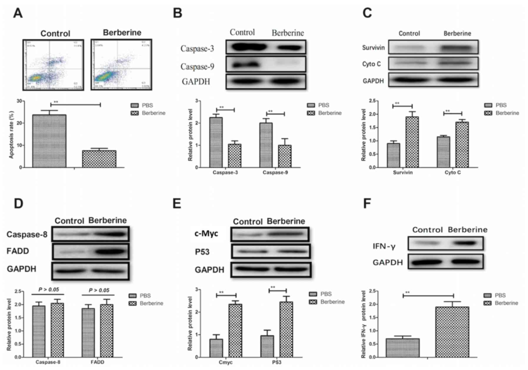
Berberine inhibits the apoptosis of
epithelial cells via the mitochondrial apoptosis pathway
The anti-apoptotic effects of berberine on
epithelial cells in a mouse model of NEC were analyzed. Berberine
significantly decreased the rate of apoptosis in epithelial cells
compared with PBS-treated mice (Fig.
2A). Furthermore, expression of the pro-apoptosis genes
caspase-3 and −9 were significantly decreased in epithelial cells
following the administration of berberine (Fig. 2B). It was also demonstrated that the
expression of survivin and cytochrome c were significantly
upregulated in the epithelial cells of berberine-treated mice
compared with PBS-treated mice; upregulated survivin and cytochrome
c may inhibit the apoptosis of epithelial cells (Fig. 2C). The expression of caspase-8 and
Fas-associated protein with death domain were not notably altered
in the epithelial cells of berberine-treated mice compared with
control mice (Fig. 2D). Furthermore,
the expression of anti-apoptotic genes, including proto-oncogene
c-Myc, tumor suppressor protein p53 and interferon (IFN)-γ were
significantly upregulated following treatment with berberine
(Fig. 2E and F). These results
indicate that berberine significantly inhibits the apoptosis of
epithelial cells by downregulating the expression of proteins in
the mitochondrial apoptosis pathway.
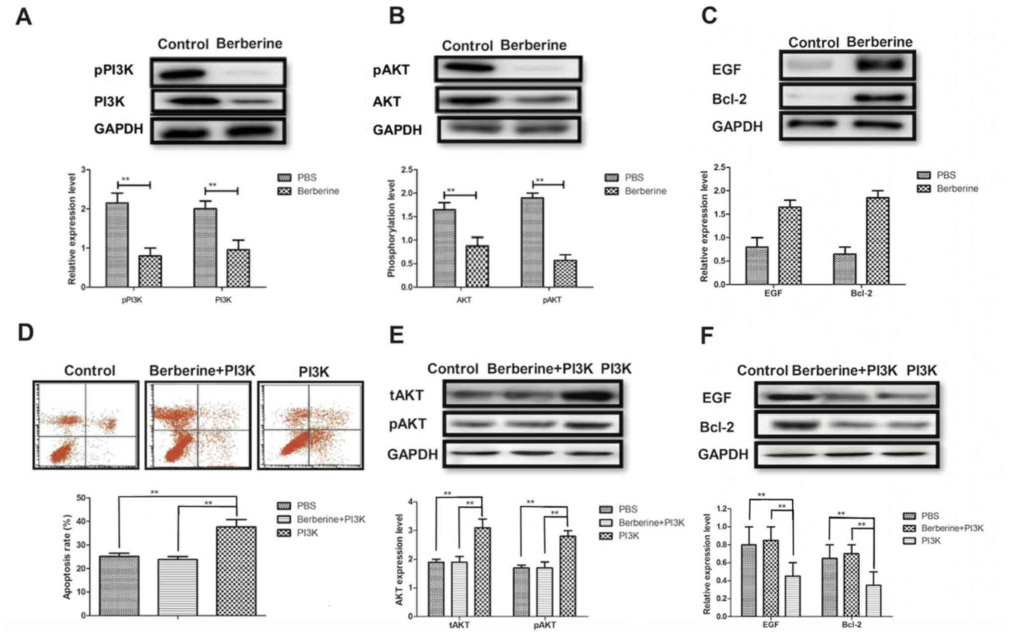
Berberine reduces the apoptosis of
epithelial cells by influencing the PI3K/AKT signaling pathway
The molecular mechanism of berberine-mediated
attenuation of epithelial cell apoptosis in a mouse model of NEC
was investigated. Berberine treatment significantly inhibited the
expression and phosphorylation of PI3K and AKT in epithelial cells
compared with controls (Fig. 3A and
B). Furthermore, EGF and Bcl-2 expression was markedly
upregulated in the epithelial cells of mice treated with berberine
(Fig. 3C). The results of in
vitro assays revealed that treatment with PI3K alone
significantly promoted the apoptosis of epithelial cells isolated
from mice with NEC compared with controls; however the apoptosis
rate of cells treated with berberine did not differ significantly
from that of the control (Fig. 3D).
Furthermore, treatment with PI3K alone promoted the expression and
phosphorylation of AKT; however, this effect was eliminated when
PI3K and berberine were administered together (Fig. 3E). PI3K treatment alone significantly
inhibited the expression of EGF and Bcl-2, and berberine treatment
significantly increased EGF and Bcl-2 expression in epithelial
cells compared to PI3K treatment (Fig.
3F). These results suggest that berberine reduces the apoptosis
of epithelial cells by mediating the PI3K/AKT signaling
pathway.
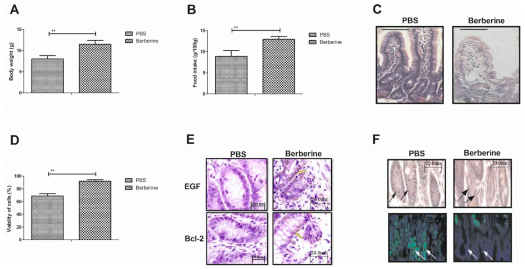
Berberine improves the physical
activity and physiological functions of mice with neonatal NEC
To analyze the efficacy of berberine in a mouse
model of neonatal NEC, the effect of berberine on the physical
activity and physiological functions of mice was investigated. The
body weight (Fig. 4A) and food
intake (Fig. 4B) of mice treated
with berberine were significantly increased compared with untreated
mice with neonatal NEC. Berberine treatment notably decreased the
area of infarction in the small intestine compared with the PBS
control group (Fig. 4C). The
viability of epithelial cells was also significantly increased in
mice treated with berberine compared with those treated with PBS
(Fig. 4D). In addition, treatment
with berberine markedly improved the expression of EGF and Bcl-2,
as determined by histological analysis (Fig. 4E). Immunohistochemistry indicated
that the apoptosis rate of epithelial cells was also notably
decreased following treatment with berberine compared with the
control (Fig. 4F). These results
indicate that berberine improves the physical activity and
physiological functions in a mouse model of neonatal NEC.
Discussion
NEC is an acquired disease that occurs in immature
small intestinal tissues and is caused by an insufficient blood
supply, dietary disorders and bacterial infection (20). The poor prognosis of individuals with
neonatal NEC contributes to the high mortality rate of premature
babies. At present, methods of treating NEC ameliorate necrosis in
the small intestine and aim to attenuate its destructive
progression (21). The pathogenesis
of NEC is not yet comprehensively understood; therefore the
treatment options available are not specifically targeted, meaning
that they are not particularly effective and that there is
potential for improvement. In the present study, the potential
mechanisms by which treatment with berberine had a positive effect
on NEC in a mouse model were explored. The anti-inflammatory and
anti-apoptotic effects of berberine in a mouse model of neonatal
NEC were determined. The results demonstrated that berberine
treatment significantly downregulated inflammation and apoptosis
while markedly improving the physical activity and physiological
functions of mice. Notably, the results indicated that berberine
treatment inhibits inflammation and apoptosis by inhibiting the
PI3K/AKT signaling pathway.
Inflammation serves an essential role in the
development of neonatal NEC (22).
Previous studies have reported that preventing inflammation is an
effective method of protecting against myocardial
ischemia-reperfusion injury and have highlighted it as a novel
technique of diagnosing and treating patients with cardiac disease
(23,24). Loubele et al (24) reported that the inhibition of
apoptosis and inflammation contributes to the rehabilitation of
myocardial ischemia/reperfusion injury. In addition, Wang et
al (25) demonstrated that
berberine acts as anti-arrhythmic drug in a rat type ІІ diabetic
myocardial infarction model by repressing the inward rectifier
potassium channel 2. Berberine also exhibits anti-inflammatory
effects on patients with acute coronary syndrome that have
experienced percutaneous coronary intervention (26). Additionally, berberine prevents
postsurgical intestinal adhesion and inhibits inflammation in a rat
model by inhibiting the transforming growth factor β-activated
kinase 1 (TAK1)/Jun N-terminal kinase and TAK1/NF-κB signaling
pathways (22). Therefore, berberine
may regulate inflammation in the progression of neonatal NEC via
the PI3K/AKT signaling pathway. These results indicate that
berberine significantly reduces levels of the inflammatory
cytokines TLR4, MD-2, TNF-α, NF-κB, IL-6 and Cxcl-1 in the
epithelial cells of a mouse model of neonatal NEC. These cytokines
contribute to the activity of epithelial cells and when they are
decreased, the apoptosis rate of the cells also decreases.
Berberine is able to reduce the apoptosis of epithelial cells in a
mouse model of neonatal NEC via this mechanism.
Neonatal NEC is characterized as the extensive
apoptosis and necrosis of the intestinal epithelium. It has been
reported that inhibitors of apoptosis can regulate intestinal
immunity and inflammation, and ameliorate bowel diseases (21). Additionally, inhibiting NF-κB
expression improves resolution of mucosal inflammation in the
intestinal epithelium (20).
Furthermore, it has been demonstrated that berberine inhibits
ischemia-induced apoptosis by activating the PI3K/PKB signaling
pathway (22). Therefore, various
strategies aiming at preventing or mitigating the extent of
apoptosis have been performed to try to protect the small intestine
against apoptosis or necrosis (23,27,28). In
the present study, it was hypothesized that berberine may regulate
the apoptosis of epithelial cells in a mouse model of neonatal NEC
by regulating the PI3K/AKT signaling pathway. The results confirm
this hypothesis and reveal that berberine suppresses the
mitochondrial apoptosis pathway in epithelial cells by inhibiting
the PI3K/AKT signaling pathway.
In conclusion, the present study investigated the
potential therapeutic effects of berberine and its ability to
protect the small intestine against necrosis. It was demonstrated
that berberine reduces epithelial cell apoptosis and tissue
necrosis by inhibiting inflammation and downregulating the PI3K/AKT
signaling pathway. The therapeutic effect of berberine during
recovery may be attributed to its upregulation of EGF and Bcl-2
expression. The results also indicated that physical activity and
physiological functions of mice with neonatal NEC were improved
following treatment with berberine, suggesting that berberine may
be a potential novel therapeutic agent for the treatment of
neonatal NEC.
References
|
1
|
Barak S, Riskin A, Kugelman A,
Abend-Weinger M, Chistyakov I and Bader D: Necrotizing
enterocolitis in a premature infant as the presenting symptom of
familial dysautonomia in the neonatal period: Case report and
review of the literature. Am J Perinatol. 22:353–355. 2005.
View Article : Google Scholar : PubMed/NCBI
|
|
2
|
Claud EC: Neonatal Necrotizing
enterocolitis-inflammation and intestinal immaturity. Antiinflamm
Antiallergy Agents Med Chem. 8:248–259. 2009. View Article : Google Scholar : PubMed/NCBI
|
|
3
|
Demirbag S: Peer review report 1 on ‘Gene
expression profile of necrotizing enterocolitis model in neonatal
mice’. Int J Surg. 13 Suppl 1:S1832015. View Article : Google Scholar
|
|
4
|
Sharma D and Shastri S: Lactoferrin and
neonatology-role in neonatal sepsis and necrotizing enterocolitis:
Present, past and future. J Maternal Fetal Neonatal Med.
29:763–770. 2016. View Article : Google Scholar
|
|
5
|
Egan CE, Sodhi CP, Good M, Lin J, Jia H,
Yamaguchi Y, Lu P, Ma C, Branca MF, Weyandt S, et al: Toll-like
receptor 4-mediated lymphocyte influx induces neonatal necrotizing
enterocolitis. J Clin Invest. 126:495–508. 2016. View Article : Google Scholar : PubMed/NCBI
|
|
6
|
Ginzel M, Yu Y, Klemann C, Feng X, von
Wasielewski R, Park JK, Hornef MW, Torow N, Vieten G, Ure BM, et
al: The viral dsRNA analogue poly (I:C) induces necrotizing
enterocolitis in neonatal mice. Pediatr Res. 79:596–602. 2016.
View Article : Google Scholar : PubMed/NCBI
|
|
7
|
Caplan MS, Russell T, Xiao Y, Amer M, Kaup
S and Jilling T: Effect of polyunsaturated fatty acid (PUFA)
supplementation on intestinal inflammation and necrotizing
enterocolitis (NEC) in a neonatal rat model. Pediatr Res.
49:647–652. 2001. View Article : Google Scholar : PubMed/NCBI
|
|
8
|
Nanthakumar NN, Fusunyan RD, Sanderson I
and Walker WA: Inflammation in the developing human intestine: A
possible pathophysiologic contribution to necrotizing
enterocolitis. Proc Natl Acad Sci USA. 97:pp. 6043–6048. 2000;
View Article : Google Scholar : PubMed/NCBI
|
|
9
|
Chan KL, Wong KF and Luk JM: Role of
LPS/CD14/TLR4-mediated inflammation in necrotizing enterocolitis:
Pathogenesis and therapeutic implications. World J Gastroenterol.
15:4745–4752. 2009. View Article : Google Scholar : PubMed/NCBI
|
|
10
|
Guner YS, Franklin AL, Chokshi NK, Castle
SL, Pontarelli E, Wang J, Wang L, Prasadarao NV, Upperman JS,
Grishin AV and Ford HR: P-glycoprotein induction by breast milk
attenuates intestinal inflammation in experimental necrotizing
enterocolitis. Lab Invest. 91:1668–1679. 2011. View Article : Google Scholar : PubMed/NCBI
|
|
11
|
Arciero JC, Ermentrout GB, Upperman JS,
Vodovotz Y and Rubin JE: Using a mathematical model to analyze the
role of probiotics and inflammation in necrotizing enterocolitis.
PloS One. 5:e100662010. View Article : Google Scholar : PubMed/NCBI
|
|
12
|
Jiang Q, Liu P, Wu X, Liu W, Shen X, Lan
T, Xu S, Peng J, Xie X and Huang H: Berberine attenuates
lipopolysaccharide-induced extracelluar matrix accumulation and
inflammation in rat mesangial cells: Involvement of NF-κB signaling
pathway. Mol Cell Endocrinol. 331:34–40. 2011. View Article : Google Scholar : PubMed/NCBI
|
|
13
|
Huang LG, Zhou W, Rong X, Tao L and Lu WN:
Effects of glycomacropeptide on damage to intestinal tissue and
apoptosis of intestinal epithelial cells in neonatal rats with
necrotizing enterocolitis. Zhonghua Er Ke Za Zhi. 50:536–542.
2012.(In Chinese). PubMed/NCBI
|
|
14
|
Jilling T, Lu J, Jackson M and Caplan MS:
Intestinal epithelial apoptosis initiates gross bowel necrosis in
an experimental rat model of neonatal necrotizing enterocolitis.
Pediatr Res. 55:622–629. 2004. View Article : Google Scholar : PubMed/NCBI
|
|
15
|
Clark JA, Lane RH, Maclennan NK, Holubec
H, Dvorakova K, Halpern MD, Williams CS, Payne CM and Dvorak B:
Epidermal growth factor reduces intestinal apoptosis in an
experimental model of necrotizing enterocolitis. Am J Physiol
Gastrointest Liver Physiol. 288:G755–G762. 2005. View Article : Google Scholar : PubMed/NCBI
|
|
16
|
Feng J, El-Assal ON and Besner GE:
Heparin-binding epidermal growth factor-like growth factor reduces
intestinal apoptosis in neonatal rats with necrotizing
enterocolitis. J Pediatr Surg. 41:742–747. 2006. View Article : Google Scholar : PubMed/NCBI
|
|
17
|
Chen K, Li G, Geng F, Zhang Z, Li J, Yang
M, Dong L and Gao F: Berberine reduces ischemia/reperfusion-induced
myocardial apoptosis via activating AMPK and PI3K-Akt signaling in
diabetic rats. Apoptosis. 19:946–957. 2014. View Article : Google Scholar : PubMed/NCBI
|
|
18
|
Medical Ethics Committee of Zhongnan
Hospital of Wuhan University. Medical ethics committee (Internet).
http://www.znhospital.cn/llwyh/4882.jhtmlOctober
28–2017
|
|
19
|
Jung K, Kim JH, Cheong HS, Shin E, Kim SH,
Hwang JY, Lee E, Yoon MO, Kim SH, Sio CA, et al: Gene expression
profile of necrotizing enterocolitis model in neonatal mice. Int J
Surg. 23:28–34. 2015. View Article : Google Scholar : PubMed/NCBI
|
|
20
|
Hammers AL, Sanchez-Ramos L and Kaunitz
AM: Antenatal exposure to indomethacin increases the risk of severe
intraventricular hemorrhage, necrotizing enterocolitis, and
periventricular leukomalacia: A systematic review with
metaanalysis. Am J Obste Gynecol. 212:505.e1–13. 2015. View Article : Google Scholar
|
|
21
|
Pedersen J, LaCasse EC, Seidelin JB,
Coskun M and Nielsen OH: Inhibitors of apoptosis (IAPs) regulate
intestinal immunity and inflammatory bowel disease (IBD)
inflammation. Trends Mol Med. 20:652–665. 2014. View Article : Google Scholar : PubMed/NCBI
|
|
22
|
Kim M, Shin MS, Lee JM, Cho HS, Kim CJ,
Kim YJ, Choi HR and Jeon JW: Inhibitory effects of Isoquinoline
Alkaloid Berberine on ischemia-induced apoptosis via activation of
phosphoinositide 3-kinase/protein kinase B signaling pathway. Int
Neurourol J. 18:115–125. 2014. View Article : Google Scholar : PubMed/NCBI
|
|
23
|
Liu LL, Lin LR, Lu CX, Fu JG, Chao PL, Jin
HW, Zhang ZY and Yang TC: Expression of inflammatory and apoptosis
factors following coronary stent implantation in coronary heart
disease patients. Int Immunopharmacol. 11:1850–1854. 2011.
View Article : Google Scholar : PubMed/NCBI
|
|
24
|
Loubele ST, Spek CA, Leenders P, van Oerle
R, Aberson HL, Hamulyák K, Ferrell G, Esmon CT, Spronk HM and ten
Cate H: Activated protein C protects against myocardial
ischemia/reperfusion injury via inhibition of apoptosis and
inflammation. Arterioscler Thromb Vasc Biol. 29:1087–1092. 2009.
View Article : Google Scholar : PubMed/NCBI
|
|
25
|
Wang LH, Yu CH, Fu Y, Li Q and Sun YQ:
Berberine elicits anti-arrhythmic effects via IK1/Kir2.1 in the rat
type 2 diabetic myocardial infarction model. Phytother Res.
25:33–37. 2011. View
Article : Google Scholar : PubMed/NCBI
|
|
26
|
Zhang Y, Li X, Zhang Q, Li J, Ju J, Du N,
Liu X, Chen X, Cheng F, Yang L, et al: Berberine hydrochloride
prevents postsurgery intestinal adhesion and inflammation in rats.
J Pharmacol Exp Ther. 349:417–426. 2014. View Article : Google Scholar : PubMed/NCBI
|
|
27
|
Salmina AB, Shul'man VA, Nikulina SY,
Trufanova LV, Fursov AA, But'yanov PA, Kuskaev AP, Bol'shakova EV
and Kotlovskii MY: Apoptosis of leukocytes as a marker of
neutrophil-endotheliocyte interaction in coronary heart disease.
Bull Exp Biol Med. 144:39–41. 2007.(In English, Russian).
View Article : Google Scholar : PubMed/NCBI
|
|
28
|
Geng YJ: Molecular mechanisms for
cardiovascular stem cell apoptosis and growth in the hearts with
atherosclerotic coronary disease and ischemic heart failure. Ann N
Y Acad Sci. 1010:687–697. 2003. View Article : Google Scholar : PubMed/NCBI
|