Introduction
Bone defects are a common problem in the field of
orthopedic medicine, and are usually the result of causes such as
trauma, infection, congenital pseudarthrosis or cancer (1,2). If the
lesion is small in size, a bone defect can repair and heal itself,
but otherwise healing is difficult. So far autogenous bone remains
the ideal bone grafting material for different kinds of bone graft
surgery, but this is hindered by the shortage of donor tissue. The
effect of other bone grafting methods, such as the use of
decalcified bone grafts or allografts, are all worse than
autogenous bone grafts, and are associated with problems including
exudation, infection, immunological rejection, infection, bone
graft non-fusion and other complications (3,4).
Currently in clinical practice it is possible to inject growth
factors into the lesion to enhance the ability to form new bone,
but these are expensive, unpopular and have other shortcomings.
Consequently it is important to find a
minimally-invasive, rapid and easy clinical technique for the
promotion of bone healing. Tissue-engineered bone has self-renewal
and reconstruction ability, as well as good biomechanical
properties, so it is expected to be an ideal bone substitute
(5–7). Cartilage seed cells and bone scaffold
are core components of tissue-engineered bone, and study has shown
that different components of tissue-engineered bone have different
properties. To date however, a method of creating perfect and
mature tissue-engineered bone has not been perfected. Currently
bone marrow-derived mesenchymal stem cells (BMSCs) are the most
widely-used cartilage seed cells in bone tissue engineering, with a
series of advantages such as convenience, minimal damage to the
donor, and good adhesion performance with a biological scaffold
(8,9). There are a variety of different methods
of transforming BMSCs into osteoblasts, but most of these center on
use of cell growth factors in vitro or in vivo, or
gene transfection.
Our study objectives are focusing on growth factors,
including members of the bone morphogenetic protein (BMP) family,
which contains the most important bone growth factors that play
important roles in promoting formation, growth and repair of
osseous tissue. Meanwhile the BMP family is also closely related to
heterotopic ossification and plays an improtant role in
tissue-engineered bone. In this study, we constructed a
BMP7-overexpressing adenovirus vector and transfected it into BMSCs
to enhance their differentiation capacity. We then used these to
study the therapeutic effect of BMP7-overexpressiong BMSCs on
healing of rabbit bone defects.
Materials and methods
Animal experiments
All animal experiments were conducted in accordance
with the National Institutes of Health Guide for the Care and Use
of Laboratory Animals and approved by Ethics Committee of 455th
hospital of PLA (20121220). Six week-old, male and female New
Zealand white rabbits were divided into three groups: A simple
infusion of nano-HAp/Collagen (NHAC) group, an NHAC composite BMSC
group and an NHAC composite highly-expressing MSC group. Each
rabbit was depilated on both forelegs and weighed, then injected
with 3% pentobarbital sodium 30 mg/kg, and a 3 cm skin incision was
made along the radius. After the radius was exposed and the
periosteum was peeled off, a 2 cm long cut was made in the bone in
the middle of the radius using an electric saw, and the defect was
implanted with NHAC/BMSCs or NHAC/BMP7-MSCc according to the
groups. The control group was implanted with NHAC scaffold alone.
The bone defect area was covered with muscle, the skin was sutured
and the rabbits were returned to their cages to feed.
Three-dimensional computed tomography (3D CT; GE Healthcare, Little
Chalfont, UK) was used for regular observation of bone
integration.
Preparation and culture of bone
marrow-derived MSCs
Six-week-old male New Zealand white rabbits were
used for this experiment. Rabbits were anesthetized intravenously
with 30 mg/kg sodium pentobarbital (3%). After sterilizing the
site, the greater trochanter of the femur was punctured under
aseptic conditions with a no. 12 puncture needle wetted with 100
U/ml heparin. This was connected to a 10 ml injector which
contained 0.2 ml 600 U/ml heparin. After mixing the cells
thoroughly with the heparin solution, the mixture was centrifuged
for 5 min at a speed of 800 r/min, the supernatant was discarded,
and the cells were resuspended in L-DMEM (Gibco; Thermo Fisher
Scientific, Inc., Waltham, MA, USA) and dispersed to create a
single cell suspension. This was then layered onto the liquid
surface of an isopyknic lymphocyte separation medium with density
of 1.077, centrifuged for 20 min at a speed of 2,000 × g, and the
cloudy mononuclear cell layer at the interface was removed to a new
tube. The isolated mononuclear cells were washed twice in D-Hanks
solution, resuspended in L-DMEM containing 10% fetal calf serum,
cell vitality was determined through trypan blue exclusion and the
number of karyocytes was counted. Aliquots containing
1×10⁵ cells/cm2 were inoculated into 25
cm2 culture bottles (Corning Incorporated, Corning, NY,
USA), and cultured in a CO2 incubator under conditions
of 37°C, 5% CO2 and saturated humidity. After 48 h the
medium was changed to remove any suspended cells, and an inverted
phase contrast microscope was used to examine the cell cultures.
The purified BMSCs were further cultured with daily observation,
and culture medium was replaced every 3 to 4 days. When the cells
reached 70–80% confluence, cultures were digested with 0.25%
trypsin (Sigma-Aldrich; Merck KGaA, Darmstadt, Germany) for 2–5 h
at 37°C, then passaged at a ratio of 1:2.
MTT assay
MSCs infected with Ad-BMP7-green fluorescent protein
(GFP) or Ad-GFP were inoculated into 24-well microtiter plates, 5
replicates per group, and cultured for 2, 4, 6, 8, 10 or 12 days.
Four hours before the end of the culture period, 50 µl of
3-(4,5-dimethyl-2-thiazolyl)-2,5-diphenyl-2-H-tetrazolium bromide
(MTT) (5 mg/ml; Sigma-Aldrich; Merck KGaA) was added to each well
and incubated for the final 4 h. The supernatant was discarded, the
wells were shaken for 10 min after the addition of DMSO to each
well, and the optical density (OD) value was read at 490 nm.
Determination of ALP activity
Following 72 h exposure to medium conditioned by
Ad-BMP7-infected cells, BMSCs were rinsed twice with PBS then fixed
for 10 min in 95% ethanol at room temperature. Staining was
performed with an alkaline phosphatase kit (Sigma-Aldrich; Merck
KGaA) used according to the manufacturer's instructions. Uninfected
MSCs were used as the control group.
Growth curve analysis
Monolayer mesenchymal stem cells were resuspended in
complete medium [DMEM + 10% FBS (Gibco; Thermo Fisher Scientific,
Inc.,)], then the cells (approximately 1×10⁴/ml) were
inoculated into 96-well microtiter plates at 200 µl/well. Cells
were cultured in a 5% CO2 incubator at 37°C with
saturated humidity, and a growth curve was drawn from the results
of the MTT colorimetric assay, with time plotted against average
absorbance in each phase.
RNA extraction and reverse
transcription-polymerase chain reaction (RT-PCR)
Total RNA was extracted using TRIzol reagent
(Invitrogen; Thermo Fisher Scientific, Inc.). Two hundred
microliters of cell suspension was added to 1 ml of TRIzol reagent
and shaken for 15 sec before centrifuging at 12,000 × g at 4°C for
15 min. The colorless supernatant was then transferred into a new
centrifuge tube with the same volume of isopropanol and allowed to
stand for 10 min at room temperature, after which the cells were
centrifuged at 12,000 × g for 10 min at 4°C, washed with 1 ml
precooled 75% ethanol and centrifuged again at 7,500 × g. The
supernatant was discarded and the RNA precipitate was dried at room
temp. then dissolved in DEPC-treated H2O at 65°C for 10
min. The concentration of RNA was determined, and cDNA was
synthesized by reverse transcription using a PrimeScript RT reagent
kit (Takara Bio, Inc., Otsu, Japan), and analyzed by qPCR.
Construction of human BMP7 adenovirus
plasmids and preparation of recombinant adenovirus
BMP7 amplification primers were designed and
synthesized according to the gene sequence in GenBank. The primer
sequences were as follows: BMP7 upstream primer was: 5′-gc aga tct
atg cac gtg cgc tca ectg cg-3′, and a BglII restriction site
was introduced, while the downstream primer was: 5′-gc gtc gac tta
gtg gca gcc aca ggc cc-3′, and a SalI restriction site was
introduced. Then the PCR products were cloned into the sequencing
vector pGEM-T-Easy. The BMP7 gene fragment was successfully
constructed and cloned into the site between BglII and
SalI on the adenovirus shuttle vector Ad-Track-CMV. A DNA
fragment between site XhoI and XbaI was separated from the IRES
sequence in the pIRES vector, then cloned into plasmid
pAdTrack-CMV, establishing the recombinant shuttle plasmid
pAdTrack-CMV-BMP7 through the IRES sequence. AdEasier-1 cells and
E. coli DH5α electroporation competent cells were prepared in 10%
sterile glycerol (in an ice-bath), the recombinant plasmid
pAdTrack-CMV-BMP7 was extracted and linearized by PmeI, and 1 µg of
the fragment was obtained by electrophoresis. The isolated fragment
was mixed with 20 µl adenovirus skeleton plasmid-competent
AdEasier-1 cells, co-transformed into competent bacteria by
electroporation, then screened in kanamycin LB medium, and the
clones were picked out and plasmids extracted. Plasmids which were
similar to pAdEasy-1 by agarose gel electrophoresis were selected
and transferred into competent bacteria by electro-transformation.
Recombinant adenovirus plasmids were named pAd-BMP7 after large
scale isolation and Pad enzyme digestion. Then, 293 cells
(2×10⁶/well) were inoculated into culture dishes 24 h
before transfection. When the cells reached 60–80% confluence,
pAd-BMP7 plasmids were constructed and identified by PacI
(NEB) restriction enzyme digestion, and 4 µg of the linearized
plasmids were used for transfection of 293 cells. Transfection was
performed using lipofectamine® 2000 according to the
manual supplied with the kit, medium was replaced with fresh bovine
serum after culture for 24 h, and the expression product of the GFP
gene was observed by fluorescence microscopy 3 days later. Seven
days later, 293 cells were collected and lysed by repeated freezing
and thawing between liquid nitrogen and a 37°C water bath four
times. The 293 cells were then re-transfected by viral supernatant
and amplified, and 5 days later the cells were collected, then the
precipitate was collected by centrifugation (1,500 × g, 7 min) and
resuspended in 2 ml PBS per culture dish. After repeated freezing
and thawing another four times, the infection and collection
procedures were repeated, and the PBS-resuspended viral supernatant
was collected and purified by cesium chloride (CsCl) gradient
centrifugation. The 293 cells were inoculated into 6-well culture
plates at 5×10⁵ cells/well, and cultured until they
reached 60–80% confluence. Next day, the cells were infected by
doubling dilution with viral supernatant, and the expression
product of the GFP gene was observed by fluorescence microscopy and
the titer was determined (efu/l) 72 h later.
Determination of protein expression by
western blotting
Fourteen days after viral transfection, two groups
of BMSCs were collected and total protein was extracted using
protein SDS PAGE loading buffer (Takara Bio, Inc.). The total
protein was blotted onto a nitrocellulose filter after 10% SDS-PAGE
electrophoresis. The nitrocellulose filter was blocked in 5%
skimmed milk powder for 1 h, then a goat anti-human hBMP7 antibody,
(Cell Signaling Technology, Inc., Danvers, MA, USA) diluted 1:200,
was added and incubated overnight at 4°C. The filter was washed and
a rabbit anti-goat IgG secondary antibody (Thermo Fisher
Scientific, Inc.), diluted 1:800, was added and incubated for 2 h
at room temperature. After washing the filter, the expression of
hBMP7 protein in the cells of the two groups was determined by the
chemiluminescence detection method using a SuperSignal western
blotting chemiluminescence detection kit (Pierce; Thermo Fisher
Scientific, Inc.).
Determination of MSC surface markers
by flow cytometry
MSCs were digested with pancreatin-containing EDTA,
dead cells were removed with 7-AAD, FcR sealing was performed with
FcR sealant and cells were centrifuged for 10 min (1,500 × g), then
the supernatant was discarded and 20 µl of antibody labeled with a
corresponding fluorophore were obtained. After separately adding
monoclonal antibodies against CD29, CD44, CD34, and CD45 (BD
Biosciences PharMingen, San Diego, CA), the tubes were incubated in
the dark in a 4°C refrigerator for 30 min then PBS was added before
centrifugation and washing twice, after which they were resuspended
in 300 µl PBS and analyzed by flow cytometry (BD Biosciences, San
Jose, CA, USA). A control was set at the same time. In addition,
all the cells and antibodies were stained with PI (BD Pharmingen,
San Jose, CA, USA) after 30 min incubation. The quantity of dead
cells was observed to assess whether or not it was significantly
increased.
Examination of the MSC-scaffold
composite by scanning electron microscopy (SEM)
The NHAC scaffold was kindly provided by Dr. Ly,
Fudan University. The NHAC scaffold was soaked in culture medium,
placed into a 24-well culture plate, and 0.2 ml of a suspension of
osteoblasts at 2×10⁵ cells/ml obtained from marrow were
dropped into the carrier in each well. After 3 h when the cells had
begun to adhere, residual culture medium was removed. Cells were
co-cultured with carrier for 10 days, with the culture medium
changed every 2–3 days, and the growth of the cells was measured
under an inverted phase contrast microscope. At 3, 6 and 10 days,
cocultured complexes were digested for 2–5 min with 0.25% trypsin
at 37°C then the digestion was terminated with culture medium, the
detached cells were pipetted to generate a uniform cell suspension,
then the cells were counted using a blood cell counting method and
the proliferation rates were calculated. Cell proliferation in the
wells without scaffold was used as the control group. Complexes
cocultured for 10 d were washed in D-Hank's solution, and stored
under 2.5% glutaraldehyde fixed with 3% pentodialdehyde, then
dehydrated through gradient ethanol, and gold-coated specimens were
observed by scanning electron microscopy to assess cell growth in
the scaffold with a JsM-5900 SEM (Hitachi, Ltd., Tokyo, Japan).
Statistical analysis
SPSS 20.0 software (IBM Corp., Armonk, NY, USA) was
used for statistical analysis. Data are presented as mean ± SE. The
F-test was used to compare differences between groups (repeated
measurement data). The χ2-test was used to compare the
incidence of bone non-union between the different groups. P<0.05
was considered to indicate a statistically significant
difference.
Results
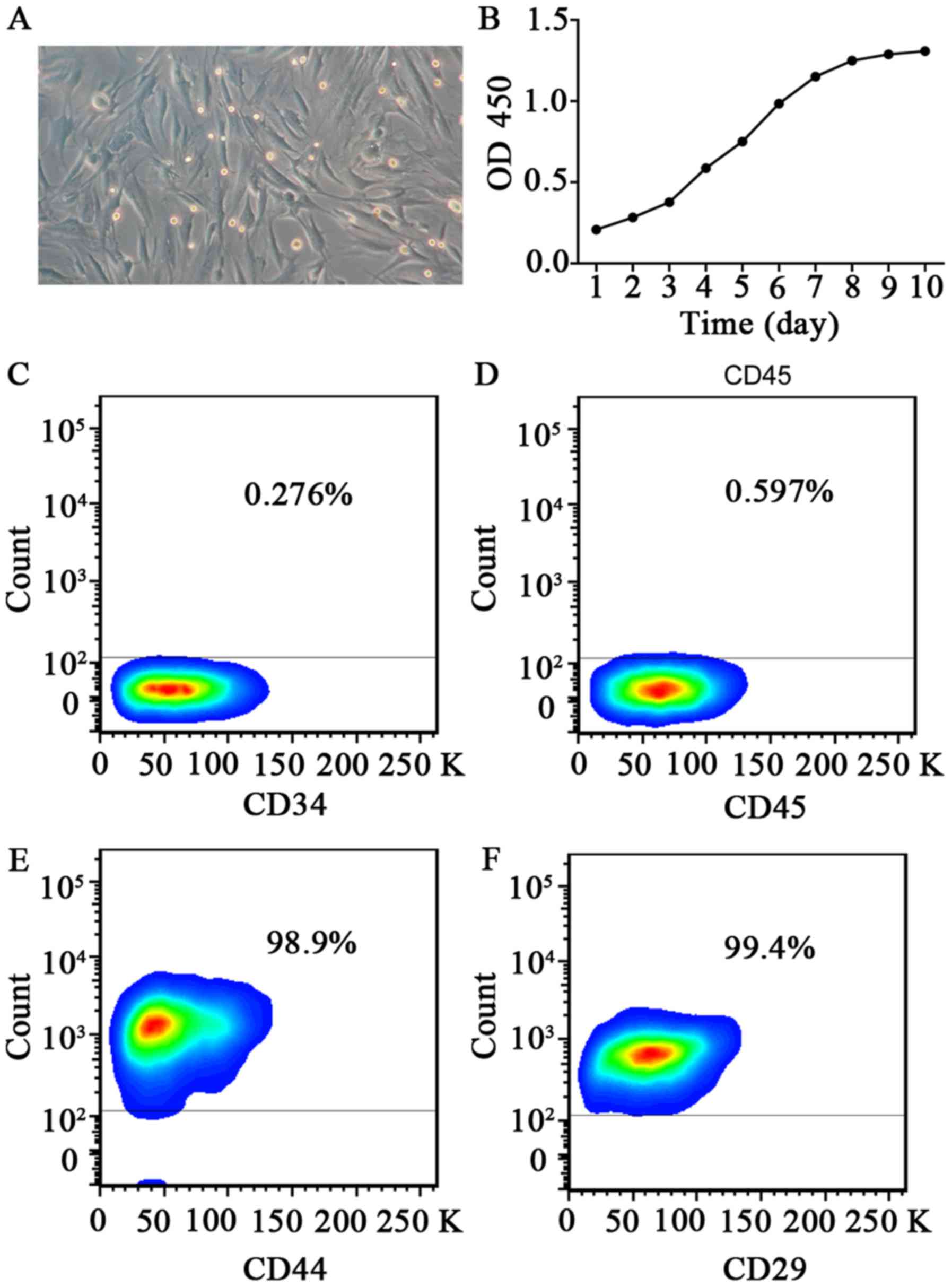
Morphology of rabbit MSCs
At 24 h after inoculation, cell adhesion was
detected from bone marrow cell suspension. After approximately 10
days, colonies of adherent cells started to appear. After
passaging, the cells exhibited a long spindle-like appearance, with
clones distributed as radial clusters (Fig. 1A).
Growth curve
Cells in primary culture showed a long period of
latency, generally lasting about 7 to 8 days, entered the
logarithmic growth phase after 10 days, and then reached the growth
plateau after 12 days. Analysis of the growth curve suggests that
the passaged cells have a quickened rate of cell growth and a
shortened incubation period, with an average doubling time of 70 h
(Fig. 1B). With the increase in the
number of generations, the cell proliferation rate levelled off,
and the proliferative capacity declined.
Detection of MSC surface markers
Cells of passage 6 were used for flow cytometric
analysis to detect molecular markers on the cell surface. Results
show that CD29 and CD44 were expressed on the cell surface, while
there was only a very weak expression of CD34 of CD34 or CD45
(Fig. 1C-F). Flow cytometry results
also showed that cultured cells were of uniform size, with
consistent expression of antigen molecules on the cell surface.
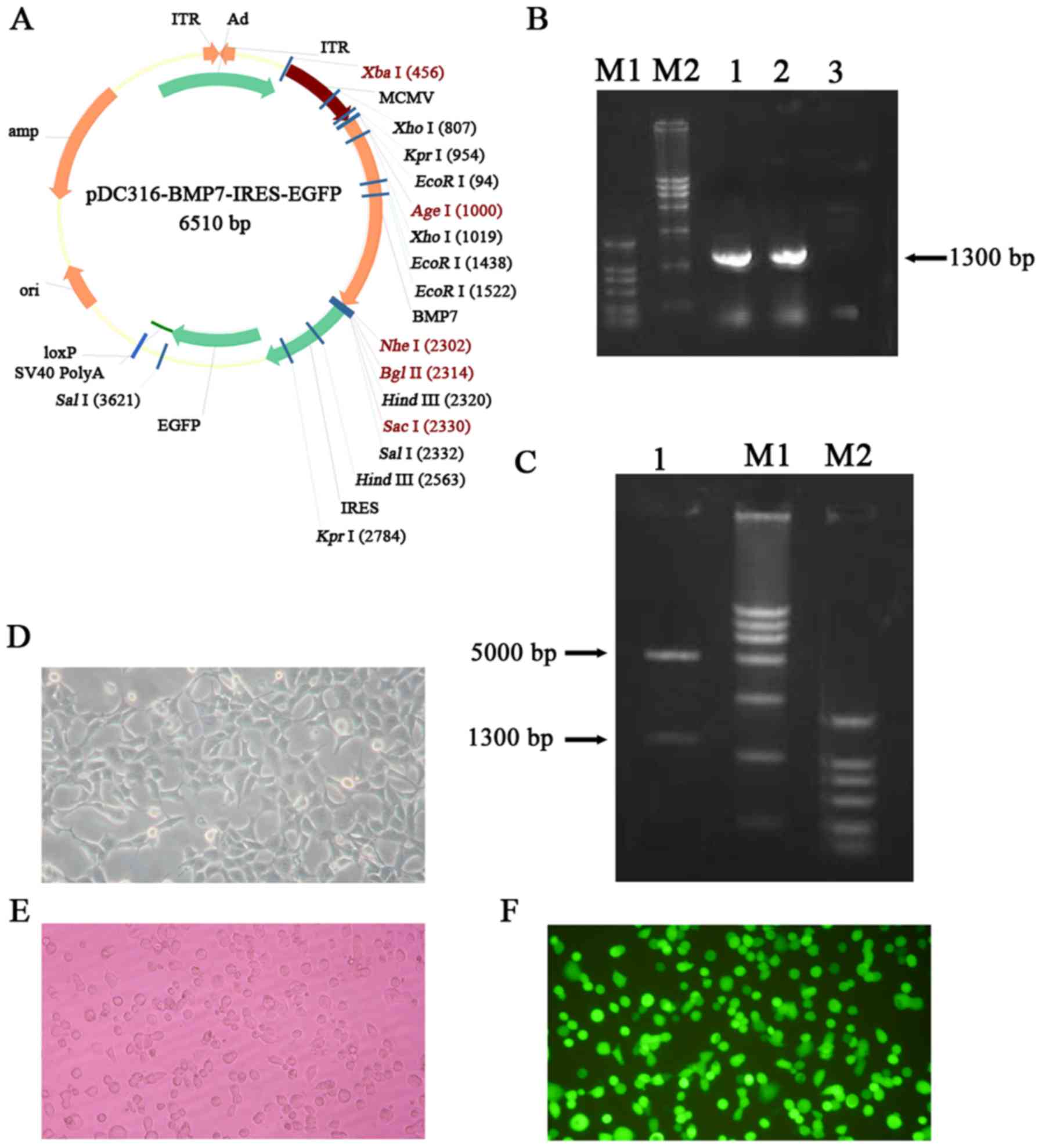
Construction of a recombinant
adenovirus vector carrying BMP7
The plasmid structure of pDC316-BMP7-IRES-enhanced
GFP (EGFP) is shown in Fig. 2A.
According to the results of electrophoresis against DNA markers,
the recombinant plasmid had the expected band at the desired size
(Fig. 2B). After colony PCR, 3 ml of
microbial liquid was used to extract the plasmid which was then
cleaved with appropriate restriction enzymes. After digestion, the
product was loaded onto a 1% agarose gel for gel electrophoresis to
detect a clear band, and the size was consistent with the size of
the BMP 7 gene (Fig. 2C). The
recombinant plasmid was transformed into 293 cells to generate the
recombinant adenovirus. After 8 days, botryoid cells were found and
obvious plaques subsequently appeared which can be regarded as
indicative of successfully-recombined adenovirus. After 11 days,
most of the lesions had formed and cells began to detach from the
bottom. Meanwhile, green fluorescence was observed under the
fluorescence microscope (Fig. 2D-F).
Detection of GFP indicates that virus particles with infection
ability have been successfully packaged. The results of MTT assay
showed that virus transfection has no obvious effects at MOI=100 on
the proliferation of bone marrow mesenchymal stem cells (P>0.05)
(Table I).
 | Table I.Ad-BMP7-MSCs viability was measured
by MTT assay (MOI=100). |
Table I.
Ad-BMP7-MSCs viability was measured
by MTT assay (MOI=100).
|
| OD450
value (mean ± SD) |
|
|---|
|
|
|
|
|---|
|
| 2 day | 4 day | 6 day | 8 day | 10 day | 12 day | P-value |
|---|
| Control | 0.360±0.002 | 0.540±0.004 | 0.540±0.003 | 0.620±0.003 | 0.640±0.004 | 0.630±0.002 |
|
| Ad-BMP7-MSCs | 0.360±0.003 | 0.550±0.003 | 0.560±0.002 | 0.590±0.002 | 0.640±0.002 | 0.620±0.004 | P>0.05 |
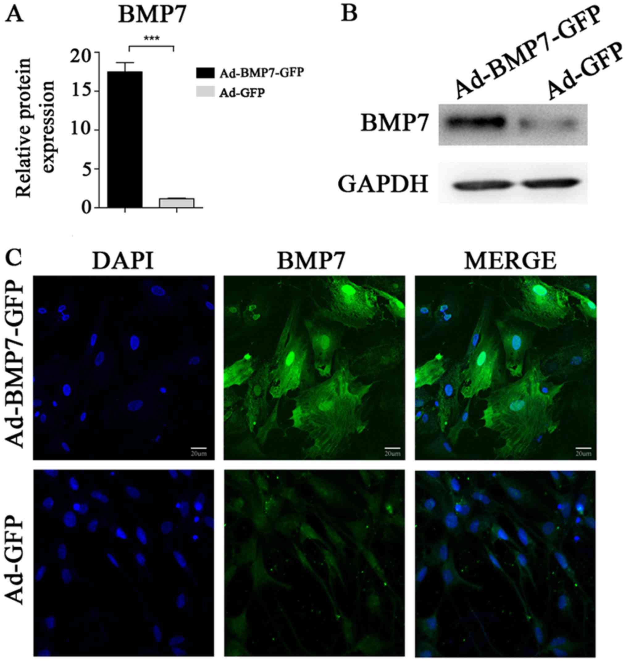
After transfection with Ad-BMP7, BMSCs
have high expression of BMP7
RT-PCR was performed to compare the difference in
BMP7 gene expression between the Ad-BMP7-GFP-transfected group
(experimental group) and the Ad-GFP group (control group). The
results showed that the gene expression level of BMP7 in the
Ad-BMP7-BMP7-GFP-transfected group was significantly higher than
the expression level of the corresponding parental cells (Fig. 3A). Moreover, the results of the
immunofluorescence assay showed that in the positive cells of the
experimental group, parts of cells with BMP7 expression were green
after labelling with the GFP tag, as observed under an inverted
fluorescence microscope (Fig. 3C),
while no green fluorescence could be seen in the control group.
Western blot results showed a positive band of the desired size
which is consistent with hBMP7, while the control group did not
have any positive bands (Fig. 3B).
After transfection, ALP activity was increased because of the
expression of BMP7, and induced differentiation of BMSCs in the
osteoblastic direction (Table
II).
| Table II.Alkaline phosphatase activity of
Ad-BMP7-MSCs at different time. |
Table II.
Alkaline phosphatase activity of
Ad-BMP7-MSCs at different time.
|
| Alkaline
phosphatase activity (mean ± SD) |
|
|---|
|
|
|
|
|---|
|
| 2 day | 4 day | 6 day | 8 day | 10 day | 12 day | P-value |
|---|
| Control | 11.26±0.77 | 12.46±0.55 | 14.61±0.35 | 16.32±0.90 | 19.35±0.42 | 20.92±0.49 | P<0.05 |
| Ad-BMP7-MSCs | 12.27±0.58 | 14.37±0.65 | 22.44±0.64 | 27.05±0.42 | 30.62±0.47 | 34.01±0.32 |
|
MSCs with high expression of BMP7
combined with an NHAC scaffold can effectively repair a rabbit
radius defect
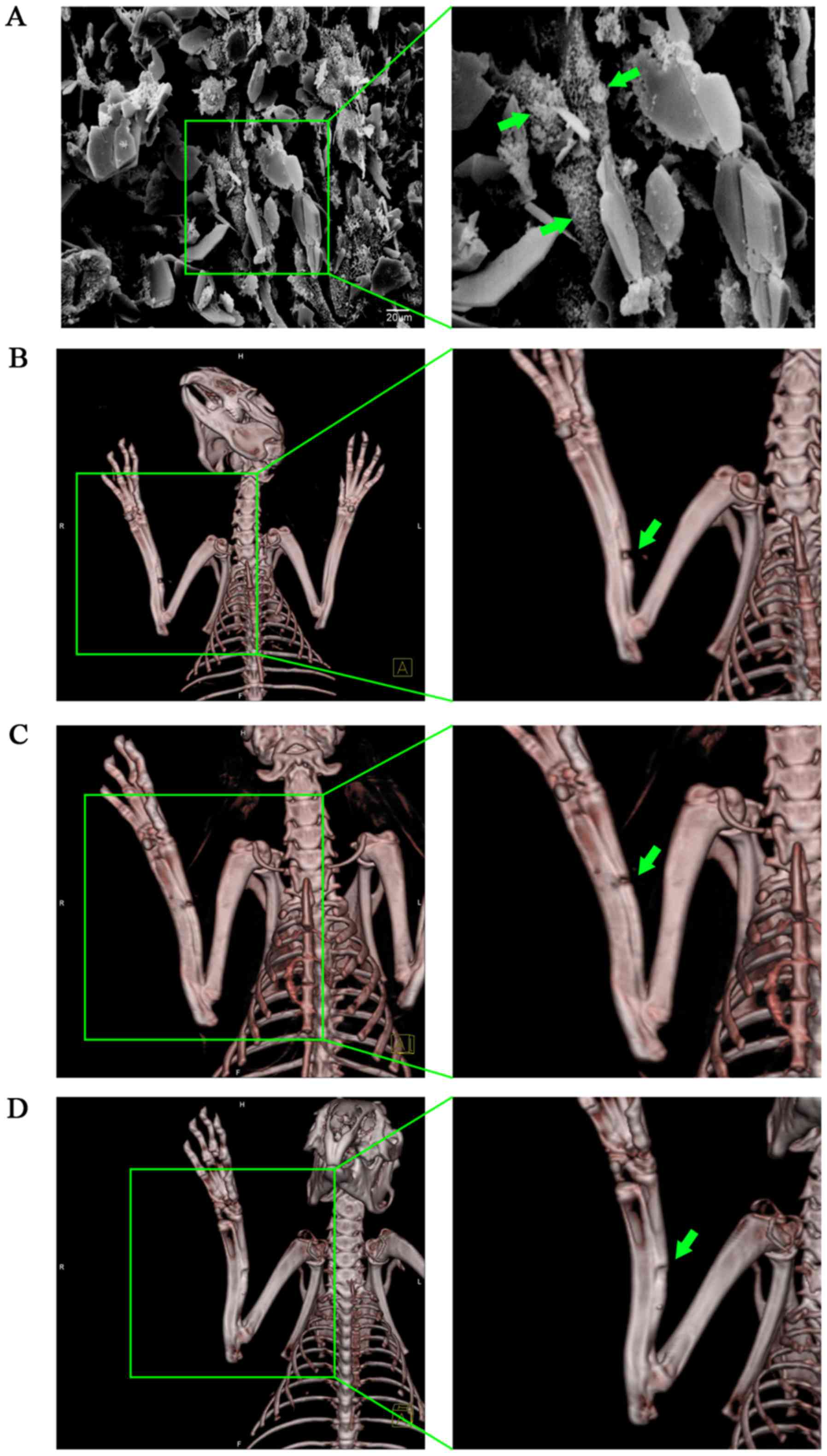
Cells were co-cultured with scaffold materials and
after 10 h, cells were observed under SEM (Fig. 4A). The surface of the scaffold
material appeared rough, with an average porosity of 70%, tiny
holes attached to the microporous wall, and with interpenetrated
microspores inside. MSCs on the scaffolds extended pseudopodia,
creeping over the pore surface of the material, then the
pseudopodia appeared to bulge and touch each other, surrounded by a
large amount of extracellular matrix, forming a woven mesh. Most
cells were fusiform, triangular or polygonal, with pseudopodia of
different lengths.
Three months later, repair of rabbit radius defects
was determined with 3D-CT. The results showed that the speed of
repair of the control group which was filled with NHAC alone was
slow, with 80% of all subjects having no bone union (Fig. 4B). In the BMSC-loaded NHAC group,
although the defect was narrowed, obvious bone nonunion could still
be seen, with only 40% of all defects healing completely, although
we did not find any cases of bone nonunion (Fig. 4C). In the group filled with BMSCs
with high expression of BMP7 combined with NHAC, callus formation
could be seen clearly (Fig. 4D).
Only one case was still not completely healed, but the bone defect
was well repaired. Compared with the other two treatments, NHAC
plus Ad-BMP7-BMSCs was more effective in repairing bone defects
(P<0.05) (Table III). This
suggests that using a BMSC-composite scaffold material transfected
with BMP7 for bone defect repair can effectively promote MSC growth
and differentiation, and improve bone repair.
| Table III.Incidence of bone defect in different
groups. |
Table III.
Incidence of bone defect in different
groups.
| Group | Bone defect
incidence | P-value |
|---|
| NHAC | 8/10 | P=0.003 |
| NHAC plus
Ad-BMP7-BMSCs | 1/10 |
|
| NHAC plus
BMSCs | 6/10 | P=0.029 |
| NHAC plus
Ad-BMP7-BMSCs | 1/10 |
|
Discussion
Bone defects and fracture non-union are common
problems, affecting many patients every year, and are difficult to
heal using current therapies (10–13). For
a long time, the accepted treatment for a tissue defect has been
tissue transplantation such as autografts or allografts (14–16).
Orthopedists often face problems such as a shortage of donor
tissue, immune rejection and so on. Such treatment can only be used
in a small range of tissue defects and the problem of large tissue
defects remains a challenge in the medical field. Here we have
devised a strategy to treat such defects using ex vivo gene
therapy. Unlike regular in vivo gene therapy, this strategy
is MSC-based and has a number of advantages including continuous
BMP7 protein secretion but also enables precise implantation into
target sites, and easy preparation of numbers of transduced
cells.
Using different inducers and methods such as
dexamethasone, vitamin C, vitamin D, BMPs, TGF-β, FGF, or insulin,
can induce MSCs to differentiate into different lineages such as
osteoblasts, chondrocytes, fibroblasts, myoblasts and so on
(9,17–22). The
direction has been set but differentiated cells can reverse the
differentiation induced under certain conditions. Cultured BMSCs
retain their normal phenotype and telomerase activity up to passage
12 and still maintain their potential for osteogenic
differentiation up to passage 15, and therefore are considered to
be the ideal seed cells (23–25).
Preliminary studies of their biological characteristics have been
performed, which provide the experimental basis for further
research into BMSCs (26–28). In view of the above advantages, BMSCs
have gradually been replacing osteoblasts as the seed cells in
subsequent research. We combined the density gradient separation
and sidewall separation methods, using the growth characteristics
of MSCs and the differences of digestion time from other cells in
the process of passage, to purify the cells gradually. In this
experiment, like other cells in vitro, the cultured bone
marrow stromal cells also experienced three stages of growth, the
lag phase, logarithmic phase and growth plateau. BMSCs have strong
proliferative ability in vitro, with an average doubling
time of approximately 70 h. However, cell proliferation ability
decreased with the increasing number of passages.
As another element in tissue engineering, growth
factors are peptides that transmit information between cells and
play an important role in controlling cell growth. However, due to
the factors' intrinsic characteristics, they are easily affected by
external conditions, such as temperature, pH and organic reagents,
and vulnerable to hemodilution, enzymatic hydrolysis, release time
and concentration, making it difficult to control their use in
defects (29–31). In recent years, the development of
genetic engineering has led to developments in bone tissue
engineering, since genetic engineering provides a good alternative
method for solving the above problems. It can be used to achieve
sustained or slow-release of gene product and multiple genes can be
transduced and regulated. An endogenously-synthesized factor may
have much stronger biological activities than an exogenous
recombinant factor. This combination of genetic engineering
technology and tissue engineering is known as ‘gene-enhanced tissue
engineering’.
Currently, growth factors such as BMP, PDGF, FGF and
IGF, which can promote tissue differentiation, are used for tissue
engineering (32–34). Results of animal experiments and
clinical studies have shown that BMP family proteins can obviously
promote the growth and healing of bone tissue (35–37).
Among the BMP family members, BMP-2 has the strongest bone
induction activity, and it can induce mesenchymal cells to
irreversibly differentiate into bone and cartilage (38). However, BMP-2 only induces bone
formation, and has no further effect on promoting proliferation for
the differentiation of osteoblasts (39). BMP7 has a strong ability to induce
bone as well as maintaining the phenotype of cartilage cells and
promoting cell proliferation and extracellular matrix proteoglycan
and collagen type II synthesis (40). The main biological function of BMP7
is to induce mesenchymal cells to differentiate into osteoblasts
(41,42). Recombinant human BMP7 shows efficient
bone induction activity both in vitro and in vivo,
promoting cartilage proteoglycan expression and articular cartilage
defect repair (43). Consequently
expression of the BMP7 gene will be more conducive to cell
differentiation from BMSCs to osteoblasts and to cell
proliferation.
Using gene transfection technology to strengthen or
transform the function of seed cells is a hot topic in tissue
engineering. It is considered that using the method of gene
transfection, rather than direct use of recombinant proteins, has
more advantages. This is because it can achieve a more ‘natural’
kind of action with long-term, high efficiency and localized
effects, and avoid the excessive reaction and systemic side effects
which may occur with the direct application of growth factors.
Using this strategy, we were able to not only obtain seed cells
with strong physiological functions, but also make the seed cells
express the target protein needed for tissue construction.
Currently, in genetic engineering, carriers can be divided into
plasmids, bacteria, phagocytosed particles, artificial chromosomes,
viruses and so on, according to the basic elements of the different
sources. Adenovirus vectors (Adv) are one of the most promising and
widely-used carriers in genetically-engineered tissue engineering
(44–46). Compared with other gene carriers,
they have many advantages, such as the wide range of transfected
cell types and their ability to efficiently infect cells both at
dividing and non-dividing stages. Our data show that MSCs can be
easily transduced with Adv-hBMP7. In our study, the transduction
efficiency of our generated Adv-hBMP7 can reach 90–100%. It cannot
replicate in the host, has good security, no carcinogenicity or
viral genome drifts outside the host genome, and no risk of
insertion mutation. Gene transfection can generally maintain
expression in the body for more than 6 weeks.
Our data suggest that MSCs may perform a role as
delivery vehicles as well as having osteogenic potential
themselves. Based on our previous experiment, which provided
preliminary data and defined the best conditions and the influence
of adenovirus-transfected BMSCS, we used the application package to
harvest the recombinant adenovirus from BMSCs transfected with
Adv-hBMP7 in vitro. Using RT-PCR to detect the expression of
hBMP7, we found that the transfection group expressed significantly
higher mRNA levels. The results of immunofluorescence staining and
western blotting showed that the protein is expressed successfully.
Therefore, by using BMSCs with enhanced BMP7 expression combined
with a suitable scaffold, we achieved very good repair of a rabbit
radial defect, demonstrating the feasibility of the application of
this method to repair bone defects in the clinic. Our work lays the
foundation for clinical application in the future.
References
|
1
|
Vander Have KL, Hensinger RN, Caird M,
Johnston C and Farley FA: Congenital pseudarthrosis of the tibia. J
Am Acad Orthop Surg. 16:228–236. 2008. View Article : Google Scholar : PubMed/NCBI
|
|
2
|
McCollum GA, Myerson MS and Jonck J:
Managing the cystic osteochondral defect: Allograft or autograft.
Foot Ankle Clin. 18:113–133. 2013. View Article : Google Scholar : PubMed/NCBI
|
|
3
|
Chahal J, Gross AE, Gross C, Mall N, Dwyer
T, Chahal A, Whelan DB and Cole BJ: Outcomes of osteochondral
allograft transplantation in the knee. Arthroscopy. 29:575–588.
2013. View Article : Google Scholar : PubMed/NCBI
|
|
4
|
Espana EM, Shah S, Santhiago MR and Singh
AD: Graft versus host disease: Clinical evaluation, diagnosis and
management. Graefes Arch Clin Exp Ophthalmol. 251:1257–1266. 2013.
View Article : Google Scholar : PubMed/NCBI
|
|
5
|
Tate Knothe ML: Top down and bottom up
engineering of bone. J Biomech. 44:304–312. 2011. View Article : Google Scholar : PubMed/NCBI
|
|
6
|
Marolt D, Knezevic M and Novakovic GV:
Bone tissue engineering with human stem cells. Stem Cell Res Ther.
1:102010. View
Article : Google Scholar : PubMed/NCBI
|
|
7
|
Fröhlich M, Grayson WL, Wan LQ, Marolt D,
Drobnic M and Vunjak-Novakovic G: Tissue engineered bone grafts:
Biological requirements, tissue culture and clinical relevance.
Curr Stem Cell Res Ther. 3:254–264. 2008. View Article : Google Scholar : PubMed/NCBI
|
|
8
|
Zhu H, Yang F, Tang B, Li XM, Chu YN, Liu
YL, Wang SG, Wu DC and Zhang Y: Mesenchymal stem cells attenuated
PLGA-induced inflammatory responses by inhibiting host DC
maturation and function. Biomaterials. 53:688–698. 2015. View Article : Google Scholar : PubMed/NCBI
|
|
9
|
Sun H and Yang HL: Calcium phosphate
scaffolds combined with bone morphogenetic proteins or mesenchymal
stem cells in bone tissue engineering. Chin Med J (Engl).
128:1121–1127. 2015. View Article : Google Scholar : PubMed/NCBI
|
|
10
|
Siddiqui NA and Owen JM: Clinical advances
in bone regeneration. Curr Stem Cell Res Ther. 8:192–200. 2013.
View Article : Google Scholar : PubMed/NCBI
|
|
11
|
Fisher DM, Wong JM, Crowley C and Khan WS:
Preclinical and clinical studies on the use of growth factors for
bone repair: A systematic review. Curr Stem Cell Res Ther.
8:260–268. 2013. View Article : Google Scholar : PubMed/NCBI
|
|
12
|
Mills LA and Simpson AH: In vivo models of
bone repair. J Bone Joint Surg Br. 94:865–874. 2012. View Article : Google Scholar : PubMed/NCBI
|
|
13
|
Shekkeris AS, Jaiswal PK and Khan WS:
Clinical applications of mesenchymal stem cells in the treatment of
fracture non-union and bone defects. Curr Stem Cell Res Ther.
7:127–133. 2012. View Article : Google Scholar : PubMed/NCBI
|
|
14
|
Papakostidis C, Bhandari M and Giannoudis
PV: Distraction osteogenesis in the treatment of long bone defects
of the lower limbs: Effectiveness, complications and clinical
results; a systematic review and meta-analysis. Bone Joint J.
95-B:1673–1680. 2013. View Article : Google Scholar : PubMed/NCBI
|
|
15
|
Berner A, Reichert JC, Müller MB, Zellner
J, Pfeifer C, Dienstknecht T, Nerlich M, Sommerville S, Dickinson
IC, Schütz MA and Füchtmeier B: Treatment of long bone defects and
non-unions: From research to clinical practice. Cell Tissue Res.
347:501–519. 2012. View Article : Google Scholar : PubMed/NCBI
|
|
16
|
Boos AM, Arkudas A, Kneser U, Horch RE and
Beier JP: Bone tissue engineering for bone defect therapy. Handchir
Mikrochir Plast Chir. 42:360–368. 2010.(In German). View Article : Google Scholar : PubMed/NCBI
|
|
17
|
Contador D, Ezquer F, Espinosa M,
Arango-Rodriguez M, Puebla C, Sobrevia L and Conget P:
Dexamethasone and rosiglitazone are sufficient and necessary for
producing functional adipocytes from mesenchymal stem cells. Exp
Biol Med (Maywood). 240:1235–1246. 2015. View Article : Google Scholar : PubMed/NCBI
|
|
18
|
Castro FO, Torres A, Cabezas J and
Rodríguez-Alvarez L: Combined use of platelet rich plasma and
vitamin C positively affects differentiation in vitro to mesodermal
lineage of adult adipose equine mesenchymal stem cells. Res Vet
Sci. 96:95–101. 2014. View Article : Google Scholar : PubMed/NCBI
|
|
19
|
Zhang HT, Zha ZG, Cao JH, Liang ZJ, Wu H,
He MT, Zang X, Yao P and Zhang JQ: Apigenin accelerates
lipopolysaccharide induced apoptosis in mesenchymal stem cells
through suppressing vitamin D receptor expression. Chin Med J
(Engl). 124:3537–3545. 2011.PubMed/NCBI
|
|
20
|
Wu Y, Peng Y, Gao D, Feng C, Yuan X, Li H,
Wang Y, Yang L, Huang S and Fu X: Mesenchymal stem cells suppress
fibroblast proliferation and reduce skin fibrosis through a
TGF-β3-dependent activation. Int J Low Extrem Wounds. 14:50–62.
2015. View Article : Google Scholar : PubMed/NCBI
|
|
21
|
Hagmann S, Moradi B, Frank S, Dreher T,
Kämmerer PW, Richter W and Gotterbarm T: FGF-2 addition during
expansion of human bone marrow-derived stromal cells alters MSC
surface marker distribution and chondrogenic differentiation
potential. Cell Prolif. 46:396–407. 2013. View Article : Google Scholar : PubMed/NCBI
|
|
22
|
Thakkar UG, Trivedi HL, Vanikar AV and
Dave SD: Insulin-secreting adipose-derived mesenchymal stromal
cells with bone marrow-derived hematopoietic stem cells from
autologous and allogenic sources for type 1 diabetes mellitus.
Cytotherapy. 17:940–947. 2015. View Article : Google Scholar : PubMed/NCBI
|
|
23
|
Zheng YH, Xiong W, Su K, Kuang SJ and
Zhang ZG: Multilineage differentiation of human bone marrow
mesenchymal stem cells in vitro and in vivo. Exp Ther Med.
5:1576–1580. 2013. View Article : Google Scholar : PubMed/NCBI
|
|
24
|
Abbas Mashhadi F, Fallahi Sichani H,
Khoshzaban A, Mahdavi N and Bagheri SS: Expression of odontogenic
genes in human bone marrow mesenchymal stem cells. Cell J.
15:136–141. 2013.PubMed/NCBI
|
|
25
|
Song K, Huang M, Shi Q, Du T and Cao Y:
Cultivation and identification of rat bone marrow-derived
mesenchymal stem cells. Mol Med Rep. 10:755–760. 2014. View Article : Google Scholar : PubMed/NCBI
|
|
26
|
Yang Z, Zhu L, Li F, Wang J, Wan H and Pan
Y: Bone marrow stromal cells as a therapeutic treatment for
ischemic stroke. Neurosci Bull. 30:524–534. 2014. View Article : Google Scholar : PubMed/NCBI
|
|
27
|
Hang HL and Xia Q: Role of BMSCs in liver
regeneration and metastasis after hepatectomy. World J
Gastroenterol. 20:126–132. 2014. View Article : Google Scholar : PubMed/NCBI
|
|
28
|
Liu J, Liu X and Cao Y: Progress of
methods of inducing bone marrow mesenchymal stem cells into
chondrocytes in vitro. Zhongguo Xiu Fu Chong Jian Wai Ke Za Zhi.
25:618–623. 2011.(In Chinese). PubMed/NCBI
|
|
29
|
Fishero BA, Kohli N, Das A, Christophel JJ
and Cui Q: Current concepts of bone tissue engineering for
craniofacial bone defect repair. Craniomaxillofac Trauma Reconstr.
8:23–30. 2015.PubMed/NCBI
|
|
30
|
Sharma AK and Cheng EY: Growth factor and
small molecule influence on urological tissue regeneration
utilizing cell seeded scaffolds. Adv Drug Deliv Rev. 82–83:86–92.
2015. View Article : Google Scholar
|
|
31
|
Gothard D, Smith EL, Kanczler JM, Rashidi
H, Qutachi O, Henstock J, Rotherham M, El Haj A, Shakesheff KM and
Oreffo RO: Tissue engineered bone using select growth factors: A
comprehensive review of animal studies and clinical translation
studies in man. Eur Cell Mater. 28:166–208. 2014. View Article : Google Scholar : PubMed/NCBI
|
|
32
|
Yamamoto M, Hokugo A, Takahashi Y, Nakano
T, Hiraoka M and Tabata Y: Combination of BMP-2-releasing
gelatin/β-TCP sponges with autologous bone marrow for bone
regeneration of X-ray-irradiated rabbit ulnar defects.
Biomaterials. 56:18–25. 2015. View Article : Google Scholar : PubMed/NCBI
|
|
33
|
Khoshkam V, Chan HL, Lin GH, Mailoa J,
Giannobile WV, Wang HL and Oh TJ: Outcomes of regenerative
treatment with rhPDGF-BB and rhFGF-2 for periodontal intra-bony
defects: A systematic review and meta-analysis. J Clin Periodontol.
42:272–280. 2015. View Article : Google Scholar : PubMed/NCBI
|
|
34
|
Mullen LM, Best SM, Ghose S, Wardale J,
Rushton N and Cameron RE: Bioactive IGF-1 release from collagen-GAG
scaffold to enhance cartilage repair in vitro. J Mater Sci Mater
Med. 26:53252015. View Article : Google Scholar : PubMed/NCBI
|
|
35
|
Wang X, Li Y, Han R, He C, Wang G, Wang J,
Zheng J, Pei M and Wei L: Demineralized bone matrix combined bone
marrow mesenchymal stem cells, bone morphogenetic protein-2 and
transforming growth factor-β3 gene promoted pig cartilage defect
repair. PLoS One. 9:e1160612014. View Article : Google Scholar : PubMed/NCBI
|
|
36
|
Yang DH, Lee DW, Kwon YD, Kim HJ, Chun HJ,
Jang JW and Khang G: Surface modification of titanium with
hydroxyapatite-heparin-BMP-2 enhances the efficacy of bone
formation and osseointegration in vitro and in vivo. J Tissue Eng
Regen Med. 9:1067–1077. 2015. View Article : Google Scholar : PubMed/NCBI
|
|
37
|
Noh SS, Bhang SH, La WG, Lee S, Shin JY,
Ma YJ, Jang HK, Kang S, Jin M, Park J and Kim BS: A dual delivery
of substance P and bone morphogenetic protein-2 for mesenchymal
stem cell recruitment and bone regeneration. Tissue Eng Part A.
21:1275–1287. 2015. View Article : Google Scholar : PubMed/NCBI
|
|
38
|
Guo P, Shi ZL, Liu A, Lin T, Bi F, Shi M
and Yan SG: Effects of cartilage oligomeric matrix protein on bone
morphogenetic protein-2-induced differentiation of mesenchymal stem
cells. Orthop Surg. 6:280–287. 2014. View Article : Google Scholar : PubMed/NCBI
|
|
39
|
Chen Y, Roohani-Esfahani SI, Lu Z, Zreiqat
H and Dunstan CR: Zirconium ions up-regulate the BMP/SMAD signaling
pathway and promote the proliferation and differentiation of human
osteoblasts. PLoS One. 10:e01134262015. View Article : Google Scholar : PubMed/NCBI
|
|
40
|
Abula K, Muneta T, Miyatake K, Yamada J,
Matsukura Y, Inoue M, Sekiya I, Graf D, Economides AN, Rosen V and
Tsuji K: Elimination of BMP7 from the developing limb mesenchyme
leads to articular cartilage degeneration and synovial inflammation
with increased age. FEBS Lett. 589:1240–1248. 2015. View Article : Google Scholar : PubMed/NCBI
|
|
41
|
Santos A, Bakker AD, Willems HM,
Bravenboer N, Bronckers AL and Klein-Nulend J: Mechanical loading
stimulates BMP7, but not BMP2, production by osteocytes. Calcif
Tissue Int. 89:318–326. 2011. View Article : Google Scholar : PubMed/NCBI
|
|
42
|
Ren Y, Han C, Jia Y, Yin H and Li S:
Expression of human bone morphogenetic protein 7 gene in
adipose-derived stem cells and its effects on osteogenic phenotype.
Zhongguo Xiu Fu Chong Jian Wai Ke Za Zhi. 25:848–853. 2011.(In
Chinese). PubMed/NCBI
|
|
43
|
Stöve J, Schneider-Wald B, Scharf HP and
Schwarz ML: Bone morphogenetic protein 7 (bmp-7) stimulates
proteoglycan synthesis in human osteoarthritic chondrocytes in
vitro. Biomed Pharmacother. 60:639–643. 2006. View Article : Google Scholar : PubMed/NCBI
|
|
44
|
Zhang X and Godbey WT: Viral vectors for
gene delivery in tissue engineering. Adv Drug Deliv Rev.
58:515–534. 2006. View Article : Google Scholar : PubMed/NCBI
|
|
45
|
Partridge KA and Oreffo RO: Gene delivery
in bone tissue engineering: Progress and prospects using viral and
nonviral strategies. Tissue Eng. 10:295–307. 2004. View Article : Google Scholar : PubMed/NCBI
|
|
46
|
Warnock JN, Daigre C and Al-Rubeai M:
Introduction to viral vectors. Methods Mol Biol. 737:1–25. 2011.
View Article : Google Scholar : PubMed/NCBI
|