Introduction
As one of the most common and aggressive cancers in
male, prostate cancer (PC) contributes to a great number of
cancer-related mortality around the world (1,2).
Although surgery combined with chemotherapy seems to be effective
for treatment of localized PC, most diagnosed PC patients have
metastases because of no effective diagnostic biomarkers (3,4). Due to
the distant metastasis of PC, the 5-year survival rate of PC
remains unsatisfactory (5). The
incidence of PC is still quickly increasing and PC remains an
important health problem worldwide (6). Therefore, it is urgently required to
determine the molecular mechanism of PC development and
progression.
MicroRNAs (miRNAs) are about 20 nucleotides in
length and regulate the degradation of target mRNAs by associating
with the 3′-URT region of mRNAs (7).
miRNAs have been proven to regulate all kinds of biological
processes, such as development, proliferation, apoptosis and
metastasis (8–10). Dysregulation of miRNA expression is
often observed in human cancers, including PC (11). For example, miR-141 suppresses PC
stem cells and metastasis by targeting a cohort of pro-metastasis
genes (12). miR-194 suppresses PC
cell migration and invasion by downregulating human nuclear
distribution protein (13).
miR-141-3p promotes PC cell proliferation through inhibiting
kruppel-like factor-9 expression (14). Downregulated miR-26a modulates PC
cell proliferation and apoptosis by targeting COX-2 (15). miRNAs have been widely demonstrated
to be important regulators in PC. Therefore, investigating the
regulatory mechanism of miRNAs will benefit for the treatment of
PC.
Emerging evidence shows that miR-202 acts as a tumor
suppressor in some tumors. For instance, Yang et al
(16) reported that miR-202 inhibits
cell proliferation, migration and invasion of glioma by directly
targeting metadherin. Meng et al (17), shown that miR-202 promotes cell
apoptosis in esophageal squamous cell carcinoma. However, the role
of miR-202 in PC is largely unknown. In this study, we found that
the expression of miR-202 was significantly downregulated in PC
tissues and cell lines. And overexpression of miR-202 markedly
inhibited the proliferation, migration and invasion of PC cells.
Moreover, miR-202 overexpression promoted the apoptosis of PC
cells. Mechanistically, we found that
phosphatidylinositol-4,5-bisphosphate 3-kinase catalytic subunit α
(PIK3CA) is the target gene of miR-202 in PC cells. Overexpression
of miR-202 inhibited the mRNA and protein levels of PIK3CA.
Finally, we found that restoration of PIK3CA rescued the
proliferation, migration and invasion of miR-202-overexpressing PC
cells. Taken together, our results indicated that miR-202 acts as a
tumor suppressor in PC by targeting PIK3CA.
Materials and methods
Patient samples
A total of 54 PC tissue specimens and 11 adjacent
normal prostate tissues were obtained from primary PC patients in
the Third Affiliated Hospital of Sun Yat-Sen University. The
resected tissues were immediately frozen in liquid nitrogen and
stored at −80°C before use. All patients agreed and signed a
written informed consent for tissue donation for research purposes
before tissue collection. The study was approved by the
Institutional Human Experiment and Ethic Committee of the Third
Affiliated Hospital of Sun Yat-Sen University.
Cell lines and cell culture
Human PC cell lines including LNCaP, 22Rv1, PC-3 and
DU145 and normal prostate epithelial cells (RWPE-1) were purchased
from Cell Bank of Chinese Academy of Sciences (Shanghai, China). PC
cells were grown in RPMI 1640 (Gibco; Thermo Fisher Scientific,
Inc., Waltham, MA, USA) supplemented with 10% fetal bovine serum
(FBS; Gibco; Thermo Fisher Scientific, Inc.), 100 U/ml Penicillin
and 0.1 µg/ml Streptomycin. RWPE-1 cells were cultured in
keratinocyte-serum free medium (Gibco; Thermo Fisher Scientific,
Inc.) containing human recombinant epidermal growth factor (5
ng/ml; Thermo Fisher Scientific, Inc.) and bovine pituitary extract
(0.05 mg/ml; Thermo Fisher Scientific, Inc.). Cells were cultured
in a humidified atmosphere of 5% CO2 at 37°C.
Xenograft tumor formation
Six-week-old BALB/c male nude mice were obtained
from Shanghai Laboratory Animal Center (Chinese Academy of
Sciences, Shanghai, China). Tumor-bearing mice were maintained
under specific pathogen-free conditions in Experimental Animal
Center of the Third Affiliated Hospital of Sun Yat-Sen University.
All animal experiments were performed in accordance with the Guide
for Care and Use of Laboratory Animal and all experimental
protocols were approved by the Animal Ethics Committee of the Third
Affiliated Hospital of Sun Yat-Sen University. For the subcutaneous
xenograft mouse model, DU145 cells (5×10⁶) that were
transiently transfected with miR-202 mimics or miR-NC were
suspended in 100 µ medium and subcutaneously injected into the left
and right backside flanks of nude mice (n=6 mice/group)
respectively. The volume of the in vivo tumor xenograft was
calculated. Five weeks after initial injection, mice were
sacrificed and PC xenograft was weighed.
Cell proliferation
Cell proliferation was measured by Cell Counting
Kit-8 (CCK-8; Sigma-Aldrich; Merck KGaA, Darmstadt, Germany).
Briefly, cells were seeded into a 96-well plate at 1×10⁴
cells/well and cultured for 48 h after transfection. Cells were
then treated with CCK-8 reagent (10 µl/well) at 37°C for 4 h.
Absorbance at 450 nm was detected using a microplate reader (BioTek
Instruments, Inc., Winooski, VT, USA).
Flow cytometric analysis of
apoptosis
After double staining with FITC-Annexin V and
propidium iodide (PI) using Annexin V-FITC apoptosis detection kit
(BD Biosciences, Franklin Lakes, NJ, USA) at room temperature,
cells were analyzed with a flow cytometry (BD LSRFortessa flow
cytometer; BD Biosciences) equipped with CellQuest software (BD
Biosciences).
Transwell assay
Cells were suspended in serum-free medium, and
3×10⁴ cells (100 µl) was plated into the upper chambers
of Transwell inserts (8.0-mm pore size; Corning Incorporated,
Corning, NY, USA) for invasion (with Matrigel) or migration
(without Matrigel) assays. The inserts were placed in 24-well
plates containing 600 µl media with 10% FBS. After incubation for
24 h for migration and 48 h for invasion, cells were fixed for 20
min and stained with 1% crystal violet. The cell numbers were
calculated and imaged under a microscope from five random
fields.
Real-time quantitative PCR
Total RNA from PC tissues and cells were extracted
using Trizol (Invitrogen; Thermo Fisher Scientific, Inc.). For
miRNA expression, RT reactions were performed with a One Step
PrimeScript miRNA cDNA Synthesis kit, followed by PCR with
SYBR® Premix Ex Taq (Takara Biotechnology Co., Ltd,
Dalian, China). For mRNA, cDNA was synthesized from the total RNA
using a PrimeScript RT Reagent Kit (TTakara Biotechnology Co.,
Ltd., Dalian, China). qPCR amplification reactions were performed
with SYBR® Premix Ex Taq II with ROX (Takara
Biotechnology Co., Ltd.). U6 or GAPDH was used for normalization
and all data were calculated by the 2−ΔΔCq method
(18).
Statistical analysis
Data are presented as mean ± SD and analyzed using
GraphPad Prism 5 (GraphPad Software Inc., San Diego, USA) and SPSS
v.21.0 software (IBM Corp., Armonk, NY, USA). Statistical
significance was investigated through a two-tailed, unpaired
Student's t-test, one-way ANOVA followed by Tukey's post hoc test
or a chi-square test as appropriate. P<0.05 was considered to
indicate a statistically significant difference.
Results
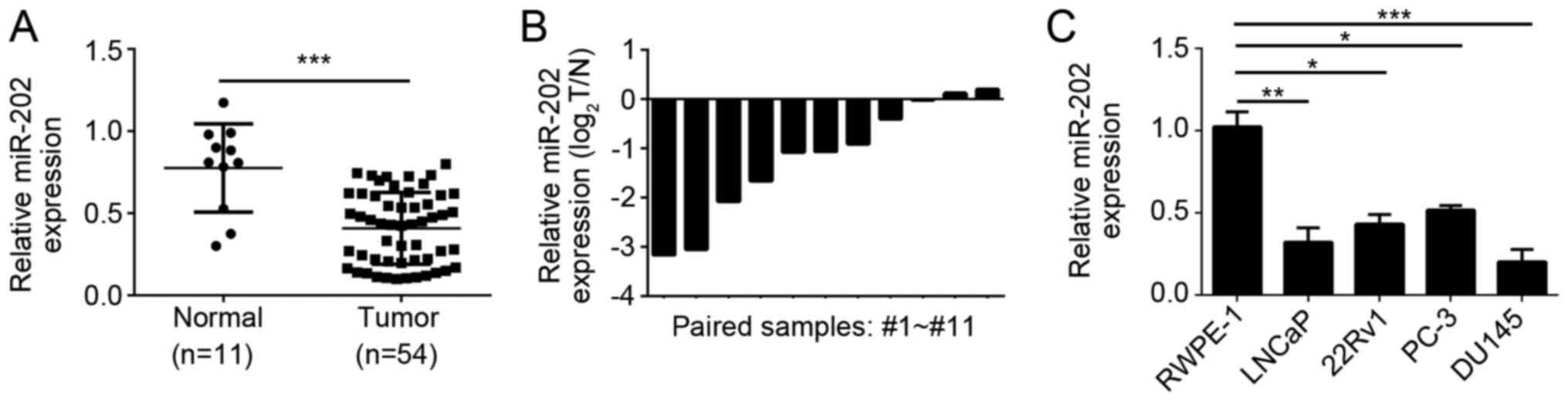
miR-202 is downregulated in PC tissues
and cells
To determine the role of miR-202 in PC, we used
qRT-PCR to analyze the expression of miR-202 in PC tissues. We
found that miR-202 was significantly downregulated in PC tissues
compared to adjacent normal tissues (Fig. 1A). Then we checked the expression of
miR-202 in 11 paired PC tissues. And we found that most PC tissues
showed lower expression of miR-202 than paired normal tissues
(Fig. 1B). We also examined the
expression of miR-202 in PC cell lines. We found that miR-202 was
downregulated in PC cell lines (LNCaP, 22Rv1, PC-3 and DU145 cells)
compared to normal RWPE-1 (Fig.
1C).
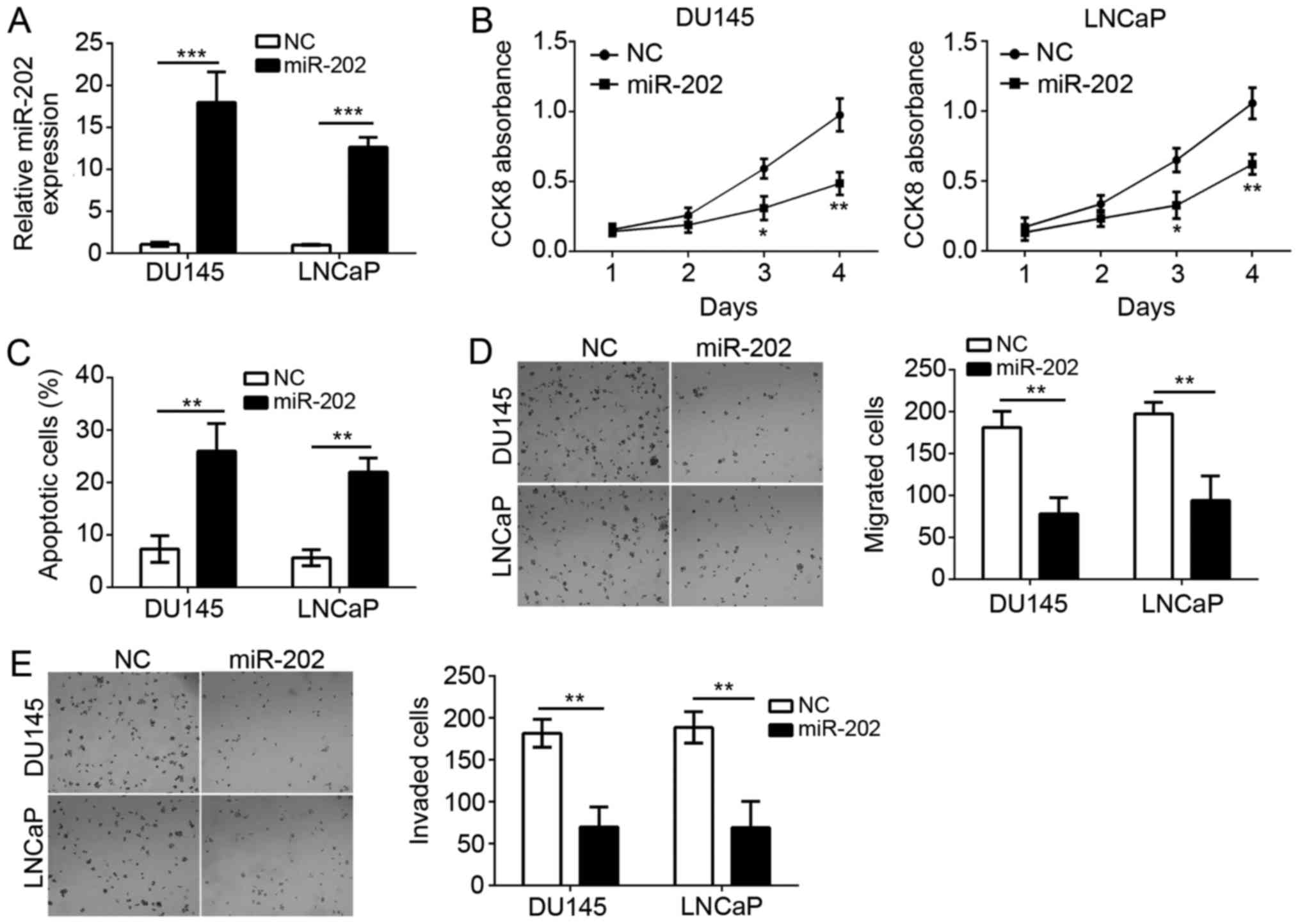
miR-202 overexpression inhibits cell
proliferation, migration and invasion of PC
To further explore the function of miR-202, we
overexpressed miR-202 in DU145 and LNCaP cells by transfection with
miR-202 mimics (Fig. 2A). Then we
performed CCK8 assay to check the proliferation of DU145 and LNCaP
cells. We found that overexpression of miR-202 significantly
inhibited the proliferatioin of DU145 and LNCaP cells (Fig. 2B). Notably, we also analyzed the
effect of miR-202 overexpression in RWPE-1 cell growth, migration
and invasion. However, the results were not significant compared to
control group (data not shown). Moreover, by staining with
AnnexinV/PI, we found that miR-202 overexpression markedly
increased the apoptotic cells (Fig.
2C). PC metastasis greatly contributes to the malignance.
Therefore, we evaluated the effect of miR-202 on cellular migration
and invasion with transwell assay. As shown in Fig. 2D-F, we found that overexpression of
miR-202 significantly suppressed the numbers of migrated and
invaded cells. Taken together, our findings indicated that miR-202
suppressed that proliferation, migration and invasion of PC cells
but induced cellular apoptosis.
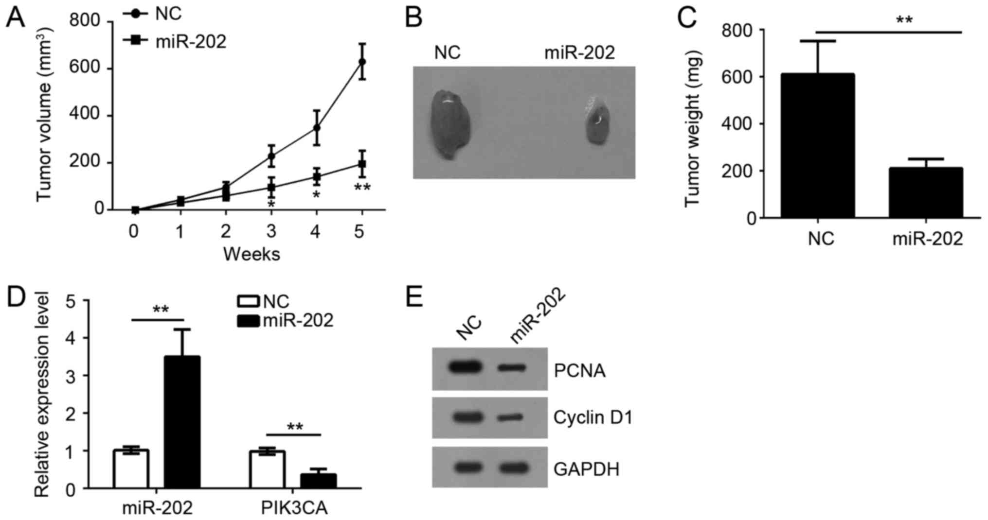
miR-202 overexpression suppresses
tumor growth in vivo
To further investigate the function of miR-202 in
vivo, we performed a xenograft experiment by subcutaneously
injecting DU145 cells into the dorsal flank of nude mice. Tumor
growth was closely monitored for 5 weeks. Results showed that
overexpression of miR-202 significantly inhibited the tumor growth
in vivo (Fig. 3A). Similarly,
the tumor weight was also significantly decreased by overexpression
of miR-202 (Fig. 3B and C). Notably,
the expression of miR-202 was still upregulated compared to control
tumor tissues while PIK3CA expression was significantly
downregulated (Fig. 3D). Then we
evaluated the cell proliferation of tumor tissues by western blot.
We found that tumor tissues derived from miR-202-overexpressing
DU145 cells showed less proliferation as the expression of Cyclin
D1 and PCNA was lower than that in control group (Fig. 3E). Taken together, our findings
demonstrated that overexpression of miR-202 inhibited tumor growth
in vivo.
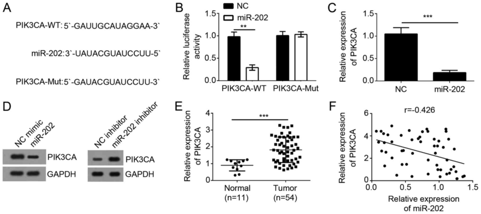
PIK3CA is a direct target of
miR-202
It has been widely acknowledged that miRNAs target
the 3′-UTR region of specific mRNAs to exert essential biological
functions. To explore miR-202-mediated mechanism, we made a
computer-based sequence analysis by TargetScan website and
identified PIK3CA as a potential target gene of miR-202. There was
a possible binding site of miR-202 in the 3′-UTR region of PIK3CA
(Fig. 4A). To determine whether
miR-202 could target the mRNA of PIK3CA, we constructed luciferase
reporter plasmid and performed luciferase activity reporter assay.
As shown, overexpression of miR-202 significantly inhibited the
luciferase activity in DU145 cells (Fig.
4B). And mutation of the binding site in the 3′-UTR region of
PIK3CA abrogated the inhibition of luciferase activity by miR-202
(Fig. 4B). To illustrate that the
endogenous miR-202 could regulate the expression of PIK3CA, we
overexpressed miR-202 in DU145 cells and found that its
overexpression significantly inhibited the mRNA and protein levels
of PIK3CA in PC cells (Fig. 4C and
D). Besides, we found that PIK3CA expression was significantly
upregulated in PC tissues compared to adjacent normal tissues
(Fig. 4E) and inversely correlated
with that of miR-202 (Fig. 4F),
suggesting PIK3CA was a target of miR-202.
PIK3CA is involved in miR-202-mediated
regulation of PC cell proliferation, apoptosis, migration and
invasion
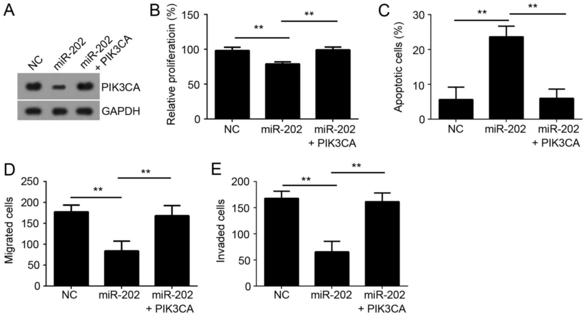
To determine whether miR-202 inhibited PC cell
proliferation, migration and invasion by targeting PIK3CA, we
performed a rescue assay. We restored the expression of PIK3CA in
miR-202-overexpressed DU145 cells by transfection with PIK3CA
ectopically expressing plasmid (Fig.
5A). Moreover, the effects of miR-202 overexpression on
cellular proliferation, apoptosis, migration and invasion were
significantly reversed by PIK3CA overexpression (Fig. 5B-E). These results indicated that
miR-202 suppressor the development and progression of PC by
downregulation of PIK3CA.
Discussion
PC is one of the most common and aggressive cancers
in male and contributes to a great number of cancer-related
mortality around the world (1,2).
Nowadays, the mechanism of PC development still remains elusive.
And PC is still a major challenge for people's health. Therefore,
it is of vital importance to explore the molecular mechanism
contributing to PC progression. In this study, we explored the
function of miR-202 and its mechanism involved in PC.
miRNAs have been widely acknowledged as important
regulators in human cancers by serving as oncogenes or tumor
suppressors (19,20). miR-202 was also proven to regulate
the occurrence of some tumors. For instance, Sun et al
(21) reported that miR-202
suppresses proliferation and induces apoptosis of osteosarcoma
cells by downregulating Gli2. Zhang et al (22), showed that miR-202 suppresses cell
proliferation in human hepatocellular carcinoma by downregulating
LRP6 post-transcriptionally. Wang et al (23), reported that miR-202-3p inhibits cell
proliferation by targeting ADP-ribosylation factor-like 5A in human
colorectal carcinoma. Additionally, Zhao et al (24) identified miR-202-3p was downregulated
in gastric cancer and acted as a novel tumor suppressor.
Nevertheless, the physiological function of miR-202 remains largely
unknown in PC. In the present study, we found that the expression
of miR-202 was downregulated in PC tissues and cell lines by
qRT-PCR. Moreover, through CCK8, FACS and transwell assays, we
found that overexpression of miR-202 significantly inhibited the
proliferation, migration and invasion but induced apoptosis in PC.
Moreover, xenograft experiments indicated that miR-202 suppressed
tumor growth in vivo. Collectively, our results demonstrated
that up-regulation of miR-202 in PC cells significantly inhibits
cell proliferation, migration and invasion, which is consistent
with previous reports about the function of miR-202 in other
cancers.
PIK3CA is an essential subunit of
phosphatidylinositol 3-kinase (PI3K), which regulates the
activation of AKT (25,26). PI3K/AKT pathway is a vital regulator
in cell proliferation, metastasis and apoptosis by modulating
downstream targets, such as mammalian target of rapamycin (mTOR)
and Cyclin-dependent kinase inhibitors (27). Increasing evidence shows that
PI3K/AKT pathway is closely related with the tumorigenesis, such as
breast cancer (28), hepatocellular
carcinoma (29) and PC (30). Dysregulated expression of PIK3CA led
to abnormal activation of PI3K/AKP pathway and consequently
development of cancers. In prostate, many studies reported that
PIK3CA expression is linked to the progression of PC. For example,
Agell et al (31) reported
that PI3K signaling pathway is activated by PIK3CA mRNA
overexpression and copy gain in prostate tumors. However, how miRNA
regulates the expression of PIK3CA in PC remains elusive. In the
present study, we found that PIK3CA was a direct target of miR-202
by bioinformatics prediction. Luciferase reporter demonstrated
their interaction. Besides, we showed overexpression of miR-202
significantly inhibited the mRNA and protein levels of PIK3CA in PC
cells. What's more, through functional experiments, we found that
overexpression of PIK3CA significantly rescued the inhibitory
effects of miR-202 on cellular proliferation, migration and
invasion, and reduced cellular apoptosis.
In summary, our results demonstrated that miR-202
served as a tumor suppressor by targeting PIK3CA in PC, which
provided a new sight on the development of therapeutic targets for
PC treatment.
Acknowledgements
Not applicable.
Funding
The present study was supported by grants from key
laboratory of Longhua, Shenzhen (grant no. 20150925A0410013). The
authors declare no competing financial interests.
Availability of data and materials
All data generated or analyzed during the present
study are included in this published article.
Authors' contributions
SZ, JC and FY initiated, designed this work,
analyzed, interpreted the results and wrote this manuscript. WX and
HL performed certain experiments. All authors read and approved the
final manuscript.
Ethics approval and consent to
participate
For the use of human samples, the protocol for this
study was approved by the Institutional Ethics Committee of The
People's hospital of Longhua and all enrolled patients signed a
written informed consent document. In addition, all procedures
involving animals conformed to the national guidelines of and were
approved by the Animal Care Ethics Committee of The People's
hospital of Longhua.
Patient consent for publication
All patients within this study provide consent for
the publication of their data.
Competing interests
The authors declare that they have no competing
interests.
References
|
1
|
Okato A, Goto Y, Kurozumi A, Kato M,
Kojima S, Matsushita R, Yonemori M, Miyamoto K, Ichikawa T and Seki
N: Direct regulation of LAMP1 by tumor-suppressive microRNA-320a in
prostate cancer. Int J Oncol. 49:111–122. 2016. View Article : Google Scholar : PubMed/NCBI
|
|
2
|
Jemal A, Bray F, Center MM, Ferlay J, Ward
E and Forman D: Global cancer statistics. CA Cancer J Clin.
61:69–90. 2011. View Article : Google Scholar : PubMed/NCBI
|
|
3
|
Coleman RE: Clinical features of
metastatic bone disease and risk of skeletal morbidity. Clin Cancer
Res. 12:6243s–6249s. 2006. View Article : Google Scholar : PubMed/NCBI
|
|
4
|
Edlind MP and Hsieh AC: PI3K-AKT-mTOR
signaling in prostate cancer progression and androgen deprivation
therapy resistance. Asian J Androl. 16:378–386. 2014. View Article : Google Scholar : PubMed/NCBI
|
|
5
|
Watson PA, Arora VK and Sawyers CL:
Emerging mechanisms of resistance to androgen receptor inhibitors
in prostate cancer. Nat Rev Cancer. 15:701–711. 2015. View Article : Google Scholar : PubMed/NCBI
|
|
6
|
Hu Y, Zhao Q, Rao J, Deng H, Yuan H and Xu
B: Longitudinal trends in prostate cancer incidence, mortality, and
survival of patients from two Shanghai city districts: A
retrospective population-based cohort study, 2000–2009. BMC Public
Health. 14:3562014. View Article : Google Scholar : PubMed/NCBI
|
|
7
|
Bartel DP: MicroRNAs: Genomics,
biogenesis, mechanism, and function. Cell. 116:281–297. 2004.
View Article : Google Scholar : PubMed/NCBI
|
|
8
|
Khan S, Brougham CL, Wall D, Newell J,
Kerin MJ and Dwyer RM: Mir-339-5p acts as A tumour suppressor in
breast cancer, mediated at least in part through regulation of cell
proliferation. Ir J Med Sci. 182:S3432013.
|
|
9
|
Wang T, Ren Y, Liu R, Ma J, Shi Y, Zhang L
and Bu R: miR-195-5p suppresses the proliferation, migration and
invasion of oral squamous cell carcinoma by targeting TRIM14.
Biomed Res Int. 2017:73781482017. View Article : Google Scholar : PubMed/NCBI
|
|
10
|
Alipour A, Mojdehfarahbakhsh A, Tavakolian
A, Morshedzadeh T, Asadi M, Mehdizadeh A and Nami M: Neural
communication through theta-gamma cross-frequency coupling in a
bistable motion perception task. J Integra Neurosci. 15:539–551.
2016. View Article : Google Scholar
|
|
11
|
Wang D, Lu G, Shao Y and Xu D:
microRNA-802 inhibits epithelial-mesenchymal transition through
targeting flotillin-2 in human prostate cancer. Biosci Rep. 37:pii:
BSR20160521. 2017. View Article : Google Scholar
|
|
12
|
Liu C, Liu R, Zhang D, Deng Q, Liu B, Chao
HP, Rycaj K, Takata Y, Lin K, Lu Y, et al: MicroRNA-141 suppresses
prostate cancer stem cells and metastasis by targeting a cohort of
pro-metastasis genes. Nat Commun. 8:142702017. View Article : Google Scholar : PubMed/NCBI
|
|
13
|
Kong Q, Chen XS, Tian T, Xia XY and Xu P:
MicroRNA-194 suppresses prostate cancer migration and invasion by
downregulating human nuclear distribution protein. Oncol Rep.
37:803–812. 2017. View Article : Google Scholar : PubMed/NCBI
|
|
14
|
Li JZ, Li J, Wang HQ, Li X, Wen B and Wang
YJ: MiR-141-3p promotes prostate cancer cell proliferation through
inhibiting kruppel-like factor-9 expression. Biochem Biophys Res
Commun. 482:1381–1386. 2017. View Article : Google Scholar : PubMed/NCBI
|
|
15
|
Zhang J, Liang J and Huang J:
Downregulated microRNA-26a modulates prostate cancer cell
proliferation and apoptosis by targeting COX-2. Oncol Lett.
12:3397–3402. 2016. View Article : Google Scholar : PubMed/NCBI
|
|
16
|
Yang J, Fan B, Zhao Y and Fang J:
MicroRNA-202 inhibits cell proliferation, migration and invasion of
glioma by directly targeting metadherin. Oncol Rep. 38:1670–1678.
2017. View Article : Google Scholar : PubMed/NCBI
|
|
17
|
Meng X, Chen X, Lu P, Ma W, Yue D, Song L
and Fan Q: miR-202 promotes cell apoptosis in esophageal squamous
cell carcinoma by targeting HSF2. Oncol Res. 25:215–223. 2017.
View Article : Google Scholar : PubMed/NCBI
|
|
18
|
Livak KJ and Schmittgen TD: Analysis of
relative gene expression data using real-time quantitative PCR and
the 2(-Delta Delta C(T)) method. Methods. 25:402–408. 2001.
View Article : Google Scholar : PubMed/NCBI
|
|
19
|
Damavandi Z, Torkashvand S, Vasei M,
Soltani BM, Tavallaei M and Mowla SJ: Aberrant expression of breast
development-related MicroRNAs, miR-22, miR-132, and miR-212, in
breast tumor tissues. J Breast Cancer. 19:148–155. 2016. View Article : Google Scholar : PubMed/NCBI
|
|
20
|
Han SY, Han HB, Tian XY, Sun H, Xue D,
Zhao C, Jiang ST, He XR, Zheng WX, Wang J, et al: MicroRNA-33a-3p
suppresses cell migration and invasion by directly targeting PBX3
in human hepatocellular carcinoma. Oncotarget. 7:42461–42473.
2016.PubMed/NCBI
|
|
21
|
Sun Z, Zhang T, Hong H, Liu Q and Zhang H:
miR-202 suppresses proliferation and induces apoptosis of
osteosarcoma cells by downregulating Gli2. Mol Cell Biochem.
397:277–283. 2014. View Article : Google Scholar : PubMed/NCBI
|
|
22
|
Zhang Y, Zheng DY, Xiong Y, Xue C, Chen G,
Yan B and Ye Q: miR-202 suppresses cell proliferation in human
hepatocellular carcinoma by downregulating LRP6
post-transcriptionally. FEBS Lett. 588:1913–1920. 2014. View Article : Google Scholar : PubMed/NCBI
|
|
23
|
Wang Q, Huang Z, Guo W, Ni S, Xiao X, Wang
L, Huang D, Tan C, Xu Q, Zha R, et al: microRNA-202-3p inhibits
cell proliferation by targeting ADP-ribosylation factor-like 5A in
human colorectal carcinoma. Clin Cancer Res. 20:1146–1157. 2014.
View Article : Google Scholar : PubMed/NCBI
|
|
24
|
Zhao Y, Li CL, Wang M, Su L, Qu Y, Li J,
Yu B, Yan M, Yu Y, Liu B and Zhu Z: Decrease of miR-202-3p
expression, a novel tumor suppressor, in gastric cancer. PLoS One.
8:e697562013. View Article : Google Scholar : PubMed/NCBI
|
|
25
|
Chang F, Lee JT, Navolanic PM, Steelman
LS, Shelton JG, Blalock WL, Franklin RA and McCubrey JA:
Involvement of PI3K/Akt pathway in cell cycle progression,
apoptosis, and neoplastic transformation: A target for cancer
chemotherapy. Leukemia. 17:590–603. 2003. View Article : Google Scholar : PubMed/NCBI
|
|
26
|
Osaki M, Oshimura M and Ito H: PI3K-Akt
pathway: Its functions and alterations in human cancer. Apoptosis.
9:667–676. 2004. View Article : Google Scholar : PubMed/NCBI
|
|
27
|
Nicholson KM and Anderson NG: The protein
kinase B/Akt signalling pathway in human malignancy. Cell Signal.
14:381–395. 2002. View Article : Google Scholar : PubMed/NCBI
|
|
28
|
Woo SU, Sangai T, Akcakanat A, Chen H, Wei
C and Meric-Bernstam F: Vertical inhibition of the PI3K/Akt/mTOR
pathway is synergistic in breast cancer. Oncogenesis. 6:e3852017.
View Article : Google Scholar : PubMed/NCBI
|
|
29
|
Chai R, Fu L, Zheng Z, Liu T, Ji S and Li
G: Resveratrol inhibits proliferation and migration through SIRT1
mediated post-translational modification of PI3K/AKT signaling in
hepatocellular carcinoma cells. Mol Med Report. 16:8037–8044. 2017.
View Article : Google Scholar
|
|
30
|
Chen R, Li Y, Buttyan R and Dong X:
Implications of PI3K/AKT inhibition on REST protein stability and
neuroendocrine phenotype acquisition in prostate cancer cells.
Oncotarget. 8:84863–84876. 2017.PubMed/NCBI
|
|
31
|
Agell L, Hernández S, Salido M, de Muga S,
Juanpere N, Arumí-Uria M, Menendez S, Lorenzo M, Lorente JA,
Serrano S and Lloreta J: PI3K signaling pathway is activated by
PIK3CA mRNA overexpression and copy gain in prostate tumors, but
PIK3CA, BRAF, KRAS and AKT1 mutations are infrequent events. Mod
Pathol. 24:443–452. 2011. View Article : Google Scholar : PubMed/NCBI
|