Introduction
Ankle surgery is a common orthopedic surgery with
various methods for anesthesia. Currently, controversy exists
regarding the anesthetic methods in clinical practice of lower limb
surgery. Traditional anesthesia is based on lumbar plexus-epidural
anesthesia. Although this method may meet the surgical requirement,
it has multiple postoperative complications, and may lead to
greater hemodynamic changes, including rapid blood pressure
decrease or even respiratory and circulatory inhibition (1,2).
Therefore, ensuring intraoperative anesthetic safety and reducing
surgical and anesthetic complications are essential (3,4). In
recent years, the application of peripheral nerve block technology
has attracted much attention (5–7). Lumbar
plexus-sciatic nerve block (LSB) has less interference on patients'
respiration and circulation in lower extremity surgery, and does
not affect the gastrointestinal and urinary functions (5–7). In
addition, it may avoid lumbar puncture injury and achieve accurate
positioning in peripheral nerve block anesthesia when using a nerve
stimulator, as well as long anesthetic duration and satisfactory
results (3–5). Ropivacaine (ROP) is a long-acting amide
local anesthetic that is widely used within clinics due to its low
toxicity towards the central nervous system and cardiovascular
system (8). Furthermore, it is able
to achieve sensory and motor anesthesia, separately (9).
It has been demonstrated that clonidine, an
α2-adrenergic receptorα (2AR) agonist, may enhance analgesic and
anesthetic effects when used for peripheral nerve block (10). Dexmedetomidine (DEX) is a novel
highly selective α2AR agonist. Its binding ratio of α2: α1 receptor
is 1,620: 1, which is eight times that of clonidine (11). Its distribution and elimination
half-lives when intravenously injected are 6 min and 2 h,
respectively (11). Meanwhile, it
also serves roles including analgesia, sedation, stress inhibition
and stabilization of hemodynamics (11). It has been reported that the local
application of DEX combined with anesthetics may prolong the
effects of nerve block, reduce the local anesthetic dosage, prolong
the effect time and enhance the analgesic effects; furthermore, it
has no neurotoxicity (12–16). However, the impact of DEX dose on
ROP-induced LSB is not clear. The present study evaluated the
impact of different doses of DEX on ROP-induced LSB. The objective
of the present study was to provide a reference for further
application of DEX to clinical local anesthesia.
Patients and methods
Patients
A total of 80 patients undergoing selective or acute
LSB-based ankle surgery (American Society of Anesthesiologists
(ASA) grading I–II) (17) at the
Central Hospital of Cangzhou (Cangzhou, China) from January 2013 to
August 2013 were enrolled in the present study. There were 47 males
and 33 females, with an age range of 18–63 years and body weight
range of 47–83 kg. The preoperative heart, lung, liver and kidney
functions were normal. The exclusion criteria were as follows:
Neuromuscular diseases, coagulation disorders, diabetes, sinus
bradycardia or atrioventricular block, mental disorders or taking
analgesics recently and infection at the nerve block site. The
patients were double-blindly and randomly divided into group R
[applied 30 ml 5% ROP (batch no. NACL; AstraZeneca, Cambridge,
UK)], group Dex1 [a total of 30 ml of 0.5% ROP + 1 µg/kg DEX (batch
no. 1512066211; Cisen Pharmaceutical Co., Ltd., Jining, China)],
group Dex2 (a total of 30 ml 0.5% ROP + 1.5 µg/kg DEX) and group
Dex3 (a total of 30 ml of 0.5% ROP + 2 µg/kg DEX), with 20 cases in
each group. The applied drugs were not prepared by the physicians
involved in the present study. The present study was approved by
the Medical Ethics Committee of the Central Hospital of Cangzhou.
Written informed consent was obtained from all patients.
Anesthetic methods
All patients had not received preoperative
medication. During the surgery, one intravenous channel was firstly
established for the infusion of Ringer's solution (6–8 ml/kg).
Additionally, electrocardiogram, heart rate (HR), oxygen saturation
(SpO2) and mean arterial pressure (MAP) were routinely
monitored. The positive electrode of one nerve stimulator was
connected to the skin electrode of the patient's leg, and the
negative electrode was connected to the nerve stimulation needle
(0.8×100 mm). The stimulation current intensity started from 1 mA
together with a stimulation frequency of 2 Hz, and the pulse was
set as 0.1 msec. Lumbar plexus block utilized the method of
intra-psoas major muscle block as follows: The patient was placed
on the lateral side with knees and hip flexed and the block side
upwards. The vertical line between the middle line (formed by
lining along the spinous processes of the lumbar spine) and the
posterior superior iliac crest was divided into three equal parts,
and the needling point was at the mediolateral one-third site and
1-cm deflecting toward the head, so that it could induce the shrink
of the quadriceps femoris. Sciatic nerve block used the posterior
approach as follows: The patient was placed in the improved Sims
supine position (the non-surgical side was fully extended, the
surgical side was flexed at the hip and the knees were upward). The
line connecting the posterior iliac crest and the posterior edge of
the greater trochanter was drawn at mid-normal, and the needling
point was located at the intersection where the above line extended
3–5 cm and intersected with the line of the greater trochanter and
sacral hiatus, so it could induce strephenopodia of ankle
metatarsal flexure or strephexopodia of ankle dorsal flexure. If
the corresponding induced response was obvious, the intensity was
gradually reduced to 0.3–0.4 mA. If there was sustained induced
motor response, and the suction of the syringe connecting to the
end of the needle indicated no blood or cerebrospinal fluid, 20 ml
anesthetic (as aforementioned) for lumbar plexus block was injected
together with sciatic nerve block using 10 ml of anesthetic.
Observation indexes
Visual Analogue Scale (VAS) pain scores obtained by
needling the femoral innervation area (the patella in front of the
thigh) and the sciatic innervation area (the lateral side of the
dorsum pedis) every 3 min after block were used to evaluate the
block effects (0 points, painless; 10 points, unendurable pain).
Meanwhile, the onset time of block (from the end of the injection
to VAS score <4 points) and the duration of sensory block (from
the end of the injection to VAS score >4 points) were also
recorded. The movements of the knee and ankle joints were
evaluated, and the effects of motor block were evaluated using the
modified Bromage Muscle Relaxation score (0 points, no motor nerve
block, the knee and tibia joints could freely move; 1 point, the
lower limb could not be raised high and straight, but the knee and
ankle joints could be active; 2 points, the lower limb could not be
raised high and straight, the knee joints could not be bent, but
the ankle joints could be active; 3 points, the lower limbs were
completely blocked, could not be raised high and straight, and the
knee and ankle joints could not flex). The onset time of block
(from the end of the injection to Bromage score=1 point) and
duration of motor nerve block (from the block onset to the recovery
of ankle motor function) were recorded.
Sedation was evaluated using the Ramsay score (1
point, irritable; 2 points, quiet, cooperative and with good
orientation; 3 points, drowsy, but still responsive to commands; 4
points, light sleep, but still active when tapping the forehead; 5
points, sleep, and dull to the forehead tapping stimulation; 6
points, deep sleep, having no response to the forehead tapping
stimulation). The values of MAP, HR, SpO2, Ramsay score
and serum vascular endothelial growth factor (VEGF) level (18) were recorded at the time of entry into
the surgery room (T0), immediately after anesthesia (T1), 10 min
after anesthesia (T2), 30 min after anesthesia (T3), 1 h after
anesthesia (T4) and end of surgery (T5).
A total of 10 mg intravenous ephedrine (30 mg/ml;
Northeast Pharmaceutical Group Co., Ltd., Shenyang, China) or 0.5
mg atropine (0.5 mg/ml; Jiangsu Lianshui Pharmaceutical Co., Ltd.,
Lianshui, China) was applied if intraoperative hypotension (basal
value reduction >30% or systolic pressure <90 mmHg) or
bradycardia (HR<50 beats/min) occurred; mask-assisted breathing
was applied in the cases of respiratory depression (respiratory
rate <12 times/min), poor breathing or SpO2 <90%.
The patients with intraoperative pain complaint were intravenously
administered with 0.2 µl sufentanil (75 µg/ml; Yichang Renfu
Pharmaceutical Co., Ltd., Yichang, China). If the pain remained too
strong for the patient to endure the surgery, general anesthesia
was applied; however, in this case, the patient should be excluded
from the study. The adverse reactions during and following surgery,
including over-sedation (Ramsay score ≥5 points), bradycardia, dry
mouth, hypotension, respiratory depression, nerve root stimulation,
urine retention and local anesthetic toxicity, were recorded.
Statistical analysis
All statistical analysis was performed using SPSS
13.0 (SPSS, Inc., Chicago, IL, USA). The enumeration data were
presented as number, and were compared using the χ2
test. The measurement data were presented as the mean ± standard
deviation, and were compared using one-way analysis of variance
followed by Student-Newman-Keuls-q test. P<0.05 was considered
to indicate a statistically significant difference.
Results
General data of patients
The general data of patients are demonstrated in
Table I. There was no significant
difference in age, gender, body mass index, ASA grade or surgery
time among the four groups (P>0.05).
 | Table I.General data of patients in the four
groups (n=20/group). |
Table I.
General data of patients in the four
groups (n=20/group).
|
| Group |
|
|---|
|
|
|
|
|---|
| Characteristic | R | Dex1 | Dex2 | Dex3 | P-value |
|---|
| Age, years | 43.11±12.36 | 39.67±14.38 | 41.09±10.56 | 36.77±13.45 | >0.05 |
| Gender, n
(male/female) | 10/10 | 12/8 | 9/11 | 11/9 | >0.05 |
| Body mass index,
kg/m2 | 20.78±4.34 | 21.82±4.56 | 19.56±3.45 | 22.56±4.90 | >0.05 |
| ASA grade, n
(I/II) | 8/12 | 9/11 | 7/13 | 8/12 | >0.05 |
| Surgery time,
min | 72.67±4.45 | 71.34±6.76 | 73.67±8.32 | 72.06±7.89 | >0.05 |
Overall outcome
All patients successfully completed the surgery. No
patient required sufentanil administration or was switched to other
anesthetic methods.
Comparison of onset time, and duration
of sensory and motor block among the four groups
As demonstrated in Table
II, there was no significant difference in the onset time of
sensory or motor block at the lumbar plexus or sciatic nerve among
the four groups (P>0.05). Compared with group R, the durations
of sensory and motor block at the lumbar plexus or sciatic nerve in
groups Dex1-3 were significantly prolonged (P<0.01).
Furthermore, the durations in groups Dex3 and Dex2 were
significantly longer than those in group Dex1 (P<0.05), and the
duration in group Dex3 was significantly longer than that in group
Dex2 (P<0.05).
| Table II.Comparison of onset time and duration
of sensory and motor block among the four groups (n=20/group). |
Table II.
Comparison of onset time and duration
of sensory and motor block among the four groups (n=20/group).
|
|
| Sensory block | Motor block |
|---|
|
|
|
|
|
|---|
| Site | Group | Onset time, min | Duration, min | Onset time, min | Duration, min |
|---|
| Lumbar plexus | R | 13.35±4.67 | 457.47±213.19 | 17.28±4.79 | 329.70±52.60 |
|
| Dex1 | 12.87±5.42 |
862.33±297.49a | 16.65±4.33 |
649.20±187.51a |
|
| Dex2 | 13.16±3.65 |
1,232.74±209.38a, b | 16.57±5.46 |
831.32±232.20a, b |
|
| Dex3 | 12.34±4.86 |
1,564.44±261.54a–c | 16.63±6.22 |
1,200.78±241.10a–c |
| Sciatic nerve | R | 12.34±4.59 | 379.44±65.54 | 15.66±5.46 | 310.10±67.43 |
|
| Dex1 | 11.55±4.22 |
741.74±217.21a | 15.71±5.53 |
587.18±249.63a |
|
| Dex2 | 11.33±5.56 |
979.33±206.13a, b | 16.88±4.64 |
782.50±251.73a, b |
|
| Dex3 | 11.72±5.31 |
1,242.5±187.63a–c | 16.52±5.69 |
1,106.53±249.77a–c |
Comparison of MAP, HR and
SpO2 among the four groups
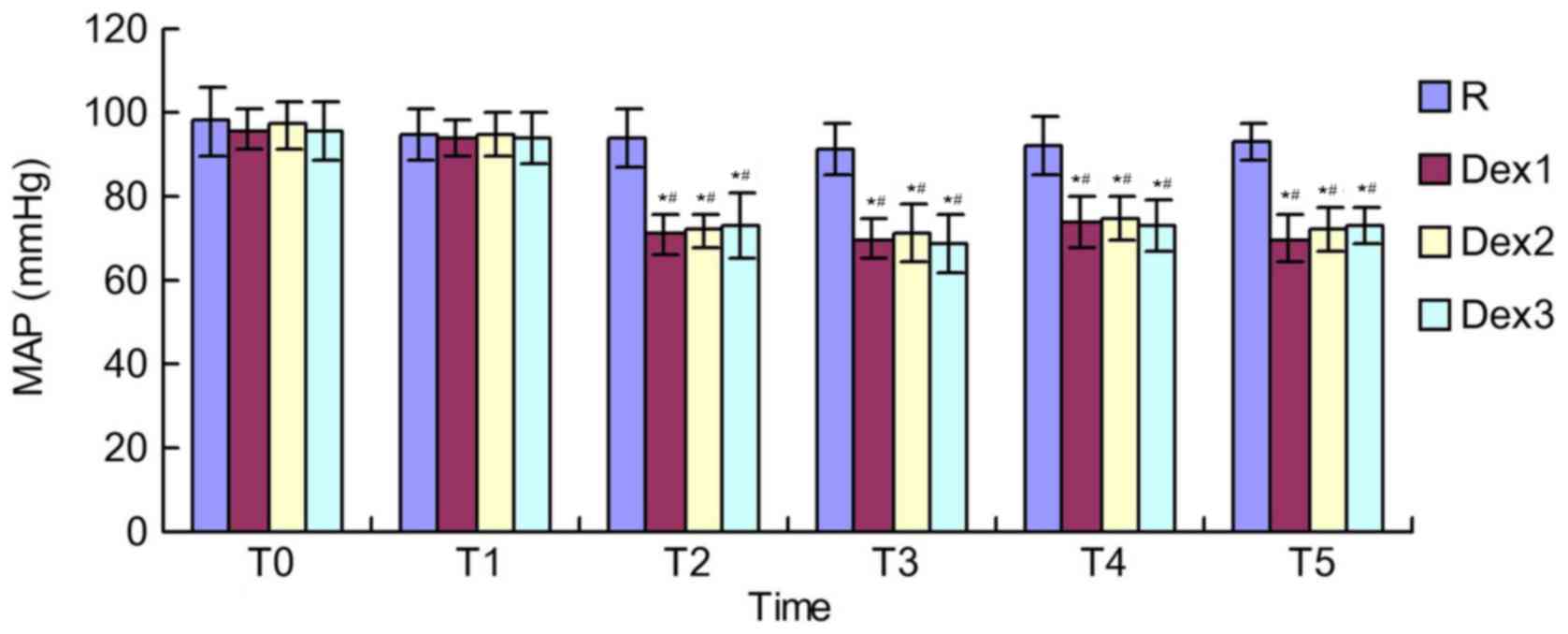
MAP in groups Dex1-3 at T2-T5 was significantly
lower than that of the same group at T0 and T1, respectively
(P<0.01). There was no significant difference in MAP between the
different time points in group R (P>0.05). The intergroup
comparison revealed that MAP in groups Dex1-3 at T2-T5 was
significantly lower than that in group R (P<0.01). There was no
significant difference in each index at each time point between
groups Dex1-3 (P>0.05) (Fig. 1).
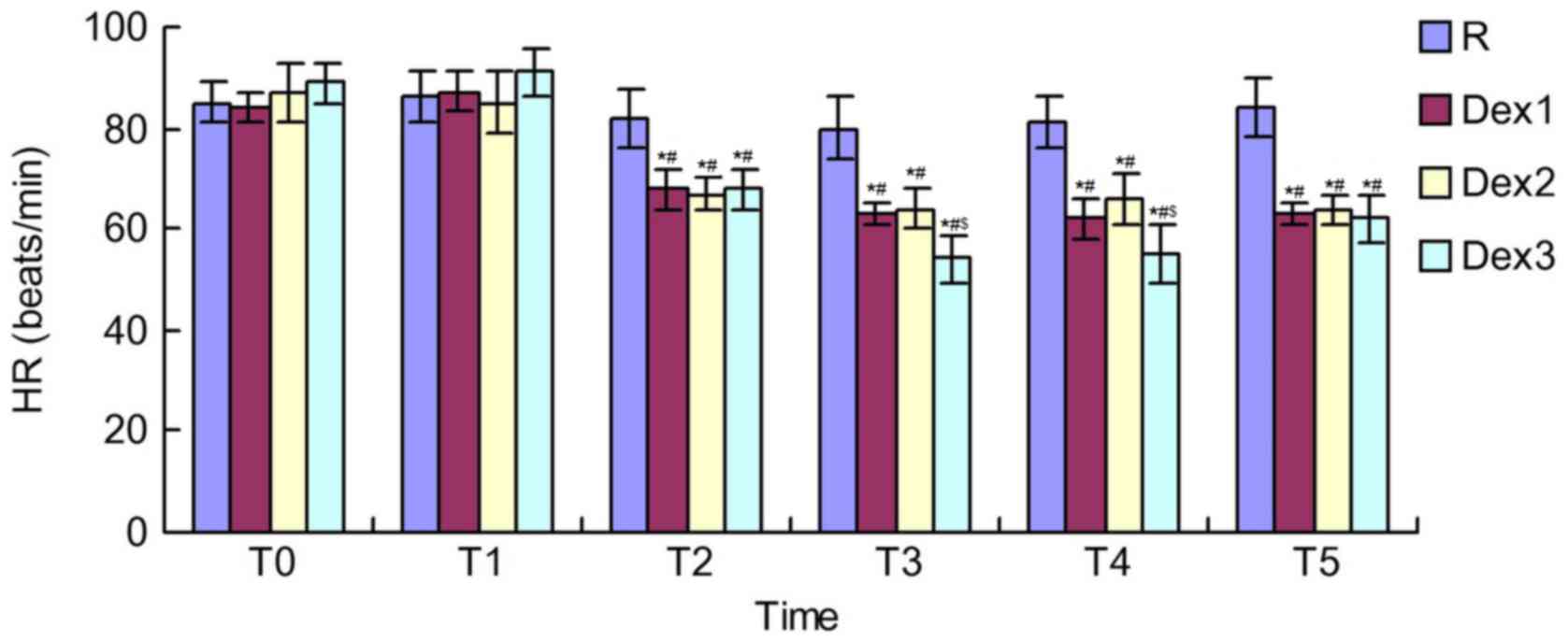
HR in groups Dex1-3 at T2-T5 was also significantly lower than
those in the same group at T0 and T1, respectively (P<0.01).
There was no significant difference in HR between the different
time points in group R (P>0.05). Compared with group R, HR in
groups Dex1-3 at T2-T5 was significantly reduced, respectively
(P<0.01). In addition, HR in group Dex3 at T3 and T4 was
significantly lower than that in groups Dex1 and Dex2, respectively
(P<0.05). There was no significant difference in each index at
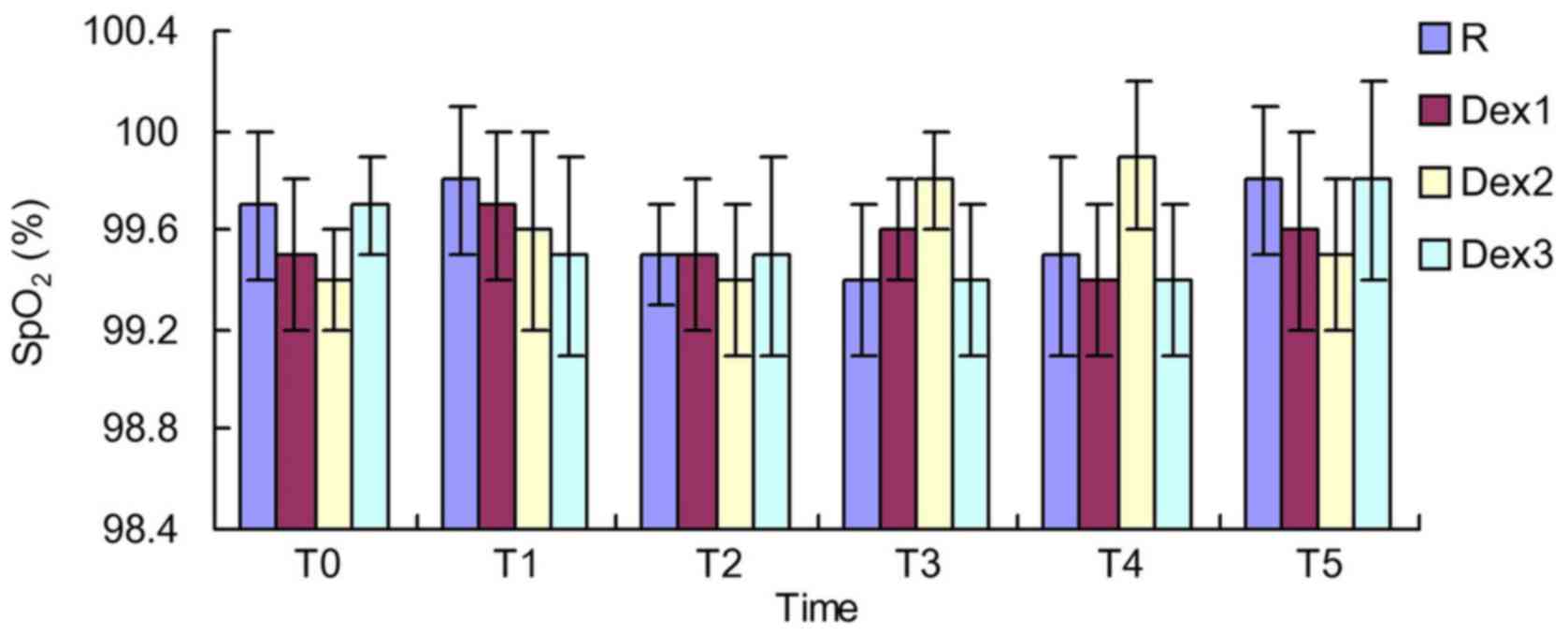
each time point between group Dex1 and Dex2 (P>0.05) (Fig. 2). There was no significant difference
in SpO2 between the different time points in each group
or among the different groups at each time point (P>0.05;
Fig. 3).
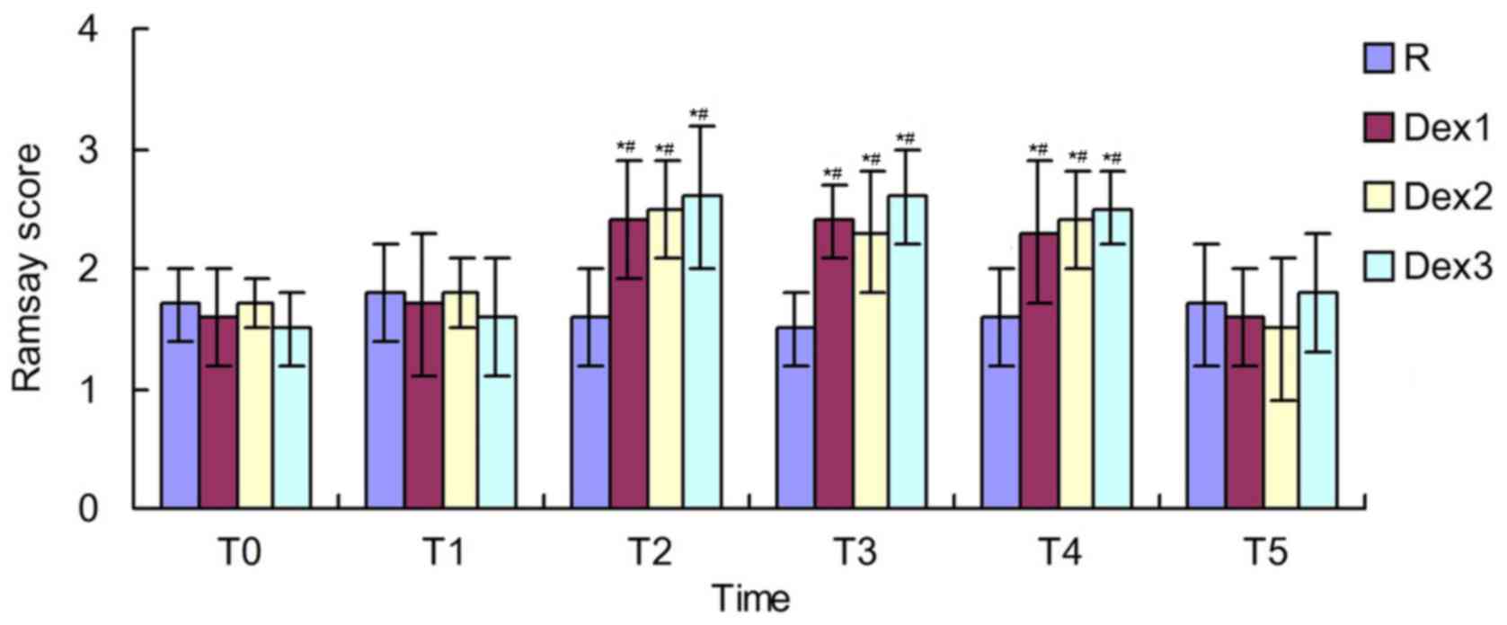
Comparison of Ramsay score among the
four groups
The Ramsay score in groups Dex1-3 at T2-T4 were
significantly higher than those in the same group at T0 and T1,
respectively (P<0.01). There was no significant difference in
the Ramsay score between the different time points in group R
(P>0.05). The Ramsay score in groups Dex1-3 at T2-T4 was
significantly higher than that in group R (P<0.05). There was no
significant difference in each index at each time point among
groups Dex1-3 (P>0.05) (Fig.
4).
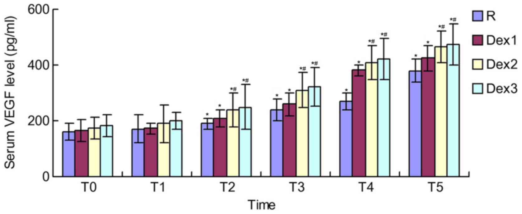
Comparison of serum VEGF level among
the four groups
The serum VEGF level in group R and Dex1-3 at T2-T5
was significantly higher than that of the same group at T0 and T1,
respectively (P<0.01). The VEGF level in groups Dex2 and Dex3 at
T2-T5 was significantly higher than that in group R, respectively
(P<0.01). There was no significant difference in each index at
each time point between group Dex2 and Dex3 (P>0.05) (Fig. 5).
Comparison of adverse reactions during
and following surgery among the four groups
There was no obvious adverse reaction during or
following surgery in groups R, Dex1 or Dex2, respectively. In group
Dex3, there was 1 (5%) case of over-sedation, 2 (10%) cases of
bradycardia and 1 (5%) case of dry mouth during and following
surgery, respectively (Table
III).
| Table III.Comparison of adverse reactions
during and following surgery among the four groups
(n=20/group). |
Table III.
Comparison of adverse reactions
during and following surgery among the four groups
(n=20/group).
|
| Group |
|---|
|
|
|
|---|
| Adverse
reaction | R | Dex1 | Dex2 | Dex3 |
|---|
| Over-sedation | 0 | 0 | 0 | 1 (5)a |
| Bradycardia | 0 | 0 | 0 | 2 (10)a |
| Dry mouth | 0 | 0 | 0 | 1 (5)a |
| Hypotension | 0 | 0 | 0 | 0 |
| Respiratory
depression | 0 | 0 | 0 | 0 |
| Nerve root
stimulation | 0 | 0 | 0 | 0 |
| Urine
retention | 0 | 0 | 0 | 0 |
| Local anesthetic
toxicity | 0 | 0 | 0 | 0 |
Discussion
It has been demonstrated that the addition of drugs,
including opioids, clonidine and DEX, to local anesthetics prolongs
the duration of nerve block, thus delaying the appearance of
patients' postoperative pain (19).
In the United States and European countries, DEX has been applied
outside the ranges regulated by the Food and Drug Administration's
provisions and instructions, known as the ‘off label’ applications
(20). The mechanism of DEX
prolonging nerve block is currently not clear; therefore, further
study is required. A study by Brummett et al (14) indicated in animal experiments that
DEX combined with bupivacaine enhanced the duration of sensory and
motor block of the sciatic nerve in mice. Furthermore, pathology
has revealed that DEX has no long-term effects on postoperative
axonal and myelin structure in the sciatic nerve of mice, and α2AR
antagonists cannot reverse its analgesic effects (21). Therefore, the present experiment
demonstrated that DEX is able to extend the nerve block time by
acting on the peripheral nerve system. Another study revealed that
DEX and ROP are effective in blocking nerve fiber conduction;
however, ROP is more dominant and DEX serves simulative roles
relative to ROP, which enhances the sensation and motor function of
the blocked nerves (22). However,
Kroin et al (23)
demonstrated that the mechanism of α2AR in extending the duration
of local anesthetics with the participation of
hyperpolarization-activated cation current.
Brummett et al (20) recommended the clinical dose of DEX in
human peripheral nerve block as 2 µg/kg. The present study
investigated the impact of various doses of DEX on ROP-induced LSB.
The results indicated that DEX combined with ROP significantly
prolonged the duration of sensory and motor block compared with the
use of ROP alone, and the effects were in the order of group
Dex3>Dex2>Dex1. The onset time of nerve block among the four
groups had no significant difference. Duma et al (24) demonstrated that 150 µg clonidine
combined with 40 mg bupivacaine for brachial plexus block had no
significant difference on the onset time of sensory and motor
block; however, this combination significantly prolonged the block
duration compared with that induced by the application of
bupivacaine alone. This was consistent with the results of the
present study. The present study also revealed that DEX had
synergistic effects with local anesthetics, which prolonged the
duration of sensory and motor block. Therefore, the present results
suggest that DEX has the effects of a local anesthetic, consistent
with the study of Marhofer et al study (15), which may be caused by the fact that
1.0 µg/kg DEX may have reached the maximum local anesthesia-like
effect. With the increasing dose of DEX in the present study, the
duration of sensory and motor block was gradually extended, and so
the appearance of postoperative pain in patients was delayed and
the anesthetic effects were improved. When the dose of DEX reached
2 µg/kg, some patients exhibited over-sedation and circulatory
suppression. Marhofer et al (15) further demonstrated that the duration
of sensory block prolonged by DEX combined with ROP was six times
that of the intravenous infusion of DEX, and it also indicates that
DEX has good local anesthetic effects.
The results of the present study revealed that MAP
and HR in groups Dex1-3 at T2-T5 were significantly lower than
those in group R. This may be derived from the anti-sympathetic
effects of DEX, which may inhibit the sympathetic nerve terminal to
release norepinephrine and enhance the activity of the vagus nerve,
thus contributing to the intraoperative hemodynamic stability in
patients (25). HR in group Dex3 at
T3 and T4 was significantly lower than group Dex1 and Dex2,
respectively. This indicated that the incidence of bradycardia at
T3 and T4 in group Dex3 is high, and it may be related to the
inhibitory effect of DEX on sympathetic tension (25). This suggests that, in clinical
applications, monitoring the cardiovascular system in patients must
be strengthened so as to actively prevent and treat complications.
The present study also demonstrated the strong sedative effects of
DEX, while inducing no respiratory depression or other
complications (26). The mechanism
may be that DEX acts on α2AR in the locus ceruleus of the brain
stem, thus inhibiting neuronal discharging and resulting in natural
non-REM sleep status (26).
In the present study, compared with group R, the
Ramsay scores in groups Dex1-3 at T2-T4 were increased. VEGF is a
special growth factor that acts on vascular endothelial cells
(27). As the most important factor
of angiogenesis, VEGF may promote the healing of injured joints
(28). As the VEGF level in groups
Dex2 and Dex3 at T2-T5 was significantly higher than that in group
R, this indicates that DEX may promote the secretion of VEGF.
However, different effects between the various DEX doses were not
found.
In the present study, no obvious adverse reactions
were observed during or following surgery in groups R, Dex1 and
Dex2. In group Dex3, there was 1 (5%) case of over-sedation, 2
(10%) cases of bradycardia and 1 (5%) case of dry mouth during and
following surgery. This indicates that a dose of DEX that is too
high may lead to more adverse reactions.
In conclusion, 1, 1.5 and 2 µg/kg DEX extends the
duration of 0.5% ROP-induced LSB, among which the latter two doses
demonstrate a superior effect in prolonging the effect time of ROP.
However, 2 µg/kg DEX exhibits a higher probability of inducing
transient hypertension or bradycardia, which is not conducive to
maintaining the stability of hemodynamics. Therefore, 1.5 µg/kg DEX
may be recommended for obtaining the greatest effects for improving
ROP-induced LSB. In the present study, the patients were limited to
young and middle-aged populations; therefore, the appropriate doses
of DEX for children and elderly patients require further
investigation.
Acknowledgements
Not applicable.
Funding
No funding was received.
Availability of data and materials
The datasets used and/or analyzed during the current
study are available from the corresponding author on reasonable
request.
Authors' contributions
JY and SS designed the study. SS and YN participated
in data collection. JY and SS performed the experiments and
statistical analysis. JY drafted the manuscript. All authors
critically revised the manuscript. All authors read and approved
the final manuscript.
Ethics approval and consent to
participate
The present study was approved by the Medical Ethics
Committee of the Central Hospital of Cangzhou. Written informed
consent was obtained from all patients.
Consent for publication
Not applicable.
Competing interests
The authors declare that they have no competing
interests.
References
|
1
|
Holzman RS: Unilateral Horner's syndrome
and brachial plexus anesthesia during lumbar epidural blockade. J
Clin Anesth. 4:464–466. 2002. View Article : Google Scholar
|
|
2
|
Marshall N and Watts S: Comparison of
epidural anaesthesia with continuous lumbar plexus block for total
hip arthroplasty. Region Anesth Pain Med. 33:e1552008. View Article : Google Scholar
|
|
3
|
Wiis JT, Jensen-Gadegaard P, Altintas Ü,
Seidelin C, Martusevicius R and Mantoni T: One-week postoperative
patency of lower extremity in situ bypass graft comparing epidural
and general anesthesia: Retrospective study of 822 patients. Ann
Vasc Surg. 28:295–300. 2014. View Article : Google Scholar : PubMed/NCBI
|
|
4
|
Karpel E, Marszolek P, Pawlak B and Wach
E: Effectiveness and safety of unilateral spinal anaesthesia.
Anestezjol Intens Ter. 41:33–36. 2009.(In Polish). PubMed/NCBI
|
|
5
|
Hamilton TW, Athanassoglou V, Trivella M,
Strickland LH, Mellon S, Murray D and Pandit HG: Liposomal
bupivacaine peripheral nerve block for the management of
postoperative pain. Cochrane Database Syst Rev.
2016:CD0114762016.
|
|
6
|
Jeon YH: Easier and safer regional
anesthesia and peripheral nerve block under ultrasound guidance.
Korean J Pain. 29:1–2. 2016. View Article : Google Scholar : PubMed/NCBI
|
|
7
|
Salinas FV: Evidence basis for ultrasound
guidance for lower-extremity peripheral nerve block: Update 2016.
Reg Anesth Pain Med. 41:261–274. 2016. View Article : Google Scholar : PubMed/NCBI
|
|
8
|
Stewart J, Kellett N and Castro D: The
central nervous system and cardiovascular effects of
levobupivacaine and ropivacaine in healthy volunteers. Anesth
Analg. 97:412–416. 2003. View Article : Google Scholar : PubMed/NCBI
|
|
9
|
Cherng CH, Yang CP and Wong CS: Epidural
fentanyl speeds the onset of sensory and motor blocks during
epidural ropivacaine anesthesia. Anesth Analg. 101:1834–1837. 2005.
View Article : Google Scholar : PubMed/NCBI
|
|
10
|
Kohane DS, Lu NT, Cairns BE and Berde CB:
Effects of adrenergic agonists and antagonists on
tetrodotoxin-induced nerve block. Reg Anesth Pain Med. 26:239–345.
2001. View Article : Google Scholar : PubMed/NCBI
|
|
11
|
Bhana N, Goa KL and Mcclellan KJ:
Dexmedetomidine. Drugs. 59:263–270. 2000. View Article : Google Scholar : PubMed/NCBI
|
|
12
|
Obayah GM, Refaie A, Aboushanab O,
Ibraheem N and Abdelazees M: Addition of dexmedeto-midine to
Bupivacaine for greater palatine nerve block prolongs
post-operative analgesia after cleft palate repair. Eur J
Anaesthesiol. 27:280–284. 2010. View Article : Google Scholar : PubMed/NCBI
|
|
13
|
Talke P, Xu M, Paloheimo M and Kalso E:
Effects of intrathecally administered dexmedetomidine, MPV-2426 and
tizanidine on EMG in rats. Acta Anaesthesiol Scand. 47:347–354.
2003. View Article : Google Scholar : PubMed/NCBI
|
|
14
|
Brummett CM, Padda AK, Amodeo FS, Welch KB
and Lydic R: Pefineural dexmedeto-midine added to ropivacaine
causes a dose-dependent increase in the duration of thermal
antinociception in sciatic nerve block in rat. Anesthesiology.
111:1111–1119. 2009. View Article : Google Scholar : PubMed/NCBI
|
|
15
|
Marhofer D, Kettner SC, Marhofer P, Pils
S, Weber M and Zeitlinger M: Dexmedetomidine as an adjuvant to
ropivacaine prolongs peripheral nerve block: A volunteer study. Br
J Anaesth. 110:438–442. 2013. View Article : Google Scholar : PubMed/NCBI
|
|
16
|
Kaya FN, Yavascaoglu B, Turker G, Yildirim
A, Gurbet A, Mogol EB and Ozcan B: Intravenous dexmedetomidine, but
not midazolam, prolongs bupivacaine spinal anesthesia. Can J
Anasesth. 57:39–45. 2010. View Article : Google Scholar
|
|
17
|
Fu KM, Smith JS, Polly DW Jr, Ames CP,
Berven SH, Perra JH, McCarthy RE, Knapp DR Jr and Shaffrey CI;
Scoliosis Research Society Morbidity and Mortality Committee:
Correlation of higher preoperative American Society of
Anesthesiology grade and increased morbidity and mortality rates in
patients undergoing spine surgery. J Neurosurg Spine. 14:470–474.
2011. View Article : Google Scholar : PubMed/NCBI
|
|
18
|
Poon RT, Ng IO, Lau C, Zhu LX, Yu WC, Lo
CM, Fan ST and Wong J: Serum vascular endothelial growth factor
predicts venous invasion in hepatocellular carcinoma: A prospective
study. Ann Surg. 233:227–235. 2001. View Article : Google Scholar : PubMed/NCBI
|
|
19
|
Crystal CS and Blankenship RB: Local
anesthetics and peripheral nerve blocks in the emergency
department. Emerg Med Clin North Am. 23:477–502. 2005. View Article : Google Scholar : PubMed/NCBI
|
|
20
|
Brummett CM, Norat MA, Palmisano JM and
Lydic R: Perineural administration of dexmedetomidine in
combination with bupivacaine enhances sensory and motor blockade in
sciatic nerve block without inducing neurotoxicity in rat.
Anesthesiology. 109:502–511. 2008. View Article : Google Scholar : PubMed/NCBI
|
|
21
|
Rangel RA, Marinho BG, Fernandes PD, de
Moura RS and Lessa MA: Pharmacological mechanisms involved in the
antinociceptive effects of dexmedetomidine in mice. Fundam Clin
Pharmacol. 28:104–113. 2014. View Article : Google Scholar : PubMed/NCBI
|
|
22
|
Erlacher W, Schuschnig C, Koinig H,
Marhofer P, Melischek M, Mayer N and Kapral S: Clonidine as
adjuvant for meprivacaine, ropivacaine and bupivacaine in axillary,
perivascular brachial plexus blaock. Can J Anaesth. 48:522–525.
2001. View Article : Google Scholar : PubMed/NCBI
|
|
23
|
Kroin JS, Buvonendran A, Beck DR, Topic
JE, Watts DE and Tuman KJ: Clonidine prolongation of lidocaine
analgesia after sciatic nerve block in rats is mediated via the
hyperpolarization-activated cation current, not by
alpha-adrenoreceptors. Anesthesiology. 101:488–494. 2004.
View Article : Google Scholar : PubMed/NCBI
|
|
24
|
Duma A, Urbanek B, Sitzwohl C, Kreiger A,
Zimpfer M and Kapral S: Clonidine as an adjuvant to local
anaesthetic axillary brachial plexus block: A randomized,
controlled study. Br J Anaesth. 94:112–116. 2005. View Article : Google Scholar : PubMed/NCBI
|
|
25
|
Penttilä J, Helminen A, Anttila M, Hinkka
S and Scheinin H: Cardiovascular and parasympathetic effects of
dexmedetomidine in healthy subjects. Can J Physiol Pharmacol.
82:359–362. 2004. View
Article : Google Scholar : PubMed/NCBI
|
|
26
|
Jung HS, Joo JD, Jeon YS, Lee JA, Kim DW,
In JH, Rhee HY and Choi JW: Comparison of an itraoperative infusion
of dexmedetomidine or remifentanil on perioperative haemodynamics,
hypnosis and sedation, and postoperative pain control. J Int Med
Res. 39:1890–1899. 2011. View Article : Google Scholar : PubMed/NCBI
|
|
27
|
Gerber HP, Dixit V and Ferrara N: Vascular
endothelial growth factor induces expression of the antiapoptotic
proteins Bcl-2 and A1 in vascular endothelial cells. J Biol Chem.
273:13313–13316. 1998. View Article : Google Scholar : PubMed/NCBI
|
|
28
|
Becker R, Pufe T, Kulow S, Giessmann N,
Neumann W, Mentlein R and Petersen W: Expression of vascular
endothelial growth factor during healing of the meniscus in a
rabbit model. J Bone Joint Surg Br. 86:1082–1087. 2004. View Article : Google Scholar : PubMed/NCBI
|