Introduction
Acute lung injury (ALI) is a common syndrome in the
clinic characterized by an abnormity of hypoxemia, epithelial
integrity, non-cardiogenic lung edema, neutrophil and leukocyte
accumulation, and an intense inflammatory response in lung
(1). Despite of the development of
medical technologies and medicines, ALI remains the leading cause
of morbidity and mortality in critically ill patients (2). Thus, it is of importance to further
explore the mechanism of ALI. Numerous causes are considered to
contribute to ALI, including trauma, pneumonia, acid aspiration,
and sepsis (3). Among them,
bacterial infection is one of the most important inducer for
sepsis. Lipopolysaccharide (LPS), a component of Gram negative
bacterial cell membrane, is considered to play an important role in
inflammatory response and immune dysfunction (4), but the mechanism of LPS-induced ALI is
not fully elucidated.
Considering of the aforementioned information,
multiple researches have been conducted to explore the mechanism of
LPS-induced ALI, as well as potential therapies. Jiang et al
(4) have identified that trillin can
exert a protective effect on LPS-induced ALI via regulating the
Nrf2/NF-κB signaling pathway. Lee et al (5), have demonstrated that
1-hexadecyl-3-(trifluoroethyl)-sn-glycero-2-phosphomethanol (MJ33)
can serve as an inhibitor for NADPH oxidase (type 2) to against ALI
associated inflammation. Besides, Do-Umehara et al (6), have documented that transcription
factor Miz1 inhibits the expression of C/EBP-δ to suppress the
inflammation during ALI.
Activated protein C (APC) is reported to enhance
autophagy with rapamycin against sepsis-induced ALI (7). Moreover, APC prevents LPS-induced
pulmonary vascular injury via attenuating the expression of
cytokine (8). Inhaled APC protects
mice from ventilator-induced lung injury via inhibiting the
activation of cytosolic phospholipase A2 (9). In addition, increased APC mediates
acute traumatic coagulopathy in mice (10). However, the detailed mechanism of APC
in lung injury remains fully understood. In the present study, the
potential mechanism of APC in ALI pathogenesis was identified, so
that could provide a deeper understanding, or new insights for
LPS-induced ALI.
Materials and methods
Lung injury animal model
This animal experimental protocol was authorized by
the Ethics Committee of Southeast University Affiliated Zhongda
Hospital (Jiangsu, China). Adult female Sprague Dawley rats
weighted 280–320 g (n=50) aged 8~10 weeks were purchased from the
Shanghai Laboratory Animal Research Center (Shanghai, China) to
apply for the following research of this study. Rats were housed in
a SPF condition at a temperature of 22–24°C and humidity of 40–70%
with a 12 h light/dark cycles, and kept with free access to food
and water. After adapted for 1 week, rats were utilized to
construct a lung injury using LPS as previously described (11). Briefly, rats were randomly divided
into five groups with 10 mice in each: i) Control group (saline),
ii) model group (LPS), iii) low-dose group (LPS + 0.1 mg/Kg
recombined human APC (rhAPC, Xigris; Eli Lilly Nederland BV,
Houten, The Netherlands)), iv) median-dose group (LPS + 0.3 mg/Kg
rhAPC), and v) high-dose group (LPS + 0.5 mg/Kg rhAPC). Rats were
administered intravenously with saline or rhAPC. At 15 min after
treatment, rats were anesthetized with 3% of chloral hydrate, and
then intratracheally administrated with 20 µg of LPS dissolved in
50 µl of phosphate buffer saline (PBS) to induce ALI. For the
control group, equal volume of PBS was used to instead of LPS
solution in the process of lung injury.
Isolation of Bronchoalveolar
lavage
After treated with LPS for 6 h, rats were
anesthetized and sacrificed. Followed by this, bronchoalveolar
lavage was collected using 0.5 ml of sterile PBS for 3 times (total
volume=1.5 ml) to obtain the bronchoalveolar lavage fluid (BALF).
Then, total leukocyte count and neutrophil count were estimated
using a hemocytometer (Qiujing, Shanghai, China). Subsequently,
BALF samples were centrifuged at 1,500 rpm at 4°C for 10 min, and
superoxide dismutase (SOD) activity of the supernatants was
measured using a test kit purchased from Nanjing Jiancheng
Bioengineering Institute (Nanjing, Jiangsu, China) according to
manufacturers' protocol.
Analysis of inflammatory cytokines
contained in BALF
The expression levels of IL-1β (catalogue no.:
RLB00), IL-6 (catalogue no.: R6000B), and tumor necrosis factor
(TNF)-α (catalogue no.: RTA00) were detected using enzyme-linked
immune sorbent assay according to manufacture's protocols (R&D
Systems, Inc., Minneapolis, MN, USA). All the experiments were
conducted in triplicate, and mean value of them was computed as the
final result.
Hematoxylin & eosin (H&E)
staining
To estimate the inflammation in lung tissue, H&E
staining was conducted for paraffin embedded sections. Briefly, the
right lungs were removed at the end of the experiment, and parts of
tissues were dehydrated using decreasing concentrations of ethanol,
embedded in paraffin wax, and cut into slices with thick of 5 µm.
Then, slices were deparaffinized, and rehydrated in decreasing
concentrations of ethanol. Sections were heated for 3 min at 110°C
in 10 mmol/l Tris/1 mmol/l EDTA (pH 9.0) antigen retrieval buffer
followed by 10 min at 95°C and then cooled to 20°C.
Western blot analysis
Lung tissues were homogenized in a 5 volumes of
pre-cold lysis buffer (BioSource International Inc.; Thermo Fisher
Scientific, Inc., Waltham, MA, USA) containing protease inhibitor
cocktail (0.01%; Sigma-Aldrich; Merck KGaA, Darmstadt, Germany).
Then, tissue samples were centrifuged at 12,000 g at 4°C for 10
min. Followed by this, the supernatants were collected, and the
concentrations were determined using the BCA method (Thermo Fisher
Scientific, Inc.). Then, supernatants were boiled with equal volume
of loading buffer for 10 min. Total 10 µg protein was loaded into
12% SDS-PAGE gel, and transferred electrophoretically on a PVDF
membrane. After blocked with 5% skim milk, blots were incubated
with specific antibodies (p38 (catalogue no.: 8690), p-p38 (4511),
ERK1/2 (9194), p-ERK1/2 (9101), c-Jun N-terminal kinase (JNK)
(9252), and p-JNK(9255)) purchased from Cell Signaling Technology,
Inc., (Danvers, MA, USA). After washing, blots were incubated with
horseradish peroxidase-conjugated second antibodies. Subsequently,
blots were washed, and visualized using ECL-detection system
(PerkinElmer, Inc., Waltham, MA, USA).
Statistical analyses
In the present study, GraphPad Prism v6.0 software
(GraphPad Software, Inc., La Jolla, CA, USA) was applied for
statistical analyses. Continuous data was presented as mean ±
standard deviation (SD). Comparison among groups was estimated
using one-way analysis of variance followed by multiple comparisons
using the least significant difference post hoc test. P<0.05 was
considered to indicate a statistically significant difference.
Results
Effect of rhAPC on lung injury induced
by LPS
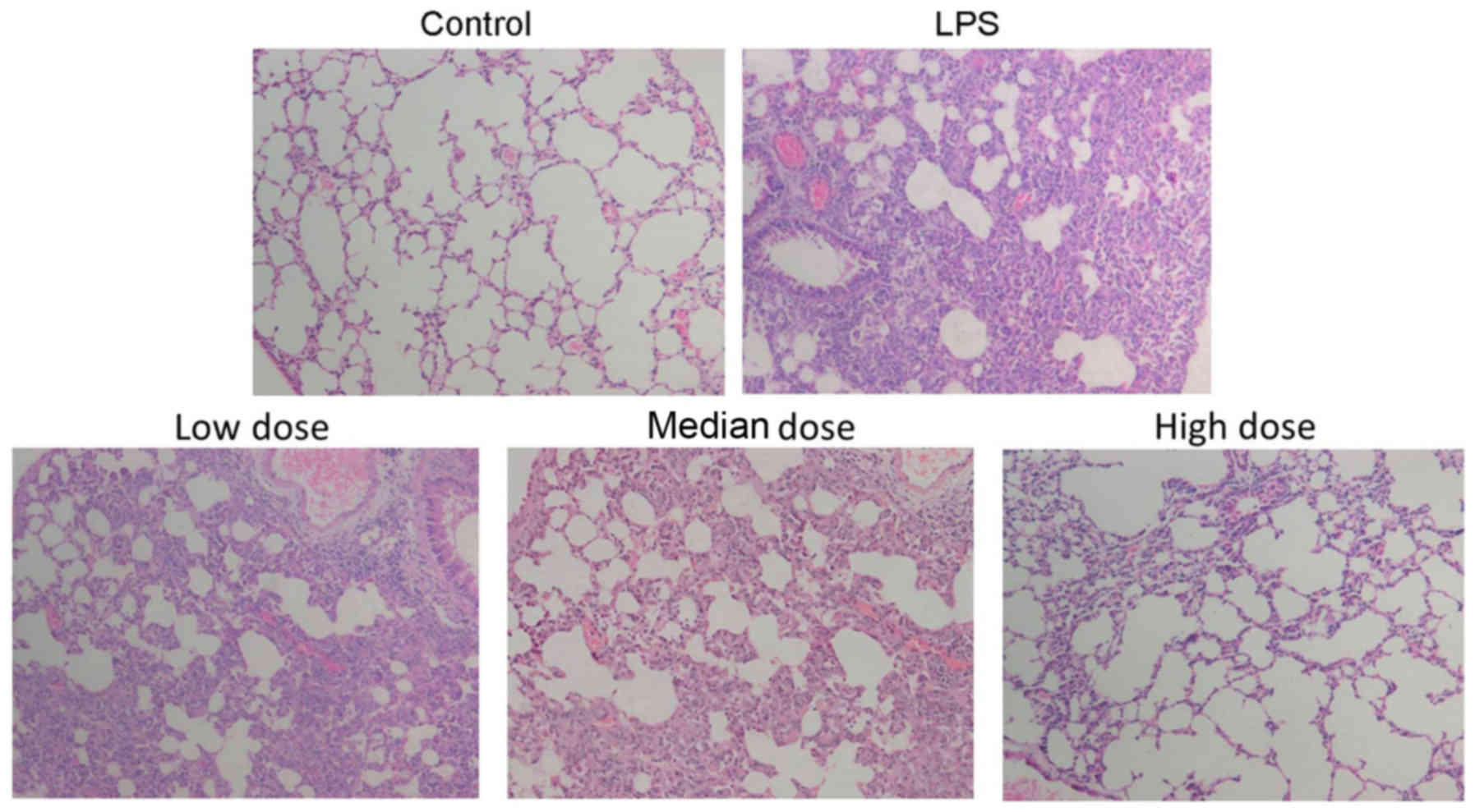
After rats sacrificed, the morphologic changes of
lung tissues in different groups were estimated using H&E
staining. The staining results showed that a larger number of
neutrophil infiltration was around the lung vessel and airway, and
distributed in the alveolar and interstitial in the LPS group
compared with the control group. After pre-treated with rhAPC, the
infiltration of inflammatory cells were obviously reduced,
indicating that rhAPC could relieve the inflammatory level in lung
injury tissues caused by LPS (Fig.
1).
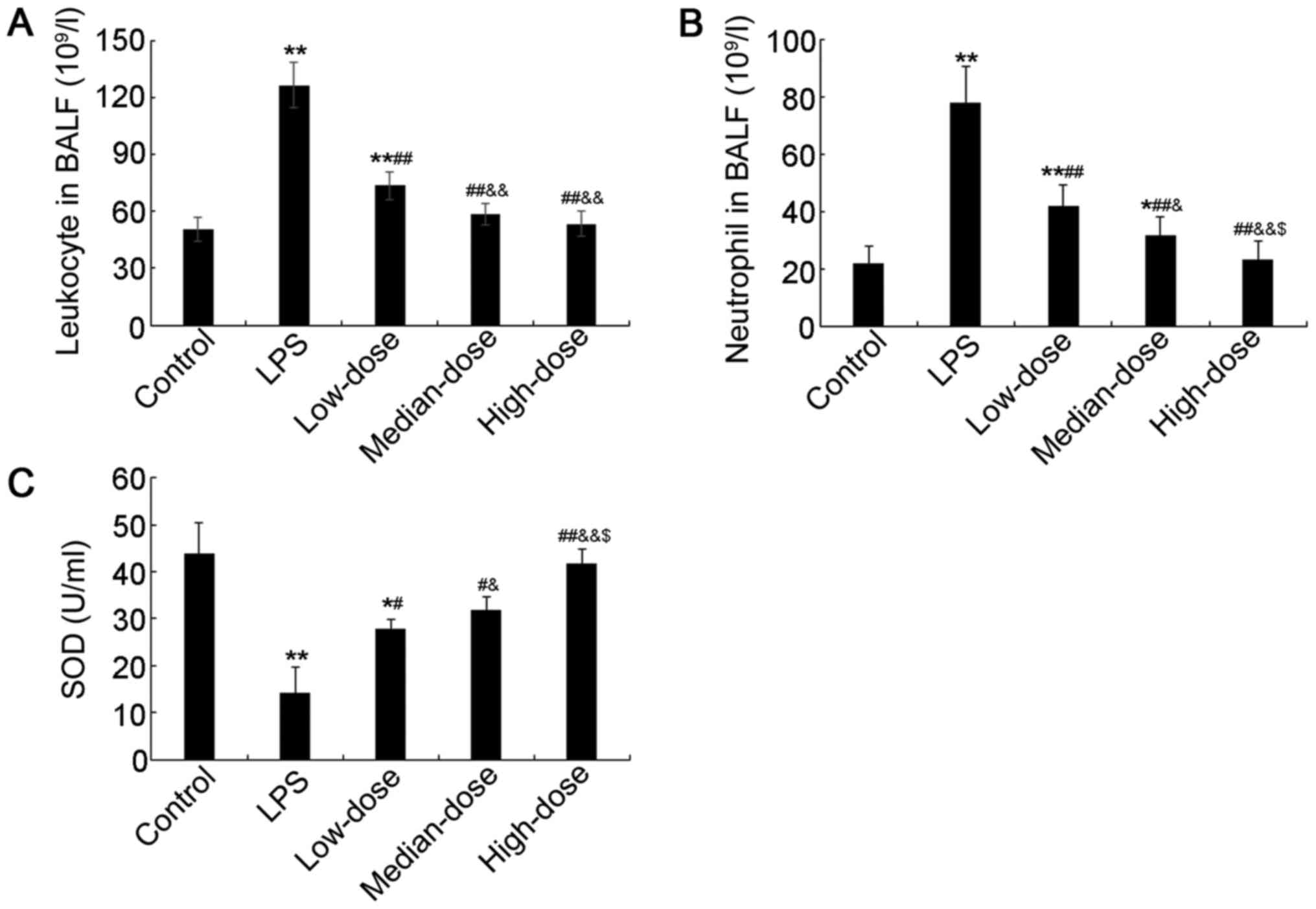
Variations of Leukocyte, neutrophil,
and SOD levels in BALF
The leukocytes and neutrophil counts, and SOD
activity in BALF were also investigated in the present study. The
counting results showed that the number of leukocyte was
significantly higher in the LPS group than in control group, and
rhAPC treatment could significantly decrease the number of
leukocyte with a dose-dependent manner (P<0.01; Fig. 2A). Meanwhile, the number of
neutrophil in BALF was also remarkably elevated in the LPS group
compared with the control group, and rhAPC also could obviously
decrease the number of neutrophil in BALF with a dose-dependent
manner (P<0.05, Fig. 2B).
However, the SOD activity level was significantly decreased in the
LPS group compared with the control group, and rhAPC could
evidently abort this elevation with a dose-dependent manner
(P<0.05, Fig. 2C).
Variations of inflammatory cytokines
in BALF
Meanwhile, inflammatory cytokines, including IL-1β,
IL-6, and TNF-α, were also investigated in LPS induced lung injury.
The detection showed that the expression level of IL-1β was
evidently increased in the LPS group compared with the control
group, but rhARC could significant abort this change with a
dose-dependent manner (P<0.05; Fig.
3A). Meanwhile, the expression levels of IL-6 and TNF-α were
also elevated in LPS-treated group than in the control group, and
pre-treated with rhAPC could significantly inhibit these elevation
with a dose-dependent manner (P<0.05; Fig. 3B and C), which were consistent with
the trend in IL-1β.
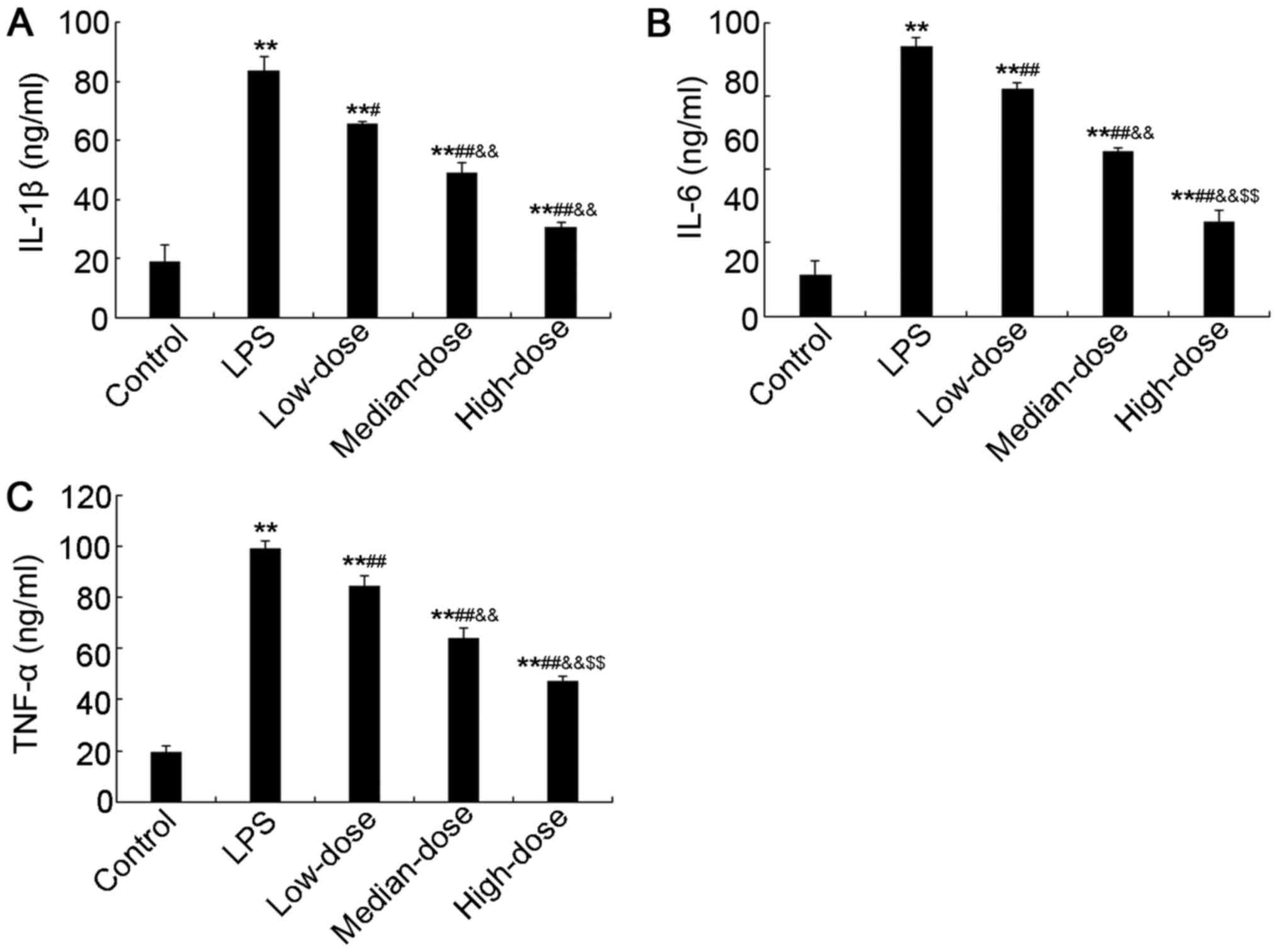 | Figure 3.Expression of IL-1β, IL-6 and TNF-α in
BALF determined using ELISA. Expression of (A) IL-1β, (B) IL-6 and
(C) TNF-α. The treatments applied for the different groups were as
follows: Control group (saline), model group (LPS), low-dose group
(LPS + 0.1 mg/kg rhAPC), median-dose group (LPS + 0.3 mg/kg rhAPC)
and high-dose group (LPS + 0.5 mg/kg rhAPC). **P<0.01 vs.
control; #P<0.05 and ##P<0.01 vs. LPS;
&&P<0.01 vs. Low-dose group;
$$P<0.01 vs. Median-dose group. IL, interleukin; TNF,
tumor necrosis factor; BALF, bronchoalveolar lavage fluid; LPS,
lipopolysaccharide; rhAPC, recombined human activated protein
C. |
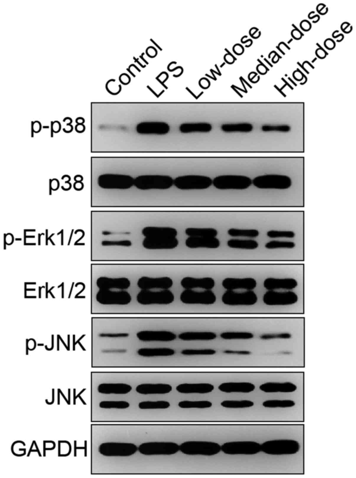
Pathway of rhAPC involved in lung
injury
To further investigate the mechanism of rhAPC,
expression and phosphorylation of participators involved in the
MAPK signaling pathway, including P-38, Erk1/2, and JNK were
determined. The results showed that there were no significantly
differences identified in the expression levels of P-38, Erk1/2,
and JNK in LPS and rhAPC treated groups compared with the control
group, but the phosphorylation levels of P-38, Erk1/2, and JNK were
significantly up-regulated in LPS treated group, and rhAPC could
significantly attenuate these elevations with a dose-dependent
manner (Fig. 4).
Discussion
In the present study, based on a LPS-induced ALI
model, the mechanism of rhAPC in the regulation of ALI associated
inflammation was investigated. The results showed that rhAPC could
significantly attenuate the accumulation and infiltration of
inflammatory cells, as well as the inflammatory cytokines,
including IL-1β, IL-6, and TNF-α induced by LPS with a
dose-dependent manner. Meanwhile, rhAPC also could evidently
reverse the reduction of SOD activity level caused by LPS with a
dependent manner. Further investigation showed that the
phosphorylation of P-38, Erk1/2, and JNK might involve in the
process of rhAPC against ALI associated inflammatory response.
As aforementioned, LPS is a common pathogen for the
occurrence of ALI (12). Several
researchers have identified that LPS can significantly up-regulated
the accumulation of neutrophil and mononuclear leukocytes, and the
expression levels of cytokines, including TNF-α, IL-1β, and IL-6
(13–15). In the present study, significant
elevations were also identified in the recruitment of neutrophil
and mononuclear leukocytes and the expression of TNF-α, IL-1β, and
IL-6, indicating that ALI model had been successfully induced.
Meanwhile, the SOD activity was significantly reduced in
LPS-treated group compared with the control group. Oxidative stress
is commonly identified in ALI and acute respiratory disease
syndrome (16), and SOD is an
important approach to revise the anomality of oxidative stress
(17,18). Thus, a significant reduction of SOD
activity might further contribute to the development of the
inflammation during ALI. Further analysis showed that LPS could
significantly increase the phosphorylation of P-38, Erk1/2, and
JNK, indicating that LPS might drive the inflammation in ALI via
MAPK signaling pathway. Park et al (19) have identified that mitochondrial ROS
regulates MAPK and NF-κB signaling pathways to participate in the
regulation of LPS-induced pro-inflammatory response in microglia.
He et al (20) have
documented that Baicalein attenuates inflammatory response induced
by LPS via suppressing TLR4 mediated NF-κB signaling pathway. These
findings indicated that LPS might enhance the inflammatory response
via regulating MAPK signaling pathway.
APC, a serine protease, is considered to play an
important role in the maintenance of hemostasis (21), inhibitions of cytokines release, and
reduction of leucocyte recruitment (22). In the past years, several studies
have recognized that APC has an anti-inflammatory effect that is
beneficial for stroke (23), sepsis
(24), ischemia-reperfusion injury
(25) in humans. In the present
study, APC was identified to significantly attenuate the
accumulations and infiltrations of leukocyte and neutrophil, and
the release of cytokines, including TNF-α, IL-6 and IL-1β, as well
as the reduction of SOD activity, in LPS-induced ALI with a
dose-dependent manner. These findings confirmed that APC indeed
performed a protective role against the increased inflammation
induced by LPS in the lung. Nick et al (26), have found that recombinant human
activated protein C could attenuate human endotoxin-induced lung
inflammation via inhibiting neutrophil chemotaxis, which was
consistent with the identification observed in the present study.
However, the mechanism of this process is still unclear. In the
present study, APC was identified to evidently decrease the
phosphorylation levels of P-38, Erk1/2, and JNK induced by LPS with
a dose-dependent manner, indicating APC could attenuate the
inflammation induced by LPS via MAPK signaling pathway. Shi et
al (27), have confirmed that
geniposide can suppress LPS-induced inflammation by inhibiting
NF-κB, MAPK and AP-1 signaling pathway. Meanwhile, Liang et
al (28), have also observed
that thymol attenuates LPS-induced inflammation via down-regulating
NF-κB and MAPK signaling pathways. All of these findings indicated
that inhibition of MAPK signaling pathway might perform a crucial
role in suppressing inflammation induced by LPS. Considering these
identification, it is supposed that APC also conduct a protective
effect against LPS-induced ALI via MAPK signaling pathway, but the
cross talk between MAPK and NF-κB was still needed to be further
investigated.
In conclusion, APC could significantly attenuate the
increase infiltrations and accumulations of leukocyte and
neutrophil, releases of IL-1β, IL-6, and TNF-α, and reduction of
SOD activity in LPS-induced ALI with a dose-dependent manner via
the MAPK signaling pathway. However, the cross talk between MAPK
and other potential signaling pathways was still needed further
exploration.
Acknowledgements
The authors would like to thank Southeast University
(Jiangsu, China).
Funding
No funding was received.
Availability of data and materials
All data generated and analyzed during this study
were included in this published article.
Authors' contributions
JZ conducted the majority of the experiments and
wrote the manuscript. RH, SJ and XX collected and analyzed the
data. WT designed the study and approved the final manuscript for
submission. All authors have read and approved the final
manuscript.
Ethics approval and consent to
participate
The animal experimental protocol was authorized by
the Ethics Committee of Southeast University Affiliated Zhongda
Hospital (Jiangsu, China).
Consent for publication
Not applicable.
Competing interests
All of the authors have no conflict of interest in
this research.
References
|
1
|
Chen T, Mou Y, Tan J, Wei L, Qiao Y, Wei
T, Xiang P, Peng S, Zhang Y, Huang Z and Ji H: The protective
effect of CDDO-Me on lipopolysaccharide-induced acute lung injury
in mice. Int Immunopharmacol. 25:55–64. 2015. View Article : Google Scholar : PubMed/NCBI
|
|
2
|
Tao W, Su Q, Wang H, Guo S, Chen Y, Duan J
and Wang S: Platycodin D attenuates acute lung injury by
suppressing apoptosis and inflammation in vivo and in vitro. Int
Immunopharmacol. 27:138–147. 2015. View Article : Google Scholar : PubMed/NCBI
|
|
3
|
Zhong W, Cui Y, Yu Q, Xie X, Liu Y, Wei M,
Ci X and Peng L: Modulation of LPS-stimulated pulmonary
inflammation by Borneol in murine acute lung injury model.
Inflammation. 37:1148–1157. 2014. View Article : Google Scholar : PubMed/NCBI
|
|
4
|
Jiang W, Luo F, Lu Q, Liu J, Li P, Wang X,
Fu Y, Hao K, Yan T and Ding X: The protective effect of Trillin
LPS-induced acute lung injury by the regulations of inflammation
and oxidative state. Chem Biol Interact. 243:127–134. 2016.
View Article : Google Scholar : PubMed/NCBI
|
|
5
|
Lee I, Dodia C, Chatterjee S, Feinstein SI
and Fisher AB: Protection against LPS-induced acute lung injury by
a mechanism-based inhibitor of NADPH oxidase (type 2). Am J Physiol
Lung Cell Mol Physiol. 306:L635–L644. 2014. View Article : Google Scholar : PubMed/NCBI
|
|
6
|
Do-Umehara HC, Chen C, Urich D, Zhou L,
Qiu J, Jang S, Zander A, Baker MA, Eilers M, Sporn PH, et al:
Suppression of inflammation and acute lung injury by Miz1 via
repression of C/EBP-δ. Nat Immunol. 14:461–469. 2013. View Article : Google Scholar : PubMed/NCBI
|
|
7
|
Yen YT, Yang HR, Lo HC, Hsieh YC, Tsai SC,
Hong CW and Hsieh CH: Enhancing autophagy with activated protein C
and rapamycin protects against sepsis-induced acute lung injury.
Surgery. 153:689–698. 2013. View Article : Google Scholar : PubMed/NCBI
|
|
8
|
Murakami K, Okajima K, Uchiba M, Johno M,
Nakagaki T, Okabe H and Takatsuki K: Activated protein C prevents
LPS-induced pulmonary vascular injury by inhibiting cytokine
production. Am J Physiol. 272:L197–L202. 1997.PubMed/NCBI
|
|
9
|
Maniatis NA, Letsiou E, Orfanos SE,
Kardara M, Dimopoulou I, Nakos G, Lekka ME, Roussos C, Armaganidis
A and Kotanidou A: Inhaled activated protein C protects mice from
ventilator-induced lung injury. Crit Care. 14:R702010. View Article : Google Scholar : PubMed/NCBI
|
|
10
|
Chesebro BB, Rahn P, Carles M, Esmon CT,
Xu J, Brohi K, Frith D, Pittet JF and Cohen MJ: Increase in
activated protein C mediates acute traumatic coagulopathy in mice.
Shock. 32:659–665. 2009. View Article : Google Scholar : PubMed/NCBI
|
|
11
|
Yunhe F, Bo L, Xiaosheng F, Fengyang L,
Dejie L, Zhicheng L, Depeng L, Yongguo C, Xichen Z, Naisheng Z and
Zhengtao Y: The effect of magnolol on the Toll-like receptor
4/nuclear factor κB signaling pathway in lipopolysaccharide-induced
acute lung injury in mice. Eur J Pharmacol. 689:255–261. 2012.
View Article : Google Scholar : PubMed/NCBI
|
|
12
|
Zhu T, Wang DX, Zhang W, Liao XQ, Guan X,
Bo H, Sun JY, Huang NW, He J, Zhang YK, et al: Andrographolide
protects against LPS-induced acute lung injury by inactivation of
NF-κB. PLoS One. 8:e564072013. View Article : Google Scholar : PubMed/NCBI
|
|
13
|
Jiang Q, Yi M, Guo Q, Wang C, Wang H, Meng
S, Liu C, Fu Y, Ji H and Chen T: Protective effects of polydatin on
lipopolysaccharide-induced acute lung injury through
TLR4-MyD88-NF-κB pathway. Int Immunopharmacol. 29:370–376. 2015.
View Article : Google Scholar : PubMed/NCBI
|
|
14
|
Wang B, Gong X, Wan JY, Zhang L, Zhang Z,
Li HZ and Min S: Resolvin D1 protects mice from LPS-induced acute
lung injury. Pulm Pharmacol Ther. 24:434–441. 2011. View Article : Google Scholar : PubMed/NCBI
|
|
15
|
Liu Y, Wu H, Nie YC, Chen JL, Su WW and Li
PB: Naringin attenuates acute lung injury in LPS-treated mice by
inhibiting NF-κB pathway. Int Immunopharmacol. 11:1606–1612. 2011.
View Article : Google Scholar : PubMed/NCBI
|
|
16
|
Ward PA: Oxidative stress: Acute and
progressive lung injury. Ann N Y Acad Sci. 1203:53–59. 2010.
View Article : Google Scholar : PubMed/NCBI
|
|
17
|
Kim Y, Kim BH, Lee H, Jeon B, Lee YS, Kwon
MJ and Kim TY: Regulation of skin inflammation and angiogenesis by
EC-SOD via HIF-1α and NF-κB pathways. Free Radic Biol Med.
51:1985–1995. 2011. View Article : Google Scholar : PubMed/NCBI
|
|
18
|
Howard MD, Greineder CF, Hood ED and
Muzykantov VR: Endothelial targeting of liposomes encapsulating
SOD/catalase mimetic EUK-134 alleviates acute pulmonary
inflammation. J Control Release. 177:34–41. 2014. View Article : Google Scholar : PubMed/NCBI
|
|
19
|
Park J, Min JS, Kim B, Chae UB, Yun JW,
Choi MS, Kong IK, Chang KT and Lee DS: Mitochondrial ROS govern the
LPS-induced pro-inflammatory response in microglia cells by
regulating MAPK and NF-κB pathways. Neurosci Lett. 584:191–196.
2015. View Article : Google Scholar : PubMed/NCBI
|
|
20
|
He X, Wei Z, Zhou E, Chen L, Kou J, Wang J
and Yang Z: Baicalein attenuates inflammatory responses by
suppressing TLR4 mediated NF-κB and MAPK signaling pathways in
LPS-induced mastitis in mice. Int Immunopharmacol. 28:470–476.
2015. View Article : Google Scholar : PubMed/NCBI
|
|
21
|
Hirose K, Okajima K, Taoka Y, Uchiba M,
Tagami H, Nakano K, Utoh J, Okabe H and Kitamura N: Activated
protein C reduces the ischemia/reperfusion-induced spinal cord
injury in rats by inhibiting neutrophil activation. Ann Surg.
232:272–280. 2000. View Article : Google Scholar : PubMed/NCBI
|
|
22
|
Bischofberger AS, Tsang AS, Horadagoda N,
Dart CM, Perkins NR, Jeffcott LB, Jackson CJ and Dart A: Effect of
activated protein C in second intention healing of equine distal
limb wounds: A preliminary study. Aust Vet J. 93:361–366. 2015.
View Article : Google Scholar : PubMed/NCBI
|
|
23
|
Amar AP, Griffin JH and Zlokovic BV:
Combined neurothrombectomy or thrombolysis with adjunctive delivery
of 3K3A-activated protein C in acute ischemic stroke. Front Cell
Neurosci. 9:3442015. View Article : Google Scholar : PubMed/NCBI
|
|
24
|
Zhang Z: The efficacy of activated protein
C for the treatment of sepsis: Incorporating observational evidence
with a Bayesian approach. BMJ Open. 5:e0065242015. View Article : Google Scholar : PubMed/NCBI
|
|
25
|
Allison SJ: Acute kidney injury: Activated
protein C protective in IRI. Nat Rev Nephrol. 11:4452015.
View Article : Google Scholar : PubMed/NCBI
|
|
26
|
Nick JA, Coldren CD, Geraci MW, Poch KR,
Fouty BW, O'Brien J, Gruber M, Zarini S, Murphy RC, Kuhn K, et al:
Recombinant human activated protein C reduces human
endotoxin-induced pulmonary inflammation via inhibition of
neutrophil chemotaxis. Blood. 104:3878–3885. 2004. View Article : Google Scholar : PubMed/NCBI
|
|
27
|
Shi Q, Cao J, Fang L, Zhao H, Liu Z, Ran
J, Zheng X, Li X, Zhou Y, Ge D, et al: Geniposide suppresses
LPS-induced nitric oxide, PGE2 and inflammatory cytokine by
downregulating NF-κB, MAPK and AP-1 signaling pathways in
macrophages. Int Immunopharmacol. 20:298–306. 2014. View Article : Google Scholar : PubMed/NCBI
|
|
28
|
Liang D, Li F, Fu Y, Cao Y, Song X, Wang
T, Wang W, Guo M, Zhou E, Li D, et al: Thymol inhibits
LPS-stimulated inflammatory response via down-regulation of NF-κB
and MAPK signaling pathways in mouse mammary epithelial cells.
Inflammation. 37:214–222. 2014. View Article : Google Scholar : PubMed/NCBI
|