Variations in ductus arteriosus Doppler parameters in different sonographic views during the second half of gestation

  • Authors:
    • Zhongshan Gou
    • Jie Zhang
    • Xinxin Yan
    • Zhenqi Wang
    • Shaolei Li
    • Xuedong Deng
  • View Affiliations

  • Published online on: November 8, 2018     https://doi.org/10.3892/etm.2018.6943
  • Pages: 502-506
Metrics: Total Views: 0 (Spandidos Publications: | PMC Statistics: )
Total PDF Downloads: 0 (Spandidos Publications: | PMC Statistics: )


Abstract

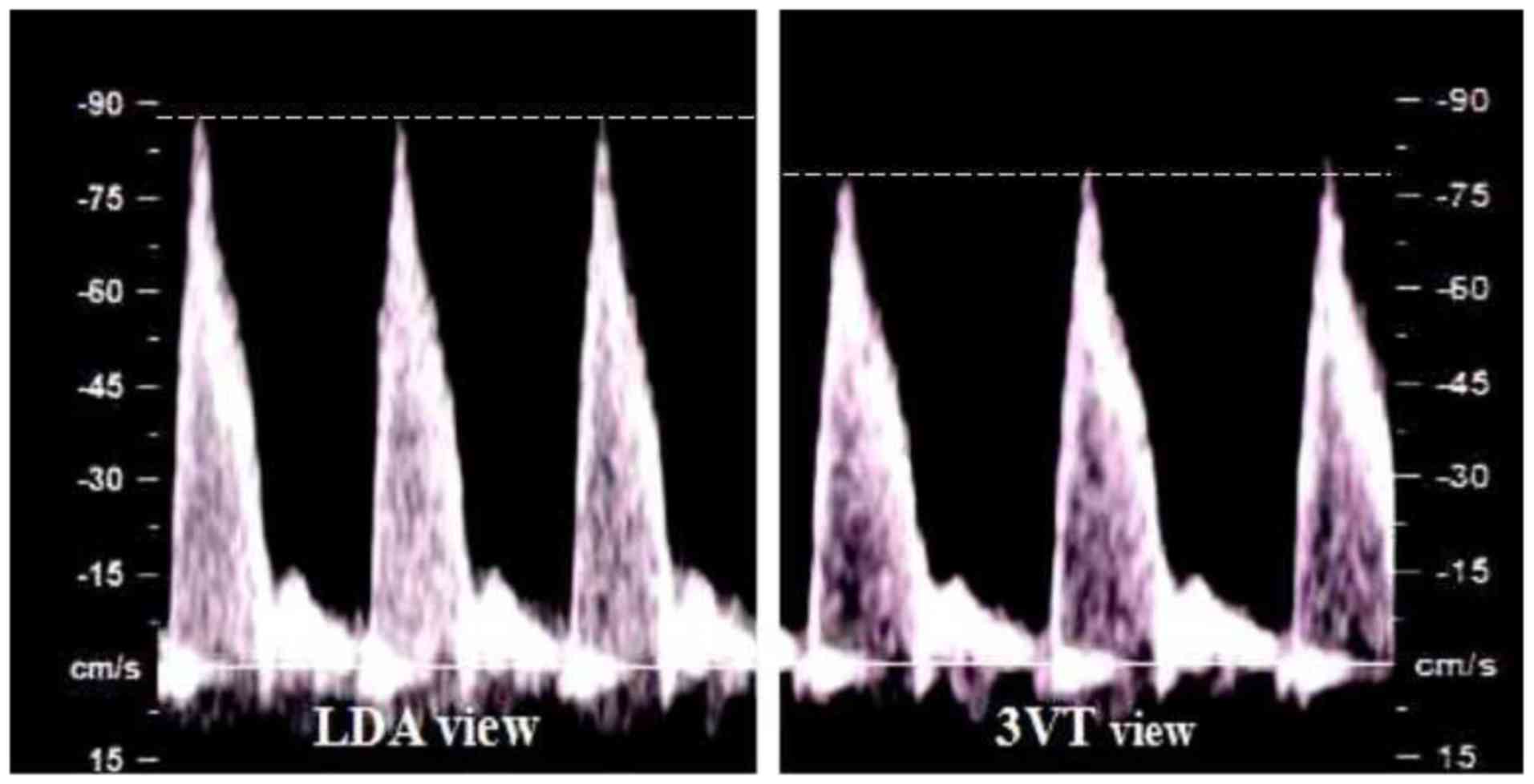
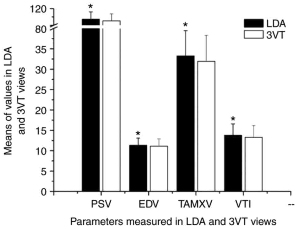
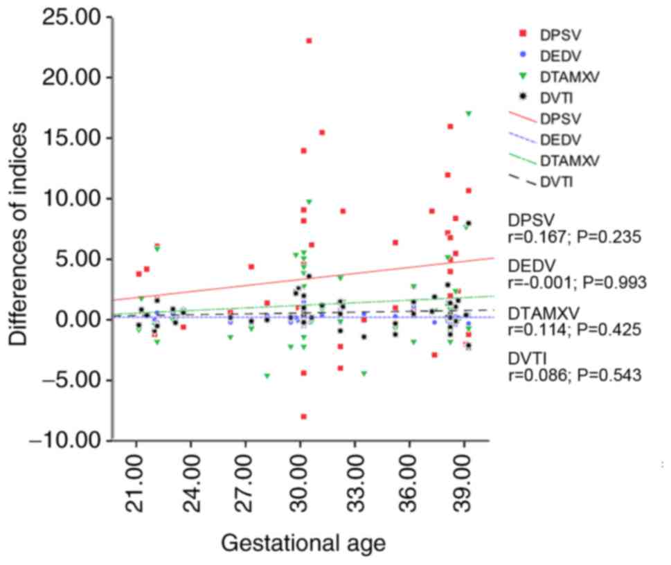
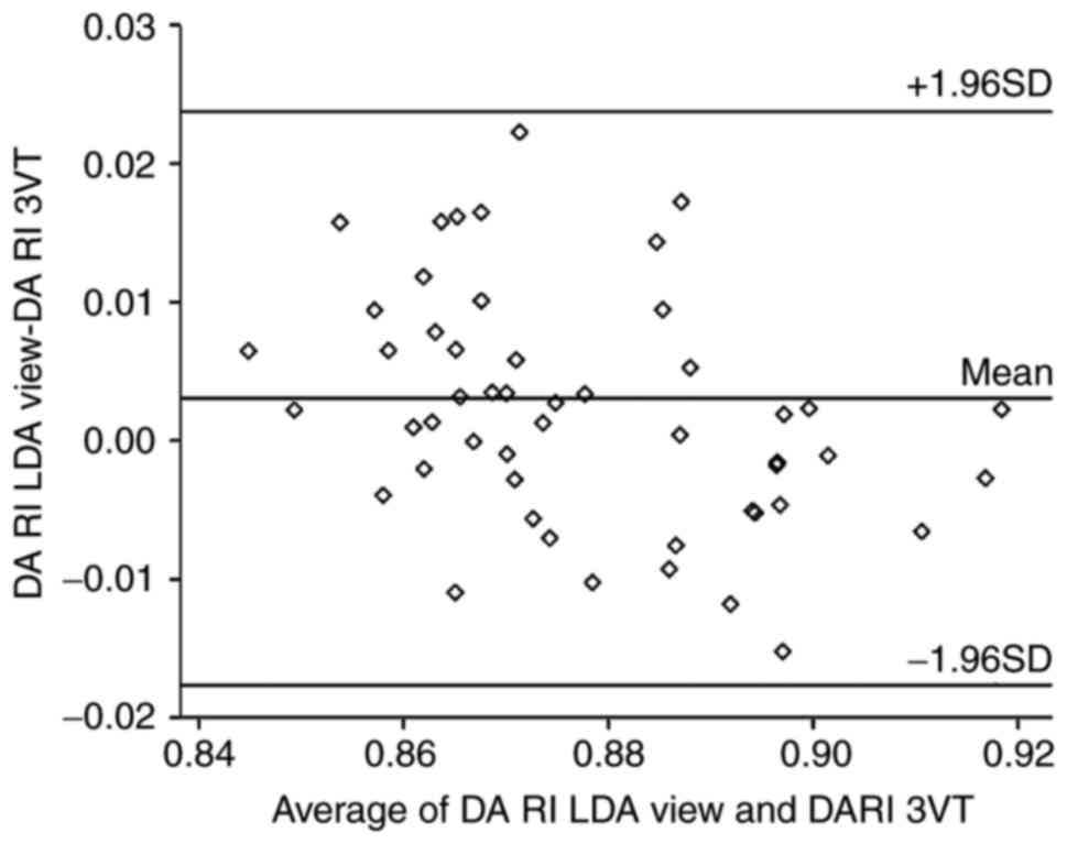
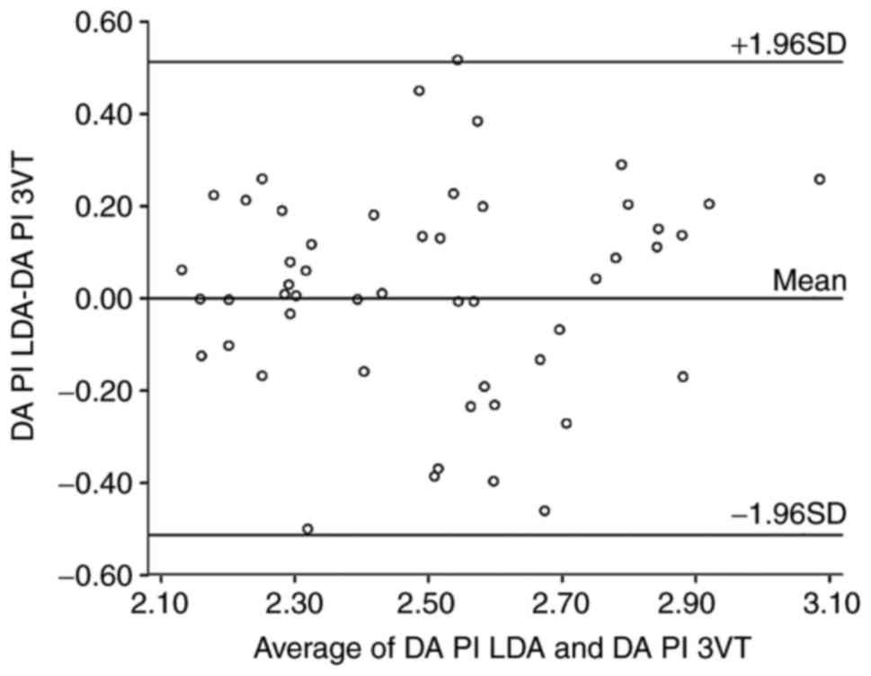
Abstract. The aim of the present study was to compare the Doppler parameters of the fetal ductus arteriosus (DA) measured in the traditional longitudinal ductal arch (LDA) view and the newly introduced three vessels and trachea (3VT) plane of the fetal upper mediastinum. The peak systolic velocity (PSV), end‑diastolic velocity (EDV), time‑averaged maximum velocity (TAMXV) and velocity‑time integral (VTI) measurements were taken for 52 fetuses with normal growth (including 29 females). The pulsatility index (PI) and resistance index (RI) were calculated. All parameters for each fetus were measured three times by the same sonographer in the LDA and the 3VT view, and the averages were taken to obtain the final value. Differences in the above values obtained from the LDA and 3VT views were analyzed and the correlation between the differences of all indices and the gestational age (GA) was evaluated using Pearson's linear coefficient of correlation. All of the values were characterized as normally distributed continuous variables by homogeneity of variance analysis. Slight increases in the PSV, EDV, TAMXV and VTI determined in the LDA view were identified compared with those in the 3VT view (P<0.05). Furthermore, these increases were identified to be independent of the GA (P>0.05). However, no significant differences in the impedance indices PI and RI were observed between the two sonographic planes (P>0.05). In conclusion, the LDA view provides a better chance than the 3VT view to obtain higher values of velocity parameters (PSV, EDV, TMAXV and VTI) within the fetal DA, and the differences are independent of the GA. However, no significant variations in the impedance indices PI and RI were observed between these two sonographic planes.
View Figures
View References

Related Articles

Journal Cover

January-2019
Volume 17 Issue 1

Print ISSN: 1792-0981
Online ISSN:1792-1015

Sign up for eToc alerts

Recommend to Library

Copy and paste a formatted citation
x
Spandidos Publications style
Gou Z, Zhang J, Yan X, Wang Z, Li S and Deng X: Variations in ductus arteriosus Doppler parameters in different sonographic views during the second half of gestation. Exp Ther Med 17: 502-506, 2019
APA
Gou, Z., Zhang, J., Yan, X., Wang, Z., Li, S., & Deng, X. (2019). Variations in ductus arteriosus Doppler parameters in different sonographic views during the second half of gestation. Experimental and Therapeutic Medicine, 17, 502-506. https://doi.org/10.3892/etm.2018.6943
MLA
Gou, Z., Zhang, J., Yan, X., Wang, Z., Li, S., Deng, X."Variations in ductus arteriosus Doppler parameters in different sonographic views during the second half of gestation". Experimental and Therapeutic Medicine 17.1 (2019): 502-506.
Chicago
Gou, Z., Zhang, J., Yan, X., Wang, Z., Li, S., Deng, X."Variations in ductus arteriosus Doppler parameters in different sonographic views during the second half of gestation". Experimental and Therapeutic Medicine 17, no. 1 (2019): 502-506. https://doi.org/10.3892/etm.2018.6943