Introduction
The incidence rate of colorectal cancer (CRC) varies
widely between different regions of the world (1). In Western countries, the incidence rate
of CRC is the second highest among all malignant tumors (2). In China, the incidence and mortality
rates of CRC ranks fourth among all malignant tumors, with 310,244
diagnoses made in 2011 alone (3).
The incidence and mortality rates of CRC vary significantly among
countries and different regions of China, as well as between urban
and rural areas (3).
Although improvements in diagnosis and treatment
have contributed to the early detection and good prognosis of
patients with CRC, those with more advanced lesions, particularly
those with distant metastasis exhibit a poor prognosis and
efficient therapeutic methods are lacking (4). Metastasis occurs when tumor cells
detach from the primary tumor site, invade the circulatory and
lymphatic vessels and travel to distant organs or tissues where
they invade and proliferate forming new tumors that affect multiple
organs, which may result in mortality (5). Therefore, an effective therapeutic
strategy is needed and targeting the molecular and cellular
mechanism underlying cancer cell invasion and metastasis may be an
appropriate therapeutic strategy.
MicroRNAs (miRNAs) are small, non-coding RNAs 18–25
nucleotides in length and are widely expressed in mammals (6). miRNAs are regulators of gene
expression, which affect cell proliferation, differentiation and
death (7,8). miR-338-3p is located on chromosome
17q25.3 within the 8th intron of the apoptosis-associated tyrosine
kinase gene, which produces two forms: miR-338-3p and miR-338-5p
(9–14). It has been reported that chromosome
17q23-25 is a region associated with mutations in several malignant
tumors (15,16). In addition, this locus is associated
with many malignant biological behaviors which include tumor
vascular invasion and distant metastasis (13). Previous studies have demonstrated
that miR-338-3p inhibits glioblastoma proliferation (9,12), renal
carcinoma invasion (10), gastric
cancer progression (11,17), ovarian epithelial carcinoma growth
(18) and hepatocellular carcinoma
proliferation (14). However,
studies investigating the cellular function of miR-338-3p in CRC
are lacking.
Metastasis-associated in colon cancer-1 (MACC1) was
identified by Stein et al (19) in 2009 and is located on chromosome 7
(7p21.1). A previous study demonstrated that MACC1 exhibits 49.3
and 43.7% homology with SH3 domain-binding protein 4 between
nucleotide and amino acid sequences, respectively (20) The protein structure of MACC1 includes
a SH3 domain and a P-X-X-P motif, which is required for binding to
SH3 domains (20). In addition,
there are multiple phosphorylation sites, which indicate that MACC1
may be required for important signal transduction pathways
(20). MACC1 expression in CRC is an
independent predictor of metastasis (19). A previous study demonstrated that
miR-338-3p regulates MACC1 expression in cervical cancer as well as
cancer progression by targeting MACC1 via the mitogen-activated
protein kinase (MAPK) signaling pathway (21). miR-338-3p also inhibits
epithelial-mesenchymal transition (EMT) by targeting zinc finger
E-box-binding homeobox 2 (ZEB2) and MACC1 (11). The aggressive features associated
with glioma cells are suppressed by miR-338-3p by directly
silencing MACC1 expression (12).
However, whether there is an association between miR-338-3p and
MACC1 in CRC remains unknown.
Therefore, it was hypothesized that the
miR-338-3p/MACC1 axis may participate in the occurrence and
development of CRC. The aim of the current study was to investigate
miR-338-3p expression in CRC tissues and its underlying mechanism.
The results of the current study may facilitate the identification
of a potential therapeutic target for the treatment of CRC.
Materials and methods
Patient samples
The present study analyzed tissue samples from 15
patients (9 males, 6 females; mean age, 52.5±9.3 years) with CRC
who underwent surgery at the Second Affiliated Hospital of Kunming
Medical University (Kunming, China) between January 2015 and May
2015. CRC was confirmed in all patients following pathological
examination and patients did not receive neoadjuvant chemotherapy
or radiotherapy. CRC tissue and adjacent normal colon tissue (≥2
cm) were collected and stored in liquid nitrogen until further use.
The study was approved by the Ethics Committee of the Second
Affiliated Hospital of Kunming Medical University and all patients
provided their written informed consent.
Cell culture
The human CRC cell line SW480 and the 293T cell line
were provided by the Shanghai Institute of Biochemistry and Cell
Biology, Chinese Academy of Sciences (Shanghai, China). Cells were
cultured in high glucose Dulbecco's modified Eagle's medium (Gibco;
Thermo Fisher Scientific, Inc., Waltham, MA, USA) supplemented with
10% fetal bovine serum (FBS; Gibco; Thermo Fisher Scientific, Inc.)
and maintained at 37°C in a 5% CO2-humidified incubator.
Cells were passaged every 2–3 days, according to cell density.
Experiments were performed when cells had reached a confluence of
80% confluence.
MACC1 siRNA lentivirus construction,
packaging and transfection
The siRNA fragments targeting MACC1 were identified
and amplified as previously described (21). MACCI siRNA was cloned into the
lentiviral transfer plasmid LV-3 (pGLVH1/GFP/Furo; cat. no. c06003,
Shanghai GenePharma Co. Ltd., Shanghai, China). 293T cells were
subsequently transfected with the transfer plasmid (10 µg) encoding
MACC1 siRNA, lentiviral packaging plasmid pRSV-Rev (2 µg; cat. no.
#12253) and the envelope plasmid pMD2.G (2 µg; #12259; both
Addgene, Inc., Cambridge, MA, USA) using Lipofectamine®
2000 (Invitrogen; Thermo Fisher Scientific, Inc.), according to the
manufacturer's protocol. Following 48-h incubation at 37°C, the
lentiviral supernatant was harvested and pelleted by centrifugation
at 4,000 × g for 10 min at 4°C. The lentiviral supernatant was
subsequently filtered using 0.4 µm cellulose acetate filters and
used to transfect SW480 cells. SW480 cells were seeded at a density
of 1×10⁴ cells/well prior to transfection with lentivirus.
Following 48-h transfection, SW480 cells were in subsequent
experimentation.
Cell proliferation assay
miR-338-3p mimic (5′-UUUGAGCAGCACUCAUUUUUGC-3′),
miR-338-3p inhibitor (5′-CAACAAAAUCACUGAUGCUGGA-3′), and miR
control (5′-CAGUACUUUUAGUGUGUACAA-3′) were purchased from Shanghai
GenePharma Co., Ltd. (Shanghai, China). Briefly, SW480 cells were
seeded into 96-well plates at a density of 2×10⁴ cells/well. Once
at 70–80% confluency, cells were transfected with 50 nM miR control
(transfection control), miR-338-3p mimic or miR-338-3p inhibitor
using Lipofectamine® 2000 (Invitrogen; Thermo Fisher
Scientific, Inc.), according to the manufacturer's protocol.
Following 24-h transfection, 10 µg of MTT was added to each well
and incubated for a further 4 h at 37°C. Following incubation, 50
µl DMSO was added to each well and plates were shaken for 10 min.
DMSO was used as control. Cell proliferation was determined by
measuring absorbance at a wavelength of 570 nm using a microplate
reader.
Cell apoptosis assay
Cell apoptosis was analyzed using an Annexin
V-fluorescein isothiocyanate (FITC) Apoptosis Detection kit (cat.
no. 401003; BestBio, Shanghai, China), according to the
manufacturer's protocol. Following 48 h transfection with controls,
miR-338-3p mimics and miR-338-3p inhibitors, SW480 cells were
washed twice with PBS, dissociated with trypsin and pelleted using
centrifugation at 1,500 × g for 5 min at 4°C. Cells were
resuspended in 500 µl 1X binding buffer and subsequently stained
with 1 µl Annexin V-FITC for 15 min at 4°C in the dark followed by
5 µl propidium iodine (PI) for 15 min at 4°C in the dark. Apoptotic
cells were detected using a BD FACS Calibur flow cytometer (BD
Biosciences, Franklin Lakes, NJ, USA). Data were analyzed using
CellQuest Pro software (version 5.1; BD Biosciences).
Cell cycle analysis
Following 48 h transfection with controls,
miR-338-3p mimics and miR-338-3p inhibitors, SW480 cells were
washed twice with PBS, dissociated with trypsin, and pelleted using
centrifugation at 1,500 × g for 5 min at 4°C. Cells were then
resuspended in 100 µl of RNase A and incubated for 30 min at 37°C.
Cells were subsequently stained with 400 µl PI (BestBio) and
incubated for 30 min at 4°C for 30 min in the dark. Flow cytometry
was used to detect the cell cycle distribution.
Transwell migration assay
Migration assays were performed using a 24-well
Transwell chambers with 8.0 µm pore size inserts (Corning
Incorporated, Corning, NY, USA). Following 24-h transfection with
controls, miR-338-3p mimics and miR-338-3p inhibitors, SW480 cells
were seeded in the upper chambers in serum-free medium at a density
of 3×10⁴ cells/well, whilst medium (containing 10% FBS) was added
to the lower chambers. Following 24-h incubation at 37°C, the
chamber was removed and cells that migrated through the membrane
were fixed with 4% formaldehyde (Sigma-Aldrich; Merck KGaA,
Darmstadt, Germany) and acetic acid for 15 min at 37°C, washed with
PBS and stained with crystal violet solution for 10 min at 37°C.
The number of cells were observed and counted from five randomly
selected visual fields under a light microscope (magnification,
×200).
Dual-luciferase reporter assay
TargetScan Bioinformatics software (www.targetscan.org/vert_72) was used to predict
the putative target genes of miR-338-3p. To confirm direct target
binding, the wild-type (wt) or mutant (mut) 3′-untranslated region
(UTR) MACC1 were cloned into the pMIR-REPORT™ Luciferase miRNA
Expression reporter vector (cat. no. AM5795; Ambion; Thermo Fisher
Scientific, Inc.) to generate pmir-MACC1-wtUTR and
pmir-MACC1-mutUTR. 293T cells were co-transfected with miR-338-3p
mimics or miR-negative controls (NCs; Wuhan Biofavor Biotech
Services Co., Ltd., Wuhan, China) and pmir-MACC1-wtUTR or
pmir-MACC1-mutUTR using Lipofectamine® 2000 (cat. no.
15596026; Invitrogen; Thermo Fisher Scientific, Inc.) for 24 h at
37°C. Following 24-h incubation, cells were collected and
luciferase activity was detected using the Dual-Luciferase Assay
system (Promega Corporation, Madison, WI, USA). Firefly luciferase
activity was normalized to Renilla luciferase activity.
Reverse transcription-quantitative
polymerase chain reaction (RT-qPCR)
RT-qPCR was used to detect the expression of
miR-338-3p in CRC and adjacent normal tissue samples. Total RNA was
extracted from tissue samples using TRIzol® reagent
(Invitrogen; Thermo Fisher Scientific, Inc.). Total RNA was reverse
transcribed into cDNA using a PrimeScript™ II. 1st strand DNA
Synthesis kit (cat. no. 6210A; Takara Bio, Inc., Otsu, Japan). qPCR
was subsequently performed using SYBR® Green PCR Master
mix (Takara Bio, Inc.) with the FQ-PCR detection system (Applied
Biosystems; Thermo Fisher Scientific, Inc.). The following primer
pairs were used for the qPCR: miR-338-3p forward,
5′-GGTCCAGCATCAGTGA-3′ and reverse, 5′-GAGCAGGCTGGAGAA-3′; and U6
forward, 5′-CTCGCTTCGGCAGCACATA-3′ and reverse,
5′-CGCTTCACGAATTTGCGTG-3′. The following thermocycling conditions
were used for the qPCR: Initial denaturation at 94°C for 4 min; 40
cycles of 95°C for 20 sec, 57°C for 30 sec and 72°C for 30 sec.
miR-338-3p levels were quantified using the 2−ΔΔCq
method (22) and normalized to the
internal reference gene U6.
Western blot analysis
Total protein was extracted from SW480 cells
following 48 h transfection with controls, miR-338-3p inhibitors
and miR-338-3p mimics using radioimmunoprecipitation assay buffer.
Total protein was quantified using a bicinchoninic acid assay and
50 µg protein/lane was separated via SDS-PAGE on a 15% gel. The
separated proteins were subsequently transferred onto
polyvinylidene difluoride membranes and blocked for 1 h at 37°C
with the 5% skimmed milk (Sangon Biotech Co., Ltd., Shanghai,
China). Membranes were then incubated with primary antibodies
against MACC1 (1:1,000; cat. no. ab106579) and GAPDH (1:2,000; cat.
no. ab9485; both Abcam, Cambridge, UK) overnight at 4°C. Following
primary incubation, membranes were incubated with horse radish
peroxidase (HRP)-labeled goat anti-rabbit IgG secondary antibody
(1:5,000; cat. no. AQ132P; Sigma-Aldrich; Merck KGaA) for 1 h at
37°C. Protein bands were visualized using the Pierce™ ECL Western
Blotting Substrate (cat. no. 32109; Pierce; Thermo Fisher
Scientific Inc.). Protein expression was analyzed using Labworks™
Analaysis software (version 4.0; UVP, Upland, CA, USA) with GAPDH
as the loading control.
Immunohistochemistry
Tissue samples were fixed in 4% paraformaldehyde
(Sigma-Aldrich; Merck KGaA) at 37°C for 30 min and embedded in
paraffin. Paraffin-embedded tissue samples were cut into 3–5-µm
thick sections. The tissue sections were deparaffinized in xylene
at 55°C and rehydrated in a descending alcohol series.
Deparaffinized tissue sections were blocked for 1 h at room
temperature with 10% FBS and incubated with 3%
H2O2 (Sangon Biotech Co., Ltd.) for 10 min at
37°C. Tissue sections were washed in triplicate with distilled
water for 2 min. Tissue sections were incubated with primary
antibody against MACC1 (1:1,000; cat. no. ab106579; Abcam)
overnight at 4°C. Subsequently, the tissues were incubated with
HRP-labeled goat anti-rabbit IgG secondary antibody (1:2,000; cat.
no. AQ132P; Sigma-Aldrich; Merck KGaA) for 1 h at 37°C. Tissue
sections were subsequently stained with 3, 3′-diaminobenzidine
(DAB) solution for color development at 37°C for 5 min and observed
under a light microscope (magnification, ×200; BX51; Olympus,
Tokyo, Japan). DAB precipitates as a dark brown pigment allowing
for the visualization of positively stained cells. PBS alone was
used as a negative control. Two independent pathologists blindly
analyzed MACC1 staining. The percentage of positively stained cells
was quantified as follows: 0 (no positive cells), 1 (<10%
positive cells), 2 (10–50% positive cells) and 3 (>50% positive
cells). Staining intensity was graded as follows: 0 (no color), 1
(light yellow), 2 (claybank) and 3 (sepia). The two scores were
multiplied and MACC1 expression was determined according to the
overall score whereby: ≤4 indicates negative expression and ≥6
indicates positive expression.
Statistical analysis
Data presented as the mean ± standard deviation. All
statistical analyses were performed using SPSS software (version
13.0; SPSS, Inc., Chicago, IL, USA). A Student's t-test was used to
analyze differences between two groups. One-way analysis of
variance followed by followed by a Bonferroni post-hoc test was
used to analyze differences among multiple groups. P<0.05 was
considered to indicate a statistically significant difference.
Results
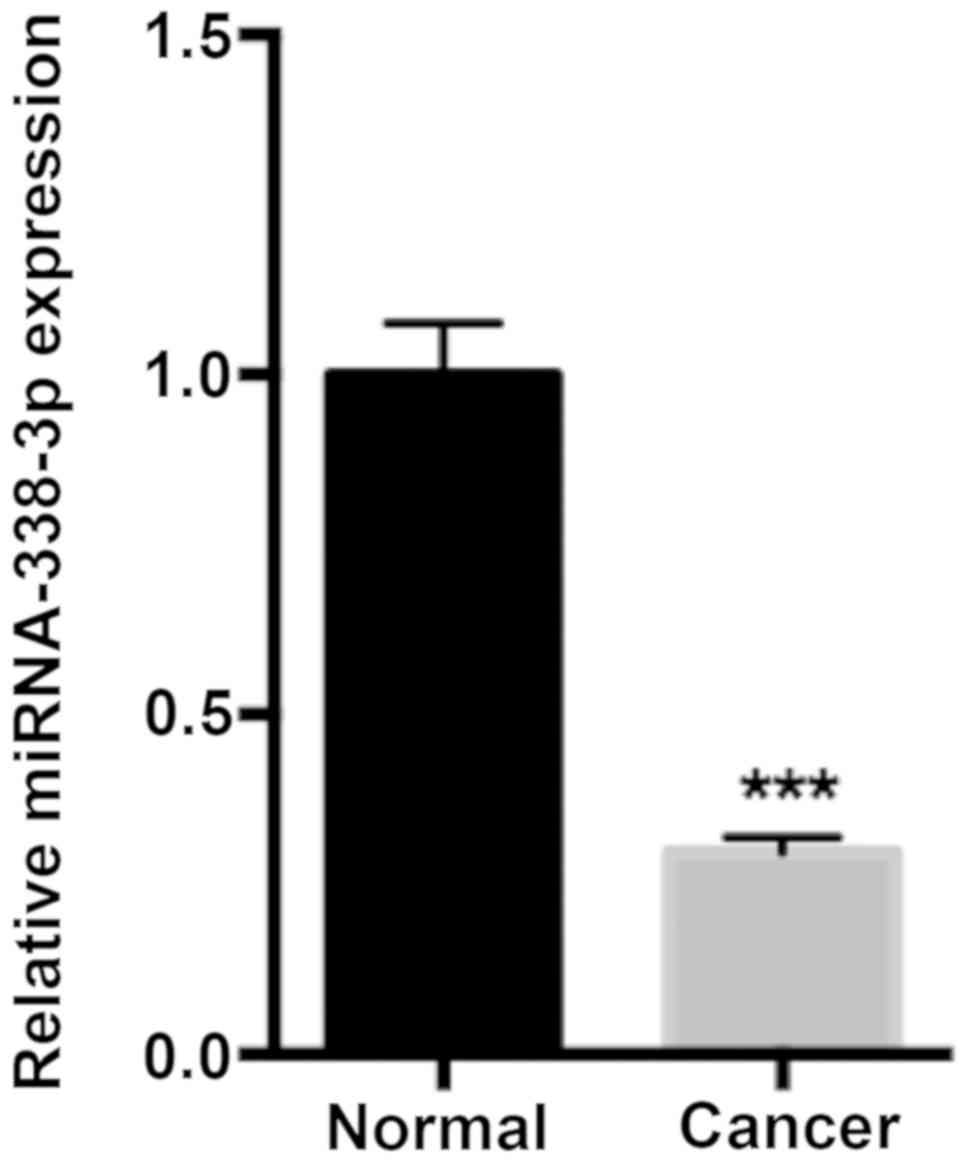
miR-338-3p expression is decreased in
CRC tissue samples
To assess the function of miR-338-3p in CRC
progression, miR-338-3p expression in human CRC and adjacent normal
tissue samples was determined via RT-qPCR. The results demonstrated
that miR-338-3p expression was significantly decreased in the CRC
tissue compared with adjacent normal tissue samples (P<0.001;
Fig. 1).
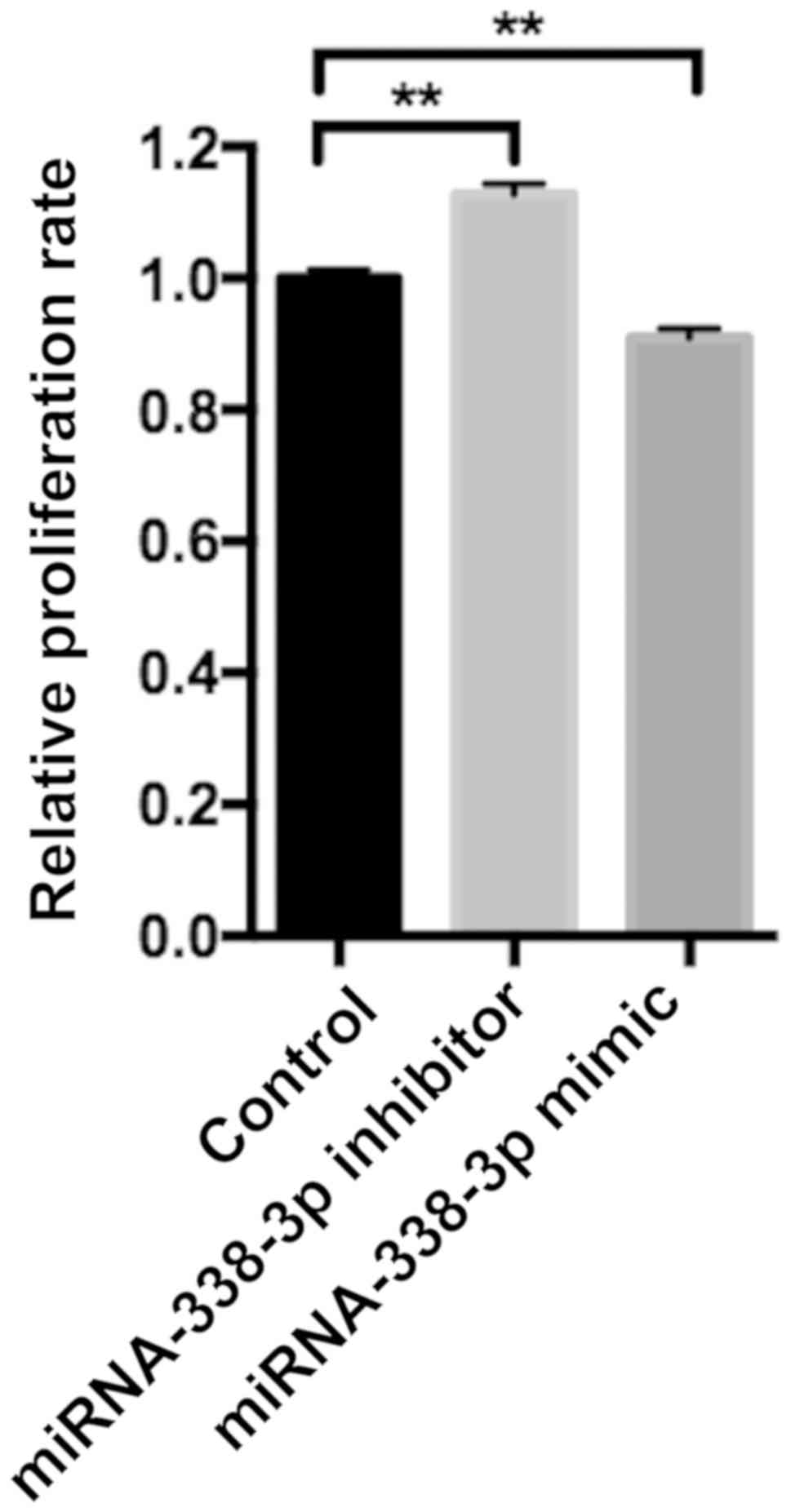
miR-338-3p regulates SW480 cell
proliferation, apoptosis and migration
SW480 cell proliferation was examined following
transfection with controls, miR-338-3p mimics or miR-338-3p
inhibitors. The miR-338-3p inhibitor significantly enhanced cell
proliferation, whilst the miR-338-3p mimic significantly inhibited
cell proliferation compared with the control group (P<0.01;
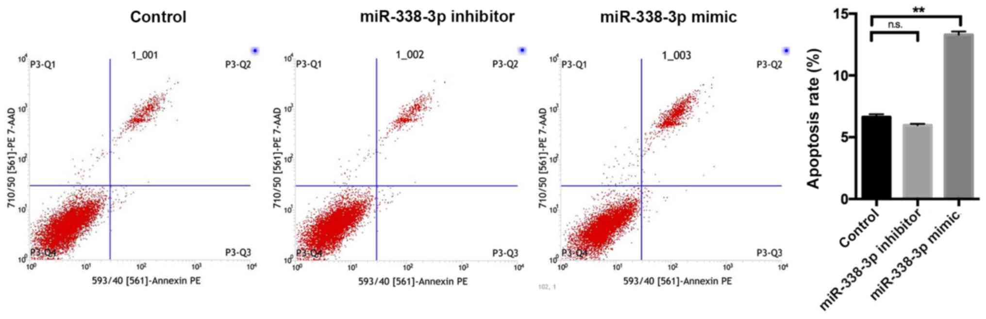
Fig. 2). Flow cytometry revealed
that the miR-338-3p inhibitor had no significant effect on cell
apoptosis, whilst the miR-338-3p mimic significantly enhanced cell
apoptosis (P<0.01; Fig. 3).
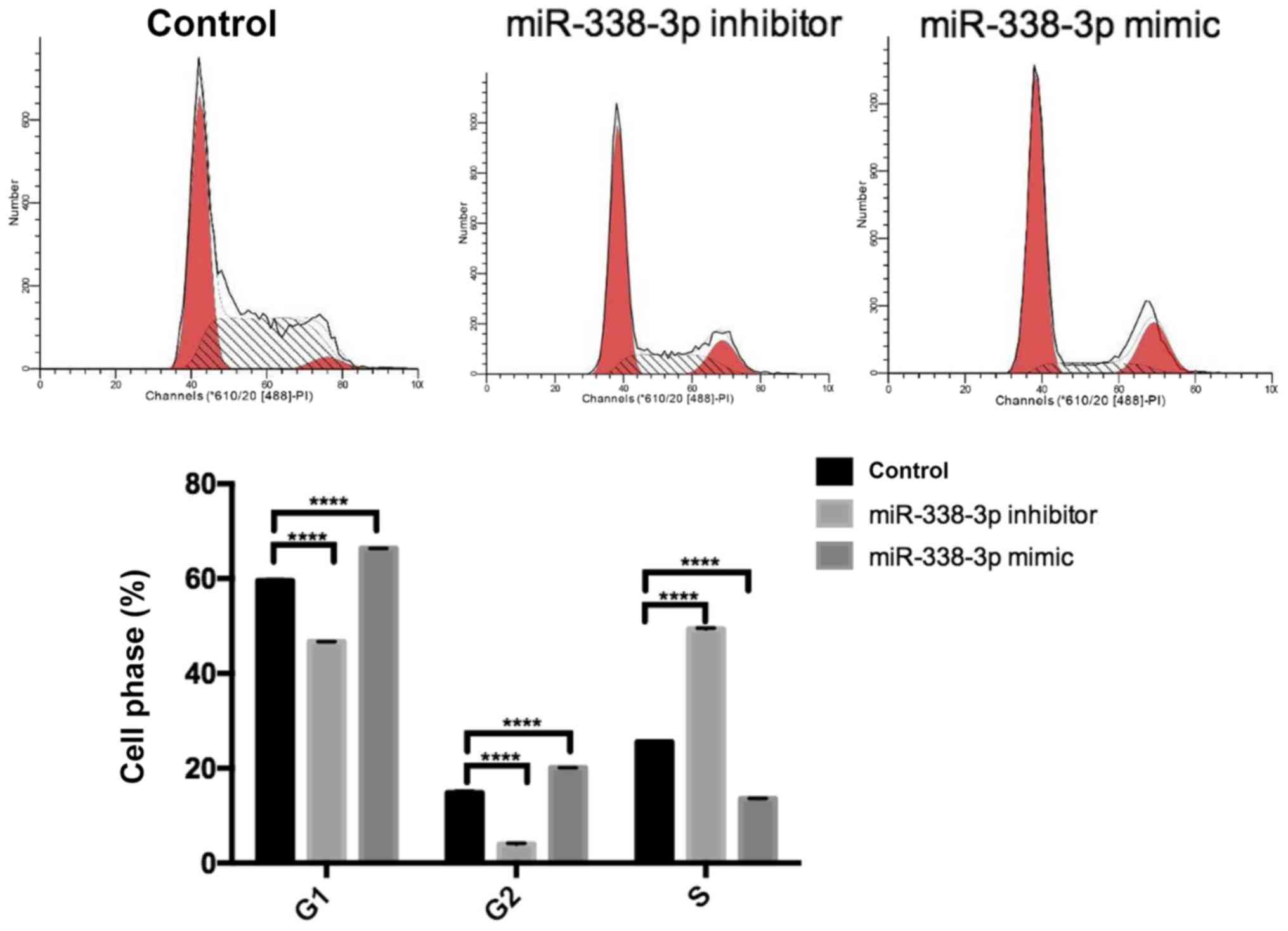
Furthermore, miR-338-3p had a significant effect on SW480 cell
cycle. The miR-338-3p inhibitor significantly increased the
proportion of cells in S phase and significantly decreased the
proportion of cells in G1/G2 phase compared with the control group
(P<0.0001; Fig. 4). However, the
miR-338-3p mimic significantly decreased the proportion of cells in
S phase and significantly increased the proportion of cells in
G1/G2 phase compared with the control group (P<0.0001; Fig. 4). Additionally, the miR-338-3p
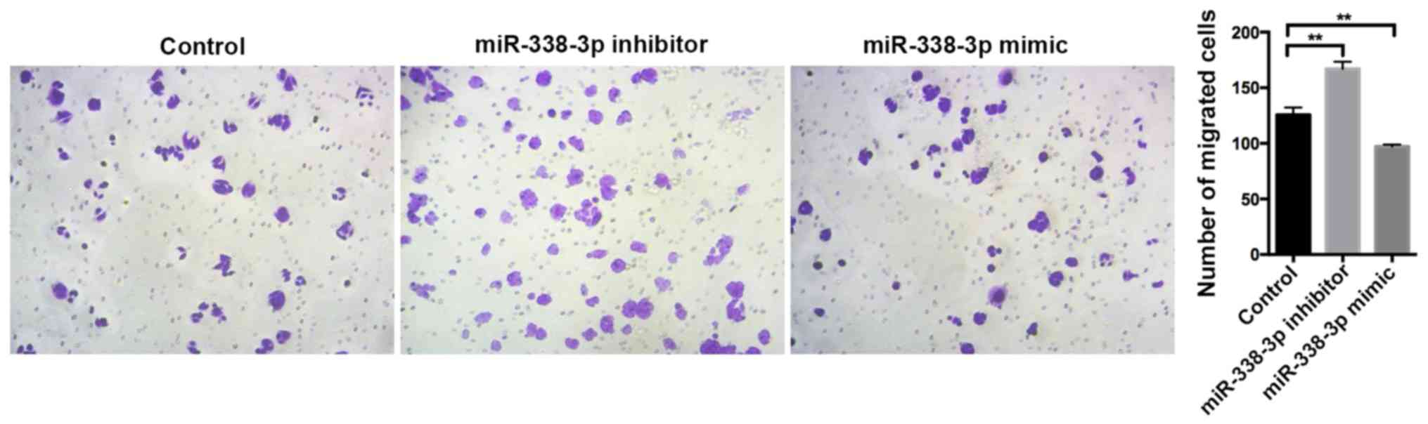
inhibitor significantly enhanced cell migration, whilst the
miR-338-3p mimic significantly suppressed cell migration compared
with the control group (P<0.01; Fig.
5), suggesting that miR-338-3p may regulate the migration of
the CRC cell line SW480. Taken together, these results indicate
that miR-338-3p may suppress the aggressive clinicopathological
features associated with CRC.
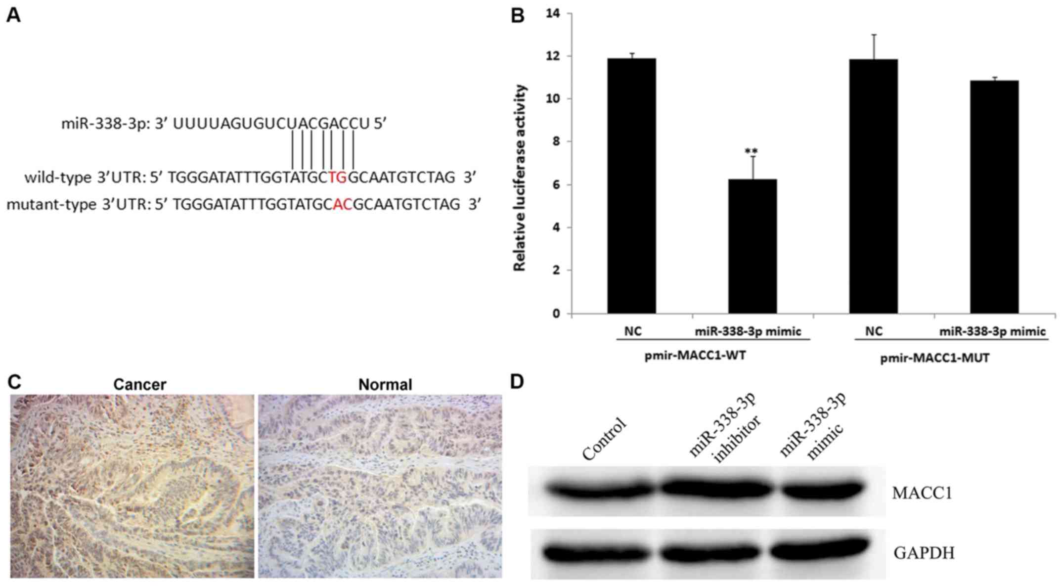
MACC1 is a direct target of
miR-338-3p
To further assess the role of miR-338-3p in human
CRC, potential targets of miR-338-3p were determined.
TargetScanHuman bioinformatics software was used to identify MACC1
as a putative target gene of miR-338-3p (Fig. 6A). The luciferase assay revealed that
miR-338-3p significantly reduced wt MACC1 expression compared with
the NC group (P<0.01; Fig. 6B).
In addition, miR-338-3p had no effect on mut MACC1 expression
compared with the NC group (Fig.
6B). However, the positive expression rate of MACC1 was
increased in CRC tissue compared with the normal adjacent tissue
samples (Fig. 6C). Among the 15 CRC
tissue samples, 10 (66.7%) were positive for MACC1 expression,
whilst in the 15 adjacent normal tissue samples, 3 (20.0%) were
positive for MACC1 expression (data not shown). The current study
demonstrated that the miR-338-3p inhibitor upregulated the level of
MACC1 protein expression compared with the control group (Fig. 6D). These results indicate that MACC1
is a direct target of miR-338-3p.
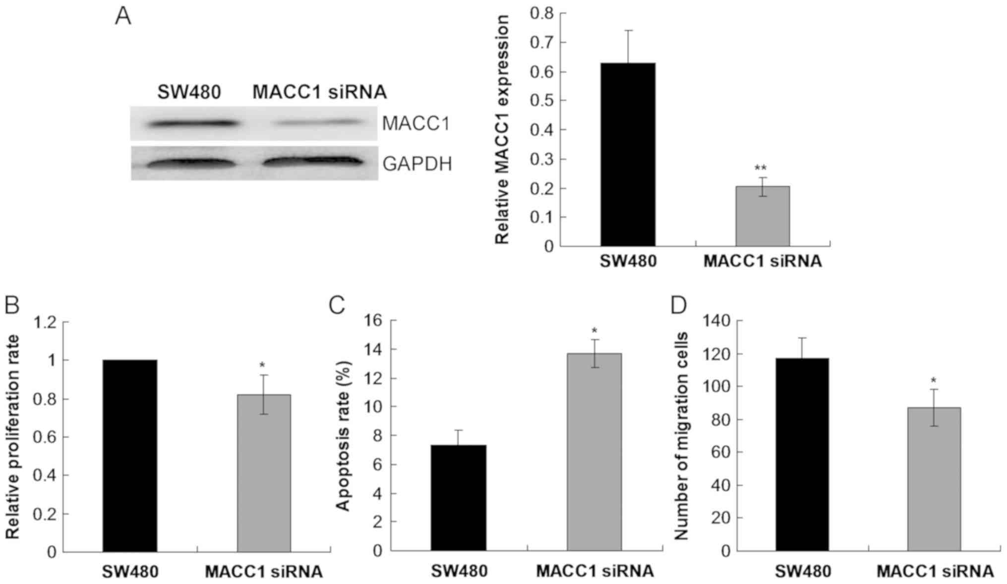
MACC1 silencing inhibits SW480 cell
proliferation, migration and induces cell apoptosis
To further investigate the role of MACC1 in CRC,
SW480 cell growth was examined following transfection with MACC1
siRNA. The current study demonstrated that MACC1 siRNA
significantly decreased the level of MACC1 protein expression
compared with the control SW480 group (P<0.01; Fig. 7A). Cell proliferation rate was
significantly reduced (P<0.05; Fig.
7B), whilst cell apoptosis was significantly enhanced
(P<0.05; Fig. 7C) in SW480 cells
following transfection with MACC1 siRNA compared with the control
SW480 group. Furthermore, cell migration was significantly reduced
in SW480 cells following transfection with MACC1 siRNA compared
with the control SW480 group (P<0.05; Fig. 7D).
Discussion
miR-338-3p has been reported to be involved in the
progression and development of various types of cancer (9–14,17,18,21).
However the cellular function of miR-338-3and its molecular
mechanism in CRC remains unknown. The aim of the current study was
to assess the cellular function of miR-338-3p in CRC, the malignant
behavior of CRC cells and the interaction between miR-338-3p and
MACC1. The current study demonstrated that miR-338-3p expression
was significantly downregulated in CRC tissues. In CRC, miR-338-3p
may act as a tumor suppressor, inhibiting CRC progression by
targeting MACC1. miR-338-3p may therefore be a potential novel
target in the treatment of CRC.
miR-338-3p has been associated with tumor
development in various types of cancer. Huang et al
(11) demonstrated that miR-338-3p
inhibits EMT in gastric cancer by targeting ZEB2 and the
MACC1/EMT/serine/threonine kinase 1 (AKT) signaling pathway.
Another study demonstrated that miR-338-3p suppressed gastric
cancer progression by targeting phosphatidylinositol
3,4,5-trisphosphate Rac exchanger 2a and inhibiting the phosphatase
and tensin homolog (PTEN)-AKT pathway (17). In normal glioblastoma, miR-338-3p
regulates the malignant biological behavior of glioma cells via
MACC1 (9,12). Wang et al (14) demonstrated that miR-338-3p regulates
the survival of hepatocellular carcinoma cells by downregulating
forkhead box P4. Hua et al (21) revealed that miR338-3p regulates
cervical cancer progression by targeting MACC1 via the MAPK
signaling pathway. In ovarian cancer, miR-338-3p suppresses tumor
growth by targeting runt related transcription factor 2 (Runx2)
(18). In renal cell carcinoma,
transforming growth factor beta receptor 1 is downregulated by
miR-338-3p (10). The current study
indicated that miR-338-3p overexpression inhibited CRC cell
proliferation and induced cell cycle arrest and apoptosis. These
results suggest that miR-338-3p may be involved in CRC progression
and development by acting as a tumor suppressor in CRC. Tang et
al (23) revealed that cell
proliferation and clone formation, as well as cell invasion and
migration capabilities of colon cancer cells were significantly
increased following miR-338-3p overexpression. Furthermore,
decreased cell proliferation, smaller and fewer clones, as well as
weakened cell migratory and invasive capabilities were observed in
SW1116 and HCT116 cells treated with MACC1 siRNA. In addition, Sun
et al (24) demonstrated that
the downregulation of miR338-3p in CRC is associated with poor
prognosis. Previous studies have also demonstrated that the effects
of miR-338-3p in tumor progression and development are mediated
through several pathways, including MET/AKT, PTEN/AKT, Runx2 and
MAPK (25,26). However, whether these pathways are
tissue-specific remains unknown.
Previous in vitro and in vivo studies
have demonstrated that MACC1 is a transduction molecule of the
hepatocyte growth factor (HGF)/MET signaling pathway and that MACC1
stimulates the proliferation and motility of CRC cells,
accelerating metastatic spread (19,23,27–29). HGF
is a ligand that, following binding with MET on the cell membrane,
activates downstream signals that positively regulate the
proliferation, invasion, vascularization and EMT of tumor cells
(23,28,29). The
HGF/MET signaling pathway is important in CRC (30). In addition, MACC1 contributes to the
basolateral polarity of epithelial cells, which indicates a
potential role of MACC1 activation in tumor neovascularization
(13). These results indicate that
miR-338-3p inhibits the development of aggressive CRC features by
directly targeting the MACC1 3′-UTR. In the current study, the
level of MACC1 protein expression was significantly higher in CRC
tissue compared with adjacent normal tissue samples. In addition,
Wang et al (27) demonstrated
that MACC1 overexpression was closely associated with survival in
solid tumors. Taken together, these results indicate that MACC1
serves a role in the progression and development of CRC. A previous
study has reported that miR338-3p regulates cervical cancer cell
growth by targeting MACC1 gene expression (21). The current study demonstrated that
miR-338-3p serves a role in CRC cell proliferation and further
verified the effects of MACC1 silencing in CRC cells.
The present study has several limitations. A
relatively small number of CRC tissue samples were utilized and
only one CRC cell line was assessed. Therefore, further studies
using several CRC cell lines that exhibit a different level of
cellular tumoral aggressiveness is required to determine the
potential pathways involved in CRC. In addition, the underlying
mechanism of miR-338-3p in CRC was not assessed in the current
study. Therefore, a comprehensive analysis of the main factors
associated with the MET/AKT, PTEN/AKT, Runx2 and MAPK signaling
pathways should be undertaken.
In conclusion, miR-338-3p expression is
significantly decreased whilst MACC1 expression is significantly
increased in CRC tissues compared with adjacent normal tissue
samples. MACC1 is a direct target gene of miR-338-3p. miR-338-3p
overexpression suppresses CRC cell proliferation and migration, and
induces apoptosis. These results indicate that miR-338-3p may
regulate CRC progression via the direct targeting of MACC1.
miR-338-3p/MACC1 may therefore be a potential therapeutic target
for patients with CRC.
Acknowledgements
Not applicable.
Funding
The present study was supported by grants obtained
from the Application and Basic Research of Yunnan Province (grant
no. 2015FB2015), the Hundred Young and Middle-aged Academic and
Technical Backbone Project of Kunming Medical University (grant no.
60117190431) and Yunsheng Yang Work station (grant no.
2017IC028).
Availability of data and materials
All datasets used and/or analyzed during the current
study are available from the corresponding author on reasonable
request.
Authors' contributions
ML, HH, YF, JY and JL made substantial contributions
to the conception and design of the current study. ML designed the
experiments. GZ, WL, XL, GL and LW performed the experiments. BS
and CZ analyzed the data; ML prepared the manuscript. ML and YF
made manuscript revisions. All authors reviewed the results and
approved the final manuscript.
Ethics approval and consent to
participate
The study was approved by the Ethics Committee of
the Second Affiliated Hospital of Kunming Medical University
(Kunming, China) and all patients provided written informed
consent.
Patient consent for publication
Not applicable.
Competing interests
The authors declare that they have no competing
interests.
Glossary
Abbreviations
Abbreviations:
|
CRC
|
colorectal cancer
|
|
EMT
|
epithelial- mesenchymal transition
|
|
MACC1
|
metastasis-associated in colon
cancer-1
|
|
miRNA
|
microRNA
|
References
|
1
|
Arnold M, Sierra MS, Laversanne M,
Soerjomataram I, Jemal A and Bray F: Global patterns and trends in
colorectal cancer incidence and mortality. Gut. 66:683–691. 2017.
View Article : Google Scholar : PubMed/NCBI
|
|
2
|
Siegel RL, Miller KD and Jemal A: Cancer
statistics, 2018. CA Cancer J Clin. 68:7–30. 2018. View Article : Google Scholar : PubMed/NCBI
|
|
3
|
Liu S, Zheng R, Zhang M, Zhang S, Sun X
and Chen W: Incidence and mortality of colorectal cancer in China,
2011. Chin J Cancer Res. 27:22–28. 2015.PubMed/NCBI
|
|
4
|
Marley AR and Nan H: Epidemiology of
colorectal cancer. Int J Mol Epidemiol Genet. 7:105–114.
2016.PubMed/NCBI
|
|
5
|
Steeg PS: Tumor metastasis: Mechanistic
insights and clinical challenges. Nat Med. 12:895–904. 2006.
View Article : Google Scholar : PubMed/NCBI
|
|
6
|
Slaby O, Svoboda M, Michalek J and Vyzula
R: MicroRNAs in colorectal cancer: Translation of molecular biology
into clinical application. Mol Cancer. 8:1022009. View Article : Google Scholar : PubMed/NCBI
|
|
7
|
Sun K, Wang W, Zeng JJ, Wu CT, Lei ST and
Li GX: MicroRNA-221 inhibits CDKN1C/p57 expression in human
colorectal carcinoma. Acta Pharmacol Sin. 32:375–384. 2011.
View Article : Google Scholar : PubMed/NCBI
|
|
8
|
Sassen S, Miska EA and Caldas C: MicroRNA:
Implications for cancer. Virchows Arch. 452:1–10. 2008. View Article : Google Scholar : PubMed/NCBI
|
|
9
|
Howe JR VIth, Li ES, Streeter SE, Rahme
GJ, Chipumuro E, Russo GB, Litzky JF, Hills LB, Rodgers KR, Skelton
PD and Luikart BW: MiR-338-3p regulates neuronal maturation and
suppresses glioblastoma proliferation. PLoS One. 12:e01776612017.
View Article : Google Scholar : PubMed/NCBI
|
|
10
|
Zhang X, Wang C, Li H, Niu X, Liu X, Pei
D, Guo X, Xu X and Li Y: miR-338-3p inhibits the invasion of renal
cell carcinoma by downregulation of ALK5. Oncotarget.
8:64106–64113. 2017.PubMed/NCBI
|
|
11
|
Huang N, Wu Z, Lin L, Zhou M, Wang L, Ma
H, Xia J, Bin J, Liao Y and Liao W: MiR-338-3p inhibits
epithelial-mesenchymal transition in gastric cancer cells by
targeting ZEB2 and MACC1/Met/Akt signaling. Oncotarget.
6:15222–15234. 2015.PubMed/NCBI
|
|
12
|
Shang C, Hong Y, Guo Y and Xue YX:
Mir-338-3p inhibits malignant biological behaviors of glioma cells
by targeting MACC1 gene. Med Sci Monit. 22:710–716. 2016.PubMed/NCBI
|
|
13
|
Tsuchiya S, Oku M, Imanaka Y, Kunimoto R,
Okuno Y, Terasawa K, Sato F, Tsujimoto G and Shimizu K:
MicroRNA-338-3p and microRNA-451 contribute to the formation of
basolateral polarity in epithelial cells. Nucleic Acids Res.
37:3821–3827. 2009. View Article : Google Scholar : PubMed/NCBI
|
|
14
|
Wang G and Sun Y, He Y, Ji C, Hu B and Sun
Y: MicroRNA-338-3p inhibits cell proliferation in hepatocellular
carcinoma by target forkhead box P4 (FOXP4). Int J Clin Exp Pathol.
8:337–344. 2015.PubMed/NCBI
|
|
15
|
Kelemen LE, Wang X, Fredericksen ZS,
Pankratz VS, Pharoah PD, Ahmed S, Dunning AM, Easton DF, Vierkant
RA, Cerhan JR, et al: Genetic variation in the chromosome 17q23
amplicon and breast cancer risk. Cancer Epidemiol Biomarkers Prev.
18:1864–1868. 2009. View Article : Google Scholar : PubMed/NCBI
|
|
16
|
Hirata Y, Murai N, Yanaihara N, Saito M,
Saito M, Urashima M, Murakami Y, Matsufuji S and Okamoto A:
MicroRNA-21 is a candidate driver gene for 17q23-25 amplification
in ovarian clear cell carcinoma. BMC Cancer. 14:7992014. View Article : Google Scholar : PubMed/NCBI
|
|
17
|
Guo B, Liu L, Yao J, Ma R, Chang D, Li Z,
Song T and Huang C: miR-338-3p suppresses gastric cancer
progression through a PTEN-AKT axis by targeting P-REX2a. Mol
Cancer Res. 12:313–321. 2014. View Article : Google Scholar : PubMed/NCBI
|
|
18
|
Wen C, Liu X, Ma H, Zhang W and Li H:
miR-338-3p suppresses tumor growth of ovarian epithelial carcinoma
by targeting Runx2. Int J Oncol. 46:2277–2285. 2015. View Article : Google Scholar : PubMed/NCBI
|
|
19
|
Stein U, Walther W, Arlt F, Schwabe H,
Smith J, Fichtner I, Birchmeier W and Schlag PM: MACC1, a newly
identified key regulator of HGF-MET signaling, predicts colon
cancer metastasis. Nat Med. 15:59–67. 2009. View Article : Google Scholar : PubMed/NCBI
|
|
20
|
Dunlevy JR, Berryhill BL, Vergnes JP,
SundarRaj N and Hassell JR: Cloning, chromosomal localization, and
characterization of cDNA from a novel gene, SH3BP4, expressed by
human corneal fibroblasts. Genomics. 62:519–524. 1999. View Article : Google Scholar : PubMed/NCBI
|
|
21
|
Hua FF, Liu SS, Zhu LH, Wang YH, Liang X,
Ma N and Shi HR: MiRNA-338-3p regulates cervical cancer cells
proliferation by targeting MACC1 through MAPK signaling pathway.
Eur Rev Med Pharmacol Sci. 21:5342–5352. 2017.PubMed/NCBI
|
|
22
|
Livak KJ and Schmittgen TD: Analysis of
relative gene expression data using real-time quantitative PCR and
the 2(-Delta Delta C(T)) method. Methods. 25:402–408. 2001.
View Article : Google Scholar : PubMed/NCBI
|
|
23
|
Tang J, Chen JX, Chen L, Tang JY, Cui Z,
Liu CH and Wang Z: Metastasis associated in colon cancer 1 (MACC1)
promotes growth and metastasis processes of colon cancer cells. Eur
Rev Med Pharmacol Sci. 20:2825–2834. 2016.PubMed/NCBI
|
|
24
|
Sun K, Su G, Deng H, Dong J, Lei S and Li
G: Relationship between miRNA-338-3p expression and progression and
prognosis of human colorectal carcinoma. Chin Med J (Engl).
127:1884–1890. 2014.PubMed/NCBI
|
|
25
|
Cohen-Solal KA, Boregowda RK and Lasfar A:
RUNX2 and the PI3K/AKT axis reciprocal activation as a driving
force for tumor progression. Mol Cancer. 14:1372015. View Article : Google Scholar : PubMed/NCBI
|
|
26
|
Walker CL, Liu NK and Xu XM: PTEN/PI3K and
MAPK signaling in protection and pathology following CNS injuries.
Front Biol (Beijing). 8:2013.doi: 10.1007/s11515-013-1255-1.
PubMed/NCBI
|
|
27
|
Wang G, Fu Z and Li D: MACC1
overexpression and survival in solid tumors: A meta-analysis.
Tumour Biol. 36:1055–1065. 2015. View Article : Google Scholar : PubMed/NCBI
|
|
28
|
Li H, Chen YX, Wen JG and Zhou HH:
Metastasis-associated in colon cancer 1: A promising biomarker for
the metastasis and prognosis of colorectal cancer. Oncol Lett.
14:3899–3908. 2017. View Article : Google Scholar : PubMed/NCBI
|
|
29
|
Ashktorab H, Hermann P, Nouraie M,
Shokrani B, Lee E, Haidary T, Brim H and Stein U: Increased MACC1
levels in tissues and blood identify colon adenoma patients at high
risk. J Transl Med. 14:2152016. View Article : Google Scholar : PubMed/NCBI
|
|
30
|
Birchmeier C, Birchmeier W, Gherardi E and
Vande Woude GF: Met, metastasis, motility and more. Nat Rev Mol
Cell Biol. 4:915–925. 2003. View
Article : Google Scholar : PubMed/NCBI
|