Introduction
Depression is a common chronic neurological disease.
It has the characteristics of high prevalence, easy recurrence,
disability, low rate of visiting and low treatment rate. It has
become the fifth most serious disease affecting human health
(1,2). The occurrence of depression is the
result of a combination of multiple factors, including heredity,
personality, endocrine, living environment and disease. It is
currently widely believed that the reduction in levels of
neurotransmitters (dopamine and serotonin) and neurotrophic factors
(neuropeptide Y) is closely related to the occurrence of depression
(3–5). Therefore, it has been found that the
purpose of alleviating or treating depression can be achieved by
increasing the levels of neurotransmitters and neurotrophic
factors. Resveratrol is a polyphenolic compound that is ubiquitous
in plants. It has been found in modern pharmacological studies to
have multiple biological functions such as anti-inflammatory,
anti-oxidation, antitumor, anti-thrombosis and anti-aging (6–10). Our
early study found that resveratrol has antidepressant effects. This
study analyzed its anti-depressant effects and possible mechanisms
to provide a new approach to the treatment of depression.
Materials and methods
Experimental materials
A total of 50 SPF grade C57BL/6 mice (female, 6–8
weeks old 20 g) were purchased from Shanghai Slac Laboratory Animal
Co., Ltd. (Shanghai, China); resveratrol was purchased from Sigma
(USA); mouse dopamine (DA), serotonin (5-HT), brain-derived nerve
growth factor (BDNF) and ELISA test kit were purchased from
BioAssay Systems LLC (Hayward, CA, USA); Neuropeptide (NPY) and
actin monoclonal antibodies were purchased from Santa Cruz,
Biotechnology, Inc. (cat. nos. sc-133080 and sc-8432; Dallas, TX,
USA); actin monoclonal antibody was purchased from Hangzhou Huaan
Biological Co., Ltd. (Hangzhou, China); HRP-labled goat anti-mouse
secondary antibody was purchased from Beijing Zhongshan Jinqiao
Biological Co., Ltd. (cat. no. ZDR-5307; Beijing, China); RIPA
tissue lysate was purchased from Beyotime Biotechnology Co., Ltd.
(Nantong, China); chemiluminescence kit was purchased from Hangzhou
Fude Biological Technology Co., Ltd. (Hangzhou, China); M200
full-band microplate reader was purchased from Tecan Group, Ltd.
(Mannedorf, Switzerland).
Construction and treatment of
depression models
A model of depression was established using social
isolation combined with chronic unpredictable stress. All modeled
mice were housed individually and randomized alternating stress
treatment was performed once a day for 21 days. The stressors were
as follows: i) fasting for 24 h; ii) water deprivation for 24 h;
iii) tilting squirrel cage for 30° for 24 h; iv) clipping 1 cm of
the root of mouse tail for 1 min; v) restraint with 9.5×3 cm
fixator for 2 h; vi) continuously soaking in 17°C cold water for 3
min; vii) continuously soaking in 40°C hot water for 3 min and
viii) day alternates with night. Each stimulus was not used
continuously. After 21 days of stimulation, the mice were randomly
divided into the model group, the low-dose group, the medium-dose
group, and the high-dose group, with 10 mice in each group, and 10
mice of the same age were selected as controls. On the 11th day
after stimulation, the low, medium and high dose groups were
intraperitoneally injected with resveratrol 10, 20 and 30 mg/kg,
respectively. Both the control and model groups were
intraperitoneally injected with an equal volume of physiological
saline once a day. After 21 days of continuous treatment, the
neurobehavioral changes of each group were analyzed by forced
swimming test, tail suspension experiment and sucrose consumption
test.
The study was approved by the Ethics Committee of
The Second Affiliated Hospital of Xinxiang Medical University
(Xinxiang, China).
Suspension experiment
The experiment was carried out in a plexiglass black
box. The distal end of the mouse tail was fixed on a crossbar 30 cm
from the ground. The head was facing down, and inverted status was
maintained. After 6 min, it was adapted for 2 min, and then the
immobility time was recorded. This result mainly reflected the
degree of acquired helplessness in mice.
Forced swimming experiment
Two hours after receiving the drug treatment, the
mice were placed in a glass cylinder of 30 cm in diameter and 25 cm
in height, with a water depth of 15 cm; The water temperature was
maintained at 24°C, and the mice were forced to soak for 6 min.
Judging criteria: All mice showed only the head exposed
horizontally, floating, and the limbs were still. The immobility
time was recorded 4 min after soaking.
Sucrose consumption test
Each group of mice was fasted and water-deprived for
24 h, then 100 ml 30 mmol/l sucrose solution and 100 ml purified
water were returned to the squirrel cage at 10:00 a.m. the next
day. After the mice were allowed to drink for 1 h, the consumption
of sucrose solution and water was measured. The sucrose partiality
was then calculated according to the following formula. Sucrose
partiality (%) = sucrose consumption/total liquid volume ×
100%.
Analysis of DA, 5-HT and BDNF levels
by ELISA
After 24 h of the last administration, the mice were
sacrificed. Brain tissue was taken, and 100 mg was weighed.
Physiological saline (200 µl) was added, and the brain tissue was
homogenized by a homogenizer, then centrifuged at 12,000 × g and
4°C for 10 min. The supernatant was taken, the levels of DA, 5-HT
and BDNF were detected according to the manufacturer's instructions
of ELISA.
Western blot analysis of NPY protein
expression levels
The mice were sacrificed 24 h after the last
administration; brain tissue was taken, and 100 mg was weighed.
Tissue lysate (200 µl) was added, and homogenized by homogenizer;
allowed to stand on ice for 30 min, and centrifuged at 12,000 × g
for 30 min at 4°C. Loading buffer 5X (50 µl, 60 mM Tris-HCl pH6.8,
2% SDS, 0.1% bromophenol blue, 25% glycerol; 14 mM
β-mercaptoethanol) was added to the supernatant, boiled for 20 min
in a metal bath, then SDS-PAGE was performed. After
electrophoresis, the gel transferred to PVDF membrane, blocked with
5% skim milk powder for 1 h, and then incubated overnight with the
primary antibody diluted with 5% skim milk powder (dilution,
1:1,000) at 4°C; then washed with PBST 3 times the next day, 5 min
each time; after incubated with HRP-labeled goat anti-mouse
secondary antibody diluted with 5% skim milk powder (dilution,
1:2,000) for 2 h at room temperature, wash 3 times with PBST, 5 min
after each time, the chemiluminescence substract was applied to the
PVDF membrane to develop color, and actin was used as an internal
reference. Image J software (National Institute of Mental Health)
was used to perform quantitative analysis.
Statistical analysis
All data were analyzed by SPSS 17.0 statistical
software (SPSS, Inc., Chicago, IL, USA). The measurement data were
expressed by mean ± standard deviation (mean ± SD), and the
measurement data were compared by Chi-square test. ANOVA was used
for comparison of multiple groups with LSD test. P<0.05
indicated that the difference was statistically significant.
Results
Comparison of behavioral indicators
after treatment in each group of mice
Compared with the control group, the immobility time
of the mice in the model group was significantly prolonged
(P<0.05) in the tail suspension and forced swimming experiment,
while the consumption of syrup water was significantly decreased at
24 h, with a statistical difference (P<0.05). Compared with the
model group, the immobility time of the tail suspension experiment
and the forced swimming experiment in the low-dose group was
significantly shortened, while the 24-h sucrose consumption was
significantly increased, and the difference was statistically
significant (P<0.05). Compared with the low-dose group, the
immobility time of the suspension experiment and forced swimming
experiment in the middle dose group was significantly reduced,
while the 24 h syrup consumption was significantly increased, the
difference was statistically significant (P<0.05). Compared with
the middle dose group, the immobility time of the tail suspension
experiment and forced swimming experiment in the high dose group
was significantly reduced, while the 24-h sucrose consumption was
significantly increased, and the difference was statistically
significant (P<0.05) (Table
I).
 | Table I.Comparison of behavioral indicators
after treatment in each group of mice. |
Table I.
Comparison of behavioral indicators
after treatment in each group of mice.
| Groups | Tail suspension
experiment(s) | Forced swimming
experiment(s) | Sucrose consumption
experiment(s) |
|---|
| Control | 89.32±12.21 | 79.33±14.20 | 21.43±6.17 |
| Model |
271.43±20.19a |
194.74±21.09a |
8.43±3.18a |
| Low-dose |
221.33±17.17b |
170.32±18.39b |
17.11±4.21b |
| Medium-dose |
174.21±14.12b,c |
141.30±16.33b,c |
14.20±3.99b,c |
| High-dose |
130.39±15.18b–d |
129.43±16.01b–d |
11.98±3.02b–d |
Comparison of DA, 5-HT and BDNF levels
in each group of mice after treatment
We further analyzed the effects of resveratrol on
neurotransmitter levels in the mice brain tissue. The ELISA results
showed that the levels of DA, 5-HT and BDNF in the brain tissue of
the model group were significantly downregulated compared with the
control group, and the difference was statistically significant
(P<0.05). After treatment with low-dose resveratrol, the levels
of DA, 5-HT and BDNF in brain tissue were significantly higher than
those in the model group, and the difference was statistically
significant (P<0.05). Compared with the low-dose group, the
levels of DA, 5-HT and BDNF in the brain tissue of mice treated
with medium dose of resveratrol were significantly increased
(P<0.05). Compared with the middle dose group, the levels of DA,
5-HT and BDNF in the brain of the high dose group were
significantly increased, and the difference was statistically
significant (P<0.05) (Table
II).
| Table II.Comparison of DA, 5-HT and BDNF levels
in each group of mice after treatment. |
Table II.
Comparison of DA, 5-HT and BDNF levels
in each group of mice after treatment.
| Groups | DA(ng/g) | 5-HT(ng/g) | BDNF(ng/g) |
|---|
| Control | 654.39±30.19 | 719.25±43.12 | 21.43±6.17 |
| Model |
198.25±21.44a |
300.18±21.44a |
8.43±3.18a |
| Low-dose |
298.33±23.56b |
381.24±25.44b |
17.11±4.21b |
| Medium-dose |
357.19±28.14b,c |
430.29±28.10b,c |
14.20±3.99b,c |
| High-dose |
490.99±30.23b–d |
551.33±32.55b–d |
11.98±3.02b–d |
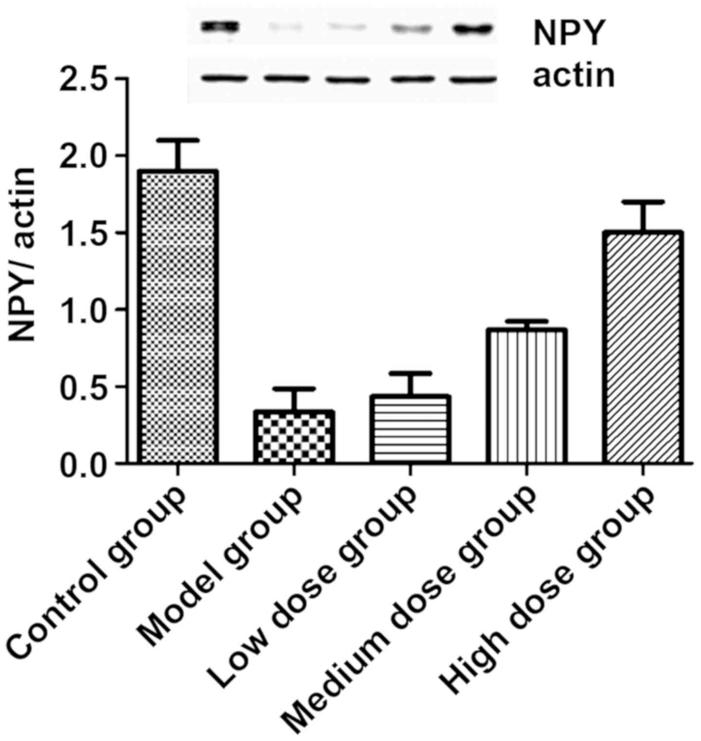
Comparison of NPY levels in brain
tissue of each group of mice
The expression levels of total NPY in the brain
tissue of each group of mice were analyzed by western blot
analysis. The results are shown in Fig.
1. Compared with the control group, the NPY levels of the model
group were significantly downregulated, and the difference was
statistically significant (P<0.05). After treatment with
resveratrol, the level of NPY in the brain tissue of mice increased
gradually, and in a dose-dependent manner. The comparison between
the levels of NPY in the brain tissue of the low-dose, medium-dose
and high-dose groups were significantly different (P<0.05).
Discussion
With the gradual increase of social and work
pressure, depression, a chronic neurological disease, seriously
threatens human cognition. Its incidence is increasing year by
year. Epidemiological analysis shows that the incidence of
different depression in young individuals is more significant and
has become a social problem (11).
Therefore, screening for highly effective and low-toxic drugs is a
prerequisite for the treatment of depression. Our previous in
vitro studies showed that after treatment of neurons with
resveratrol, the level of dopamine was significantly increased. The
study showed that the levels of DA, 5-HT and BDNF in patients with
depression were significantly reduced, which is one of the main
causes of depression. Therefore, we speculated whether resveratrol
has the effect of relieving depression.
DA and 5-HT are monoamine neurotransmitters in the
brain. In the pathogenesis of depression, the DA and 5-HT
hypotheses are recognized. Studies also showed that the level of DA
and 5-HT decreased in the hypothalamic tissue of patients with
depression, indicating that depression is closely related to the
low function of DA and 5-HT (12,13). The
levels of DA and 5-HT in the brain tissue of the mouse model of
depression established in this study were significantly lower than
those in normal mice, indicating that DA and 5-HT play an important
role in the pathogenesis of depression. After treatment with
resveratrol, the levels of DA and 5-HT increased gradually and in a
dose-dependent manner. This result suggests that resveratrol may
have the effect of treating depression. BDNF is a member of the
neurotrophic factor family, which has the effect of promoting
proliferation and differentiation of neurons, promoting neuronal
survival and development, altering neuronal morphology in the
brain, increasing synaptic terminal density, and promoting
dendritic and axon growth (14). NPY
is a known neuroendocrine polypeptide that plays an important
regulatory role in neuronal excitability (15). The current study indicated that BDNF
levels and brain NPY were significantly decreased in patients with
depression. This result is consistent with the results obtained in
the mouse model of this study. After treatment with resveratrol,
the level of BDNY increased with the increase of resveratrol
concentration, indicating that resveratrol may increase the
activity of neurons by increasing brain-derived neurotrophic
factor, thus achieving anti-depressant effect.
Behavioral analysis of mice showed that the behavior
of depression in mice after resveratrol treatment was significantly
relieved, indicating that resveratrol has a role in alleviating
depression. Early studies on resveratrol have shown that they have
important biological functions such as anti-inflammatory, antitumor
and anti-aging. In addition, this study revealed a new
anti-depressant biological function of resveratrol, which expanded
the clinical use of resveratrol.
In summary, resveratrol can significantly increase
the levels of DA, 5-HT, BDNF and NPY in the brain to achieve the
effect of treating depression.
Acknowledgements
Not applicable.
Funding
No funding was received.
Availability of data and materials
The datasets used and/or analyzed during the present
study are available from the corresponding author on reasonable
request.
Authors' contributions
ZG conceived the study and drafted the manuscript.
ZG and LC were responsible for construction and treatment of the
depression models. ZG and YH helped with the sucrose consumption
test. All authors have read and approved the final manuscript.
Ethics approval and consent to
participate
The study was approved by the Ethics Committee of
The Second Affiliated Hospital of Xinxiang Medical University
(Xinxiang, China).
Patient consent for publication
Not applicable.
Competing interests
The authors declare that they have no competing
interests.
References
|
1
|
Bennett S and Thomas AJ: Depression and
dementia: Cause, consequence or coincidence? Maturitas. 79:184–190.
2014. View Article : Google Scholar : PubMed/NCBI
|
|
2
|
Korczyn AD and Halperin I: Depression and
dementia. J Neurol Sci. 283:139–142. 2009. View Article : Google Scholar : PubMed/NCBI
|
|
3
|
Liu MY, Zhang LJ, Zhou YX and Wei WL:
5-Hydroxytryptamine changes under different pretreatments on rat
models of myocardial infarction and/or depression. Chin Med J
(Engl). 130:2219–2225. 2017. View Article : Google Scholar : PubMed/NCBI
|
|
4
|
Teo CH, Soga T and Parhar IS: Brain
beta-catenin signalling during stress and depression. Neurosignals.
26:31–42. 2018. View Article : Google Scholar : PubMed/NCBI
|
|
5
|
Martocchia A, Curto M, Scaccianoce S,
Comite F, Xenos D, Nasca C, Falaschi GM, Ferracuti S, Girardi P,
Nicoletti F, et al: Effects of escitalopram on serum BDNF levels in
elderly patients with depression: A preliminary report. Aging Clin
Exp Res. 26:461–464. 2014. View Article : Google Scholar : PubMed/NCBI
|
|
6
|
Wu X, Xu Y, Zhu B, Liu Q, Yao Q and Zhao
G: Resveratrol induces apoptosis in SGC-7901 gastric cancer cells.
Oncol Lett. 16:2949–2956. 2018.PubMed/NCBI
|
|
7
|
Li J, Xie C, Zhuang J, Li H, Yao Y, Shao C
and Wang H: Resveratrol attenuates inflammation in the rat heart
subjected to ischemia-reperfusion: Role of the TLR4/NF-κB signaling
pathway. Mol Med Rep. 11:1120–1126. 2015.PubMed/NCBI
|
|
8
|
Tan L, Wang W, He G, Kuick RD, Gossner G,
Kueck AS, Wahl H, Opipari AW and Liu JR: Resveratrol inhibits
ovarian tumor growth in an in vivo mouse model. Cancer.
122:722–729. 2016. View Article : Google Scholar : PubMed/NCBI
|
|
9
|
Tresguerres IF, Tamimi F, Eimar H,
Barralet J, Torres J, Blanco L and Tresguerres JA: Resveratrol as
anti-aging therapy for age-related bone loss. Rejuvenation Res.
17:439–445. 2014. View Article : Google Scholar : PubMed/NCBI
|
|
10
|
Subramanian M, Goswami M, Chakraborty S
and Jawali N: Resveratrol induced inhibition of Escherichia
coli proceeds via membrane oxidation and independent of
diffusible reactive oxygen species generation. Redox Biol.
2:865–872. 2014. View Article : Google Scholar : PubMed/NCBI
|
|
11
|
Miniati M, Callari A, Calugi S, Rucci P,
Savino M, Mauri M and Dell'Osso L: Interpersonal psychotherapy for
postpartum depression: A systematic review. Arch Women Ment Health.
17:257–268. 2014. View Article : Google Scholar
|
|
12
|
Segura-Aguilar J, Paris I, Muñoz P,
Ferrari E, Zecca L and Zucca FA: Protective and toxic roles of
dopamine in Parkinson's disease. J Neurochem. 129:898–915. 2014.
View Article : Google Scholar : PubMed/NCBI
|
|
13
|
Li X, Fan Y, Xiao S, Peng S, Dong X, Zheng
X, Liu CC, Li H and Xiao Z: Decreased platelet 5-hydroxytryptamin
(5-HT) levels: A response to antidepressants. J Affect Disord.
187:84–90. 2015. View Article : Google Scholar : PubMed/NCBI
|
|
14
|
Wilkinson ST, Kiselycznyk C, Banasr M,
Webler RD, Haile C and Mathew SJ: Serum and plasma brain-derived
neurotrophic factor and response in a randomized controlled trial
of riluzole for treatment resistant depression. J Affect Disord.
241:514–518. 2018. View Article : Google Scholar : PubMed/NCBI
|
|
15
|
Sun Z, Liu S, Kharlamov EA, Miller ER and
Kelly KM: Hippocampal neuropeptide Y protein expression following
controlled cortical impact and posttraumatic epilepsy. Epilepsy
Behav. 87:188–194. 2018. View Article : Google Scholar : PubMed/NCBI
|