Introduction
Colorectal cancer (CRC) has one of the highest
morbidity and mortality rates of tumors worldwide. There are
~1,000,000 newly diagnosed cases, at an increasing rate of >20%
per year (1). The 5-year survival
rate of CRC is <15% due to the poor sensitivity of radiotherapy
and chemotherapy (2). In the past
decades, progress has been achieved in the CRC field, particularly
in molecular carcinogenesis and novel targeted therapies (3–5).
However, the overall survival rate of CRC remains low (6,7). The
resistance of tumor cells to anticancer drugs is the primary factor
affecting the curative effect of CRC chemotherapy (8).
At present, the standard therapy for CRC is surgical
intervention followed by combination chemotherapy, consisting of
cisplatin and platinum-based drugs (9). Cisplatin, one of the most effective
non-specific cell cycle drugs, serves a pivotal role in the
treatment of CRC (10,11). Cisplatin is an effective primary
chemotherapeutic agent for the treatment of CRC. The development of
drug resistance has resulted in poor survival rates in CRC and has
become an increasing clinical challenge for patients with cancer
(12). Therefore, there is an urgent
requirement to develop targeted solutions to overcome drug
resistance in the chemotherapeutic treatment of CRC based on the
distinct molecular background of CRC and the mechanism of
resistance to platinum-based drugs.
MicroRNAs (miRNA) (13,14) are
a type of short single small non-coding RNA that are induced via
the specific combination with target gene mRNA (mRNA). This
interaction induces the degradation or inhibits the translation of
mRNA to change the expression of target proteins and regulate their
signaling pathways, leading to the occurrence and development of
tumors (15). miRNA can regulate the
expression of hundreds of target genes, the expression profiles of
which differ significantly between tumor and normal tissues
(16). It is also associated with
the early diagnosis and prognostic prediction of tumors, alongside
individualized treatment with novel molecular markers that exhibit
tumor specificity (17). miRNAs are
abnormally expressed or mutated in a variety of tumors (18). Therefore, it has been hypothesized
that miRNAs serve the role of an oncogene or tumor suppressor gene
in tumor development (13,19). A widened understanding of the
association between miRNA and CRC may provide novel agents for its
diagnosis, treatment and prognosis.
The change in the expression of miRNAs serves an
important role in the resistance to anticancer drugs. Thus,
determining the association between miRNA expression and drug
resistance will provide a novel mechanism to reverse resistance to
anticancer drugs. The primary mechanism used to achieve this, is
the regulation of target gene expression by directly targeting the
3′-untranslated region (UTR) of mRNA (20). Recently, numerous studies have
revealed that miRNAs are involved in the regulation of cancer
occurrence and development (21–24).
Specifically, miR-1271 serves an important role in the regulation
of CRC cell apoptosis, proliferation and metastasis (25). The present study revealed that
miR-1271 was abnormally expressed in CRC tissues; however, the
mechanism of miR-1271 function in CRC remains unclear. Therefore,
the aim of the present study was to assess the influence of
miR-1271 on the sensitivity of CRC to chemotherapeutic drugs and
the possible mechanisms of molecular regulation in CRC.
Materials and methods
Clinical sample collection
Tumor tissues and adjacent normal tissues (>2 cm
away from the tumor tissue) were extracted from 30 patients with
CRC (male, 17; female, 13; age range, 45–63 years; mean age, 56
years) who had undergone surgical resection at Shanghai Jiao Tong
University Affiliated Sixth People's Hospital (Shanghai, China).
All enrolled patients had not received chemotherapy or radiotherapy
prior to the study. Tissue samples were stored in liquid nitrogen
at −80°C until further use. Written informed consent was obtained
from each patient or their relatives prior to surgery. The present
study was approved by the Ethics Committee of Shanghai Jiao Tong
University Affiliated Sixth People's Hospital (Shanghai,
China).
Cell culture
The CRC SW480 cell line was obtained from the
American Type Culture Collection (Manassas, VA, USA). Cell lines
were cultured in RPMI-1640 medium (Gibco; Thermo Fisher Scientific,
Inc., Waltham, MA, USA), supplemented with 10% fetal bovine serum
(Hyclone; GE Healthcare Life Sciences, Logan, UT, USA) and 100 U/ml
penicillin and 100 µg/ml streptomycin (Invitrogen; Thermo Fisher
Scientific, Inc.) in an incubator with a humidified atmosphere at
5% CO2 and 37°C. Cell propagation was performed for 2
days. Cells in the logarithmic phase of growth were used for
subsequent experimentation.
Drug treatment and cell
transfection
Cells were treated with 10 µg/ml of cisplatin
(Sigma-Aldrich; Merck KGaA, Darmstadt, Germany). SW480 cells in the
logarithmic stage were were transfected with 200 nmol/l miR-1271
mimic, miR-NC mimic, 200 nmol/l miR-1271 inhibitor or miR-NC
inhibitor (all Thermo Fisher Scientific, Inc.) using Lipofectamine
RNAiMAX (Thermo Fisher Scientific, Inc.), according to the
manufacturer's protocol. Following 72-h transfection, the
cisplatin-miR-1271 mimic, cisplatin-miR-1271 inhibitor and the
cisplatin group were treated with cisplatin (10 µg/ml) for 48 h at
37°C. The cells of the control group were untreated. The sequences
of miR-1271 mimics and miR-NC mimics were as follows:
5′-CUUGGCACCUAGCAAGCACUCA-3′ and 3′-UUCUCCGAACGUGUCACGUTT-5′. The
sequences of miR-1271 inhibitor and miR-NC inhibitor were
5′-UGAGUGCUUGCUAGGUGCCAAG-3′ and 5′-UUGUACUACACAAAAGUACUG-3′,
respectively.
Drug sensitivity assay
Cell proliferation was assessed in vitro
using the cell counting kit-8 (CCK-8) assay (Dojindo Molecular
Technologies, Inc., Kumamoto, Japan). Briefly, SW480 cells were
transfected with miR-1271 mimic, inhibitor or negative control for
72 h and incubated with cisplatin for 48 h (as described
previously). Cell were seeded into 96-well plates at a density of
5.0×10³ cells/well and incubated in 100 µl RPMI-1640
medium (Gibco; Thermo Fisher Scientific, Inc.) at 37°C. Following
0, 12, 24 or 48 h incubation, 10 µl CCK-8 was added to each well
and incubated for a further 2 h at 37°C. The absorbance of each
sample was measured at a wavelength of 450 nm using a microplate
reader (Bio-Rad Laboratories Inc., Hercules, CA, USA).
Flow cytometry
Cell apoptosis was analyzed using the Annexin
V-fluorescein isothiocyanate (FITC)/propidium iodide apoptosis
detection kit (cat. no. 556570; BD Biosciences, San Jose, CA, USA).
Briefly, cells were collected and resuspended at ~5×10⁵
cells/ml in binding solution. Cells were transferred into the flow
tube (100 µl/tube) and incubated for 5 min at room temperature with
5 µl Annexin V/FITC and PI. Following incubation, cells were washed
thrice with ice-cold PBS and apoptotic cells were analyzed using a
FACS Calibur flow cytometer and CellQuest software (version 6.0; BD
Biosciences).
Reverse transcription-quantitative
polymerase chain reaction (RT-qPCR)
Total RNA was extracted from cells and tissues using
TRIzol® reagent (Invitrogen; Thermo Fisher Scientific,
Inc.). Total RNA was reverse transcribed into cDNA using the
Primer-Script™ One Step RT-PCR kit (Takara Biotechnology Co., Ltd.,
Dalian, China). qPCR was subsequently performed using
SYBR® Premix Ex Taq (Takara Biotechnology Co., Ltd.),
according to the manufacturer's protocol. The following
thermocycling conditions were used: 95°C for 5 min; 40 cycles of
95°C for 10 sec, 60°C for 20 sec and 72°C for 20 sec. Differences
in the expression of Bcl-2-associated X (Bax), B-cell lymphoma-2
(Bcl-2), caspase-3 and mammalian target of rapamycin (mTOR) in the
four groups were compared. The relative mRNA expression levels were
quantified using the 2−ΔΔCq method (26) and normalized to the internal control
GAPDH.
For the detection of miRNA, total RNA was isolated
using the miRNA Extraction kit (Qiagen GmbH, Hilden, Germany).
Total RNA was reverse transcribed into cDNA using the miRNA cDNA
Synthesis kit (Qiagen GmbH). qPCR was subsequently performed using
the SYBR® Green (Takara Biotechnology Co., Ltd.),
according to the manufacturer's protocol. The experiment was
performed as described above. U6 served as an internal control. The
sequences of the primers utilized are listed in Table I.
 | Table I.Oligonucleotide primer pairs used for
quantitative polymerase chain reaction. |
Table I.
Oligonucleotide primer pairs used for
quantitative polymerase chain reaction.
| Gene | Primer sequence
(5′-3′) |
|---|
| miR-1271 | F:
CAGCACTTGGCACCTAGCA |
|
| R:
TATGGTTGTTCTCCTCTCTGTCTC |
| U6 | F:
CTCGCTTCGGCAGCACA |
|
| R:
AACGCTTCACGAATTTGCGT |
| Bax | F:
CACCAGCTCTGAACAGATCATGA |
|
| R:
TCAGCCCATCTTCTTCCAGATGT |
| Bcl-2 | F:
CACCCCTGGCATCTTCTCCTT |
|
| R:
AGCGTCTTCAGAGACAGCCAG |
| caspase-3 | F:
AACTGGACTGTGGCATTGAG |
|
| R:
ACAAAGCGACTGGATGAACC |
| mTOR | F:
AACAACGGCTTTCCACCAGG |
|
| R:
CACCCTAAGTGAGCCCTTGGA |
| GAPDH | F:
GGAGCGAGATCCCTCCAAAAT |
|
| R:
GGCTGTTGTCATACTTCTCATGG |
Western blot analysis
Following transfection, SW480 cells from each group
were collected and lysed using radioimmunoprecipitation assay
buffer (Beyotime Institute of Biotechnology, Haimen, China) on ice.
Total protein was quantified using a bicinchoninic acid assay kit
(Beyotime Institute of Biotechnology). The supernatant was used to
detect its protein concentration following high-speed
centrifugation at 11,180 × g for 15 min at 4°C. Total protein was
quantified using a bicinchoninic acid assay (Beyotime Institute of
Biotechnology) and 50 µg protein/lane was separated via SDS-PAGE on
a 10% gel. The separated proteins were transferred onto
polyvinylidene fluoride membranes and blocked for 2 h at 37°C with
non-fat milk. The membranes were incubated with primary antibodies
against Bax (cat. no. B3428; 1:1,000), Bcl-2 (cat. no. SAB3500343;
1:1,000), caspase-3 (cat. no. C9598; 1:1,000), mTOR (cat. no.
SAB2701843; 1:1,000) and anti-GAPDH (cat. no. G9545; 1:100; all
Sigma-Aldrich; Merck KGaA) overnight at 4°C. The membranes were
subsequently incubated with horseradish peroxidase-conjugated goat
anti-rabbit secondary antibody (cat. no. ab205718; 1:5,000; Abcam,
Cambridge, UK) for 1 h at room temperature. Protein bands were
visualised using an the ECL Plus immunoblotting detection system
(Thermo Fisher Scientific, Inc.) and quantified using Gel-Pro
analyzer software (version 6.0; Media Cybernetics, Inc., Rockville,
MD, USA). The results are expressed as the ratio of objective
protein to GAPDH.
Bioinformatics analysis and
dual-luciferase reporter assay
TargetScan bioinformatics software (www.targetscan.org/vert_71) was used to predicted
mTOR was a target of miR-1271. The potential miR-1271 binding site
in the 3′UTR of mTOR and the corresponding mutant were synthesized
and cloned into the pGL3 vector (Switchgear Genomics, Menlo Park,
CA, USA). The cDNA fragment corresponding to the wild-type 3′-UTR
of mTOR were inserted downstream of the luciferase in the
psiCHECK-2 reporter plasmid (cat. no. C8021; Promega Corporation,
Madison, WI, USA). A deletion mutant lacking the seed sequence of
the miR-1271 binding site in the 3′-UTR of mTOR was created using
the QuikChange Site-directed Mutagenesis kit (Agilent Technologies,
Inc., Santa Clara, CA, USA). For the reporter assay, SW480 cells
were co-transfected with miR-1271 mimic, inhibitor, or negative
control, and WT/MUT-3′UTR of mTOR. Following 72-h transfection,
luciferase activities were detected using the Dual-Luciferase
Reporter assay system (Promega Corporation), according to
manufacturer's protocol. Firefly luciferase activity was normalized
to Renilla luciferase activity.
Statistical analysis
SPSS 20.2 software (IBM Corp., Armonk, NY, USA) was
used to analyze the data. Data are presented as the mean ± standard
deviation of ≥3 independent experiments. A two-tailed Student's
t-test was used to analyze the difference between two groups.
One-way analysis of variance followed by a Newman-Keuls post-hoc
test was employed to analyze data among multiple groups. P<0.05
was considered to indicate a statistically significant
difference.
Results
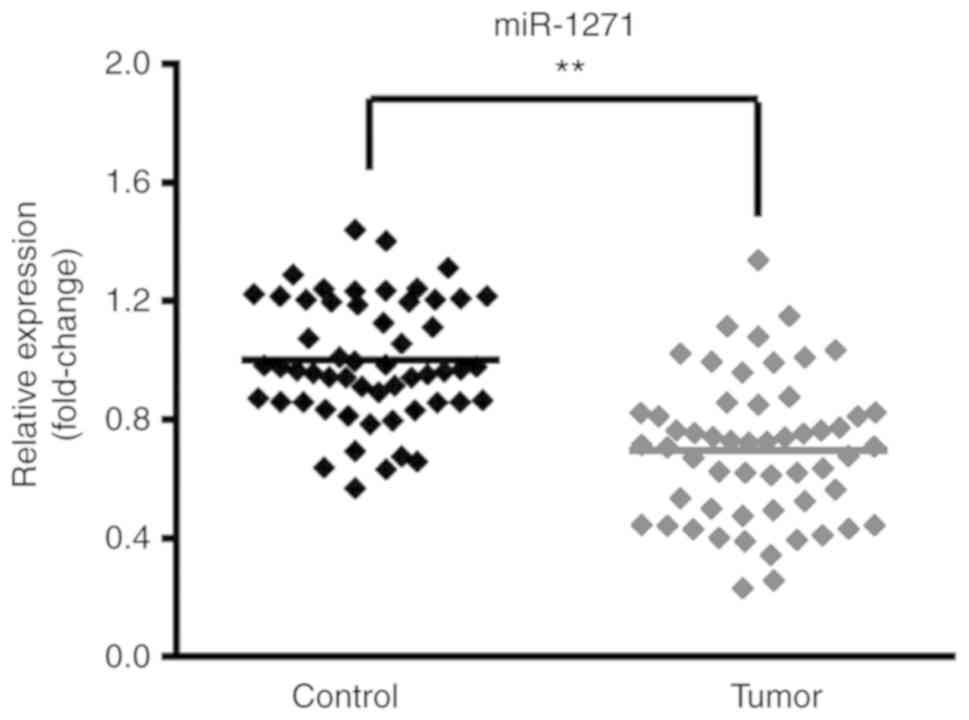
miR-1271 expression decreases in CRC
tissue samples
Tumor and matched non-tumor tissue samples were
profiled using RT-qPCR to identify the expression of miR-1271 in
CRC. The results revealed that miR-1271 expression was
significantly decreased in tumor tissues compared with non-tumor
tissues, indicating that miR-1271 was abnormally expressed at low
levels in CRC tissues (Fig. 1).
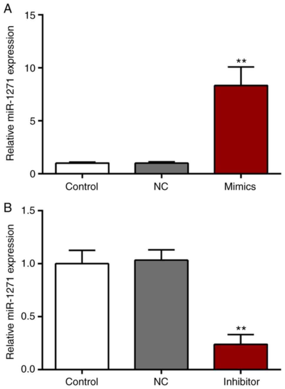
Expression of miR-1271
SW480 cells were transfected with miR-1271 mimics,
miR-NC mimics, a miR-1271 inhibitor or a miR-NC inhibitor. The
expression of miR-1271 was significantly increased and decreased
following transfection with miR-1271 mimics and the miR-1271
inhibitor, respectively (Fig. 2A and
B).
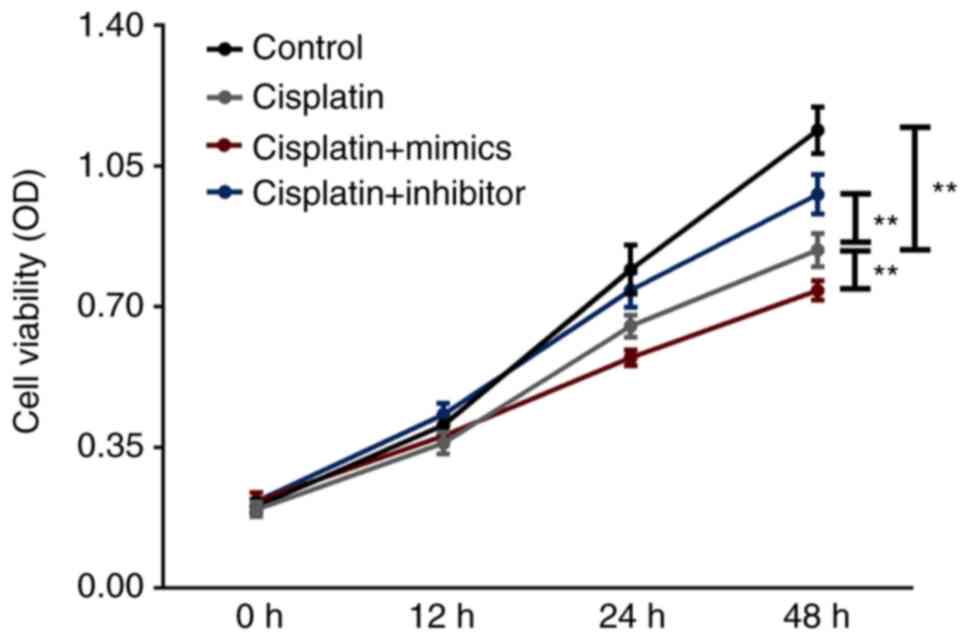
miR-1271 inhibits SW480 cell
viability
A CCK-8 was performed to assess the effect of SW480
cell activity and proliferation (Fig.
3). The results revealed that, compared with the cisplatin
group, the cisplatin + miR-1271 mimics group significantly
decreased the cell viability of SW480 cells at 48 h. However, when
compared with the cisplatin group, the transient downregulation of
miR-1271 increased the cell viability of cisplatin-treated SW480
cells.
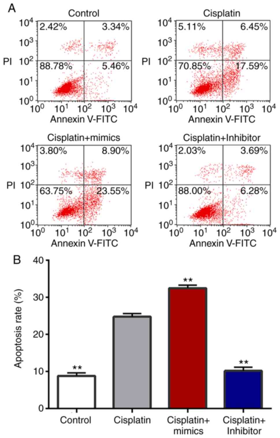
miR-1271 promotes SW480 cell
apoptosis
Flow cytometry was performed to assess the effect of
miR-1271 on the apoptosis of cisplatin-treated SW480 cells
(Fig. 4). The results demonstrated
that transfection of miR-1271 mimics induced a significant increase
in SW480 cell apoptosis compared with the cisplatin group.
Furthermore, transfection with the miR-1271 inhibitor significantly
decreased the rate of apoptosis induced by cisplatin treatment.
mTOR expression
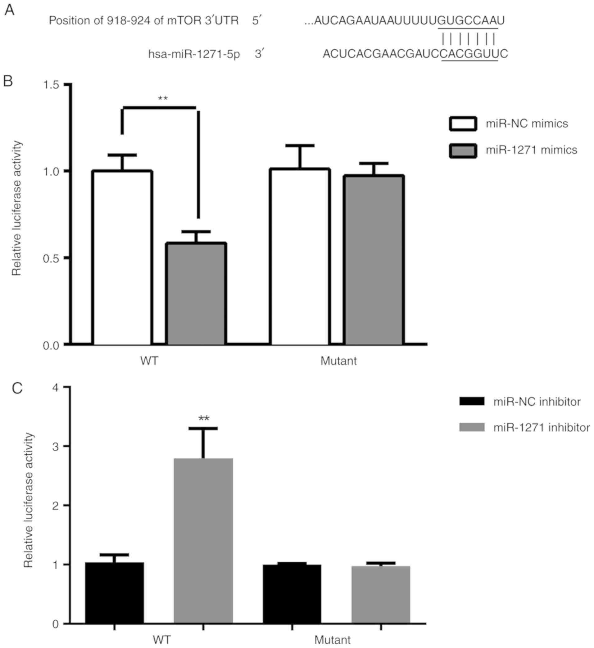
To further gain insight into the potential
underlying mechanism of miR-1271 in chemoresistance, TargetScan
(www.targetscan.org/vert_71) was used
to predict the targets of miR-1271. It was revealed that mTOR can
be directly regulated by miR-1271 and is therefore a potential
candidate target gene for further study (Fig. 5A). Dual-luciferase reporter assay
demonstrated that the luciferase activity in SW480 cells was
significantly decreased following co-transfection with miR-1271
mimic and WT 3′-UTR of mTOR (Fig.
5B). Furthermore, the luciferase activity was significantly
increased following co-transfection with miR-1271 inhibitor and WT
3′-UTR of mTOR (Fig. 5C). The
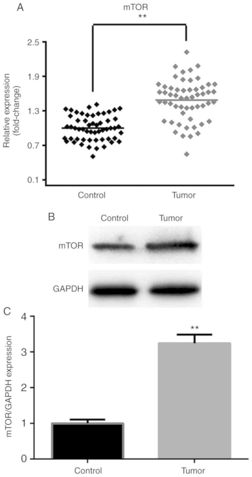
results of RT-qPCR and western blotting were used to determine the
effect of miR-1271 expression on the regulation of mTOR. miR-1271
was revealed to be overexpressed in CRC tissues (Fig. 6A-C).
miR-1271 regulated the expression of
Bcl-2, mTOR, Bax and Caspase-3
RT-qPCR results revealed that the pro-apoptosis
genes (Bax and caspase-3) were upregulated and that the
anti-apoptotic gene (Bcl-2) and mTOR were downregulated in the
cisplatin + mimics group compared with the cisplatin treatment
group (Fig. 7A-D). However,
transfection with miR-1271 inhibitor exhibited the opposite effect.
The protein expression of Bax and caspase-3 in the cisplatin +
mimics group were significantly increased, while the expression of
Bcl-2 and mTOR was significantly decreased in the cisplatin +
mimics group compared with the cisplatin treatment group (Fig. 7E and F). However, transfection with
miR-1271 inhibitor exhibited the opposite effect.
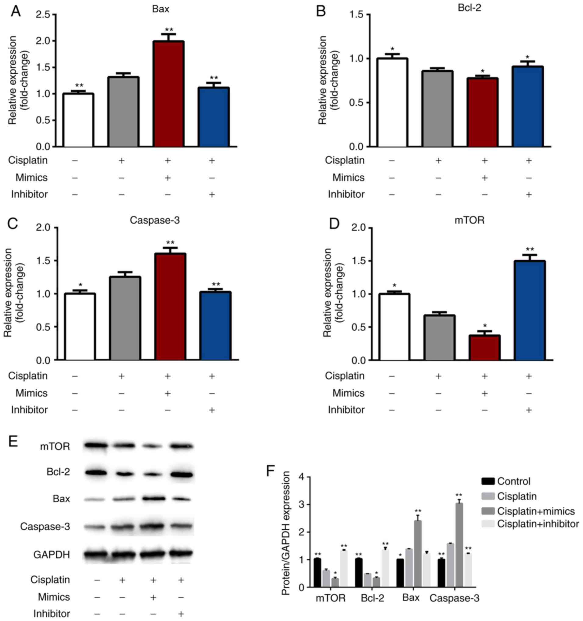 | Figure 7.miR-1271 regulates Bax, Bcl-2,
caspase-3 and mTOR expression. Reverse transcription-quantitative
polymerase chain reaction was performed to detect the transfection
efficiency of control, cisplatin, cisplatin+mimics and
cisplatin+inhibitor in SW480 cells. (A) Bax, (B) Bcl-2, (C)
caspase-3 and (D) mTOR expression. (E) Western blotting and (F)
densitometry were performed to assess the protein levels of Bax,
Bcl-2, caspase-3 and mTOR. *P<0.05 vs. Cisplatin group;
**P<0.01 vs. Cisplatin group. miR, microRNA; Bax,
Bcl-2-associated X; Bcl-2, B cell lymphoma-2; mTOR, mammalian
target of rapamycin. |
Discussion
Recently, a number of studies have reported that
abnormally expressed miRNAs are associated with cancer due to their
affect on cell proliferation, apoptosis and invasion (27–29).
miRNAs may thus serve as biomarkers for the diagnosis and prognosis
of different types of cancer, including CRC. It is therefore
necessary to identify specific miRNAs and their targets as they may
provide valuable insight for the diagnosis and treatment of
patients with CRC (30). The present
study revealed that the expression of miR-1271 was significantly
decreased in CRC tumors and cell lines compared with control
tissues. Furthermore, the expression of miR-1271 was increased
following treatment with miR-1271 mimics and decreased following
transfection with a miR-1271 inhibitor.
To the best of our knowledge, it is necessary to
identify specific miRNA target genes to elucidate the mechanism
involved in the progression and development of cancer. Previous
studies have demonstrated that miRNAs regulate their target genes
by binding to the mRNA 3′-UTR (31–33),
which is consistent with the results of the present study.
Furthermore, the relative luciferase activity of mTOR was decreased
following miR-1271 overexpression and increased following the
suppression of miR-1271 expression.
Cisplatin is the most widely used drug in the
chemotherapeutic treatment of CRC and other types of cancer,
including ovarian (8), lung
(20) and gastric cancer (31). However, the effect of chemotherapy is
not satisfactory due to drug resistance (8,34). The
resistance of tumor cells is involved in a variety of biochemical
cellular changes, including the reduced accumulation of drugs,
elevated levels of metallothionein in cells, a higher expression of
drug resistance-associated genes and enhanced DNA repair ability
(35). At present, no drug has been
able to effectively reverse cisplatin resistance in the body
(36). The drug sensitivities of
miR-1271 mimics and miR-1271 inhibitor were measured via cell
proliferation and apoptosis assays. The results indicated that
miR-1271 mimics could decrease the proliferation and increase the
apoptosis of cisplatin-treated cells, while miR-1271 inhibitor
exhibit the opposite results. In addition, the pro-apoptosis genes,
Bax and caspase-3, were upregulated, however the anti-apoptotic
gene Bcl-2, and mTOR were downregulated in miR-1271-mimic-treated
cells. The proportion of Bax and Bcl-2 can be a predictor of
apoptosis (37). In the present
study, miR-1271 served an anti-tumor role in CRC. Cells transfected
with miR-1271 mimics upregulated the pro-apoptotic genes Bax and
caspase-3 and downregulated the anti-apoptotic gene Bcl-2. In
addition, the miR-1271 inhibitor exhibited adverse effects. The
results indicated that the inhibitor reduced cisplatin sensitivity.
mTOR has been demonstrated to be involved in the progression and
metastasis of various types of cancer (38,39),
acting as an oncogene via a variety of mechanisms. mTOR expression
was associated with lung and prostate cancer, as well as
osteosarcoma (40–42). The present study revealed that
miR-1271 may regulate the chemosensitivity of CRC cells by
regulating the expression of mTOR.
In conclusion, miR-1271 may be a potential target
for antitumor therapy, particularly in the sensitivity of cells to
chemotherapy drugs. We hypothesized that miR-1271 regulates
chemotherapeutic sensitivity in CRC by targeting mTOR. In order to
further clarify the mechanism of chemotherapeutic drug sensitivity,
in vivo assays are required to verify the sensitivity of
miR-1271 to chemotherapy drugs and to lay a foundation for the
application of miR-1271 in future studies.
However, there is a limitation in our study. Since
the level of Bax in the cisplatin + mimics group was significantly
increased compared with the cisplatin group, it is possible that
there may be another mechanism mediated by Bax in these conditions,
which should be investigated in future study.
Acknowledgements
Not applicable.
Funding
No funding was received.
Availability of data and materials
The datasets used and/or analyzed during the current
study are available from the corresponding author on reasonable
request.
Authors' contributions
HY was responsible for drafting the manuscript, as
well as the acquisition, analysis and interpretation of data. JZ
collected, analyzed and interpreted the data. QS contributed to the
conception and design of the current study. All authors read and
approved the final manuscript.
Ethics approval and consent to
participate
The present study was approved by the Ethics
Committee of Shanghai Jiao Tong University Affiliated Sixth
People's Hospital (Shanghai, China). Written informed consent was
obtained from each patient or their relatives prior to surgery.
Patient consent for publication
All patients agreed with the publication of this
study.
Competing interests
The authors declare that they have no competing
interests.
References
|
1
|
Li S, Li C and Fang Z: MicroRNA 214
inhibits adipocyte enhancer-binding protein 1 activity and
increases the sensitivity of chemotherapy in colorectal cancer.
Oncol Lett. 17:55–62. 2019.PubMed/NCBI
|
|
2
|
Tie Y, Chen C, Yang Y, Qian Z, Yuan H,
Wang H, Tang H, Peng Y, Du X and Liu B: Upregulation of let-7f-5p
promotes chemotherapeutic resistance in colorectal cancer by
directly repressing repressing several pro-apoptotic proteins.
Oncol Lett. 15:8695–8702. 2018.PubMed/NCBI
|
|
3
|
Lo Russo G, Proto C and Garassino MC:
Afatinib in the treatment of squamous non-small cell CRC: A new
frontier or an old mistake? Transl Lung Cancer Res. 5:110–114.
2016.PubMed/NCBI
|
|
4
|
Kazandjian D, Suzman DL, Blumenthal G,
Mushti S, He K, Libeg M, Keegan P and Pazdur R: FDA approval
summary: Nivolumab for the treatment of metastatic non-small cell
CRC with progression on or after platinum-based chemotherapy.
Oncologist. 21:634–642. 2016. View Article : Google Scholar : PubMed/NCBI
|
|
5
|
Antonia S, Goldberg SB, Balmanoukian A,
Chaft JE, Sanborn RE, Gupta A, Narwal R, Steele K, Gu Y, Karakunnel
JJ and Rizvi NA: Safety and antitumour activity of durvalumab plus
tremelimumab in non-small cell CRC: A multicentre, phase 1b study.
Lancet Oncol. 17:299–308. 2016. View Article : Google Scholar : PubMed/NCBI
|
|
6
|
Zhou YY, Hu ZG, Zeng FJ and Han J:
Clinical profile of cyclooxygenase-2 inhibitors in treating
non-small cell CRC: A meta-analysis of nine randomized clinical
trials. PLoS One. 11:e01519392016. View Article : Google Scholar : PubMed/NCBI
|
|
7
|
Fenchel K, Sellmann L and Dempke WC:
Overall survival in non-small cell CRC-what is clinically
meaningful? Transl Lung Cancer Res. 5:115–119. 2016.PubMed/NCBI
|
|
8
|
Shi X, Xiao L, Mao X, He J, Ding Y, Huang
J, Peng C and Xu Z: miR-205-5p mediated downregulation of PTEN
contributes to cisplatin resistance in C13K human ovarian cancer
cells. Front Genet. 9:5552018. View Article : Google Scholar : PubMed/NCBI
|
|
9
|
Wu D, Lu P, Mi X and Miao J:
Downregulation of miR-503 contributes to the development of drug
resistance in ovarian cancer by targeting pi3K p85. Arch Gynecol
Obstet. 297:699–707. 2018. View Article : Google Scholar : PubMed/NCBI
|
|
10
|
Siddik ZH: Cisplatin: Mode of cytotoxic
action and molecular basis of resistance. Oncogene. 22:7265–7279.
2003. View Article : Google Scholar : PubMed/NCBI
|
|
11
|
Dasari S and Tchounwou PB: Cisplatin in
cancer therapy: Molecular mechanisms of action. Eur J Pharmacol.
740:364–378. 2014. View Article : Google Scholar : PubMed/NCBI
|
|
12
|
Huang R, Lin JY and Chi YJ: miR-519d
reduces the 5-fluorouracil resistance in colorectal cancer cells by
down-regulating the expression of CCND1. Eur Rev Med Pharmacol Sci.
22:2869–2875. 2018.PubMed/NCBI
|
|
13
|
Paliouras AR, Monteverde T and Garofalo M:
Oncogene-induced regulation of microrna expression: Implications
for cancer initiation, progression and therapy. Cancer Lett.
421:152–160. 2018. View Article : Google Scholar : PubMed/NCBI
|
|
14
|
Zhang C, Qian D, Zhao H, Lv N, Yu P and
Sun Z: miR17 improves insulin sensitivity through inhibiting
expression of ask1 and anti-inflammation of macrophages. Biomed
Pharmacother. 100:448–454. 2018. View Article : Google Scholar : PubMed/NCBI
|
|
15
|
Rupaimoole R, Calin GA, Lopez-Berestein G
and Sood AK: miRNA deregulation in cancer cells and the tumor
microenvironment. Cancer Discov. 6:235–246. 2016. View Article : Google Scholar : PubMed/NCBI
|
|
16
|
Yin W, Shi L and Mao Y: miR-194 regulates
nasopharyngeal carcinoma progression by modulating MAP3K3
expression. FEBS Open Bio. 9:43–52. 2018. View Article : Google Scholar : PubMed/NCBI
|
|
17
|
Rupaimoole R and Slack FJ: MicroRNA
therapeutics: Towards a new era for the management of cancer and
other diseases. Nat Rev Drug Discov. 16:203–222. 2017. View Article : Google Scholar : PubMed/NCBI
|
|
18
|
Kaalund SS, Venø MT, Bak M, Møller RS,
Laursen H, Madsen F, Broholm H, Quistorff B, Uldall P, Tommerup N,
et al: Aberrant expression of miR-218 and miR-204 in human mesial
temporal lobe epilepsy and hippocampal sclerosis-convergence on
axonal guidance. Epilepsia. 55:2017–2027. 2014. View Article : Google Scholar : PubMed/NCBI
|
|
19
|
Shen J and Li M: Microrna-744 inhibits
cellular proliferation and invasion of colorectal cancer by
directly targeting oncogene notch1. Oncol Res. 2018. View Article : Google Scholar
|
|
20
|
Zhang Q, Zhang B, Sun L, Yan Q, Zhang Y,
Zhang Z, Su Y and Wang C: MicroRNA-130b targets PTEN to induce
resistance to cisplatin in lung cancer cells by activating
Wnt/β-catenin pathway. Cell Biochem Funct. 36:194–202. 2018.
View Article : Google Scholar : PubMed/NCBI
|
|
21
|
Gao Y, Ma H, Gao C, Lv Y, Chen X, Xu R,
Sun M, Liu X, Lu X, Pei X and Li P: Tumor-promoting properties of
miR-8084 in breast cancer through enhancing proliferation,
suppressing apoptosis and inducing epithelial-mesenchymal
transition. J Transl Med. 16:382018. View Article : Google Scholar : PubMed/NCBI
|
|
22
|
Hu Y, Guo X, Wang J, Liu Y, Gao H, Fan H,
Nong X, Yang X, Liu M, Li S and Tang H: A novel microrna identified
in hepatocellular carcinomas is responsive to lef1 and facilitates
proliferation and epithelial-mesenchymal transition via targeting
of nfix. Oncogenesis. 7:222018. View Article : Google Scholar : PubMed/NCBI
|
|
23
|
Xia D, Tian S, Chen Z, Qin W and Liu Q:
miR302a inhibits the proliferation of esophageal cancer cells
through the mapk and PI3K/Akt signaling pathways. Oncol Lett.
15:3937–3943. 2018.PubMed/NCBI
|
|
24
|
Yu Z, Xu N, Yang W, Liu Y and Yan F:
Microrna-411 promoted the osteosarcoma progression by suppressing
MTSS1 expression. Environ Sci Pollut Res Int. 25:12064–12071. 2018.
View Article : Google Scholar : PubMed/NCBI
|
|
25
|
Sun X, Zhai H, Chen X, Kong R and Zhang X:
MicroRNA-1271 suppresses the proliferation and invasion of
colorectal cancer cells by regulating metadherin/Wnt signaling. J
Biochem Mol Toxicol. 32:2018.Doi: 10.1002/jbt.22028. View Article : Google Scholar
|
|
26
|
Livak KJ and Schmittgen TD: Analysis of
relative gene expression data using real-time quantitative PCR and
the 2(-Delta Delta C(T)) method. Methods. 25:402–408. 2001.
View Article : Google Scholar : PubMed/NCBI
|
|
27
|
Xing F, Wang S and Zhou J: The expression
of microRNA-598 inhibits ovarian cancer cell proliferation and
metastasis by targeting URI. Mol Ther Oncolytics. 12:9–15. 2018.
View Article : Google Scholar : PubMed/NCBI
|
|
28
|
Wang Y and Qin H: miR-338-3p targets RAB23
and suppresses tumorigenicity of prostate cancer cells. Am J Cancer
Res. 8:2564–2574. 2018.PubMed/NCBI
|
|
29
|
Weng L, Ma J, Jia YP, Wu SQ, Liu BY, Cao
Y, Yin X, Shang MY and Mao AW: miR-4262 promotes cell apoptosis and
inhibits proliferation of colon cancer cells: Involvement of
GALNT4. Am J Transl Res. 10:3969–3977. 2018.PubMed/NCBI
|
|
30
|
Toiyama Y, Okugawa Y, Fleshman J, Richard
Boland C and Goel A: MicroRNAs as potential liquid biopsy
biomarkers in colorectal cancer: A systematic review. Biochim
Biophys Acta Rev Cancer. 1870:274–282. 2018. View Article : Google Scholar : PubMed/NCBI
|
|
31
|
Yang M, Shan X, Zhou X, Qiu T, Zhu W, Ding
Y, Shu Y and Liu P: miR-1271 regulates cisplatin resistance of
human gastric cancer cell lines by targeting IGF1R, IRS1, mTOR, and
BCL2. Anticancer Agents Med Chem. 14:884–891. 2014. View Article : Google Scholar : PubMed/NCBI
|
|
32
|
Shao Q, Zhang P, Ma Y, Lu Z, Meng J, Li H,
Wang X, Chen D, Zhang M, Han Y, et al: Microrna-139-5p affects
cisplatin sensitivity in human nasopharyngeal carcinoma cells by
regulating the epithelial-to-mesenchymal transition. Gene.
652:48–58. 2018. View Article : Google Scholar : PubMed/NCBI
|
|
33
|
Yang Y, Liu L, Zhang Y, Guan H, Wu J, Zhu
X, Yuan J and Li M: miR-503 targets pi3K p85 and IKK-beta and
suppresses progression of non-small cell CRC. Int J Cancer.
135:1531–42. 2014. View Article : Google Scholar : PubMed/NCBI
|
|
34
|
Chen Z, Gao YJ, Hou RZ, Ding DY, Song DF,
Wang DY and Feng Y: MicroRNA-206 facilitates gastric cancer cell
apoptosis and suppresses cisplatin resistance by targeting MAPK2
signaling pathway. Eur Rev Med Pharmacol Sci. 23:171–180.
2019.PubMed/NCBI
|
|
35
|
Baguley BC: Multiple drug resistance
mechanisms in cancer. Mol Biotechnol. 46:308–316. 2010. View Article : Google Scholar : PubMed/NCBI
|
|
36
|
Li J, Sun H, Liu T and Kong J:
MicroRNA-423 promotes proliferation, migration and invasion and
induces chemoresistance of endometrial cancer cells. Exp Ther Med.
16:4213–4224. 2018.PubMed/NCBI
|
|
37
|
Wei Y, Wu S, Xu W, Liang Y, Li Y, Zhao W
and Wu J: Depleted aldehyde dehydrogenase 1A1 (ALDH1A1) reverses
cisplatin resistance of human lung adenocarcinoma cell A549/DDP.
Thorac Cancer. 8:26–32. 2017. View Article : Google Scholar : PubMed/NCBI
|
|
38
|
Li L, Sun JX, Wang XQ, Liu XK, Chen XX,
Zhang B, He ZD, Liu DZ, Chen LX, Wang LW and Huang Z:
Notoginsenoside R7 suppresses cervical cancer via
PI3K/PTEN/Akt/mTOR signaling. Oncotarget. 8:109487–109496. 2017.
View Article : Google Scholar : PubMed/NCBI
|
|
39
|
Fujishita T, Kojima Y, Kajino-Sakamoto R,
Taketo MM and Aoki M: Tumor microenvironment confers mTOR inhibitor
resistance in invasive intestinal adenocarcinoma. Oncogene.
36:6480–6489. 2017. View Article : Google Scholar : PubMed/NCBI
|
|
40
|
Wang C, Liu E, Li W, Cui J and Li T:
miR-3188 inhibits non-small cell lung cancer cell proliferation
through FOXO1-mediated mTOR-p-PI3K/AKT-c-JUN signaling pathway.
Front Pharmacol Dec. 9:13622018. View Article : Google Scholar
|
|
41
|
Gao S, Zhao Z, Wu R, Wu L, Tian X and
Zhang Z: miR-146b inhibits autophagy in prostate cancer through
affecting PTEN/AKT/mTOR signaling pathway. Aging (Albany NY).
11:2842019. View Article : Google Scholar : PubMed/NCBI
|
|
42
|
Song L, Zhou Z, Gan Y, Li P, Xu Y, Zhang
Z, Luo F, Xu J, Zhou Q and Dai F: Long noncoding RNA OIP5-AS1
causes cisplatin resistance in osteosarcoma through inducing the
LPAATβ/PI3K/AKT/mTOR signaling pathway by sponging the miR-340-5p.
J Cell Biochem. Dec 11–2018.(Epub ahead of print). doi:
10.1002/jcb.28244.
|