Introduction
Metabolic syndrome (MetS) comprises a group of
metabolic abnormalities, including central obesity, impaired
glucose tolerance, insulin resistance (IR), lipid metabolism
disorders and hypertension, which markedly increase the risk of
diabetes mellitus and cardio-cerebrovascular disease (1–3).
Approximately one-quarter of the population worldwide have been
reported to suffer from MetS (2).
Chronic inflammation was previously suggested to be implicated in
MetS (4).
Helicobacter pylori (H. pylori)
infection is one of the most common infections globally, affecting
>50% of the world's population, particularly in developing
countries. Its prevalence varies across different cohorts with
differences in age and developmental status (5,6). H.
pylori infection may cause chronic gastritis, peptic ulcers and
gastric cancer (7–9). More recently, H. pylori
gastritis was considered as an infectious disease (10). H. pylori infection has been
indicated to increase systemic inflammation by producing
inflammatory factors, including C-reactive protein (CRP), tumor
necrosis factor-α, interferon-γ and interleukin-1, −6 and −8, and
these factors were confirmed to be involved in the pathogenesis of
IR (11,12). In addition to gastrointestinal
infections, H. pylori has also been reported to be
associated with various extra-intestinal diseases, including MetS
(13–20).
At present, the association between H. pylori
infection and MetS determined in different cohorts of different
countries remains inconclusive (17,20–32). In
the majority of those studies, H. pylori infection was
diagnosed based on the presence of H. pylori IgG antibody in
the serum (29–31); however, serum IgG may persist after
H. pylori is eradicated and may therefore not reflect the
current infection status (33). The
13C-urea breath test (UBT) is a non-invasive method for
detecting current H. pylori infection. The sensitivity and
specificity of the UBT are ~0.96 [95% confidence interval (CI):
0.95–0.97] and 0.93 (95% CI: 0.91–0.94), respectively (34). While it may be assumed that only
current infection is able to cause systemic inflammatory reactions,
few studies have investigated the association between H.
pylori infection and MetS using UBT as the diagnostic method,
to the best of our knowledge.
Therefore, given the diversity of the incidence of
H. pylori infection across different countries and the
controversial results of the studies investigating the association
between H. pylori infection and MetS, particularly in East
Asia, it is crucial to further investigate this association in a
large population. The aim of the present study was to provide a
detailed analysis of the association between H. pylori
infection, as diagnosed by UBT, and MetS in a large community from
Zhejiang province in eastern China.
Materials and methods
Study participants
Participants who voluntarily underwent a general
health screening between January 2014 and December 2015 were
recruited at the International Health Care Center of the Second
Affiliated Hospital of Zhejiang University School of Medicine
(Hangzhou, China). Participants with any of the following
characteristics were excluded from the study: i) History of gastric
surgery; ii) history of anti-H. pylori therapy; iii) use of
antibiotics, proton pump inhibitors, H2 blockers or bismuth within
the previous 4 weeks; iv) severe mental or neurological disorders;
v) history of cancer(s). All subjects underwent a detailed physical
examination, including UBT detection of H. pylori
infection.
Diagnosis of H. pylori infection
After fasting for at least 2 h, all of the
participants underwent a 13C-UBT (Richen-Force, Beijing,
China) for the detection of H. pylori infection. After
collecting the baseline breath sample, the participants ingested a
13C-urea reagent dissolved in water. The second breath
sample was collected and analyzed after 30 min. A delta over
baseline value ≥4.0 indicated a positive result for H.
pylori infection.
Definitions of the study
variables
For each participant, age, sex, weight, height, body
mass index (BMI) and waist circumference were recorded. Blood
pressure was measured after at least 10 min of rest. Waist
circumference was measured with a measuring tape while standing,
midway between the lowest rib and the iliac crest. The BMI was
calculated as follows: Weight (kg)/[height (m)]2.
Information on the history of smoking and alcohol
consumption, as well as the medical history, including
hypertension, hyperlipidemia and/or diabetes, was also collected
using a questionnaire. The questionnaire also included the history
of the present illness, previous diagnoses of H. pylori
infection, history of anti-H. pylori therapy and
confirmation of eradication after treatment. If the questionnaire
was incomplete, the patient was contacted to ensure for integrity
of the data.
Laboratory examinations were performed after a 10-h
fast, including white blood cell (WBC) count, hemoglobin (Hb),
platelet count, high-sensitivity (HS)-CRP, total cholesterol,
low-density lipoprotein cholesterol, high-density lipoprotein
cholesterol (HDL-C), triglyceride (TG), total bilirubin, alanine
aminotransferase (ALT), γ-glutamyl transpeptidase, aspartate
aminotransferase (AST), fasting plasma glucose, glycosylated Hb
(HbA1c), 2-h postprandial plasma glucose, fasting insulin, blood
urea nitrogen, creatinine (Cr), uric acid (UA), homocysteic acid
(HCY) and serum cystatin C (CYSC).
The identification of individuals with MetS was
based on the definition of the International Diabetes Federation
(35) as follows: i) Central obesity
(waist circumference, ≥90 and ≥80 cm in Chinese males and females,
respectively); ii) a combination of any of the following four
indicators: TG level increased to >150 mg/dl (1.7 mmol/l), or
treated accordingly; HDL-C level decreased to <40 mg/dl (0.9
mmol/l) in males and <50 mg/dl (1.1 mmol/l) in females, or the
patient receiving corresponding treatment; systolic blood pressure
≥130 or diastolic blood pressure ≥85 mmHg, or treated accordingly,
or previous diagnosis of hypertension; and fasting plasma glucose
level increased to ≥100 mg/dl (5.6 mmol/l), or previous diagnosis
of type 2 diabetes mellitus, or the patient receiving corresponding
treatment.
IR is generally considered to be the most important
pathophysiological basis for MetS. The homeostasis model assessment
(HOMA) score may be used to estimate IR, which is defined as
follows: [Fasting plasma insulin (mU/l) × fasting plasma glucose
(mmol/l)]/22.5. A high HOMA-IR score denotes low insulin
sensitivity and IR (36). Based on a
previous study (17), three cut-off
values of the HOMA-IR index (2, 2.5 and 3) were adopted to evaluate
IR.
Statistical analysis
The statistical analysis was performed using
IBM-SPSS 24.0.0.0 software (IBM Corp.) and Python. Wilcoxon's
rank-sum test was used to evaluate differences between groups for
quantitative data, and the Chi-squared test was used for
qualitative data. All P-values were based on a two-sided test of
statistical significance. P<0.05 was considered to indicate a
statistically significant difference. The optimal cut-off point of
age was calculated using the maximization of Youden's index
(sensitivity+specificity-1) in the receiver operating curve
analysis. The association between H. pylori status and MetS
characteristics was evaluated by a multivariate logistic regression
(LR) analysis model following adjustment for age, sex, alcohol
consumption, smoking, WBC count, HS-CRP, ALT, AST, HbA1C, HCY,
CYSC, Cr and UA levels. The patients were further stratified into
subgroups based on age and sex.
Results
Clinical and demographic
characteristics
In the present study, 5,884 participants were
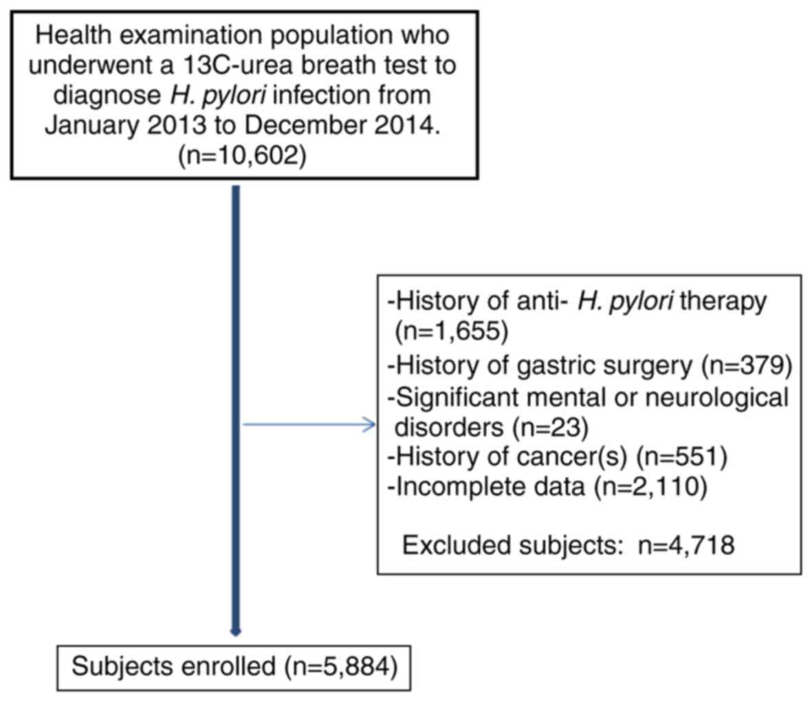
included after screening of a total of 10,602 subjects (Fig. 1). The study subjects had a mean age
of 46.81±10.13 years and 2,053 (34.9%) were female. A total of
1,265 (21.5%) had MetS, with a different percentage among males and
females (24.8 vs. 15.3%, respectively; P<0.001). In addition,
the prevalence of MetS was identified to increase with age,
particularly in females.
The overall prevalence of H. pylori infection
in this cohort was 46%, and it was higher in male compared with
female patients (47.1 vs. 43.9%, respectively; P=0.023), however,
it did not exhibit any differences across different age groups.
A statistically significant difference in the
presence of H. pylori was observed between subjects with and
those without MetS (50.4 vs. 44.8%, respectively; P<0.001;
Table I).
 | Table I.Demographic characteristics of
subjects with or without MetS. |
Table I.
Demographic characteristics of
subjects with or without MetS.
|
| MetS |
|
|---|
|
|
|
|
|---|
| Features | Yes | No | P-value |
|---|
| Sex (female%) | 315 (24.9) | 1738 (32.6) | <0.001 |
| Age (years) | 50.9±9.9 | 46.3±10.3 | <0.001 |
| Height (cm) | 167.8±8.0 | 165.7±7.8 | <0.001 |
| Weight (kg) | 75.8±10.5 | 63.8±10.2 | <0.001 |
| BMI
(kg2/m2) | 26.8±2.6 | 23.2±2.7 | <0.001 |
| Waist (cm) | 94.1±6.7 | 82.2±8.3 | <0.001 |
| WBC
(×109/l) | 6.51±1.6 | 6.1±1.6 | <0.001 |
| Hb (g/l) | 150.5±14.5 | 144.7±16.1 | <0.001 |
|
PLT(×109/l) | 214.0±55.7 | 212.5±53.6 | 0.390 |
| T-BiL (µmol/l) | 13.7±5.3 | 13.7±5.7 | 0.790 |
| ALT (U/l) | 34.5±26.99 | 23.5±23.1 | <0.001 |
| GGTP (U/l) | 56.8±54.4 | 34.0±38.99 | <0.001 |
| AST (U/l) | 27.0±12.7 | 22.9±13.2 | <0.001 |
| Hs-CRP (mg/l) | 2.1±3.1 | 1.2±2.4 | <0.001 |
| BUN (mmol/l) | 5.3±1.2 | 4.98±1.2 | <0.001 |
| Cr (mmol/l) | 65.2±14.0 | 63.1±13.97 | <0.001 |
| UA (µmol/l) | 387.2±90.9 | 336.3±86.8 | <0.001 |
| HCY (µmol/l) | 11.2±5.3 | 10.5±5.2 | <0.001 |
| CYS (mg/l) | 0.95±0.2 | 0.9±0.2 | <0.001 |
| TC (mmol/l) | 5.2±1.1 | 4.9±0.9 | <0.001 |
| TG (mmol/l) | 2.7±2.3 | 1.4±1.3 | <0.001 |
| LDL (mmol/l) | 2.9±0.8 | 2.8±0.7 | <0.001 |
| HDL (mmol/l) | 1.07±0.3 | 1.2±0.3 | <0.001 |
| FBG (mg/dl) | 5.9±1.7 | 5.0±0.8 | <0.001 |
| HbA1c (%) | 7.4±1.1 | 6.8±0.7 | <0.001 |
| PBG (mg/dl) | 9.0±4.1 | 6.5±2.5 | <0.001 |
| FINS (pmol/l) | 100.8±51.9 | 63.6±35.9 | <0.001 |
| DBP (mmHg) | 83.4±10.6 | 74.3±11.3 | <0.001 |
| SBP (mmHg) | 136.7±16.6 | 121.6±16.9 | <0.001 |
| H. pylori
infection (%) | 637 (50.4) | 2068 (44.8) | <0.001 |
| HOMA-IR | 3.8±2.3 | 2.1±1.5 | <0.001 |
| >2 (%) | 1057 (87) | 1958 (42.8) | <0.001 |
| >2.5 (%) | 888 (71.5) | 1180 (25.8) | <0.001 |
| >3 (%) | 711 (57.2) | 689 (15.3) | <0.001 |
| Carotid
atherosclerosis (%) | 441 (37.1) | 949 (22.8) | <0.001 |
| Diabetes (%) | 201 (15.9) | 174 (3.8) | <0.001 |
| Fatty liver
(%) | 1037 (82) | 1520 (32.9) | <0.001 |
| Drinking (%) | 700 (55.3) | 2055 (44.5) | <0.001 |
| Smoking (%) | 528 (42.1) | 1429 (30.9) | <0.001 |
| Hypertension
(%) | 416 (32.9) | 499 (10.8) | <0.001 |
| Hyperlipidemia
(%) | 70 (5.5) | 53 (1.1) | <0.001 |
Overall, age and sex subgroup analyses
for the risk of MetS associated with H. Pylori infection
A multivariate LR model was constructed for
predicting MetS by considering the H. pylori infection
status and other potential covariates, including sex, age and
HOMA-IR. It was observed that H. pylori infection was a
significant risk factor contributing to the prediction of MetS with
a broad-line risk of 1.2 (95% CI: 1.02–1.36, P=0.028; Table II). Subsequently, a receiver
operating characteristic curve analysis on age was performed to
segment participants, which indicated the age of 50 years as an
optimal threshold (i.e., maximizing Youden's index; Table III). It was also used to
differentiate the MetS distribution between subjects with or
without H. pylori infection. Furthermore, A multivariate LR
model analyses revealed that H. pylori infection was a
significant risk factor for MetS in male participants aged <50
years and in female participants aged ≥50 years (Table IV).
| Table II.LR analysis of MetS risk associated
with H. pylori and other factors. |
Table II.
LR analysis of MetS risk associated
with H. pylori and other factors.
| Variables | OR [95% CI] | P-value |
|---|
| H. pylori
infection | 1.21
[1.02–1.36] | 0.028 |
| HOMA-IR | 2.13
[1.98–2.20] | <0.001 |
| Age (years) | 1.11
[1.04–1.16] | <0.001 |
| Gender | 1.54
[1.27–1.75] | <0.001 |
| Table III.Sensitivity, specificity and Youden's
index at different age cut-off points. |
Table III.
Sensitivity, specificity and Youden's
index at different age cut-off points.
| Age | Sensitivity | 1-Specificity | Youden's index |
|---|
| 15 | 1 | 1 | 0.000 |
| 20 | 0.999 | 0.998 | 0.001 |
| 30 | 0.986 | 0.928 | 0.058 |
| 40 | 0.846 | 0.699 | 0.147 |
| 50 | 0.48 | 0.313 | 0.167 |
| 60 | 0.158 | 0.081 | 0.077 |
| 70 | 0.024 | 0.011 | 0.013 |
| 80 | 0.005 | 0.003 | 0.002 |
| Table IV.LR analysis of the risk of MetS
associated with H. pylori infection in different groups. |
Table IV.
LR analysis of the risk of MetS
associated with H. pylori infection in different groups.
| H. pylori
groups | OR [95% CI] | P-value |
|---|
| Males |
| <50
years | 1.28
[1.04–1.56] | 0.017 |
| ≥50
years | 1.21
[0.97–1.50] | 0.088 |
| Females |
| <50
years | 0.87
[0.58–1.30] | 0.495 |
| ≥50
years | 1.53
[1.10–2.10] | 0.010 |
Multivariate LR analysis of H. pylori
infection and other factors with MetS
A multivariate LR analysis was performed to
determine the risk of MetS associated with the H. pylori
infection status and other metabolic-associated parameters, which
were not direct variables in defining MetS, including WBC count,
HS-CRP, ALT, AST, HbA1c, HCY, CYSC, Cr and UA. It was observed that
H. pylori infection, age, sex, HS-CRP, HbA1c, UA and Cr were
also positively associated with MetS (Table V). When stratifying participants into
different gender groups and different age groups, similar patterns
among contributors for predicting MetS were revealed. It was noted
that, when the subjects were stratified by sex and further by age,
H. pylori infection was significantly associated with MetS
in female patients aged ≥50 years [odds ratio (OR)=1.29, 95% CI:
1.09–1.91; Table VI].
| Table V.Predictors of MetS by LR based on
H. pylori infection and other factors. |
Table V.
Predictors of MetS by LR based on
H. pylori infection and other factors.
| Variables | OR [95% CI] | P-value |
|---|
| Age (years) | 0.95
[0.68–0.98] | <0.001 |
| Sex | 4.40
[4.27–6.97] | <0.001 |
| HS-CRP (mg/l) | 1.94
[1.80–2.80] | <0.001 |
| ALT (U/l) | 0.92
[0.85–0.99] | <0.001 |
| AST (U/l) | 1.92
[1.19–3.15] | <0.001 |
| HbA1c (%) | 1.51
[1.32–2.92] | <0.001 |
| Cr (mmol/l) | 1.01
[1.39–1.80] | 0.001 |
| UA (µmol/l) | 1.99
[1.59–3.49] | <0.001 |
| H. pylori
infection | 1.12
[1.02–1.59] | 0.017 |
| Table VI.Predictors of MetS by LR from
subjects of sex groups with different ages. |
Table VI.
Predictors of MetS by LR from
subjects of sex groups with different ages.
|
| Males | Females |
|---|
|
|
|
|
|---|
|
| <50 years | ≥50 years | <50 years | ≥50 years |
|---|
|
|
|
|
|
|
|---|
| Variables | OR [95% CI] | P-value | OR [95% CI] | P-value | OR [95% CI] | P-value | OR [95% CI] | P-value |
|---|
| HS-CRP (mg/l) | 1.94
[1.9–1.99] | 0.010 | 1.54
[1.49–1.59] | 0.013 | 0.91
[0.81–1.03] | 0.150 | 0.97
[0.92–1.03] | 0.340 |
| ALT (U/l) | 1.89
[1.84–1.93] | <0.001 | 0.96
[0.91–1.02] | 0.180 | 1.80
[1.71–1.91] | 0.003 | 1.01
[0.92–1.11] | 0.890 |
| AST (U/l) | 2.07
[1.51–2.84] | <0.001 | 1.62
[1.15–2.27] | <0.001 | 5.53
[2.91–10.51] | <0.001 | 1.15
[0.75–1.77] | 0.520 |
| HbA1c (%) | 1.53
[1.44–1.64] | <0.001 | 1.33
[1.26–1.62] | <0.001 | 1.22
[1.12–1.39] | <0.001 | 1.27
[1.19–1.4] | <0.001 |
| Cr (mmol/l) | 1.02
[1.01–1.04] | 0.001 | 1.00
[0.99–1.01] | 0.800 | 1.06
[1.02–1.11] | 0.007 | 1.01
[0.98–1.03] | 0.700 |
| UA (µmol/l) | 1.10
[1.05–1.2] | <0.001 | 1.09
[1.01–1.12] | <0.001 | 1.02
[1.01–1.99] | <0.001 | 1.05
[1.03–1.20] | 0.003 |
| H. pylori
infection | 0.82
[0.63–1.06] | 0.130 | 0.79
[0.59–1.06] | 0.120 | 1.39
[0.80–2.41] | 0.250 | 1.29
[1.09–1.91] | 0.002 |
Discussion
The present study investigated the prevalence of
H. pylori infection in subjects with MetS, and further
explored the association of H. pylori infection and other
factors with MetS. A significant difference was identified in the
presence of H. pylori infection between subjects with and
those without MetS (50.4 vs. 44.8%, respectively; P<0.001). In a
multivariate LR analysis, H. pylori infection was determined
to be significantly associated with MetS in female patients aged
≥50 years.
Several studies have identified H. pylori
infection as a risk factor for MetS (17,20–25).
Chen et al (17), reported
that the prevalence of MetS was higher among H.
pylori-positive individuals of either gender in a Chinese adult
cohort. Upala et al (21)
demonstrated that H. pylori infection was positively
associated with IR using a meta-analysis. Yang and Xuan (25) indicated that elderly patients (aged
73.19±11.03 years) with H. pylori infection had a higher BMI
and fasting glucose levels and a higher incidence of MetS.
Furthermore, certain studies suggested that H. pylori
infection has a causative association with MetS through
pathophysiological analysis (26–28). By
contrast, other studies failed to prove this association (29–32). For
instance, Naja et al (31)
reported no suggested association of H. pylori infection
with IR or MetS in Lebanese adults. Similarly, Tamura et al
(32), identified no association
between H. pylori infection and diabetes in a Japanese
population.
In addition to national and regional factors, these
inconsistent results may be due to the different screening methods
for H. pylori infection among different studies. H.
pylori infection is diagnosed on the basis of clinical and
laboratory findings, as well as microbiological and
histopathological examinations following endoscopy. Shin et
al (37), observed that MetS was
more closely associated with histological positivity for H.
pylori (adjusted OR=1.26; 95% CI: 1.08–1.48) rather than
serological positivity (adjusted OR=1.12, 95% CI: 0.95–1.32). This
conclusion was attributed to the fact that serological positivity
for H. pylori does not necessarily indicate current
infection. Only few studies have been performed to assess the
effects of H. pylori infection on MetS in Chinese
populations, and most of those are based on the detection of IgG
antibody in the serum. Although Chen et al (17) used 13C-UBT to demonstrate
that H. pylori infection is positively associated with MetS,
the systemic association among MetS, H. pylori infection and
other variables has remained to be elucidated.
In the present study, a different prevalence of
H. pylori infection was observed between male and female
subjects (47.1 vs. 43.9%, respectively; P=0.023), whereas Chen
et al (17) reported a
similar prevalence in either gender (20%) in an adult population
from Taiwan. In addition, there was no difference in the prevalence
of H. pylori infection among different age groups; however,
they reported an increase in prevalence with advancing age in males
and females (17). Those results
indicate a different prevalence (also between the two sexes) of
MetS and H. pylori infection across the Chinese population,
suggesting that different measures have to be taken accordingly. In
addition, the present study also observed a difference in the
prevalence of MetS between sexes (24.8 vs. 15.3% in male and female
subjects, respectively), consistently with the results of a more
recent study (38).
This association was further investigated by
stratifying the subjects into males and females, and into different
age groups. An increased significance was observed in aged females,
which was consistent with a previous study reporting that H.
pylori infection was a predictor of MetS in elderly patients
(aged 73.19±11.03 years) (25).
Although in the present study, H. pylori infection was
significantly associated with MetS, it was observed that H.
pylori infection per se was only a weak predictor of MetS
(accuracy, 65%) regardless of sex and age (all these models
achieved an accuracy of ~65%). Furthermore, H. pylori
infection was not the major contributor in the LR model compared
with other metabolism-associated parameters, but it may be an
important contributor to MetS when combined with other factors. Due
to the higher incidence of hyperlipidemia, hyperglycemia or
hypertension among individuals aged >50 years, H. pylori
infection has a greater impact in this population. In addition, it
has been demonstrated that post-menopausal females have
significantly reduced estrogen levels, reduced resistance to
inflammatory reactions and increased levels of inflammatory factors
(39,40), which may explain for the more
pronounced inflammatory response of aged females to H.
pylori infection compared with males.
The health check-up population at our center mainly
comprised residents from Zhejiang province, so the economic level
is expected not to differ significantly. In fact, the questionnaire
included queries associated with socioeconomic conditions and
eating habits. However, a previous statistical analysis indicated
that their impact was not significant, so they were not included in
the present study.
Of note, the present study had certain limitations.
First, the subjects were recruited from a single center. Second,
the present study was an observational study, and conclusions may
only be drawn regarding the association between H. pylori
infection and other factors with MetS.
In conclusion, H. pylori infection increases
the risk of MetS in aged females. However, these observations are
inconsistent across different cohorts, which warrants further
investigation by prospective or biochemical studies. If confirmed,
eradication of H. pylori infection may be of therapeutic
value for MetS.
Acknowledgements
The authors would like to thank Dr Bin Ju
(Department of Epidemiology of Zhejiang University) for his advice
regarding statistics throughout the project.
Funding
The present study was supported by the Zhejiang
Provincial Natural Science Foundation of China (grant no.
LGF19H030016), the Education Department of Zhejiang Province (grant
no. Y201636053), and the Zhejiang Provincial Medical Scientific and
Technological Projects (grant nos. 2017KY387 and 2018KY413).
Availability of data and materials
All the datasets generated and analyzed in the
present study are included in this published article.
Authors' contributions
YY and JC contributed to the literature search and
the writing of the manuscript; JW and LW contributed to data
collection and analysis; ZS designed the study.
Ethics approval and consent to
participate
All of the participants provided written informed
consent prior to the examination. The present study was reviewed
and approved by the Ethics Committee of the 2nd Affiliated
Hospital, School of Medicine, Zhejiang University (Hangzhou, China;
no. 2013-218).
Patient consent for publication
Not applicable.
Competing interests
The authors declare no potential conflicts of
interest with respect to the research, authorship and/or
publication of this article.
Glossary
Abbreviations
Abbreviations:
|
H. pylori
|
Helicobacter pylori
|
|
MetS
|
metabolic syndrome
|
|
BMI
|
body mass index
|
|
WBC
|
white blood cell
|
|
Hb
|
hemoglobin
|
|
ALT
|
alanine aminotransferase
|
|
AST
|
aspartate aminotransferase
|
|
Cr
|
creatinine
|
|
UA
|
uric acid
|
|
HCY
|
homocysteic acid
|
|
CYSC
|
serum cystatin C
|
|
HS-CRP
|
high-sensitivity C-reactive
protein
|
|
HbA1c
|
glycosylated Hb
|
|
HOMA-IR
|
homeostasis model assessment of
insulin resistance
|
|
HDL-C
|
high-density lipoprotein
cholesterol
|
|
TG
|
triglyceride
|
|
95% CI
|
95% confidence interval
|
|
OR
|
odds ratio
|
References
|
1
|
Samson SL and Garber AJ: Metabolic
syndrome. Endocrinol Metab Clin North Am. 43:1–23. 2014. View Article : Google Scholar : PubMed/NCBI
|
|
2
|
Giudice A, Crispo A, Massimiliano G,
D'Arena G, Tecce MF, Grimaldi M, Amore A, Esposito E and Montella
M: Metabolic syndrome, insulin resistance, circadian disruption,
antioxidants and pancreatic carcinoma: An overview. J
Gastrointestin Liver Dis. 23:73–77. 2014.PubMed/NCBI
|
|
3
|
Hoffman EL, VonWald T and Hansen K: The
metabolic syndrome. S D Med. 9:24–28. 2015.
|
|
4
|
Festa A, D'Agostino R Jr, Howard G,
Mykkänen L, Tracy RP and Haffner SM: Chronic subclinical
inflammation as part of the insulin resistance syndrome: The
insulin resistance atherosclerosis study (IRAS). Circulation.
102:42–47. 2000. View Article : Google Scholar : PubMed/NCBI
|
|
5
|
Suerbaum S and Michetti P: Helicobacter
pylori infection. N Engl J Med. 347:1175–1186. 2002. View Article : Google Scholar : PubMed/NCBI
|
|
6
|
McColl KE: Clinical practice. Helicobacter
pylori infection. N Engl J Med. 362:1597–1604. 2010. View Article : Google Scholar : PubMed/NCBI
|
|
7
|
Qadri Q, Rasool R, Gulzar GM, Naqash S and
Shah ZA: H. pylori infection, inflammation and gastric cancer. J
Gastrointest Cancer. 45:126–132. 2014. View Article : Google Scholar : PubMed/NCBI
|
|
8
|
Atherton JC: The pathogenesis of
Helicobacter pylori-induced gastro-duodenal diseases. Annu Rev
Pathol. 1:63–96. 2006. View Article : Google Scholar : PubMed/NCBI
|
|
9
|
Lee YC, Chen TH, Chiu HM, Shun CT, Chiang
H, Liu TY, Wu MS and Lin JT: The benefit of mass eradication of
Helicobacter pylori infection: A community-based study of gastric
cancer prevention. Gut. 62:676–682. 2013. View Article : Google Scholar : PubMed/NCBI
|
|
10
|
Malfertheiner P, Megraud F, O'Morain CA,
Gisbert JP, Kuipers EJ, Axon AT, Bazzoli F, Gasbarrini A, Atherton
J, Graham DY, et al: Management of Helicobacter pylori
infection-the Maastricht V/Florence Consensus Report. Gut. 66:6–30.
2017. View Article : Google Scholar : PubMed/NCBI
|
|
11
|
Oshima T, Ozono R, Yano Y, Oishi Y,
Teragawa H, Higashi Y, Yoshizumi M and Kambe M: Association of
Helicobacter pylori infection with systemic inflammation and
endothelial dysfunction in healthy male subjects. J Am Coll
Cardiol. 45:1219–1222. 2005. View Article : Google Scholar : PubMed/NCBI
|
|
12
|
Franceschi F, Annalisa T, Teresa DR,
Giovanna D, Ianiro G, Franco S, Viviana G, Valentina T, Riccardo LL
and Antonio G: Role of Helicobacter pylori infection on nutrition
and metabolism. World J Gastroenterol. 20:12809–12817. 2014.
View Article : Google Scholar : PubMed/NCBI
|
|
13
|
Kyburz A and Muller A: Helicobacter pylori
and extragastric diseases. Curr Top Microbiol Immunol. 400:325–347.
2017.PubMed/NCBI
|
|
14
|
Goni E and Franceschi F: Helicobacter
pylori and extragastric diseases. Helicobacter. 21 (Suppl
1):S45–S48. 2016. View Article : Google Scholar
|
|
15
|
Albaker WI: Helicobacter pylori infection
and its relationship to metabolic syndrome: Is it a myth or fact?
Saudi J Gastroenterol. 17:165–169. 2011. View Article : Google Scholar : PubMed/NCBI
|
|
16
|
Chen LW, Chien CY, Yang KJ, Kuo SF, Chen
CH and Chien RN: Helicobacter pylori infection increases insulin
resistance and metabolic syndrome in residents younger than 50
years old: A community-based study. PLoS One. 10:e01286712015.
View Article : Google Scholar : PubMed/NCBI
|
|
17
|
Chen TP, Hung HF, Chen MK, Lai HH, Hsu WF,
Huang KC and Yang KC: Helicobacter pylori infection is positively
associated with metabolic syndrome in taiwanese adults: A
cross-sectional study. Helicobacter. 20:184–191. 2015. View Article : Google Scholar : PubMed/NCBI
|
|
18
|
Işıktaş Sayılar E, Çelik B and Dumlu Ş:
Relationship between Helicobacter pylori infection and metabolic
syndrome. Turk J Gastroenterol. 26:468–473. 2015. View Article : Google Scholar : PubMed/NCBI
|
|
19
|
Koklu H, Koklu S and Cinar H: Helicobacter
pylori and metabolic syndrome. Turk J Gastroenterol. 27:2012016.
View Article : Google Scholar : PubMed/NCBI
|
|
20
|
Vafaeimanesh J, Bagherzadeh M, Mirzaei A,
Parham M, Norouzinia M and Vafaee R: Effect of Helicobacter pylori
on metabolic syndrome parameters in diabetic patients.
Gastroenterol Hepatol Bed Bench. 9 (Suppl 1):S36–S41.
2016.PubMed/NCBI
|
|
21
|
Upala S, Jaruvongvanich V, Riangwiwat T,
Jaruvongvanich S and Sanguankeo A: Association between Helicobacter
pylori infection and metabolic syndrome: A systematic review and
meta-analysis. J Dig Dis. 17:433–440. 2016. View Article : Google Scholar : PubMed/NCBI
|
|
22
|
Polyzos SA and Kountouras J: Novel
advances in the association between helicobacter pylori infection,
metabolic syndrome, and related morbidity. Helicobacter.
20:405–409. 2015. View Article : Google Scholar : PubMed/NCBI
|
|
23
|
Kayar Y, Pamukçu Ö, Eroğlu H, Kalkan Erol
K, Ilhan A and Kocaman O: Relationship between Helicobacter pylori
infections in diabetic patients and inflammations, metabolic
syndrome, and complications. Int J Chronic Dis.
2015:2901282015.PubMed/NCBI
|
|
24
|
Lu LJ, Hao NB, Liu JJ, Li X and Wang RL:
Correlation between Helicobacter pylori infection and metabolic
abnormality in general population: A cross-sectional study.
Gastroenterol Res Pract. 2018:74108012018. View Article : Google Scholar : PubMed/NCBI
|
|
25
|
Yang W and Xuan C: Influence of
Helicobacter pylori infection on metabolic syndrome in old chinese
people. Gastroenterol Res Pract. 2016:69512642016. View Article : Google Scholar : PubMed/NCBI
|
|
26
|
Ando T, Ishikawa T, Takagi T, Imamoto E,
Kishimoto E, Okajima A, Uchiyama K, Handa O, Yagi N, Kokura S, et
al: Impact of Helicobacter pylori eradication on circulating
adiponectin in humans. Helicobacter. 18:158–164. 2013. View Article : Google Scholar : PubMed/NCBI
|
|
27
|
de Luis DA, Garcia Avello A, Lasuncion MA,
Aller R, Martin de Argila C, Boixeda de Miquel D and de la Calle H:
Improvement in lipid and haemostasis patterns after Helicobacter
pylori infection eradication in type 1 diabetic patients. Clin
Nutr. 18:227–231. 1999. View Article : Google Scholar : PubMed/NCBI
|
|
28
|
Gen R, Demir M and Ataseven H: Effect of
Helicobacter pylori eradication on insulin resistance, serum lipids
and low-grade inflammation. South Med J. 103:190–196. 2010.
View Article : Google Scholar : PubMed/NCBI
|
|
29
|
Devaraj S, Rosenson RS and Jialal I:
Metabolic syndrome: An appraisal of the pro-inflammatory and
procoagulant status. Endocrinol Metab Clin North Am. 33:431–453.
2004. View Article : Google Scholar : PubMed/NCBI
|
|
30
|
Gillum RF: Infection with Helicobacter
pylori, coronary heart disease, cardiovascular risk factors, and
systemic inflammation: The third national health and nutrition
examination survey. J Natl Med Assoc. 96:1470–1476. 2004.PubMed/NCBI
|
|
31
|
Naja F, Nasreddine L, Hwalla N, Moghames
P, Shoaib H, Fatfat M, Sibai A and Gali-Muhtasib H: Association of
H. pylori infection with insulin resistance and metabolic syndrome
among lebanese adults. Helicobacter. 17:444–451. 2012. View Article : Google Scholar : PubMed/NCBI
|
|
32
|
Tamura T, Morita E, Kawai S, Sasakabe T,
Sugimoto Y, Fukuda N, Suma S, Nakagawa H, Okada R, Hishida A, et
al: No association between Helicobacter pylori infection and
diabetes mellitus among a general Japanese population: A
cross-sectional study. Springerplus. 4:6022015. View Article : Google Scholar : PubMed/NCBI
|
|
33
|
Miernyk KM, Bruden DL, Bruce MG, McMahon
BJ, Hennessy TW, Peters HV, Hurlburt DA, Sacco F and Parkinson AJ:
Dynamics of Helicobacter pylori-specific immunoglobulin G for 2
years after successful eradication of Helicobacter pylori infection
in an American Indian and Alaska native population. Clin Vaccine
Immunol. 14:85–86. 2007. View Article : Google Scholar : PubMed/NCBI
|
|
34
|
Ferwana M, Abdulmajeed I, Alhajiahmed A,
Madani W, Firwana B, Hasan R, Altayar O, Limburg PJ, Murad MH and
Knawy B: Accuracy of urea breath test in Helicobacter pylori
infection: Meta-analysis. World J Gastroenterol. 21:1305–1314.
2015. View Article : Google Scholar : PubMed/NCBI
|
|
35
|
Alberti KG, Zimmet P and Shaw J; IDF
Epidemiology Task Force Consensus Group, : The metabolic syndrome-a
new worldwide definition. Lancet. 366:1059–1062. 2005. View Article : Google Scholar : PubMed/NCBI
|
|
36
|
Gayoso-Diz P, Otero-González A,
Rodriguez-Alvarez MX, Gude F, García F, De Francisco A and Quintela
AG: Insulin resistance (HOMA-IR) cut-off values and the metabolic
syndrome in a general adult population: Effect of gender and age:
EPIRCE cross-sectional study. BMC Endocr Disord. 13:472013.
View Article : Google Scholar : PubMed/NCBI
|
|
37
|
Shin DW, Kwon HT, Kang JM, Park JH, Choi
HC, Park MS, Park MS, Park SM, Son KY and Cho B: Association
between metabolic syndrome and Helicobacter pylori infection
diagnosed by histologic status and serological status. J Clin
Gastroenterol. 46:840–845. 2012. View Article : Google Scholar : PubMed/NCBI
|
|
38
|
Refaeli R, Chodick G, Haj S, Goren S,
Shalev V and Muhsen K: Relationships of H. pylori infection and its
related gastroduodenal morbidity with metabolic syndrome: A large
cross-sectional study. Sci Rep. 8:40882018. View Article : Google Scholar : PubMed/NCBI
|
|
39
|
Figueroa-Vega N, Moreno-Frías C and
Malacara JM: Alterations in adhesion molecules, pro-inflammatory
cytokines and cell-derived microparticles contribute to
intima-media thickness and symptoms in postmenopausal women. PLoS
One. 10:e01209902015. View Article : Google Scholar : PubMed/NCBI
|
|
40
|
Taleb-Belkadi O, Chaib H, Zemour L, Fatah
A, Chafi B and Mekki K: Lipid profile, inflammation, and oxidative
status in peri- and postmenopausal women. Gynecol Endocrinol.
32:982–985. 2016. View Article : Google Scholar : PubMed/NCBI
|