Introduction
Spinal cord injury (SCI) is a serious trauma causing
severe and often permanent disability. SCI induces primary
mechanical damage and causes secondary damage to the spinal cord.
Primary damage occurs by mechanical tissue disruption immediately
subsequent to trauma. Secondary damage is mediated by complex
cellular and molecular processes (1).
Apoptosis, or programmed cell death, is a highly
regulated process that enables the elimination of unwanted or
dysfunctional cells. However, inappropriate or excessive apoptosis
is a component of numerous neurodegenerative conditions, including
traumatic injury (2–5). Crowe et al (6) observed neuronal apoptosis along the
longitudinal axis of the injured spinal cord, which induced
deterioration of sensorimotor function. Contusion injury causes
apoptosis of neurons, astrocytes, oligodendroglia and microglia in
the rat spinal cord (7,8). Apoptosis in SCI is accompanied by
activation of caspase-3, a member of the cysteine protease family
(9). Apoptosis is regulated by
pro- and anti-apoptotic members of the B-cell lymphoma 2 (Bcl-2)
protein family (10). Neuronal
apoptosis occurs following traumatic brain injury, SCI, seizure and
stroke (3,11,12).
Therefore, an understanding of apoptotic mechanisms is important
for the prevention and treatment of various diseases (9,13,14).
The phosphatidylinositol 3-kinase (PI3K) signaling
pathway is implicated in cell survival and apoptosis (15). Akt, also known as protein kinase B,
is a main effector in the PI3K signaling pathway and an increase in
Akt activity blocks the mitochondrial apoptotic pathway (16,17).
The PI3K/Akt signaling pathway mediates mitogen-dependent growth
and survival, and inhibition of this pathway results in cell growth
arrest and apoptosis (18).
Phosphorylation of Akt inactivates the pro-apoptotic factors Bad
and procaspase-9, and inhibits apoptosis (19). Inhibition of the phosphorylation of
Akt induces apoptosis (20).
A number of neurotrophic factors are implicated in
the survival, differentiation and function of central nervous
system neurons. Among these factors, brain-derived neurotrophic
factor (BDNF), nerve growth factor (NGF), neurotrophin (NT)-3 and
insulin-like growth factor (IGF)-1 mediate neuroprotective effects
against apoptosis (21,22). Sustained local expression of
neurotrophic factors in the sensorimotor cortex and the spinal cord
increased axonal sprouting following SCI, providing a basis for the
development of neurotrophic factor therapy (23). Acceleration of repair following
neuronal injury is accompanied by the upregulation and release of
endogenous neurotrophic factors (24). Exogenous NGF is critical in
neuronal plasticity, regeneration and prevention of apoptosis
following traumatic brain injury (25).
Exercise exerts neuroprotective effects by enhancing
neurogenesis, increasing the expression of neurotrophic factors and
inhibiting apoptosis (3,26,27).
Griesbach et al (28)
reported that voluntary wheel exercise improved functional recovery
from traumatic brain injury via an endogenous mechanism involving
upregulation of BDNF and IGF-1 expression. Treadmill exercise may
improve functional recovery from SCI (29,30).
However, to the best of our knowledge, detailed mechanisms of the
impact of treadmill exercise on SCI have not been described. In
this study, a rat model was used to investigate the effect of
treadmill exercise on SCI-induced apoptosis and expression of
neutrotrophic factors.
Materials and methods
Animals and treatments
Male Sprague Dawley rats (180±10 g, 6 weeks old,
n=32) were purchased the Daehan Biolink Co. (Chungbuk, Korea), and
individually housed in plastic cages with a controlled temperature
(20±2°C) and a 12-h light/dark cycle (lights on from 7:00 a.m. to
7:00 p.m.). Food and water were available ad libitum. The
rats were divided into four groups: Sham surgery, sham surgery plus
exercise, SCI and SCI plus exercise (n=8/group). This study was
performed in accordance with the guidelines of the National
Institutes of Health and the Korean Academy of Medical Sciences
(Seoul, Korea), and approved by Kyung Hee University Institutional
Animal Care and Use Committee (Seoul, Korea).
Surgical procedures
The surgical procedure for inducing SCI was
conducted according to established methods (31). The rats were anesthetized with
chloral hydrate (500 mg/kg, intraperitoneal), and a laminectomy was
performed at the T9–T10 level, exposing the underlying cord without
disrupting the dura. The spinous processes of the T8–T11 level were
clamped to stabilize the spine, and the exposed dorsal surface of
the cord was subjected to contusion injury using the New York
University impactor (New York University, New York, NY, USA) as
previously described (31). A
moderate contusion was created by dropping a 10-g rod (2.5 mm in
diameter) from a height of 12.5 mm onto the exposed cord. Following
SCI, urinary bladders were emptied twice a day for one week and
thereafter when necessary. Rats in the sham surgery and the sham
surgery plus exercise groups received a T10 laminectomy without
weight-drop contusion injury.
Treadmill exercise
At one week post-surgery, the rats in the exercise
groups were trained to walk on the treadmill. When no stepping of
the hindlimb occurred in response to the moving treadmill and
stepping of the forelimb, movement was elicited by manual
stimulation of the perineum. The exercise sessions consisted of
three minutes four times per day during the first week, and five
minutes six times per day from the second to the sixth week at a
speed of 6 m/min, with five minutes resting time between each
session. Treadmill exercise was performed six days per week.
Grid-walking test
The grid-walking test was conducted as previously
described (32). This method
measures the ability of animals to control hindlimb placing and is
regarded as an indicator of corticospinal function. Prior to
surgery, the rats were trained to walk on a runway of metal grid
bars elevated from the ground. A grid pathway measuring 1.2 m in
length was used, and the rats were allowed to walk three times
voluntarily across the pathway. The number of errors (hindlimb
stepping through the grid) was counted and the three values were
averaged to produce a single error value per rat.
Western blot analysis
Western blotting was performed following standard
methods (2,3). Spinal cord tissue was collected and
immediately frozen at −70°C. The tissues were homogenized with
lysis buffer [50 mM Tris-HCl at pH 8.0, 150 mM NaCl, 10% glycerol,
1% Triton X-100, 1.5 mM MgCl2·6H2O, 1 mM
ethylene glycol tetraacetic acid (EGTA), 1 mM phenylmethylsufonyl
fluoride (PMSF), 1 mM Na2VO4 and 100 mM NaF]
and centrifuged at 900 × g for 30 min. Protein content was measured
using a colorimetric protein assay kit (Bio-Rad, Hercules, CA,
USA). Equal amounts of protein (30 μg) were loaded onto 12%
polyacrylamide gels and separated by SDS-PAGE. Separated proteins
were transferred from the gels to a nitrocellulose membrane. To
evaluate the expression of neurotrophic factors, rabbit polyclonal
anti-NT-3 antibody (1:1,000; Santa Cruz Biotechnology Inc., Santa
Cruz, CA, USA), goat polyclonal anti-IGF-1 antibody (1:1,000; Santa
Cruz Biotechnology Inc.) and rabbit polyclonal anti-NGF antibody
(1:1,000; Santa Cruz Biotechnology Inc.) were used as the primary
antibodies. In addition, mouse monoclonal anti-PI3K antibody
(1:1,000; Santa Cruz Biotechnology, Inc.), rabbit polyclonal
anti-phosphorylated (p)Akt and anti-Akt antibodies (1:1,000; Cell
Signaling Technology, Inc., Beverly, MA, USA), mouse monoclonal
anti-Bcl-2 and Bax antibodies (1:1,000; Santa Cruz Biotechnology,
Inc.) and rabbit polyclonal anti-cleaved caspase-3 antibody
(1:1,000; Cell Signaling Technology, Inc.) were used as the primary
antibodies. Horseradish peroxidase-conjugated anti-mouse antibody
for PI3K, Bcl-2 and Bax (1:3,000; Vector Laboratories, Burlingame,
CA, USA), horseradish peroxidase-conjugated anti-rabbit antibody
for NT-3, NGF, pAkt, Akt and cleaved caspase-3 (1:5,000; Vector
Laboratories), and horseradish peroxidase-conjugated anti-goat
antibody for IGF-1 (1:5,000; Vector Laboratories) were used as the
secondary antibodies. Bands were visualized using an enhanced
chemiluminescence (ECL) kit (Santa Cruz Biotechnology Inc.). To
compare the expression level of proteins, bands were calculated
densitometrically using Image-Pro® Plus software (Media
Cybernetics, Silver Spring, MD, USA).
Statistical analysis
All data were analyzed using SPSS 20.0 statistical
software (SPSS Inc., Chicago, IL, USA). The data are expressed as
the mean ± standard error of the mean (SEM). For comparisons among
the groups, one-way analysis of variance (ANOVA) and Duncan’s post
hoc test were performed. P<0.05 was considered to indicate a
statistically significant difference.
Results
Treadmill exercise improves hindlimb
motor function following SCI
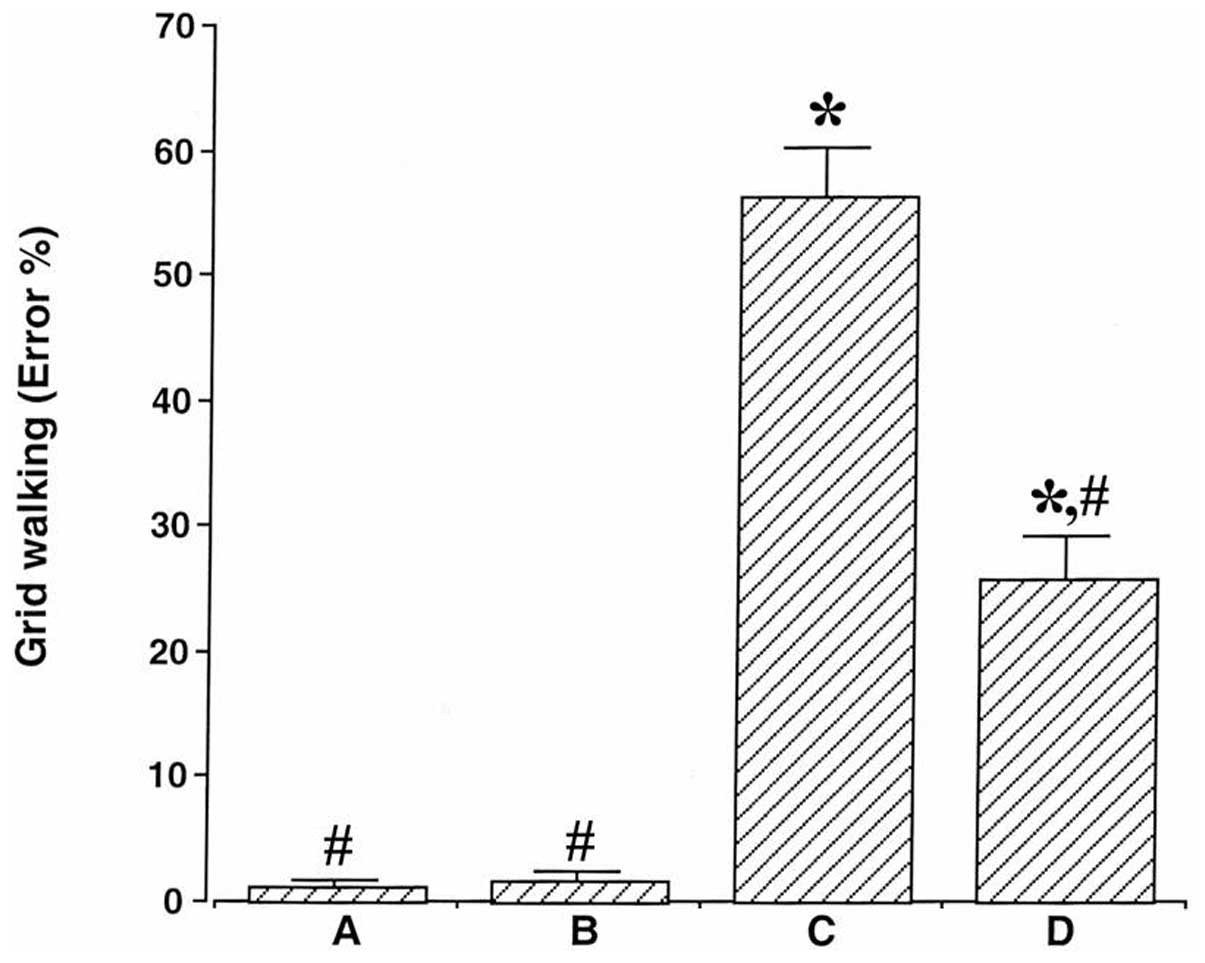
The rats’ performance was monitored by counting the
number of mis-steps and accurate footsteps. Prior to induction of
SCI, all rats had highly accurate foot placement when walking on
the grid. Following SCI, the error ratio was 1.10±0.70% in the sham
surgery group, 1.65±0.64% in the sham surgery plus exercise group,
56.30±3.86% in the SCI group and 25.63±3.52% in the SCI plus
exercise group (Fig. 1). The error
ratio increased following SCI (P<0.05). By contrast, treadmill
exercise reduced the error ratio (P<0.05), showing that
treadmill exercise improved motor function in the hindlimb
following SCI.
Treadmill exercise enhances the
expression of neurotrophic factors following SCI
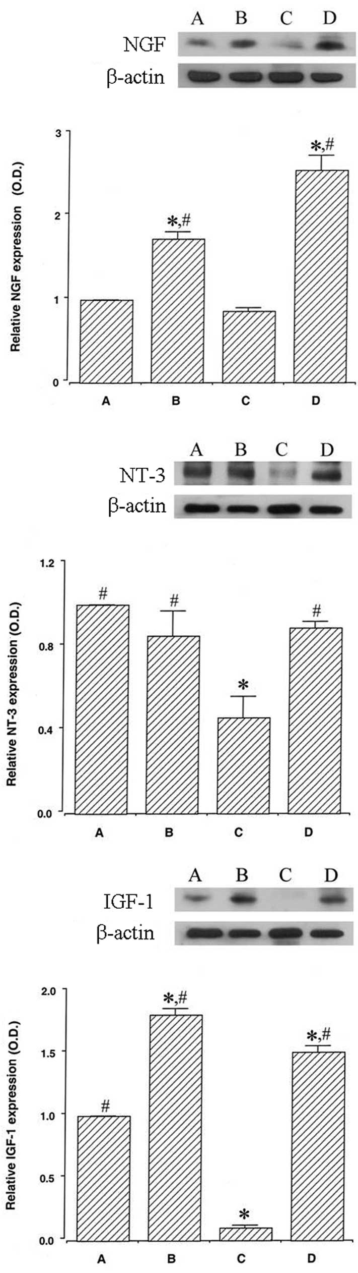
Protein expression of NGF (13 kDa) was set at 1.00
in the sham surgery group, with expression levels of 1.73±0.08 in
the sham surgery plus exercise group, 0.86±0.04 in the SCI group
and 2.55±0.18 in the SCI plus exercise group (Fig. 2, upper panel). Expression of NGF
was not changed by SCI, while treadmill exercise enhanced NGF
expression in sham-operated and SCI rats (P<0.05).
Setting the expression of NT-3 (21 kDa) in the sham
surgery group at 1.00, the expression of NT-3 was 0.85±0.12 in the
sham surgery plus exercise group, 0.46±0.10 in the SCI group and
0.89±0.03 in the SCI plus exercise group (Fig. 2, middle panel). The expression of
NT-3 was reduced following SCI (P<0.05); however, treadmill
exercise enhanced NT-3 expression in the SCI rats (P<0.05).
Setting the expression of IGF-1 (17 kDa) in the sham
surgery group at 1.00, the expression of IGF-1 was 1.81±0.05 in the
sham surgery plus exercise group, 0.10±0.02 in the SCI group and
1.51±0.05 in the SCI plus exercise group (Fig. 2, lower panel). The expression of
IGF-1 was reduced following SCI (P<0.05); however, treadmill
exercise was observed to enhance IGF-1 expression in sham-operated
and SCI rats (P<0.05).
Treadmill exercise enhances the
expression of PI3K following SCI
When the expression of PI3K (85 kDa) in the sham
surgery group was set at 1.00, the expression of PI3K was 1.20±0.03
in the sham surgery plus exercise group, 0.27±0.09 in the SCI group
and 0.46±0.08 in the SCI plus exercise group (Fig. 3). Expression of PI3K was reduced
following SCI (P<0.05); however, treadmill exercise enhanced
PI3K expression in sham-operated and SCI rats (P<0.05).
Treadmill exercise enhances the ratio of
pAkt to Akt following SCI
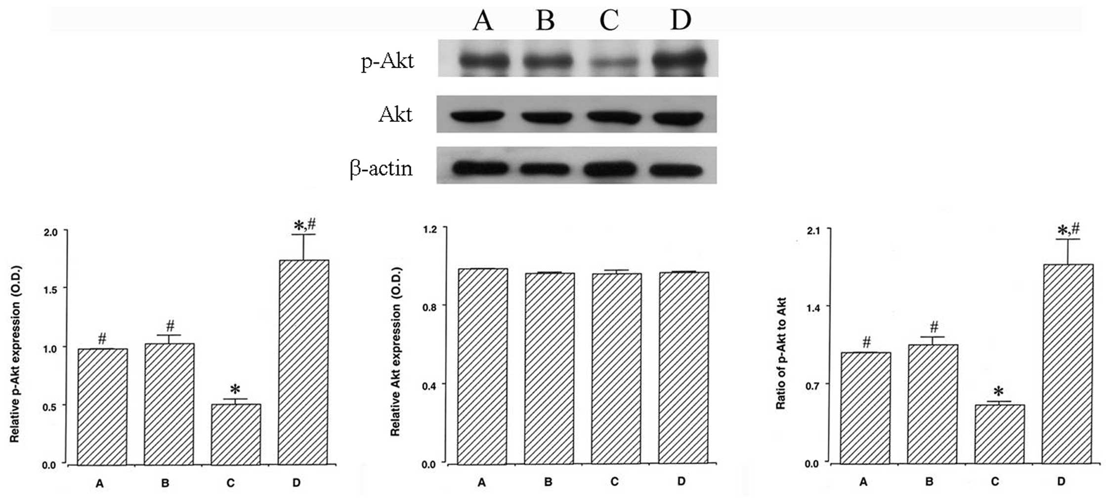
When the expression of pAkt (60 kDa) in the sham
surgery group was set at 1.00, the expression of pAkt was 1.04±0.07
in the sham surgery plus exercise group, 0.52±0.04 in the SCI group
and 1.75±0.22 in the SCI plus exercise group (Fig. 4, left panel). Expression of pAkt
was reduced following SCI (P<0.05). Treadmill exercise enhanced
pAkt expression in the SCI rats (P<0.05).
When the expression of Akt (60 kDa) in the sham
surgery group was set at 1.00, the expression of Akt was 0.97±0.01
in the sham surgery plus exercise group, 0.96±0.02 in the SCI group
and 0.97±0.01 in the SCI plus exercise group (Fig. 4, middle panel). Expression of Akt
was not changed by induction of SCI, and treadmill exercise exerted
no significant effect on Akt expression in either sham-operated or
SCI rats.
When the ratio of pAkt/Akt in the sham surgery group
was set at 1.00, the ratio of pAkt/Akt was 1.07±0.07 in the sham
surgery plus exercise group, 0.53±0.03 in the SCI group and
1.79±0.23 in the SCI plus exercise group (Fig. 4, right panel). The pAkt/Akt ratio
was reduced following SCI (P<0.05). Treadmill exercise enhanced
the pAkt/Akt ratio in SCI rats (P<0.05).
Treadmill exercise enhances the ratio of
Bcl-2 to Bax following SCI
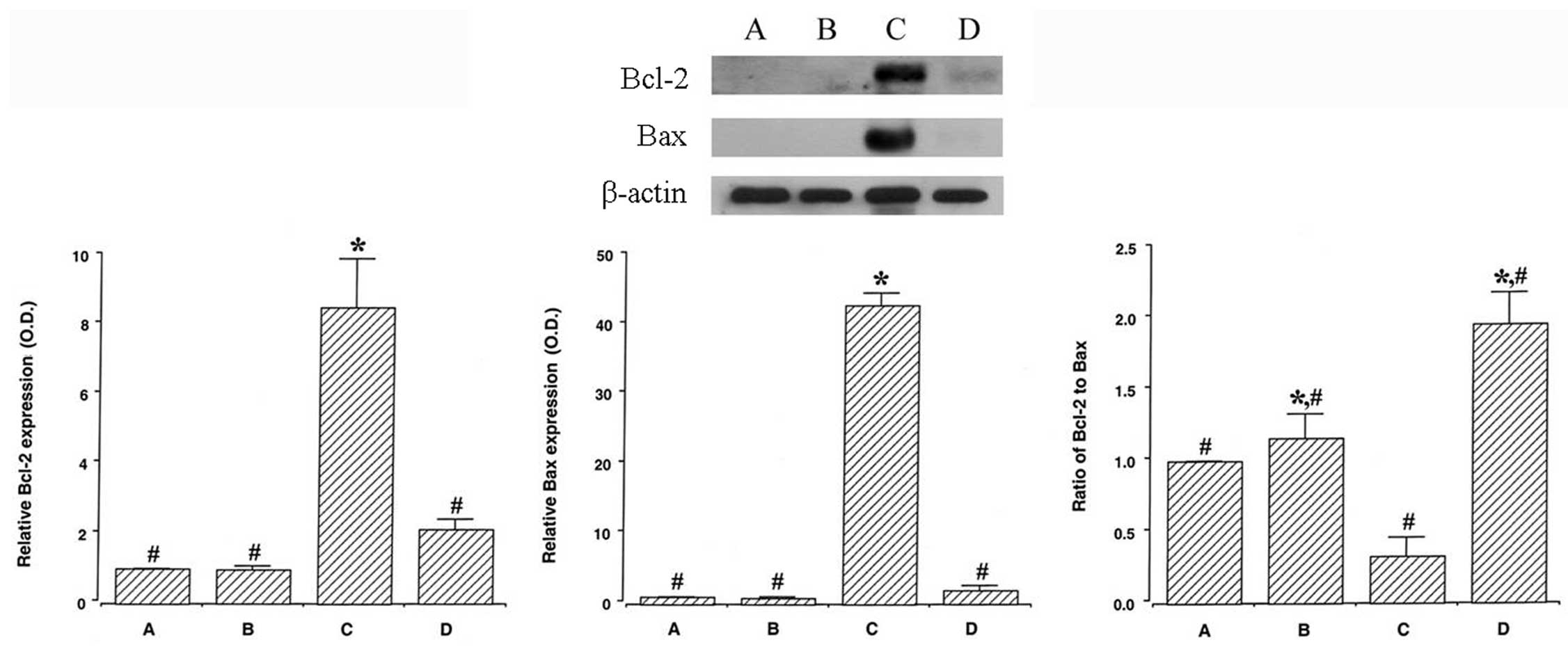
When the expression of Bcl-2 (28 kDa) in the sham
surgery group was set at 1.00, the expression of Bcl-2 was
0.99±0.09 in the sham surgery plus exercise group, 8.52±3.43 in the
SCI group and 2.12±0.74 in the SCI plus exercise group (Fig. 5, left panel). Expression of Bcl-2
increased following SCI (P<0.05), while treadmill exercise
suppressed Bcl-2 expression in the SCI rats (P<0.05).
When the expression of Bax (23 kDa) in the sham
surgery group was set at 1.00, the expression of Bax was 0.90±0.12
in the sham surgery plus exercise group, 42.88±3.31 in the SCI
group and 2.02±0.69 in the SCI plus exercise group (Fig. 5, middle panel). Expression of Bax
increased following SCI (P<0.05), while treadmill exercise
suppressed Bax expression in the SCI rats (P<0.05).
When the ratio of Bcl-2/Bax in the sham surgery
group was set at 1.00, the ratio of Bcl-2/Bax was 1.16±0.17 in the
sham surgery plus exercise group, 0.33±0.13 in the SCI group and
1.96±0.63 in the SCI plus exercise group (Fig. 5, right panel). The ratio of
Bcl-2/Bax was decreased by SCI (P<0.05) and treadmill exercise
enhanced the Bcl-2/Bax ratio in sham-operated and SCI rats
(P<0.05).
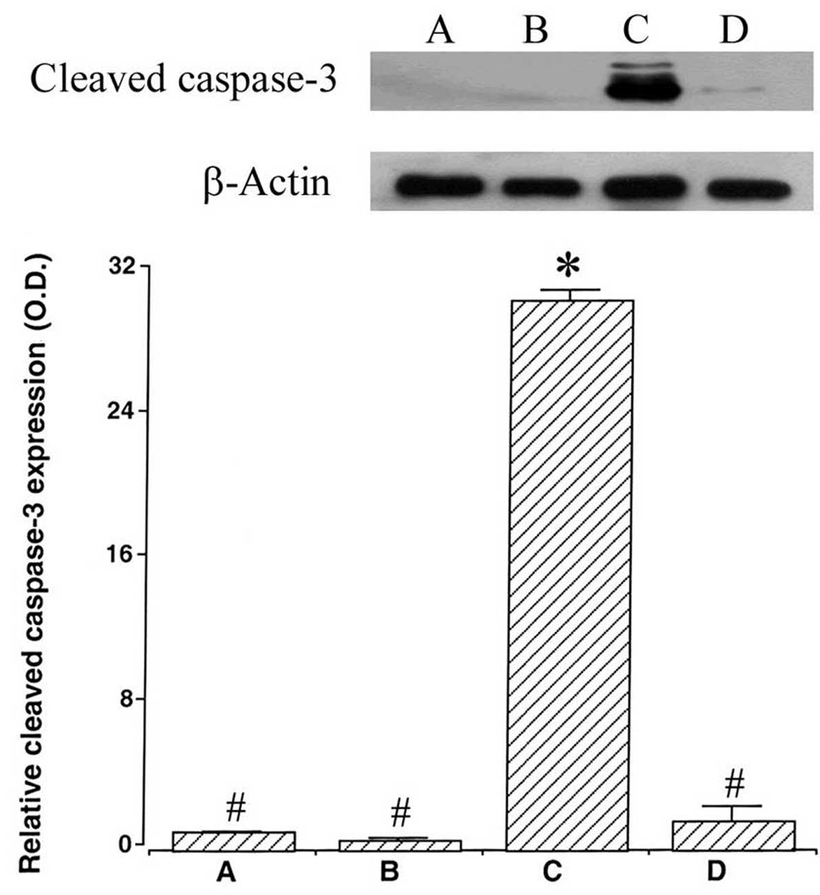
Treadmill exercise suppresses the
expression of cleaved caspase-3 following SCI
When the expression of cleaved caspase-3 (17 kDa, 19
kDa) in the sham surgery group was set at 1.00, the expression of
cleaved caspase-3 protein was 0.63±0.10 in the sham surgery plus
exercise group, 30.38±12.61 in the SCI group and 1.65±0.78 in the
SCI plus exercise group (Fig. 6).
The expression of cleaved caspase-3 was increased by SCI
(P<0.05), while treadmill exercise suppressed cleaved caspase-3
expression in the SCI rats (P<0.05).
Discussion
The exploration of different models of SCI has
suggested that physical exercise may facilitate functional recovery
(29,30). A study by de Leon and Acosta
(33) suggested that
robot-assisted training generates hindlimb sensory stimuli that
effectively enhance the ability of the lumbar spinal cord to
produce hindlimb stepping. Treadmill exercise improved the use of a
paretic hindlimb, decreased muscle atrophy and increased axonal
regrowth and collateral sprouting proximal to the lesion site in
thoracic hemisectioned mice (29).
In the present study, treadmill exercise enhanced hindlimb motor
function in the SCI rats as assessed by grid-walking, showing that
treadmill exercise improved hindlimb motor function following
SCI.
Neurotrophic factors, including NGF, NT-3 and IGF-1,
modulate neuronal growth, differentiation and survival (34). Neurotrophic factors protect against
various brain injuries, and these therapeutic effects are mediated
by the PI3K/Akt signaling pathway (35,36).
Ying et al (37) reported
that voluntary wheel running increased expression of NT-3 and its
tyrosine kinase C receptor in the spinal cord. Statins improved
neurological outcomes in an experimental intracerebral hemorrhage
model through activation of the PI3K/Akt signaling pathway
(35). The anti-apoptotic effect
of quercetin on focal cerebral ischemia may be associated with
activation of the PI3K/Akt signaling pathway (36). In the present study, expression of
NT-3 and IGF-1 was reduced in the spinal cord following SCI. NGF
levels in the SCI rats were not significantly different from those
in the sham-operated rats. By contrast, treadmill exercise
increased expression of NGF, NT-3 and IGF-1 expression in SCI rats.
These results suggested that treadmill exercise exerted a
protective effect on SCI by stimulating the expression of
neurotrophic factors.
Intracellular pro-apoptotic signals induce
mitochondrial outer membrane permeabilization followed by release
of cytochrome c into the cytosol (38). As a result, caspase-9 is activated
and induces the activation of caspase-3 (39). The PI3K/Akt pathway is involved in
apoptosis regulation (15). The
PI3K/Akt pathway is implicated in anti-apoptotic mechanisms, and
this pathway influences the balance of anti- and pro-apoptotic
proteins such as Bcl-2 and Bax, respectively (40). An ex vivo study using human
monocytes demonstrated that activation of PI3K/Akt signaling
suppressed apoptosis (40). By
contrast, in a human prostate cancer cell line, inhibition of the
PI3K/Akt signaling pathway exerted a pro-apoptotic effect (18). Exercise-induced enhancement of
neuronal survival is associated with the increased expression of
several key intermediates of the PI3K/Akt pathway (41). In the present study, PI3K
expression and the ratio of pAkt/Akt were reduced in the spinal
cord following SCI. However, treadmill exercise increased PI3K
levels and enhanced the ratio of pAkt/Akt in the injured spinal
cord. These results suggested that exercise-induced activation of
the PI3K/Akt signaling pathway may be correlated with the
anti-apoptotic effect of treadmill exercise following SCI.
The Bcl-2 family includes anti-apoptotic molecules,
including Bcl-2, and pro-apoptotic molecules, including Bax, Bid
and Bad. The Bcl-2 family is critical in determining whether
neurons survive or die (42).
Cycling exercise increased Bcl-2 mRNA expression, and high levels
of Bcl-2 mRNA expression correlated with reduced expression of
caspase-7 and caspase-9 mRNAs in SCI rats (43). Treadmill exercise suppressed Bax
expression and increased Bcl-2 expression in the hippocampus
following traumatic brain injury (3). The ratio of Bcl-2/Bax determines the
mitochondrial response to apoptotic stimuli and is crucial in
determining whether cells survive or undergo apoptosis (2,44).
In the present study, the ratio of Bcl-2/Bax in the spinal cord was
decreased by SCI, while treadmill exercise increased the ratio of
Bcl-2/Bax in the injured spinal cord, by enhancing expression of
the anti-apoptotic Bcl-2 protein. Under the experimental conditions
of the present study, SCI increased cleaved caspase-3 expression in
the injured spinal cord. By contrast, treadmill exercise suppressed
cleaved caspase-3 expression in the SCI rats. These results
suggested that treadmill exercise exerted an anti-apoptotic effect
on the injured spinal cord.
The present study demonstrated that treadmill
exercise promotes the recovery of hindlimb motor function through
suppressing apoptosis in the injured spinal cord. The
anti-apoptotic effect of exercise may result from the
exercise-mediated increase in the expression of neurotrophic
factors via activation of the PI3K/Akt pathway. Based on these
results, treadmill exercise may exert a protective effect and
improve the recovery of locomotion following SCI.
Acknowledgements
This study was supported by the 2010 Research Fund
from Kyung Hee University (grant no. KHU 20100848).
References
|
1
|
Tator CH and Fehlings MG: Review of the
secondary injury theory of acute spinal cord trauma with emphasis
on vascular mechanisms. J Neurosurg. 75:15–26. 1991. View Article : Google Scholar : PubMed/NCBI
|
|
2
|
Hwang L, Choi IY, Kim SE, Ko IG, Shin MS,
Kim CJ, Kim SH, Jin JJ, Chung JY and Yi JW: Dexmedetomidine
ameliorates intracerebral hemorrhage-induced memory impairment by
inhibiting apoptosis and enhancing brain-derived neurotrophic
factor expression in the rat hippocampus. Int J Mol Med.
31:1047–1056. 2013.
|
|
3
|
Kim DH, Ko IG, Kim BK, Kim TW, Kim SE,
Shin MS, Kim CJ, Kim H, Kim KM and Baek SS: Treadmill exercise
inhibits traumatic brain injury-induced hippocampal apoptosis.
Physiol Behav. 101:660–665. 2010. View Article : Google Scholar : PubMed/NCBI
|
|
4
|
Savitz SI and Rosenbaum DM: Apoptosis in
neurological disease. Neurosurgery. 42:555–574. 1998. View Article : Google Scholar
|
|
5
|
Sung YH, Kim SC, Hong HP, Park CY, Shin
MS, Kim CJ, Seo JH, Kim DY, Kim DJ and Cho HJ: Treadmill exercise
ameliorates dopaminergic neuronal loss through suppressing
microglial activation in Parkinson’s disease mice. Life Sci.
91:1309–1316. 2012.PubMed/NCBI
|
|
6
|
Crowe MJ, Bresnahan JC, Shuman SL, Masters
JN and Beattie MS: Apoptosis and delayed degeneration after spinal
cord injury in rats and monkeys. Nat Med. 3:73–76. 1997. View Article : Google Scholar : PubMed/NCBI
|
|
7
|
Li GL, Brodin G, Farooque M, Funa K, Holtz
A, Wang WL and Olsson Y: Apoptosis and expression of Bcl-2 after
compression trauma to rat spinal cord. J Neuropathol Exp Neurol.
55:280–289. 1996. View Article : Google Scholar : PubMed/NCBI
|
|
8
|
Yong C, Arnold PM, Zoubine MN, Citron BA,
Watanabe I, Berman NE and Festoff BW: Apoptosis in cellular
compartments of rat spinal cord after severe contusion injury. J
Neurotrauma. 15:459–472. 1998. View Article : Google Scholar : PubMed/NCBI
|
|
9
|
Emery E, Aldana P, Bunge MB, Puckett W,
Srinivasan A, Keane RW, Bethea J and Levi AD: Apoptosis after
traumatic human spinal cord injury. J Neurosurg. 89:911–920. 1998.
View Article : Google Scholar : PubMed/NCBI
|
|
10
|
Youle RJ and Strasser A: The BCL-2 protein
family: opposing activities that mediate cell death. Nat Rev Mol
Cell Biol. 9:47–59. 2008. View
Article : Google Scholar : PubMed/NCBI
|
|
11
|
Nathoo N, Narotam PK, Agrawal DK, Connolly
CA, van Dellen JR, Barnett GH and Chetty R: Influence of apoptosis
on neurological outcome following traumatic cerebral contusion. J
Neurosurg. 101:233–240. 2004. View Article : Google Scholar : PubMed/NCBI
|
|
12
|
Tsuchiya D, Hong S, Matsumori Y, Kayama T,
Swanson RA, Dillman WH, Liu J, Panter SS and Weinstein PR:
Overexpression of rat heat shock protein 70 reduces neuronal injury
after transient focal ischemia, transient global ischemia, or
kainic acid-induced seizures. Neurosurgery. 53:1179–1188. 2003.
View Article : Google Scholar
|
|
13
|
Elmore S: Apoptosis: a review of
programmed cell death. Toxicol Pathol. 35:495–516. 2007. View Article : Google Scholar : PubMed/NCBI
|
|
14
|
Jin Z and El-Deiry WS: Overview of cell
death signaling pathways. Cancer Biol Ther. 4:139–163.
2005.PubMed/NCBI
|
|
15
|
Schorey JS and Cooper AM: Macrophage
signaling upon mycobacterial infection: the MAP kinases lead the
way. Cell Microbiol. 5:133–142. 2003. View Article : Google Scholar : PubMed/NCBI
|
|
16
|
Cardone MH, Roy N, Stennicke HR, Salvesen
GS, Franke TF, Stanbridge E, Frisch S and Reed JC: Regulation of
cell death protease caspase-9 by phosphorylation. Science.
282:1318–1321. 1998. View Article : Google Scholar : PubMed/NCBI
|
|
17
|
Datta SR, Dudek H, Tao X, Masters S, Fu H,
Gotoh Y and Greenberg ME: Akt phosphorylation of BAD couples
survival signals to the cell-intrinsic death machinery. Cell.
91:231–241. 1997. View Article : Google Scholar : PubMed/NCBI
|
|
18
|
Shin DY, Kim GY, Lee JH, Choi BT, Yoo YH
and Choi YH: Apoptosis induction of human prostate carcinoma DU145
cells by diallyl disulfide via modulation of JNK and PI3K/AKT
signaling pathways. Int J Mol Sci. 13:14158–14171. 2012. View Article : Google Scholar : PubMed/NCBI
|
|
19
|
Fresno Vara JA, Casado E, de Castro J,
Cejas P, Belda-Iniesta C and González-Barón M: PI3K/Akt signalling
pathway and cancer. Cancer Treat Rev. 30:193–204. 2004.PubMed/NCBI
|
|
20
|
Li Y, Jiang Y, Wan Y, Zhang L, Tang W, Ma
J, Wu S and Cheng W: Medroxyprogestogen enhances apoptosis of
SKOV-3 cells via inhibition of the PI3K/Akt signaling pathway.
Biomed Res. 27:43–50. 2013. View Article : Google Scholar : PubMed/NCBI
|
|
21
|
Barber AJ, Nakamura M, Wolpert EB, Reiter
CE, Seigel GM, Antonetti DA and Gardner TW: Insulin rescues retinal
neurons from apoptosis by a phosphatidylinositol
3-kinase/Akt-mediated mechanism that reduces the activation of
caspase-3. J Biol Chem. 276:32814–32821. 2001. View Article : Google Scholar : PubMed/NCBI
|
|
22
|
Lobner D and Ali C: Mechanisms of bFGF and
NT-4 potentiation of necrotic neuronal death. Brain Res. 954:42–50.
2002. View Article : Google Scholar : PubMed/NCBI
|
|
23
|
Zhou L and Shine HD: Neurotrophic factors
expressed in both cortex and spinal cord induce axonal plasticity
after spinal cord injury. J Neurosci Res. 74:221–226. 2003.
View Article : Google Scholar : PubMed/NCBI
|
|
24
|
Guan J, Bennet L, Gluckman PD and Gunn AJ:
Insulin-like growth factor-1 and post-ischemic brain injury. Prog
Neurobiol. 70:443–462. 2003. View Article : Google Scholar : PubMed/NCBI
|
|
25
|
Pierchala BA, Ahrens RC, Paden AJ and
Johnson EM Jr: Nerve growth factor promotes the survival of
sympathetic neurons through the cooperative function of the protein
kinase C and phosphatidylinositol 3-kinase pathways. J Biol Chem.
279:27986–27993. 2004. View Article : Google Scholar : PubMed/NCBI
|
|
26
|
Cotman CW, Berchtold NC and Christie LA:
Exercise builds brain health: key roles of growth factor cascades
and inflammation. Trends Neurosci. 30:464–472. 2007. View Article : Google Scholar : PubMed/NCBI
|
|
27
|
Tillerson JL, Caudle WM, Reverón ME and
Miller GW: Exercise induces behavioral recovery and attenuates
neurochemical deficits in rodent models of Parkinson’s disease.
Neuroscience. 119:899–911. 2003.PubMed/NCBI
|
|
28
|
Griesbach GS, Hovda DA, Molteni R, Wu A
and Gomez-Pinilla F: Voluntary exercise following traumatic brain
injury: brain-derived neurotrophic factor upregulation and recovery
of function. Neuroscience. 125:129–139. 2004. View Article : Google Scholar
|
|
29
|
Goldshmit Y, Lythgo N, Galea MP and
Turnley AM: Treadmill training after spinal cord hemisection in
mice promotes axonal sprouting and synapse formation and improves
motor recovery. J Neurotrauma. 25:449–465. 2008. View Article : Google Scholar : PubMed/NCBI
|
|
30
|
Ying Z, Roy RR, Zhong H, Zdunowski S,
Edgerton VR and Gomez-Pinilla F: BDNF-exercise interactions in the
recovery of symmetrical stepping after a cervical hemisection in
rats. Neuroscience. 155:1070–1078. 2008. View Article : Google Scholar : PubMed/NCBI
|
|
31
|
Yune TY, Lee JY, Jung GY, Kim SJ, Jiang
MH, Kim YC, Oh YJ, Markelonis GJ and Oh TH: Minocycline alleviates
death of oligodendrocytes by inhibiting pro-nerve growth factor
production in microglia after spinal cord injury. J Neurosci.
27:7751–7761. 2007. View Article : Google Scholar : PubMed/NCBI
|
|
32
|
Chao OY, Pum ME, Li JS and Huston JP: The
grid-walking test: assessment of sensorimotor deficits after
moderate or severe dopamine depletion by 6-hydroxydopamine lesions
in the dorsal striatum and medial forebrain bundle. Neuroscience.
202:318–325. 2012. View Article : Google Scholar
|
|
33
|
de Leon RD and Acosta CN: Effect of
robotic-assisted treadmill training and chronic quipazine treatment
on hindlimb stepping in spinally transected rats. J Neurotrauma.
23:1147–1163. 2006.PubMed/NCBI
|
|
34
|
Hennigan A, O’Callaghan RM and Kelly AM:
Neurotrophins and their receptors: roles in plasticity,
neurodegeneration and neuroprotection. Biochem Soc Trans.
35:424–427. 2007. View Article : Google Scholar : PubMed/NCBI
|
|
35
|
Yang D, Han Y, Zhang J, Chopp M and
Seyfried DM: Statins enhance expression of growth factors and
activate the PI3K/Akt-mediated signaling pathway after experimental
intracerebral hemorrhage. World J Neurosci. 2:74–80. 2012.
View Article : Google Scholar
|
|
36
|
Yao RQ, Qi DS, Yu HL, Liu J, Yang LH and
Wu XX: Quercetin attenuates cell apoptosis in focal cerebral
ischemia rat brain via activation of BDNF-TrkB-PI3K/Akt signaling
pathway. Neurochem Res. 37:2777–2786. 2012. View Article : Google Scholar : PubMed/NCBI
|
|
37
|
Ying Z, Roy RR, Edgerton VR and
Gómez-Pinilla F: Voluntary exercise increases neurotrophin-3 and
its receptor TrkC in the spinal cord. Brain Res. 987:93–99. 2003.
View Article : Google Scholar : PubMed/NCBI
|
|
38
|
Tait SW and Green DR: Mitochondria and
cell death: outer membrane permeabilization and beyond. Nat Rev Mol
Cell Biol. 11:621–632. 2010. View
Article : Google Scholar : PubMed/NCBI
|
|
39
|
Taylor RC, Cullen SP and Martin SJ:
Apoptosis: controlled demolition at the cellular level. Nat Rev Mol
Cell Biol. 9:231–241. 2008. View
Article : Google Scholar : PubMed/NCBI
|
|
40
|
Chan G, Nogalski MT, Bentz GL, Smith MS,
Parmater A and Yurochko AD: Pi3k-dependent upregulation of Mcl-1 by
human cytomegalovirus is mediated by epidermal growth factor
receptor and inhibits apoptosis in short-lived monocytes. J
Immunol. 184:3213–3222. 2010. View Article : Google Scholar : PubMed/NCBI
|
|
41
|
Chen M and Russo-Neustadt AA: Exercise
activates the phosphatidylinositol 3-kinase pathway. Mol Brain Res.
135:181–193. 2005. View Article : Google Scholar : PubMed/NCBI
|
|
42
|
Cory S, Huang DC and Adams JM: The Bcl-2
family: roles in cell survival and oncogenesis. Oncogene.
22:8590–8607. 2003. View Article : Google Scholar : PubMed/NCBI
|
|
43
|
Liu G, Keeler BE, Zhukareva V and Houlé
JD: Cycling exercise affects the expression of apoptosis-associated
microRNAs after spinal cord injury in rats. Exp Neurol.
226:200–206. 2010. View Article : Google Scholar : PubMed/NCBI
|
|
44
|
Upadhyay D, Panduri V, Ghio A and Kamp DW:
Particulate matter induces alveolar epithelial cell DNA damage and
apoptosis: role of free radicals and the mitochondria. Am J Respir
Cell Mol Biol. 29:180–187. 2003. View Article : Google Scholar : PubMed/NCBI
|