Introduction
Renal interstitial fibrosis is the common pathway of
a variety of kidney diseases that develop into end-stage renal
failure (1). The process of renal
interstitial fibrosis involves the loss of renal tubules and
peritubular capillary endothelial cells, and the accumulation of
inflammatory cells, interstitial myofibroblasts and extracellular
matrix (2). A study has demonstrated
that reductions in kidney function are more closely correlated with
renal interstitial fibrosis levels than with glomerular sclerosis
(3). Renal interstitial fibrosis is
associated with a decline in renal function and is the most
important prognostic marker (1,4).
Therefore, renal interstitial fibrosis is the focus of increasing
attention and has become a popular research topic in international
nephrology in recent years.
The pathogenesis of renal fibrosis is very complex.
Studies have indicated that kidney diseases lead to renal tubular
and interstitial cell apoptosis, affect renal remodeling and
repair, and exacerbate renal interstitial fibrosis (5,6).
Furthermore, other studies have shown that apoptotic stimuli
increase Drp-1 expression, induce mitochondrial division, cause
changes in the expression and translocation of factors associated
with apoptosis, and induce cell apoptosis (7,8).
Research suggests that more severe interstitial fibrosis is
associated with increased cell apoptosis (9).
Studies suggest that erythropoietin (EPO) is a
multifunctional cytokine superfamily member with a protective
effect on multiple organs (10,11). EPO
has been demonstrated to exert a renoprotective effect in addition
to a hematopoietic effect in acute and chronic kidney injury
(12). It plays an important role in
antioxidation, antiapoptosis and anti-inflammation in many models
of kidney diseases (13,14,15). A
number of clinical studies have demonstrated that the early
treatment of anemia in chronic kidney disease patients with EPO
results in a slowing of the progressive decline in renal function
(16–18).
In the present study, the renoprotective effect of
EPO treatment was investigated in a unilateral ureteric obstruction
(UUO) rat model of renal interstitial fibrosis. The hypothesis that
EPO treatment may have a renoprotective effect that is mediated
through modifying the expression of Drp-1 was also evaluated.
Materials and methods
Animal care and model
This study was carried out in strict accordance with
the recommendations in the Guide for the Care and Use of Laboratory
Animals of the National Institutes of Health. The animal use
protocol has been reviewed and approved by the Institutional Animal
Care and Use Committee (IACUC) of Henan Provincial People's
Hospital (Zhengzhou, China). The Sprague-Dawley rats, weighing
250–300 g, were obtained from Xinxiang Medical College Animal
Center (Xinxiang, China). The rats were randomly divided into sham
surgery, UUO model and treatment groups, with 27 rats in each
group. The modeling method for the model and treatment groups was
to ligate the left ureter and cut it. The modeling method for the
sham surgery group was to free the ureter without ligation
(19). The treatment group was
administered EPO at a dose of 3,000 U/kg body weight by
subcutaneous injection after surgery. The sham surgery and model
groups were administered the same volume of saline by injection.
Nine rats in each group were selected at random for sacrifice on
each of days 7, 14 and 21 after surgery. Blood was collected from
the heart at the time of sacrifice. Serum creatinine (Cr) and blood
urea nitrogen (BUN) levels in the blood were tested. Kidney
specimens were fixed in 10% formalin solution and embedded in
paraffin as 3-µm sections for hematoxylin and eosin (H&E),
Masson's trichrome and immunohistochemical staining.
Renal histology and
immunohistochemistry
After H&E and Masson's trichrome staining, the
relative area of renal interstitial fibrosis was calculated using
an IDA-2000 high-resolution digital image analysis system (Leica,
Wetzlar, Germany). Ten non-overlapping visual fields were randomly
selected in each slice in order to determine the renal interstitial
fibrosis area as a percentage of the total interstitial area in the
same visual field. The percentage of renal interstitial fibrosis in
each slice was calculated as an average value. Renal interstitial
injury was scored using a semi-quantitative scoring system. An
average score was determined based on the score of each slice. The
scoring system was as follows: 0 points, no lesions; 1 point,
<25% lesions; 2 points, 25–50% lesions; 3 points, >50%
lesions (20).
Immunohistochemical staining
The streptavidin biotin complex method was used to
detect Drp-1 in the renal tissue. The staining procedure was
conducted as described by the instructions provided with the
Streptavidin Biotin Complex (SABC) kit (Wuhan Boster Biological
Engineering Co., Ltd., Wuhan, China). Under a light microscope
(SPZ-50 PFM; Carton Optical Industries, Ltd., Tokyo, Japan),
histological sections were observed, and areas in which brown
particles were deposited were positive staining areas. Under a
high-power lens (magnification, x400), renal interstitial areas
without glomeruli and vascular features were randomly selected in
10 non-overlapping visual fields. Images were collected and
analyzed by the IDA-2000 high resolution digital image analysis
system. The area with a gray level indicative of positive staining
was calculated as a percentage of the total visual field, and an
average value was determined (21).
Statistical analysis
SPSS software, version 16.0 (SPSS, Inc., Chicago,
IL, USA) was used. The data are expressed as the mean ± standard
deviation. The differences between groups were analyzed by single
factor variance. Relevance judgments were analyzed by Pearson
correlation. P<0.05 was considered to indicate a statistically
significant difference.
Results
Renal function
The serum Cr and BUN levels of the model and
treatment groups were increased significantly compared with those
in the sham surgery group at the same time point (P<0.05).
However, the serum Cr and BUN levels of the treatment group were
decreased significantly compared with those in the model group at
each time point. The differences were statistically significant
(P<0.05; Tables I and II).
 | Table I.Serum creatinine levels in all groups
(µmol/l). |
Table I.
Serum creatinine levels in all groups
(µmol/l).
| Groups | n | 7 days | 14 days | 21 days |
|---|
| Sham surgery | 9 | 38.36±3.52 | 43.05±5.27 | 44.096±4.68 |
| Model | 9 | 84.
19±4.27a |
108.44±5.63a,c |
157.43±5.73a,c |
| Treatment | 9 |
59.62±2.78a,b |
74.39±6.80a–c |
113.32±6.21a–c |
| F-value |
| 146.293 | 214.086 | 207.425 |
| P-value |
| <0.05 | <0.05 | <0.05 |
| Table II.Blood urea nitrogen levels in all
groups (mmol/l). |
Table II.
Blood urea nitrogen levels in all
groups (mmol/l).
| Groups | n | 7 days | 14 days | 21 days |
|---|
| Sham surgery | 9 | 5.72±0.14 | 6.03±0.35 | 6.28±0.09 |
| Model | 9 |
8.61±0.30a |
17.56±0.23a,c |
24.56±1.42a,c |
| Treatment | 9 |
6.49±1.28a,b |
11.98±0.64a–c |
16.84±0.47a–c |
| F-value |
| 79.264 | 113.489 | 161.731 |
| P-value |
| <0.05 | <0.05 | <0.05 |
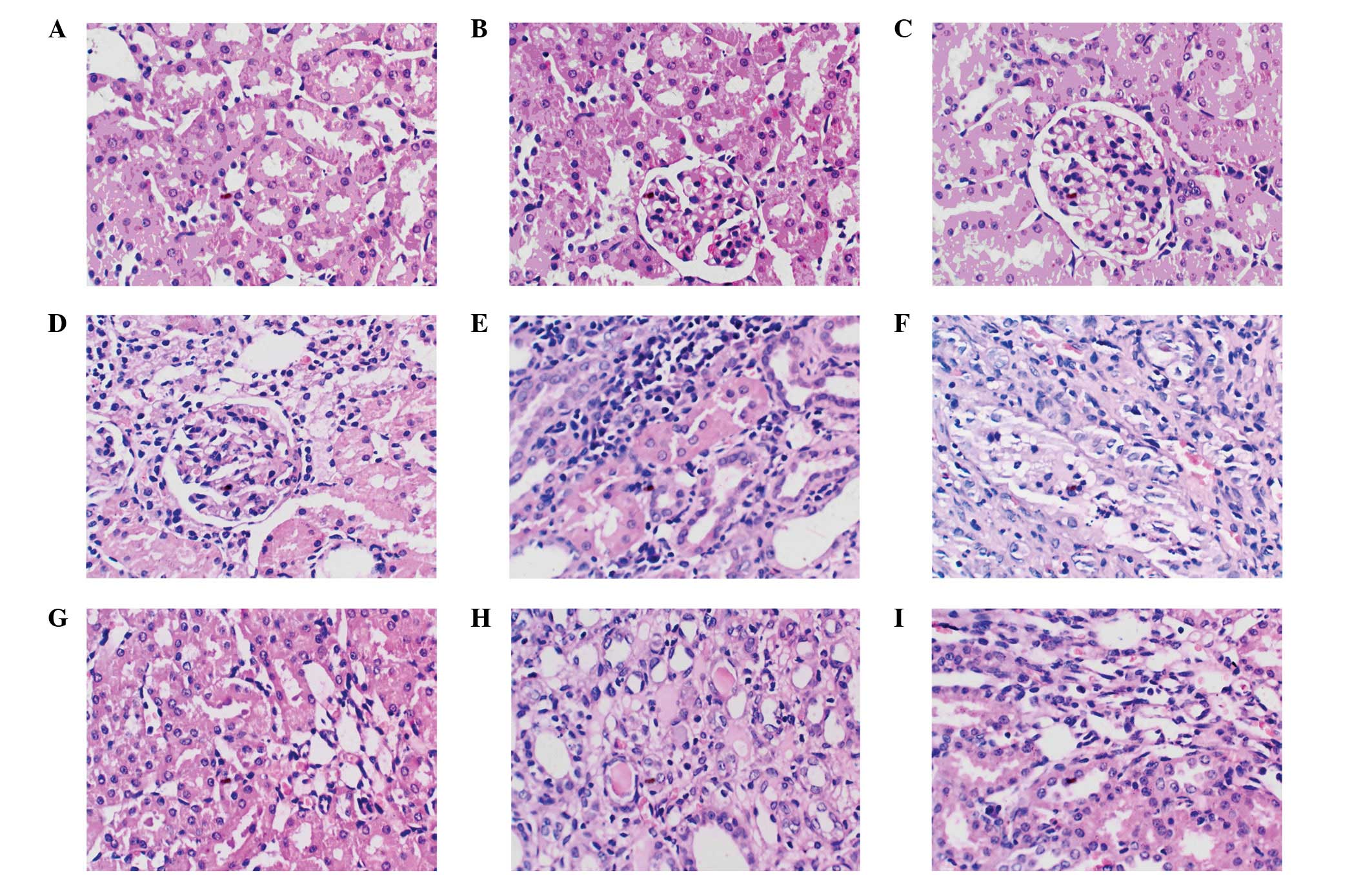
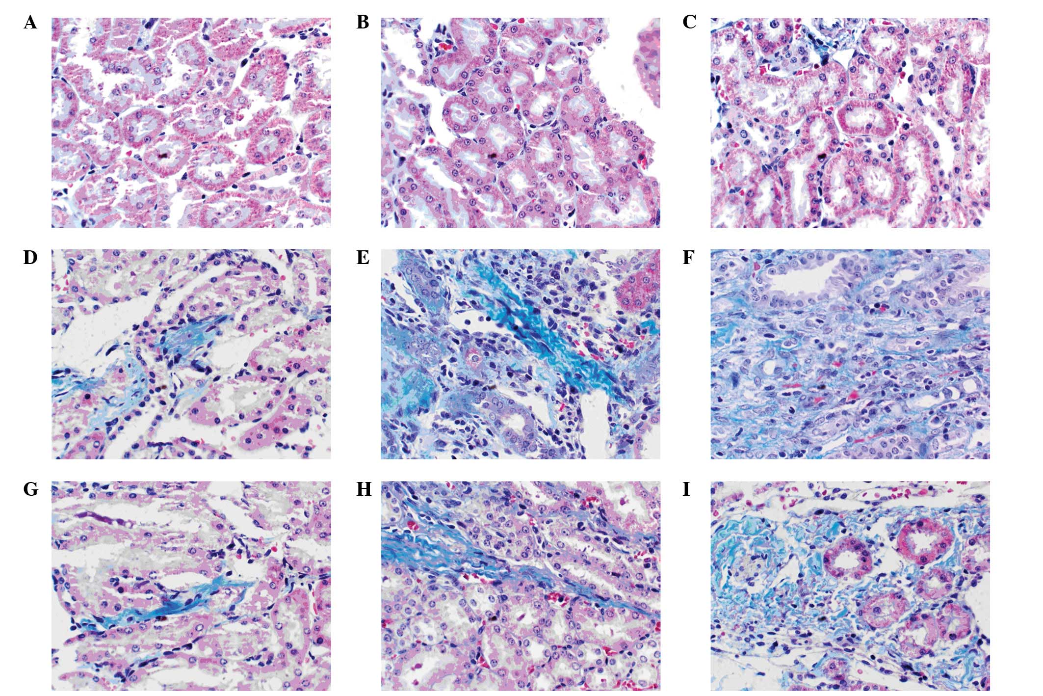
Kidney histology
H&E and Masson's trichrome staining results
showed that in the model group on day 7 after surgery, the renal
interstitium was infiltrated by inflammatory cells, the cytoplasm
of the renal tubular epithelial cells was loose with swelling and
vacuolar degeneration, partial tubular dilation had occurred, the
interstitium was widened, cells and extracellular matrix were
increased in quantity, and fibrosis was visible in the cortex and
the corticomedullary zone. On day 14 after surgery, the renal
interstitium was markedly infiltrated by inflammatory cells,
tubular dilatation was evident, partial shedding and atrophy of
tubular epithelial cells was visible, the interstitium was widened
significantly, and the cortex and corticomedullary zone were
clearly fibrotic. On day 21 after surgery, the renal interstitium
was diffusely infiltrated by inflammatory cells, the renal tubular
structures were seriously damaged, tubular dilation, deformation
and atrophy were severe, tubulointerstitial broadening was more
evident, and fibrous hyperplasia was observed. The renal tissue of
the sham surgery group showed none of these changes. The treatment
group at each time point was compared with the model group. In the
treatment group, renal interstitial infiltration by inflammatory
cells was reduced, tubular dilation and atrophy were attenuated
significantly, the area of renal interstitial fibrosis was reduced
and the renal interstitial injury score was significantly decreased
(P<0.05), although it remained higher than that in the sham
surgery group (P<0.01). The renal tubular interstitial damage
scores are shown in Table III and
Fig. 1, and the relative area of
renal interstitial fibrosis is shown in Table IV and Fig. 2.
| Table III.Renal tubulointerstitial damage score
of all groups (%). |
Table III.
Renal tubulointerstitial damage score
of all groups (%).
| Groups | n | 7 days | 14 days | 21 days |
|---|
| Sham surgery | 9 | 0.49±0.03 | 0.51±0.07 | 0.52±0.04 |
| Model | 9 |
3.41±0.18a |
5.07±0.26a,c |
6.38±0.25a,c |
| Treatment | 9 |
2.32±0.21a,b |
3.94±0.15a–c |
5.03±0.28a–c |
| F-value |
| 46.274 | 103.655 | 116.937 |
| P-value |
| <0.01 | <0.01 | <0.01 |
| Table IV.Relative area of renal interstitial
fibrosis in all groups (%). |
Table IV.
Relative area of renal interstitial
fibrosis in all groups (%).
| Groups | n | 7 days | 14 days | 21 days |
|---|
| Sham surgery | 9 | 1.63±0.09 | 1.72±0.11 | 1.86±0.15 |
| Model | 9 |
14.43±0.57a |
27.85±1.39a,c |
53.16±2.45a,c |
| Treatment | 9 |
9.77±0.62a,b |
17.36±0.82a–c |
31.29±1.94a–c |
| F-value |
| 85.033 | 126.572 | 187.836 |
| P-value |
| <0.01 | <0.01 | <0.01 |
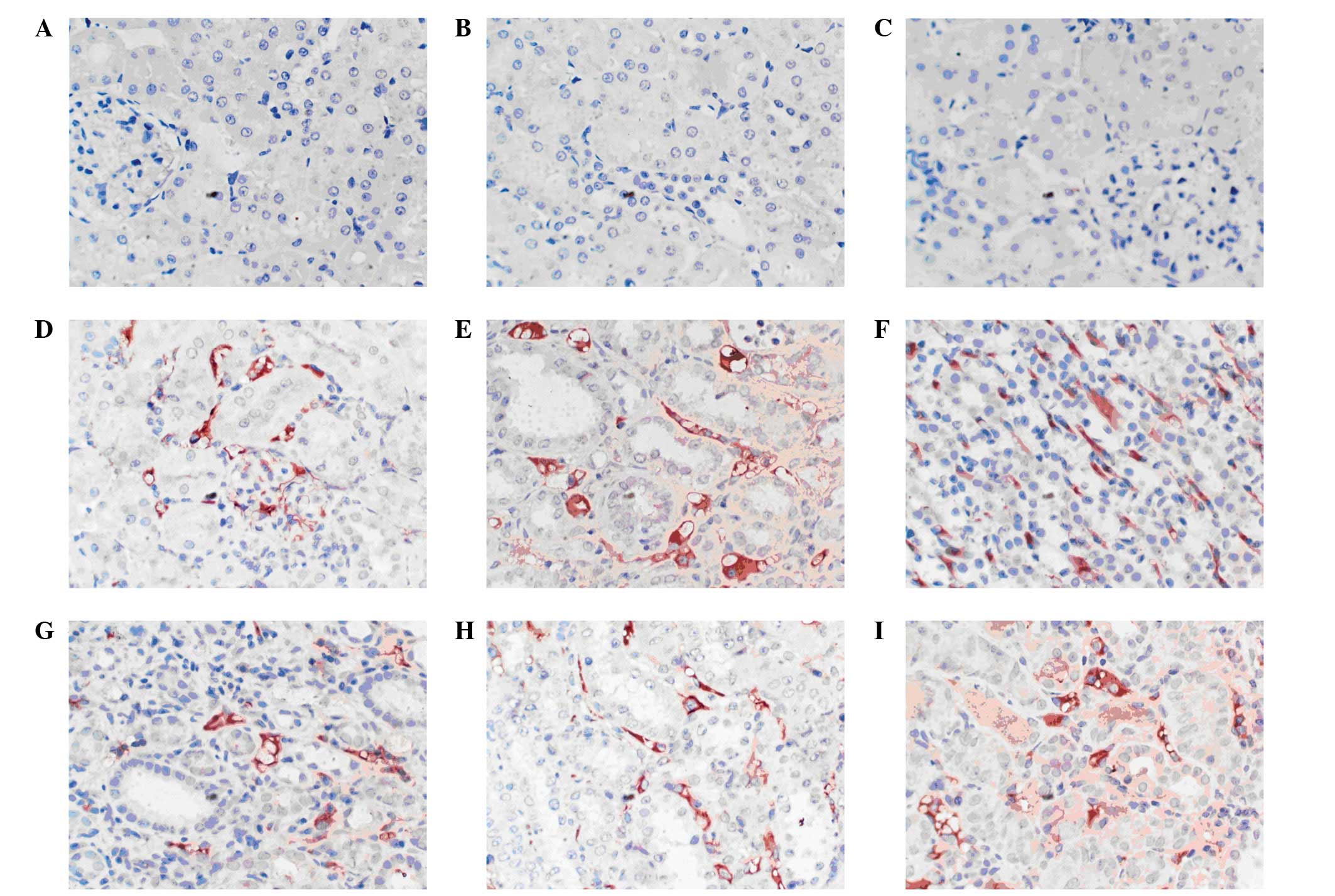
Immunohistochemistry
Drp-1 in the sham surgery group at each time point
was expressed at low levels in renal interstitial and tubular
epithelial cells. In the model group, the expression of Drp-1 was
located in the cell cytoplasm of the renal tubular epithelial cells
and interstitium, and the expression levels gradually increased
with the prolongation of UUO. In the treatment and model groups,
the sites of expression were essentially the same, but the scope
and intensity were decreased in the treatment group. Parallel
comparison showed that Drp-1 expression in the model and treatment
groups at each time point was significantly increased compared with
that in the sham surgery group (P<0.05), and that Drp-1
expression in the treatment group was reduced compared with that in
the model group. The difference was statistically significant
(P<0.05; Table V and Fig. 3).
| Table V.Relative expression area of Drp-1 in
renal tissue of the three groups at different time points (%). |
Table V.
Relative expression area of Drp-1 in
renal tissue of the three groups at different time points (%).
| Groups | n | 7 days | 14 days | 21 days |
|---|
| Sham surgery | 9 | 0.88±0.09 | 1.05±0.14 | 1.06±0.17 |
| Model | 9 |
9.92±0.37a |
16.42±0.58a,c |
25.74±0.19a,c |
| Treatment | 9 |
6.05±0.21a,b |
11.28±0.13a–c |
18.63±0.22a–c |
| F-value |
| 153.628 | 428.371 | 507.845 |
| P-value |
| <0.05 | <0.05 | <0.05 |
Correlation analysis
By analyzing the area of positive Drp-1 expression,
renal tubulointerstitial injury, and relative area of renal
interstitial fibrosis of the model and treatment groups, it was
identified that the positively stained area of Drp-1, renal
tubulointerstitial injury and relative area of renal interstitial
fibrosis were positively correlated (r=0.923, 0.895).
Discussion
Renal interstitial fibrosis is the common pathway
for a variety of chronic kidney disease as they develop to
end-stage renal failure. The pathological features include renal
interstitial fibroblast proliferation and the excessive
accumulation of extracellular matrix (22). There is evidence that, in renal
interstitial fibrosis, there is a close relationship between the
apoptosis of renal tubular epithelial cells and tubular atrophy;
the increase in the apoptosis index is one of the causes (23) of extracellular matrix formation. A
study suggests that more severe interstitial fibrosis is associated
with increased cell apoptosis, with a positive correlation existing
between the degree of interstitial fibrosis and the apoptotic cell
number (24). One of the main
processes involved in apoptosis is mitochondrial fracture. Drp-1 in
mammalian cells is an important executive molecule for
mitochondrial fission. Drp-1 is mainly located in the cytoplasm as
a polymer. It can be translocated to the outer mitochondrial
membrane and accumulated into the potential sites of mitochondrial
division when it is recruited by the molecules of the mitochondrial
outer membrane. Multiple Drp-1 molecules surrounding the
mitochondria ultimately lead to the breakdown of the mitochondria
and apoptosis (25). A previous
study found that the overexpression of Drp-1 may lead to an
acceleration of mitochondrial fission, resulting in the production
of a large number of mitochondrial fragments (26). The inhibition of endogenous Drp-1 by
gene knockout can inhibit mitochondrial fission (27). A study of the apoptosis of renal
tubular epithelial cells induced by ATP depletion found that the
activation of Drp-1 leads to the fragmentation of mitochondria,
resulting in the release of intermembrane pro-apoptotic proteins,
and induction of the apoptosis of cells (28).
Erythropoietin is bone marrow stimulation factor,
synthesized by the kidneys and liver, and a multifunctional member
of the cytokine super family (29).
A previous study found that EPO can ameliorate anemia, inhibit
inflammatory reactions, promote the growth of blood vessels,
inhibit apoptosis, and reduce the expression of cytokines and
inflammatory factors, thereby protecting organs from damage
(30). In recent years, a large
number of animal experiments have shown that EPO can delay the
progress of chronic kidney disease and improve the degree of
interstitial fibrosis, and has a certain renal protective effect
(31–33). A study conducted by Nakazawa et
al demonstrated that EPO is able to inhibit renal tubule cell
apoptosis, thereby reducing the degree of renal interstitial
fibrosis (34).
In the present study, a rat model of UUO was
established to study the expression of Drp-1 in renal interstitial
fibrosis and the ability of erythropoietin to protect against it.
The results showed that 7, 14, and 21 days after surgery, in the
treatment group at each time point compared with the model group,
the renal function was improved significantly and infiltration of
the renal interstitium by inflammatory cells was reduced. In
addition, tubular dilatation and atrophy, the relative area of
renal interstitial fibrosis, and the renal interstitial injury
score were markedly decreased. Immunohistochemical analysis showed
that in the model and treatment groups at each time point, Drp-1
expression levels were significantly increased compared with those
in the sham surgery group (P<0.05). The Drp-1 expression levels
in the kidneys of rats in the treatment group were decreased
significantly compared with those in the model group at the same
time point. The results of correlation analysis showed that the
expression of Drp-1 in renal tissue, the renal interstitial injury
score and relative area of renal interstitial fibrosis were
positively correlated. The results indicate that erythropoietin may
reduce apoptosis and decrease renal interstitial fibrosis by
inhibiting the expression of Drp-1 in renal tissue.
In summary, the overexpression of Drp-1 is
potentially one of the mechanisms underlying the development of
renal interstitial fibrosis. Erythropoietin may delay the
development of renal interstitial fibrosis and protect the kidney
by inhibiting the expression of Drp-1 in the renal tissue of rats
subjected to UUO. However, the mechanism by which erythropoietin
inhibits the expression of Drp-1 in the renal tissue in this model
requires further study.
References
|
1
|
Nangaku M: Mechanisms of
tubulointerstitial injury in the kidney: final common pathways to
end-stage renal failure. Intern Med. 43:9–17. 2004. View Article : Google Scholar : PubMed/NCBI
|
|
2
|
Campanholle G, Ligresti G, Gharib SA and
Duffield JS: Cellular mechanisms of tissue fibrosis 3. Novel
mechanisms of kidney fibrosis. Am J Physiol Cell Physiol.
304:C591–C603. 2013. View Article : Google Scholar : PubMed/NCBI
|
|
3
|
Eddy AA: Molecular basis of renal
fibrosis. Pediatric Nephrol. 15:290–301. 2000. View Article : Google Scholar
|
|
4
|
Ucero AC, Benito-Martin A, Izquierdo MC,
et al: Unilateral ureteral obstruction: beyond obstruction. Int
Urol Nephrol. 46:765–776. 2014. View Article : Google Scholar : PubMed/NCBI
|
|
5
|
Strutz F: Pathogenesis of
tubulointerstitial fibrosis in chronic allograft dysfunction. Clin
Transplant. 23 (Suppl 21):26–32. 2009. View Article : Google Scholar : PubMed/NCBI
|
|
6
|
Ucero AC, Goncalves S, Benito-Martin A, et
al: Obstructive renal injury: from fluid mechanics to molecular
cell biology. Open Access J Urol. 2:41–55. 2010.PubMed/NCBI
|
|
7
|
Otera H and Mihara K: Molecular mechanisms
and physiologic functions of mitochondrial dynamics. J Biochem.
149:241–251. 2011. View Article : Google Scholar : PubMed/NCBI
|
|
8
|
Ucero AC, Benito-Martin A, Fuentes-Calvo
I, et al: TNF-related weak inducer of apoptosis (TWEAK) promotes
kidney fibrosis and Ras-dependent proliferation of cultured renal
fibroblast. Biochim Biophys Acta. 1832:1744–1755. 2013. View Article : Google Scholar : PubMed/NCBI
|
|
9
|
Izquierdo MC, Sanz AB, Mezzano S, et al:
TWEAK (tumor necrosis factor-like weak inducer of apoptosis)
activates CXCL16 expression during renal tubulointerstitial
inflammation. Kidney Int. 81:1098–1107. 2012. View Article : Google Scholar : PubMed/NCBI
|
|
10
|
Brines M and Cerami A: Discovering
erythropoietin's extra-hematopoietic functions: biology and
clinical promise. Kidney Int. 70:246–250. 2006. View Article : Google Scholar : PubMed/NCBI
|
|
11
|
Baker JE: Enythropoietin mimics ischemic
preconditioning. Vascul Pharmacol. 42:233–241. 2005. View Article : Google Scholar : PubMed/NCBI
|
|
12
|
Chang YK, Choi DE, Na KR, et al:
Erythropoietin attenuates renal injury in an experimental model of
rat unilateral ureteral obstruction via anti-inflammatory and
anti-apoptotic effects. J Urol. 181:1434–1443. 2009. View Article : Google Scholar : PubMed/NCBI
|
|
13
|
Cassis P, Gallon L, Benigni A, et al:
Erythropoietin, but not the correction of anemia alone, protects
from chronic kidney allograft injury. Kidney Int. 81:903–918. 2012.
View Article : Google Scholar : PubMed/NCBI
|
|
14
|
Yu XQ, Wu LL, Huang XR, et al: Osteopontin
expression in progressive renal injury in remnant kidney:rule of
angiotension II. Kidney Int. 58:1469–1480. 2000. View Article : Google Scholar : PubMed/NCBI
|
|
15
|
Li C, Chen Y and Hong MY: Erythropoietin
protective effect of renal perfusion by pretreatment on acute
global ischemia in rats. Zhongguo Bingli Shengli Zazhi.
20:2336–2338. 2004.
|
|
16
|
Srisawat N, Manotham K and Eiam-Ong S,
Katavetin P, Praditpornsilpa K and Eiam-Ong S: Erythropoietin and
its non-erythropoietic derivative: do they ameliorate renal
tubulointerstitial injury in ureteral obstruction? Int J Urol.
15:1011–1017. 2008. View Article : Google Scholar : PubMed/NCBI
|
|
17
|
Sharples EJ, Patel N, Brown P, et al:
Erythropoietin protects the kidney against the injury and
dysfunction caused by ischemia-reperfusion. J Am Soc Nephrol.
15:2115–2124. 2004. View Article : Google Scholar : PubMed/NCBI
|
|
18
|
Ates E, Yalcin AU, et al: Protective
effect of erythropoietin on renal ischemia and reperfusion injury.
ANZ J Surg. 75:1100–1105. 2005. View Article : Google Scholar : PubMed/NCBI
|
|
19
|
Chevalier RL, Forbes MS and Thornhill BA:
Ureteral obstruction as a model of renal interstitial fibrosis and
obstructive nephropathy. Kidney Int. 75:1145–1152. 2009. View Article : Google Scholar : PubMed/NCBI
|
|
20
|
Pang M, Kothapally J, Mao H, et al:
Inhibition of histone deacetylase activity attenuates renal
fibroblast activation and interstitial fibrosis in obstructive
nephropathy. Am J Physiol Renal Physiol. 297:F996–F1005. 2009.
View Article : Google Scholar : PubMed/NCBI
|
|
21
|
Cregger M, Berger AJ and Rimm DL:
Immunohistochemistry and quantitative analysis of protein
expression. Arch Pathol Lab Med. 130:1026–1030. 2006.PubMed/NCBI
|
|
22
|
Hewitson TD, Ho WY and Samuel CS:
Antifibrotic properties of relaxin: in vivo mechanism of action in
experimental renal tubulointerstitial fibrosis. Endocrinol.
151:4938–4948. 2010. View Article : Google Scholar
|
|
23
|
Yang T, Vesey DA, Johnson DW, Wei MQ and
Gobe GC: Apoptosis of tubulointerstitial chronic inflammatory cells
in progressive renal fibrosis after cancer therapies. Transl Res.
150:40–50. 2007. View Article : Google Scholar : PubMed/NCBI
|
|
24
|
Izquierdo MC, Sanz AB, Mezzano S, et al:
TWEAK (tumor necrosis factor-like weak inducer of apoptosis)
activates CXCL16 expression during renal tubulointerstitial
inflammation. Kidney Int. 81:1098–1107. 2012. View Article : Google Scholar : PubMed/NCBI
|
|
25
|
Figueroa-Romero C, Iniguez-Lluhi JA,
Stadler J, et al: SUMOylation of the mitochondrial fission protein
Drpl occurs at multiple nonconsensus sites within the B domain and
is linked to its activity cycle. FASEB J. 23:3917–3927. 2009.
View Article : Google Scholar : PubMed/NCBI
|
|
26
|
Brooks C, Cho SG, et al: Fragmented
mitochondria are sensitized to Bax insertion and activation during
apoptosis. Am J Physiol Cell Physiol. 300:C447–C455. 2011.
View Article : Google Scholar : PubMed/NCBI
|
|
27
|
Wang H, Lim PJ, Karbowski M and Monteiro
MJ: Effects of overexpression of huntingtin proteins on
mitochondrial integrity. Hum Mol Genet. 18:737–752. 2009.
View Article : Google Scholar : PubMed/NCBI
|
|
28
|
Brooks C, Cho SG, Wang CY, Yang T and Dong
Z: Fragmented mitochondria are sensitized to Bax insertion and
activation during apoptosis. Am J Physiol Cell Physiol.
300:C447–C455. 2011. View Article : Google Scholar : PubMed/NCBI
|
|
29
|
Ghezzi P and Brines M: Erythropoietin as
an antiapoptotic, tissue-protective cytokine. Cell Death Differ. 11
(Suppl 1):S37–S44. 2004. View Article : Google Scholar : PubMed/NCBI
|
|
30
|
Ergur BU, Kiray M, Pekcetin C, et al:
Protective effect of erythropoietin pretreatment in testicular
ischemia-reperfusion injury in rats. J Pediatr Surg. 43:722–728.
2008. View Article : Google Scholar : PubMed/NCBI
|
|
31
|
Kitamura H, Isaka Y, Takabatake Y, et al:
Nonethropoietic derivative of erythropoietin protect against
tubulointerstitial injury in a unilateral ureteral obstruction
model. Nephrol Dial Transplant. 23:1521–1528. 2008. View Article : Google Scholar : PubMed/NCBI
|
|
32
|
Kuriyams S, Tomonari H, Tokudome G, et al:
Association of angiotensinogen gene polymorphism with
erythropoietin-induced hypertension: a preliminary report.
Hypertens Res. 24:501–505. 2001. View Article : Google Scholar : PubMed/NCBI
|
|
33
|
Jungers P, Choukroun G, Oualim Z, Robino
C, Nguyen AT and Man NK: Beneficial influence of recombinant human
erythropoietin therapy on the rate of progression of chronic renal
failure in predialysis patients. Nephrol Dial Transplant.
16:307–312. 2001. View Article : Google Scholar : PubMed/NCBI
|
|
34
|
Nakazawa Y, Nishino T, Obata K, et al:
Recombinant human erythropoietin attenuates renal
tubulointerstitial injury in murine adriamycin-induced nephropathy.
J Nephrol. 26:527–533. 2013. View Article : Google Scholar : PubMed/NCBI
|