Introduction
Stroke, a medical emergency characterized by the
rapid loss of brain function due to disturbance in brain blood
supply, is currently the leading cause of serious and long-term
disability in adults, as well as the second most common cause of
death worldwide, ranking after heart disease and before cancer
(1,2). Ischemia, which is caused by
thrombosis blocking the blood flow to brain, is one of the major
causative factors for the pathogenesis of stroke. Therapeutic
approaches of ischemic stroke include removing the blood clot by
thrombolysis or thrombectomy, minimizing clot enlargement or
preventing new clot formation with medications, recovering any lost
function with rehabilitation modalities such as physical therapy
and occupational therapy.
Acupuncture has long been used in China to treat
various diseases including stroke and currently it is also
increasingly practiced in the western society (3). A large number of studies have
demonstrated the clinical efficacy of acupuncture in stroke
rehabilitation (4–7). Recently, we reported that
electroacupuncture may alleviate neurological deficits via
promoting the proliferation and differentiation of nerve stem cells
(8). Moreover, on the basis of a
large amount of documents and materials, the Zusanli (ST36) and
Quchi (LI11) acupoints were commonly used in China to clinically
treat stroke. However, the precise mechanism of neuroprotective
effect of electroacupuncture at Quchi and Zusanli remains largely
unclear.
PI3K/Akt pathway is essential for cell growth,
proliferation, differentiation, motility, survival and
intracellular trafficking (9,10).
Phosphatidylinositol 3-kinase (PI3K) can be activated by cytokines
including neurotrophic factors, such as brain-derived neurotrophic
factor (BDNF) and glial cell line-derived neurotrophic factor
(GDNF). Activated PI3K is able to phosphorylate PI(4)P and PI(4,5)P2
at the 3 position hydroxyl group of the inositol ring to generate
PI(3,4)P2 and PI(3,4,5)
P3, respectively. These lipids serve as plasma membrane docking
sites for proteins that contain pleckstrin-homology (PH) domains,
such as Akt and its upstream activator PDK1. Upon activation of
PI3K, both Akt and PDK1 translocate to the plasma membrane, where
they bind directly to PI(3,4)P2
and PI(3,4,5)P3
via PH domain. The colocalization of PDK1 and Akt results in the
phosphorylation of Akt, which in turn promotes cell survival by
increasing the expression of anti-apoptotic Bcl-2 and
downregulating the pro-apoptotic Bax expression. It has been shown
that the activation of PI3K/Akt pathway could exert a
neuroprotective role through inhibiting cell apoptosis in cerebral
ischemia (11–13).
To elucidate the neuroprotective mechanism of
electroacupuncture at Quchi and Zusanli, in the present study we
investigate its effect on PI3K/Akt pathway in cerebral ischemia by
using a focal cerebral ischemia/reperfusion injured rat model.
Materials and methods
Materials and reagents
Trizol reagent was purchased from Invitrogen
(Carlsbad, CA, USA). SuperScript II reverse transcriptase and TUNEL
assay kit were provided by Promega (Madison, WI, USA). PI3K, Akt,
phospho-Akt (Thr308), Bcl-2, Bax and β-actin antibodies,
horseradish peroxidase (HRP)-conjugated secondary antibodies were
obtained from Cell Signaling (Beverly, MA, USA). Rat BDNF and GDNF
ELISA kits were purchased from Shanghai Xitang Biological
Technology Co., Ltd. (Shanghai, China). All the other chemicals
used, unless otherwise stated, were obtained from Sigma Chemicals
(St. Louis, MO, USA).
Animals
Male Sprague-Dawley rats (with an initial body
weight of ∼250 g) were obtained from Shanghai SLAC Laboratory
Animal Co., Ltd. (Shanghai, China) and housed under pathogen-free
conditions with a 12 h light/dark cycle. Food and water were given
ad libitum throughout the experiment. All animal treatments
were strictly in accordance with the international ethical
guidelines and the National Institutes of Health Guide concerning
the Care and Use of Laboratory Animals, and the experiments were
approved by the Institutional Animal Care and Use Committee of
Fujian University of Traditional Chinese Medicine.
Establishment of the cerebral
ischemia-reperfusion (I/R) injured rat model and animal
grouping
I/R injured model was established by middle cerebral
artery occlusion (MCAO) as previously described (14). Briefly, after a rat was
anesthetized with 10% chloral hydrate by intraperitoneal injection,
the left common carotid artery (CCA), the left external carotid
artery (ECA) and internal carotid artery (ICA) were carefully
exposed by a midline neck incision. The left middle cerebral artery
was occluded by introducing an embolus through the ICA. Focal
cerebral ischemia started until the tip of the catheter reached the
origin of the MCA (∼18–22 mm). Reperfusion was achieved by pulling
out the thread after 120 min of occlusion to restore blood supply
to the MCA area, and the left CCA and ECA were ligated. The rectal
temperature of rats was maintained at 37°C throughout the surgical
procedures. After operation the rats were allowed to recover in
pre-warmed cages.
The rats were randomly divided into 3 groups (n=8)
as follows: i) sham operation control group (SC): rats underwent a
neck dissection and coagulation of the external carotid artery, but
no occlusion of the middle cerebral artery; ii) ischemia control
group (IC): the blood flow of left middle cerebral artery was
blocked for 120 min, followed by reperfusion; iii)
electroacupuncture group (EA): the treatment of
ischemia/reperfusion (I/R) was same as that in IC group. After
recovery from operation (2 h after I/R treatment), rats received
electroacupuncture for 30 min daily. The acupuncture needles (0.3
mm diameter) were inserted 2–3 mm deep into the Quchi (LI11) and
Zusanli (ST36) acupoints on the right paralyzed limb. Then
stimulation was generated by the EA apparatus (Model G6805, SMIF,
Shanghai, China) and the stimulation parameter were set as disperse
wave of 1 and 20 Hz.
Evaluation of neurological deficit
scores
At 2 or 24 h after ischemia/reperfusion, the
neurological deficit score was examined in a blinded fashion as
described before (14): score 0
represented no neurological deficit; score 1 (failure to extend the
right forepaw fully) represented mild deficits; both score 2
(circling to the right) and score 3 (falling to the right)
represented moderate deficits; score 4 (loss of walking)
represented severe deficits. In brief, the rats scored 0 or 4 were
eliminated out of the experiment.
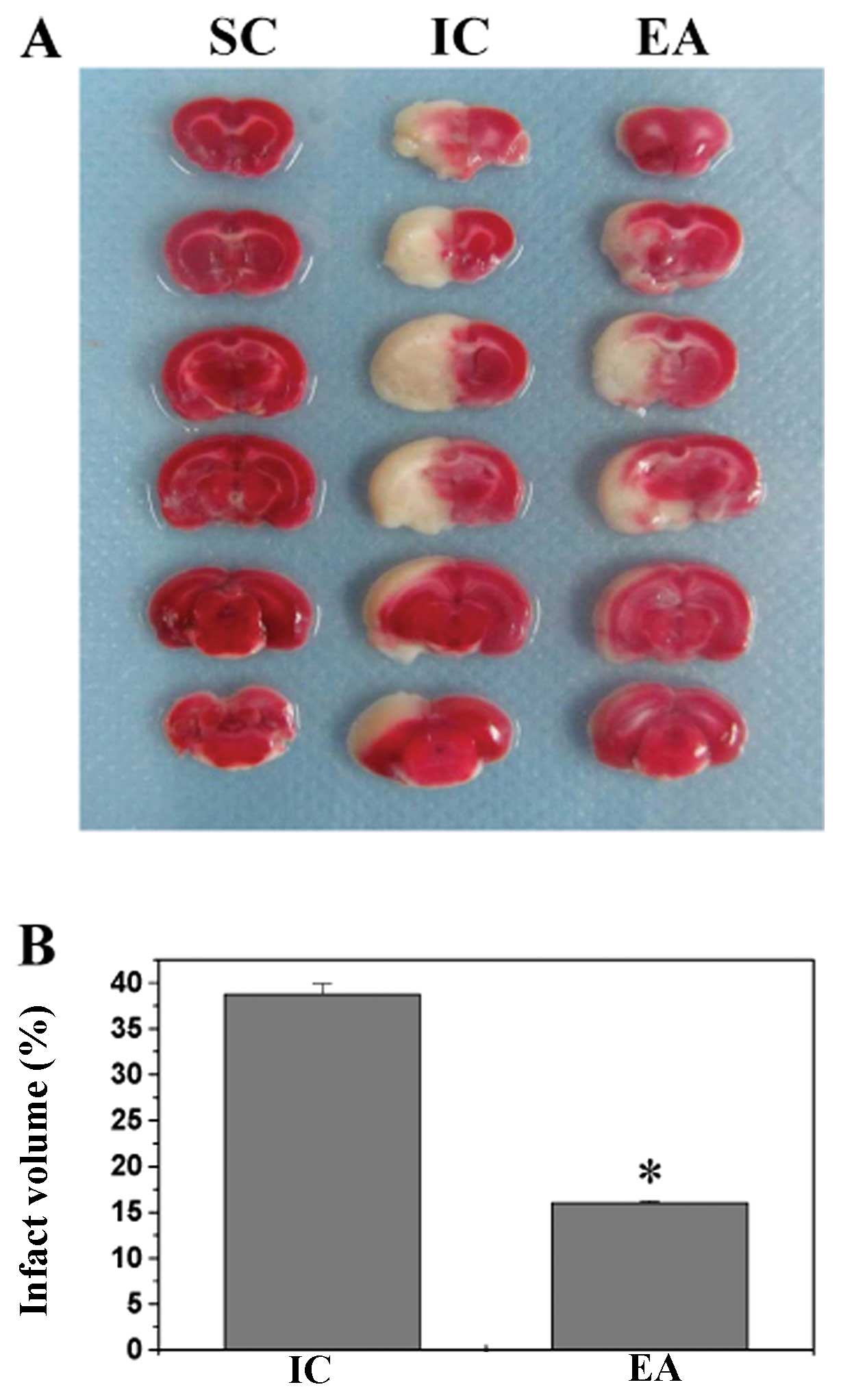
Measurement of cerebral infarct
volume
After cerebral I/R injury for 24 h, rats were
anaesthetized with 10% chloral hydrate by intraperitoneal
injection. The rat was perfused transcardiacally with 0.9% NaCl and
the brain was removed. The brain of each rat was coronally
sectioned into 2-mm-thick slices. The slices were stained with 2%
TTC solution (Sigma, St. Louis, MO, USA) at 37°C for 20 min and
then fixed with 10% buffered formalin solution. Stained slices were
photographed by a high-resolution digital camera (Cannon sx20), and
the infarct volume was quantified with Motic Med 6.0 System, which
was represented as a percentage of the total brain volume.
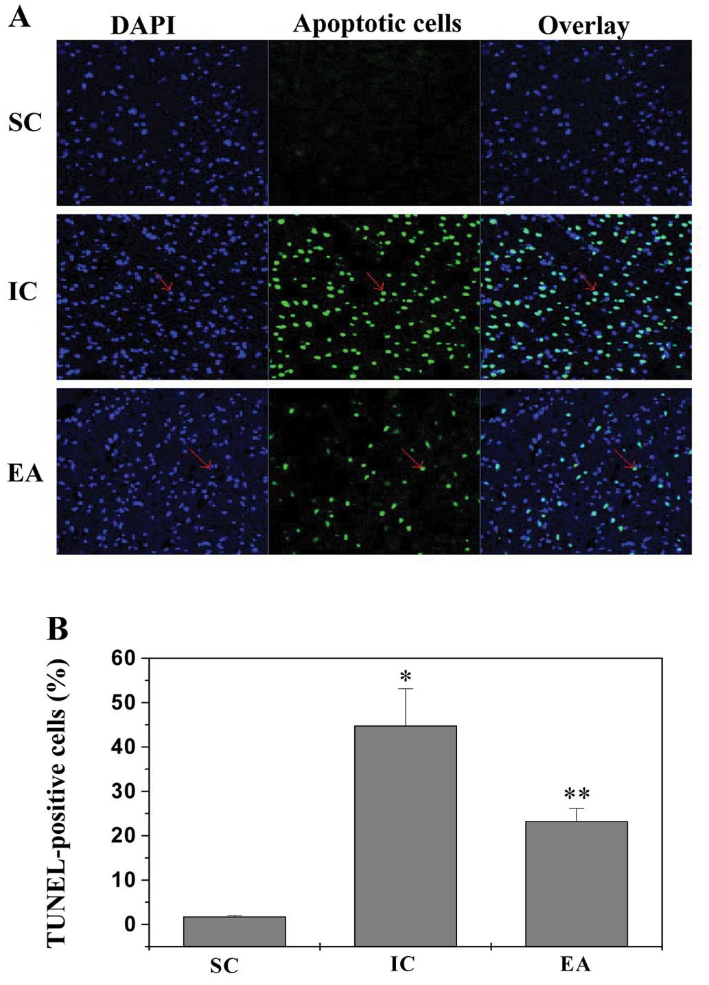
In situ apoptosis detection by TUNEL
staining
Rats were anesthetized and perfused transcardiacally
with 0.9% NaCl and 4% paraformaldehyde through the left ventricle
and the brain was removed. Samples were fixed in cold 4%
paraformaldehyde and then processed into 5-μm-thick
sections. In situ apoptosis was analyzed by TUNEL assay kit
(Promega) according to the manufacturer’s instructions. Nuclei of
all cells were visualized by DAPI staining and the green
fluorescence of apoptotic cells was detected by a confocal
fluorescence microscope (Leiss LSM 710). Apoptotic cells were
counted at four arbitrarily selected microscopic fields at a
magnification of 200x. Apoptotic rate was expressed as the ratio of
green-stained cells to the blue DAPI-stained total cells.
Western blot analysis
Ischemic cerebral tissues were homogenized in
non-denaturing lysis buffer and centrifuged at 15,000 x g for 15
min followed by determination of protein concentration in
supernatants. Protein lysates were separated by 12% SDS-PAGE gels
and then electrophoretically transferred onto PVDF membranes. The
membranes were blocked for 2 h with 5% non-fat dry milk and then
probed with primary antibodies against PI3K, Akt, pAkt, Bcl-2, Bax
and β-actin (at a dilution of 1:1,000) overnight at 4°C, followed
by incubation with appropriate HRP-conjugated secondary antibody
for 50 min. Blots were developed using enhanced chemiluminescence,
and images were taken using a Bio-Image Analysis System (Bio-Rad
Laboratories, Hercules, CA, USA).
RNA extraction and RT-PCR analysis
Total RNA was isolated from cerebral tissues with
TRIzol Reagent. Oligo(dT)-primed RNA (1 μg) was
reverse-transcribed with SuperScript II reverse transcriptase
(Promega) according to the manufacturer’s instructions. The
obtained cDNA was used to determine the mRNA amount of Bcl-2 and
Bax by PCR with Taq DNA polymerase (Fermentas). β-actin was used as
an internal control. The sequences of the primers used for
amplification of Bcl-2, Bax and β-actin transcripts are as follows:
Bcl-2 forward 5′-CGG GAG AAC AGG GTA TGA-3′ and reverse 5′-CAG GCT
GGA AGG AGA AGA T-3′; Bax forward 5′-GTT GCC CTC TTC TAC TTT GC-3′
and reverse 5′-ATG GTC ACT GTC TGC CAT G-3′; β-actin forward 5′-ACT
GGC ATT GTG ATG GAC TC-3′ and reverse 5′-CAG CAC TGT GTT GGC ATA
GA-3′. Samples were analyzed by gel electrophoresis (1.5% agarose).
The DNA bands were examined using a Gel Documentation System
(Bio-Rad Laboratories, Model Gel Doc 2000, USA).
Detection the level of BDNF and GDNF in
serum by ELISA
Animal blood was obtained aseptically from abdominal
aorta. Blood-containing tubes were allowed to stand at room
temperature for 2 h, and serums were obtained by centrifugation at
3000 x g for 20 min in 4°C. The serum level of BDNF and GDNF was
measured using ELISA kits (Xitang, Shanghai, China) according to
the manufacturer’s instructions. The wells were coated with 100
μl capture antibody diluted in coating buffer. The plate was
sealed and incubated overnight at 4°C. After three washes, the
wells were blocked with 200 μl assay diluents at room
temperature for 1 h, followed by another three washes. Then, 100
μl diluted BDNF or GDNF standard and test samples were added
and incubated for 2 h at room temperature. After repeated washes,
the substrate was added and incubated for 20 min at room
temperature, and the absorbance was measured at 450 nm using an
ELISA reader (BioTek, Model ELx 800, USA).
Statistical analysis
All data were the means of at least three
determinations and data were analyzed using the SPSS package for
Windows (Version 16.0). Statistical analysis of the data was
performed with the Student’s t-test and ANOVA. Differences with
P<0.05 were considered statistically significant.
Results
Electroacupuncture at acupoints of
Zusanli (ST36) and Quchi (LI11) improve neurological deficits and
reduce infarct volumes in cerebral ischemia-reperfusion (I/R)
injured rats
After establishing the I/R injured rat model by MCAO
on the left side, rats received electroacupuncture at Zusanli and
Quchi acupoints on the right paralyzed limbs. The neuroprotective
effect of electroacupuncture was examined by evaluating
neurological deficit scores and cerebral infarct volume. As shown
in Table I and Fig. 1, compared with sham operation
group (SC) rats that did not show any signs of cerebral injury, all
rats in both ischemia control (IC) and electroacupuncture (EA)
groups displayed obvious manifestation of neurological deficits and
cerebral infarction (P<0.05, vs. SC group), indicating the
success of model construction. There was no significant difference
in clinical evaluation between IC and EA groups before electric
stimulation. However, electroacupuncture at Zusanli and Quchi for
24 h significantly ameliorated neurological deficit scores and
reduced cerebral infarct volumes (P<0.05, vs. IC group),
demonstrating the therapeutic efficacy of electroacupuncture
against cerebral I/R injury.
 | Table INeurological deficit score. |
Table I
Neurological deficit score.
| Group (n=8) | 2 h after IR | 24 h after IR |
|---|
| SC | 0 | 0 |
| IC | 2.63±0.74 | 2.25±0.70 |
| EA | 2.38±0.52 | 1.50±0.54a |
Electroacupuncture inhibits cerebral cell
apoptosis in cerebral I/R injured rats
To determine the mechanism of neuroprotective action
of electroacupuncture, we examined its anti-apoptotic activity in
cerebral I/R injured rats via TUNEL. Data in Fig. 2 show 1.78±0.19%, 44.82±8.31% and
23.3±2.86% TUNEL-positive cells in SC, IC and EA rat groups,
respectively, suggesting that electroacupuncture at Zusanli and
Quchi inhibits ischemia-mediated cerebral cell apoptosis in
vivo.
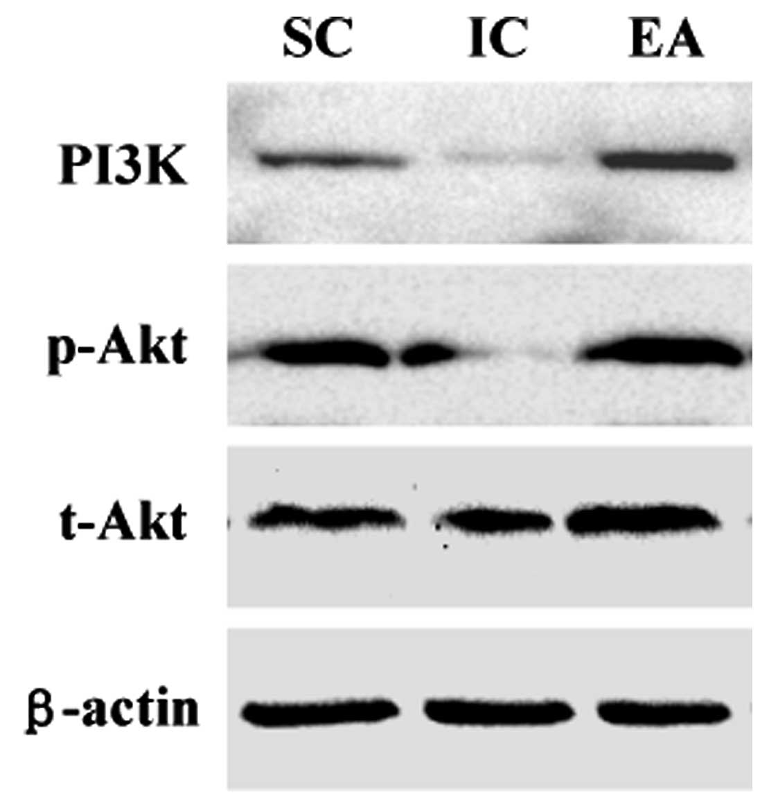
Electroacupuncture activates PI3K/Akt
pathway in cerebral I/R injured rats
PI3K/Akt signaling pathway plays an important role
in cell survival. The phosphorylation of Akt (pAkt) upon activation
of PI3K regulates the expression of various target genes, leading
to the inhibition of cell apoptosis. We therefore examined the
effect of electroacupuncture on PI3K expression and Akt
phosphoralytion in ischemic cerebral tissues using western
blotting. As shown in Fig. 3,
PI3K protein expression and the phosphorylation level of Akt in IC
group were significantly reduced compared with those in SC group
(P<0.05), consistent with previous studies that in models of
cerebral ischemia Akt phosphorylation profoundly decreases 24 h
after reperfusion (11,13). However, electroacupuncture at
Zusanli and Quchi significantly neutralized the effect of model
construction, increasing both PI3K protein expression and Akt
phosphorylation levels in ischemic cerebral tissues (P<0.05, vs.
IC group).
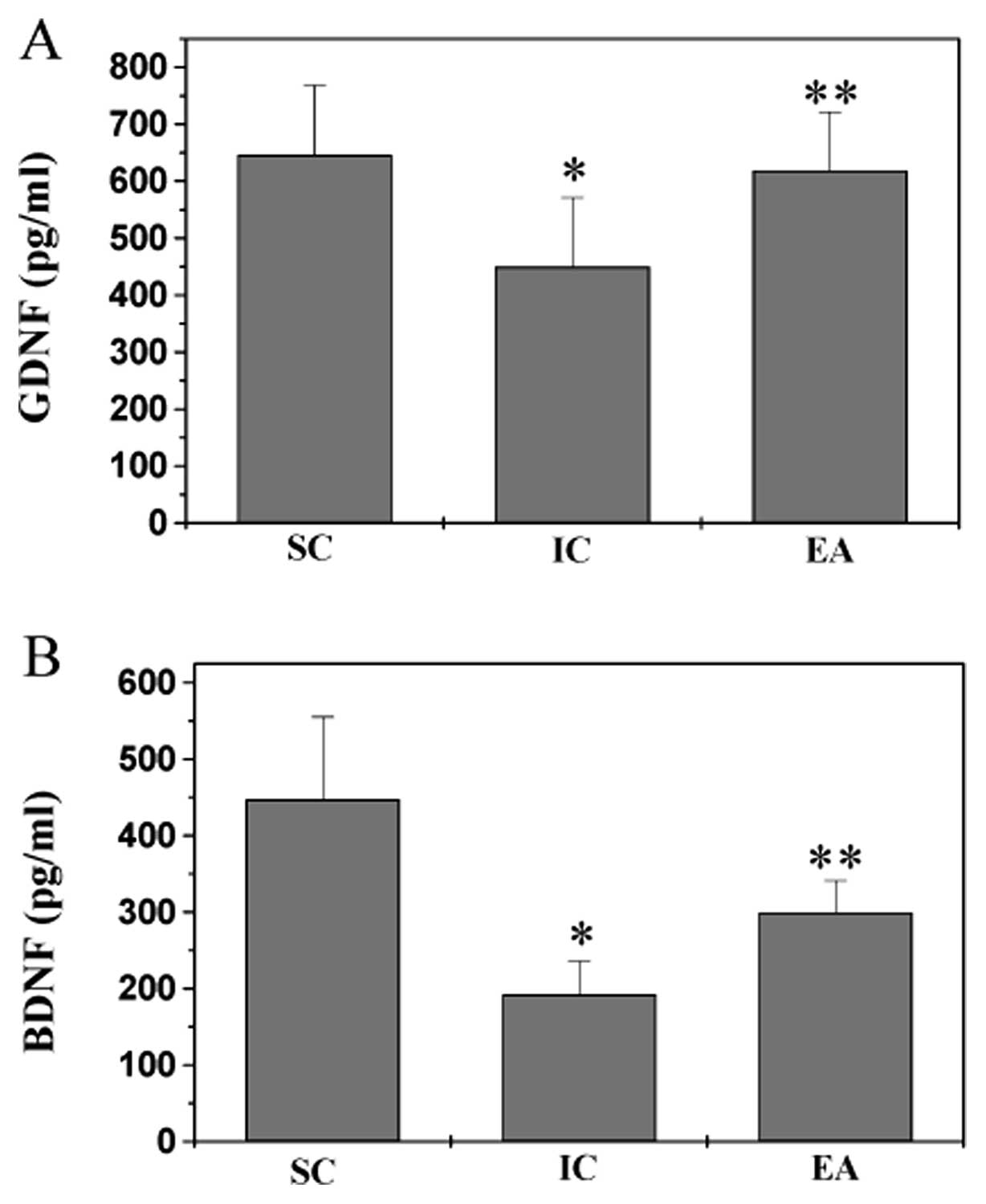
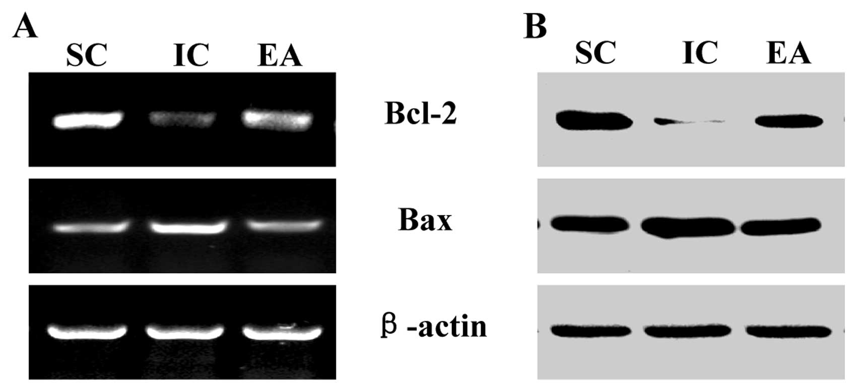
Electroacupuncture increases BDNF and
GDNF secretion levels and the anti-apoptotic Bcl-2/Bax ratio in
cerebral I/R injured rats
BDNF and GDNF are critical activators of PI3K in
cerebrum. By using ELISA we found that the serum levels of BDNF and
GDNF in IC group were significantly decreased, compared to that in
the SC group (P<0.05), which, however, was neutralized by
electroacupuncture at Zusanli and Quchi (Fig. 4; P<0.05, vs. IC group). Bcl-2
family proteins are key regulators of apoptosis, functioning as
either suppressors such as Bcl-2, or promoters such as Bax. Both
anti-apoptotic Bcl-2 and pro-apoptotic Bax are important target
genes of PI3K/Akt signaling pathway. To further explore the
mechanism of electroacupuncture’s anti-apoptotic activity, we
performed RT-PCR and western blot analyses to respectively examine
the mRNA and protein expression of Bcl-2 and Bax in ischemic
cerebral tissues. As shown in Fig. 5A
and B, electroacupuncture at Zusanli and Quchi profoundly
inhibited the model construction-mediated downregulation of
anti-apoptotic Bcl-2/Bax ratio, at both transcriptional and
translational levels.
Discussion
PI3K/Akt pathway is a critical mediator of cell
survival, which exerts anti-apoptotic function via inhibiting the
pro-apoptotic Bax/Bcl-2 ratio (15–17). A large number of reports indicate
that the suppression of PI3K/Akt pathway is strongly associated
with the progression of cerebral ischemia/reperfusion (I/R) injury,
increasing the infarct size and promoting cerebral cell death
(18,19). Therefore, inhibiting cerebral cell
apoptosis via activation of PI3K/Akt signaling has been a promising
strategy for the treatment of ischemic stroke. Acupuncture is an
alternative medicine methodology that has long been used in China
to treat various diseases. Previous studies have demonstrated the
clinical efficacy of acupuncture in stroke rehabilitation. On the
basis of a large amount of documents and materials, the Zusanli
(ST36) and Quchi (LI11) acupoints were commonly used in China to
clinically treat stroke. However, the mode of action of its
neuroprotective activities remains poorly understood.
By using a focal cerebral ischemia/reperfusion rat
model, in the present study we demonstrated that eletroacupuncture
at Zusanli and Quchi for only 24 h already displayed
neuroprotective effect as evidenced by improving neurological
deficits and reducing cerebral infarct volume. In addition, we
found that PI3K/Akt pathway was suppressed 24 h after cerebral I/R
injury, which was consistent with previous studies (11,13). However, electroacupuncture
significantly neutralized the effect of cerebral ischemia,
activating PI3K/Akt signaling in ischemic cerebral tissues.
Consequently, the upregulatory effect of electroacupuncture on
PI3K/Akt activation resulted in the inhibition of cerebral cell
apoptosis. Neurotrophic factors, such as BDNF and GDNF, are
critical activators for PI3K (20,21), and anti-apoptotic Bcl-2 and
pro-apoptotic Bax are important target genes of PI3K/Akt pathway
(22). As expected we found that
electroacupuncture increased the serum secretion levels of both
BDNF and GDNF, as well as upregulated the anti-apoptotic Bcl-2/Bax
ratio in cerebral ischemic.
In conclusion, here we reported for the first time
that electroacupuncture at Quchi (LI11) and Zusanli (ST36)
acupoints on the contralateral paralyzed limb exerts
neuroprotective function in ischemic stroke via activation of
PI3K/Akt pathway. These results suggest that electroacupuncture may
be a potential therapeutic modality for cerebral ischemia.
Abbreviations:
|
PI3K
|
phosphoinositide 3-kinase
|
|
IR
|
ischemia/reperfusion
|
|
MCAO
|
middle cerebral artery occlusion
|
|
EA
|
electroacupuncture
|
|
BDNF
|
brain-derived neurotrophic factor
|
|
GDNF
|
glial cell line-derived neurotrophic
factor
|
|
TTC
|
2,3,5-triphenyl tetra-zolium
chloride
|
|
TUNEL
|
terminal deoxynucleotidyl
transferase-mediated dUTP nick end labeling
|
Acknowledgements
This study was sponsored by the
Special Program for Key Basic Research Project of the China
Ministry of Science and Technology (973 Program, no. 2010CB534900),
and the National Natural Science Foundation of China (no.
30901935).
References
|
1.
|
VL FeiginStroke epidemiology in the
developing
worldLancet36521602161200510.1016/S0140-6736(05)66755-415978910
|
|
2.
|
GA DonnanM FisherM MacleodSM
DavisStrokeLancet3716121623200810.1016/S0140-6736(08)60694-7
|
|
3.
|
JN WuA short history of acupunctureJ
Altern Complement Med21921199610.1089/acm.1996.2.19
|
|
4.
|
HH HuC ChungT LiuA randomized controlled
trial on the treatment for acute partial ischemic stroke with
acupunctureNeuroepidemiology12106113199310.1159/0001103088232703
|
|
5.
|
G JansenT LundebergJ KjartanssonU
SamuelsonAcupuncture and sensory neuropeptides increase cutaneous
blood flow in ratsNeurosci
Lett97305309198910.1016/0304-3940(89)90615-02469996
|
|
6.
|
K JohanssonI LindgrenH WidnerI WiklundB
JohanssonCan sensory stimulation improve the functional outcome in
stroke
patients?Neurology4321892189199310.1212/WNL.43.11.21898232927
|
|
7.
|
M MagnussonK JohanssonBB JohanssonSensory
stimulation promotes normalization of postural control after
strokeStroke2511761180199410.1161/01.STR.25.6.11768202976
|
|
8.
|
J TaoXH XueLD ChenElectroacupuncture
improves neurological deficits and enhances proliferation and
differentiation of endogenous nerve stem cells in rats with focal
cerebral ischemiaNeurol Res32198204201010.1179/174313209X414506
|
|
9.
|
DP BrazilZZ YangBA HemmingsAdvances in
protein kinase B signalling: AKTion on multiple frontsTrends
Biochem Sci29233242200410.1016/j.tibs.2004.03.00615130559
|
|
10.
|
TF FrankeDR KaplanLC CantleyA TokerDirect
regulation of the Akt proto-oncogene product by
phosphatidylinositol-3,
4-bisphosphateScience275665668199710.1126/science.275.5300.6659005852
|
|
11.
|
S JanelidzeBR HuP SiesjöBK
SiesjöAlterations of Akt1 (PKBalpha) and p70(S6K) in transient
focal ischemiaNeurobiol
Dis8147154200110.1006/nbdi.2000.032511162248
|
|
12.
|
N NoshitaA LewénT SugawaraPH ChanEvidence
of phosphorylation of Akt and neuronal survival after transient
focal cerebral ischemia in miceJ Cereb Blood Flow
Metab2114421450200110.1097/00004647-200112000-0000911740206
|
|
13.
|
M ShibataT YamawakiT SasakiUpregulation of
Akt phosphorylation at the early stage of middle cerebral artery
occlusion in miceBrain
Res942110200210.1016/S0006-8993(02)02474-512031847
|
|
14.
|
EZ LongaPR WeinsteinS CarlsonR
CumminsReversible middle cerebral artery occlusion without
craniectomy in
ratsStroke208491198910.1161/01.STR.20.1.842643202
|
|
15.
|
C LuL LiuY ChenTLR2 ligand induces
protection against cerebral ischemia/reperfusion injury via
activation of phosphoinositide 3-kinase/Akt signalingJ
Immunol18714581466201110.4049/jimmunol.100342821709150
|
|
16.
|
N Produit-ZengaffinenCJ PournarasDF
SchorderetRetinal ischemia-induced apoptosis is associated with
alteration in Bax and Bcl-x(L) expression rather than modifications
in Bak and Bcl-2Mol Vis1521012110200919862336
|
|
17.
|
H XiT ZhangT TaoPropofol improved
neurobehavioral outcome of cerebral ischemia-reperfusion rats by
regulating Bcl-2 and Bax expressionBrain
Res14102432201110.1016/j.brainres.2011.06.06021783180
|
|
18.
|
X GaoH ZhangG SteinbergH ZhaoThe Akt
pathway is involved in rapid ischemic tolerance in focal ischemia
in ratsTransl Stroke
Res1202209201010.1007/s12975-010-0017-521804899
|
|
19.
|
JM TaylorU AliRC IannelloP HertzogPJ
CrackDiminished Akt phosphorylation in neurons lacking glutathione
peroxidase-1 (Gpx1) leads to increased susceptibility to oxidative
stress-induced cell deathJ
Neurochem92283293200510.1111/j.1471-4159.2004.02863.x15663476
|
|
20.
|
T BabaM KamedaT YasuharaElectrical
stimulation of the cerebral cortex exerts antiapoptotic,
angiogenic, and anti-inflammatory effects in ischemic stroke rats
through phosphoinositide 3-kinase/Akt signaling
pathwayStroke40e598e605200910.1161/STROKEAHA.109.563627
|
|
21.
|
B ChenXQ GaoCX YangNeuroprotective effect
of grafting GDNF gene-modified neural stem cells on cerebral
ischemia in ratsBrain
Res1284111200910.1016/j.brainres.2009.05.10019520066
|
|
22.
|
T GenoveseE MazzonI PaternitiE EspositoS
CuzzocreaNeuroprotective effects of olprinone after cerebral
ischemia/reperfusion injury in ratsNeurosci
Lett5039399201110.1016/j.neulet.2011.08.01521872644
|