Chondrogenic ATDC5 cells: An optimised model for rapid and physiological matrix mineralisation

  • Authors:
    • P. T. Newton
    • K. A. Staines
    • L. Spevak
    • A. L. Boskey
    • C. C. Teixeira
    • V. E. Macrae
    • A. E. Canfield
    • C. Farquharson
  • View Affiliations

  • Published online on: August 31, 2012     https://doi.org/10.3892/ijmm.2012.1114
  • Pages: 1187-1193
Metrics: Total Views: 0 (Spandidos Publications: | PMC Statistics: )
Total PDF Downloads: 0 (Spandidos Publications: | PMC Statistics: )


Abstract

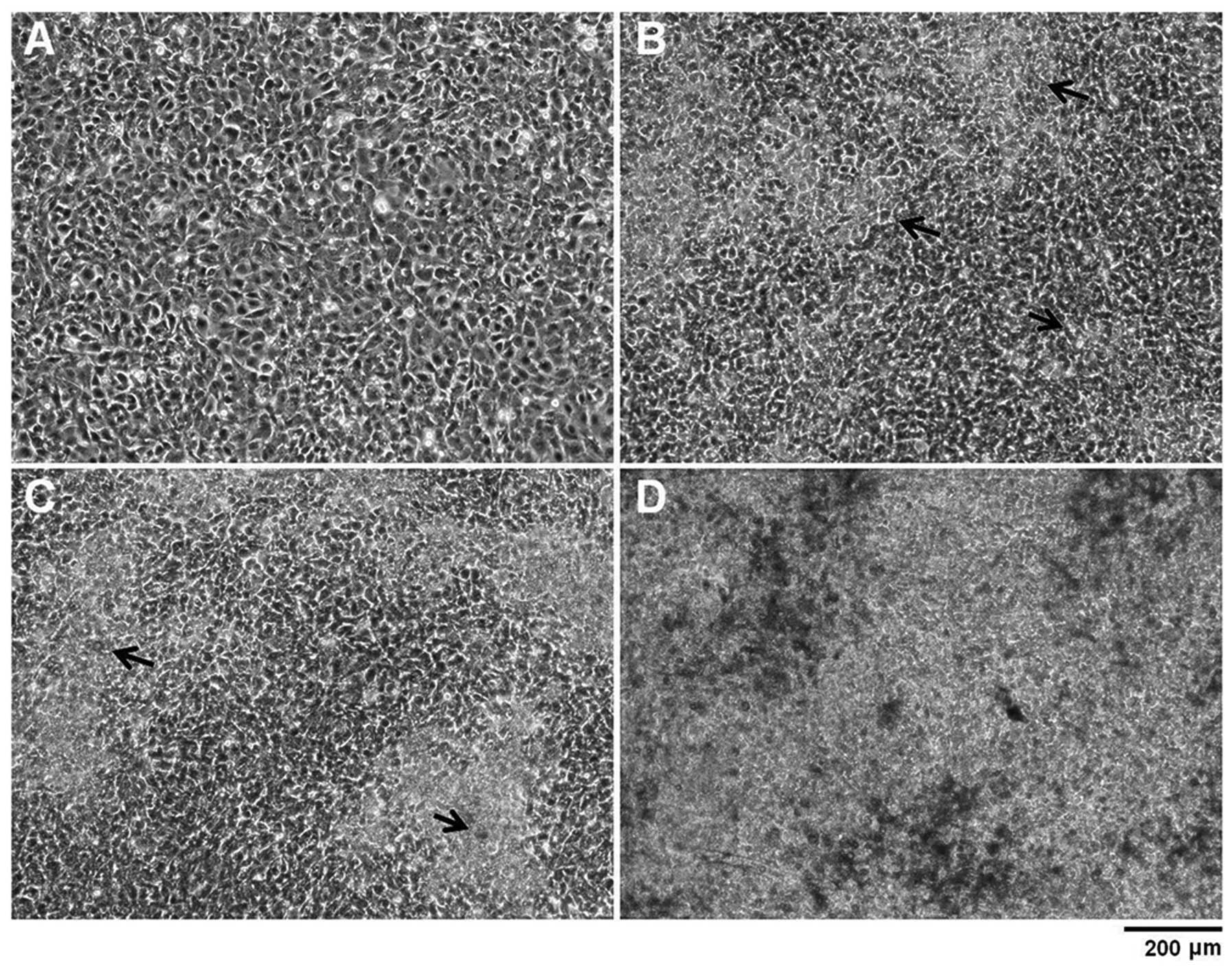
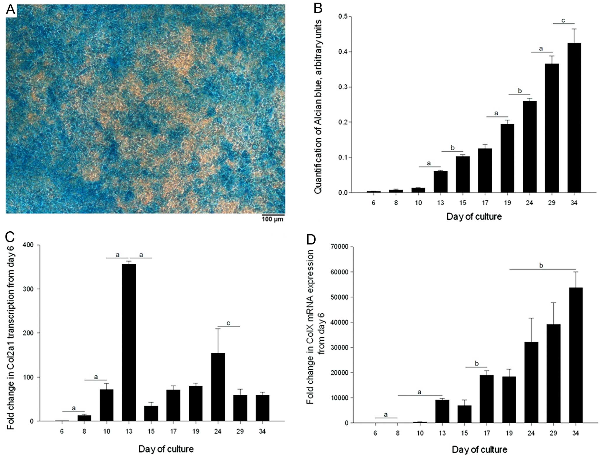
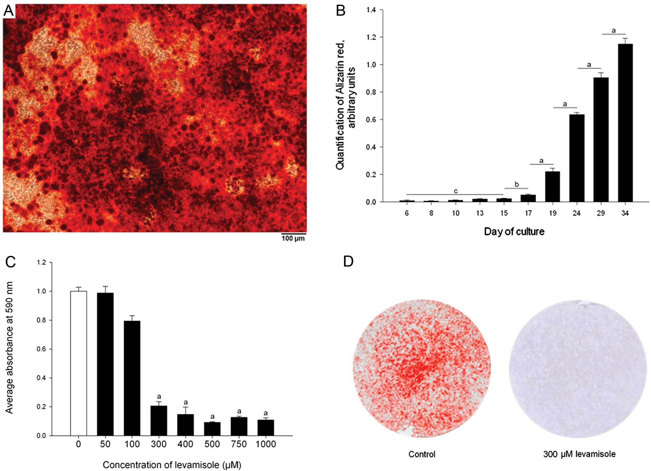
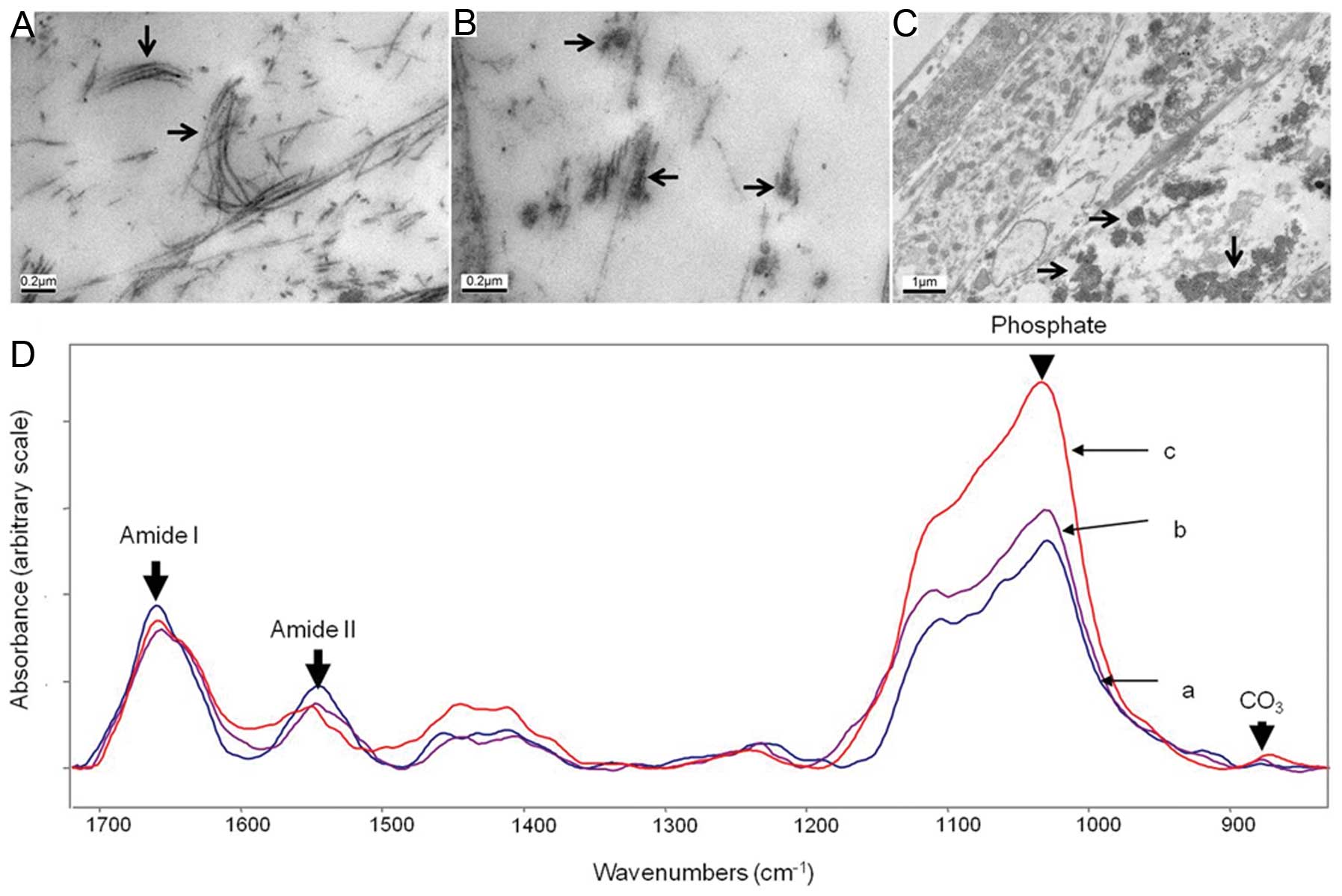
The development of chondrogenic cell lines has led to major advances in the understanding of how chondrocyte differentiation is regulated, and has uncovered many signalling pathways and gene regulatory mechanisms required to maintain normal function. ATDC5 cells are a well established in vitro model of endochondral ossification; however, current methods are limited for mineralisation studies. In this study we demonstrate that culturing cells in the presence of ascorbic acid and 10 mM β-glycerophosphate (βGP) significantly increases the rate of extracellular matrix (ECM) synthesis and reduces the time required for mineral deposition to occur to 15 days of culture. Furthermore, the specific expression patterns of Col2a1 and Col10a1 are indicative of ATDC5 chondrogenic differentiation. Fourier transform-infrared spectroscopy analysis and transmission electron microscopy (TEM) showed that the mineral formed by ATDC5 cultures is similar to physiological hydroxyapatite. Additionally, we demonstrated that in cultures with βGP, the presence of alkaline phosphatase (ALP) is required for this mineralisation to occur, further indicating that chondrogenic differentiation is required for ECM mineralisation. Together, these results demonstrate that when cultured in the presence of ascorbic acid and 10 mM βGP, ATDC5 cells undergo chondrogenic differentiation and produce a physiological mineralised ECM from Day 15 of culture onwards. The rapid and novel method for ATDC5 culture described in this study is a major improvement compared with currently published methods and this will prove vital in the pursuit of underpinning the molecular mechanisms responsible for poor linear bone growth observed in a number of chronic diseases such as cystic fibrosis, chronic kidney disease, rheumatological conditions and inflammatory bowel disease.
View Figures
View References

Related Articles

Journal Cover

November 2012
Volume 30 Issue 5

Print ISSN: 1107-3756
Online ISSN:1791-244X

Sign up for eToc alerts

Recommend to Library

Copy and paste a formatted citation
x
Spandidos Publications style
Newton PT, Staines KA, Spevak L, Boskey AL, Teixeira CC, Macrae VE, Canfield AE and Farquharson C: Chondrogenic ATDC5 cells: An optimised model for rapid and physiological matrix mineralisation. Int J Mol Med 30: 1187-1193, 2012
APA
Newton, P.T., Staines, K.A., Spevak, L., Boskey, A.L., Teixeira, C.C., Macrae, V.E. ... Farquharson, C. (2012). Chondrogenic ATDC5 cells: An optimised model for rapid and physiological matrix mineralisation. International Journal of Molecular Medicine, 30, 1187-1193. https://doi.org/10.3892/ijmm.2012.1114
MLA
Newton, P. T., Staines, K. A., Spevak, L., Boskey, A. L., Teixeira, C. C., Macrae, V. E., Canfield, A. E., Farquharson, C."Chondrogenic ATDC5 cells: An optimised model for rapid and physiological matrix mineralisation". International Journal of Molecular Medicine 30.5 (2012): 1187-1193.
Chicago
Newton, P. T., Staines, K. A., Spevak, L., Boskey, A. L., Teixeira, C. C., Macrae, V. E., Canfield, A. E., Farquharson, C."Chondrogenic ATDC5 cells: An optimised model for rapid and physiological matrix mineralisation". International Journal of Molecular Medicine 30, no. 5 (2012): 1187-1193. https://doi.org/10.3892/ijmm.2012.1114