Introduction
Isoflavones belong to the flavonoids and exist in
nature in commonly consumed plants. They have been suggested to
possess antioxidant, anticancer and anti-inflammatory activities.
Numerous reports have supported this concept, and isoflavones are
generally accepted to have the potential to benefit human
health.
Daidzein, which occurs in nature as daidzin (its
β-glycoside form), is one of the main components of isoflavones.
Gastrointestinal bacteria in humans metabolize daidzein to the
reduced [equol and O-desmethylangolensin (O-DMA)] and
oxidized (3′,4′,7-trihydroxyisoflavone and
4′,6,7-trihydroxyisoflavone) forms (1,2).
Researchers have speculated that these metabolites play an
important role in the biological mechanism of daidzein (3–5).
Studies have reported that daidzein inhibits the
growth of various types of cancer cells such as breast (6,7),
ovarian (8,9), prostate (10) and hepatocellular carcinoma (HCC)
(11,12). In addition, daidzein has been
shown to induce apoptosis by modulating molecular signaling
pathways such as DNA damage-signaling (10), cell cycle regulation (6,10),
receptor-mediated signaling (13–15) and the mitochondrial apoptotic
pathway (7).
Nevertheless, no study has investigated the effect
of the metabolite O-DMA on HCC, which is one of most common
malignant tumors diagnosed worldwide. Therefore, we investigated
the anticancer effects of O-DMA on cell cycle arrest and
apoptosis induction in human HCC Hep3B cells.
Materials and methods
Cell culture
Human HCC Hep3B cells were purchased from the Korean
Cell Line Bank (KCLB), Korea. Cells were routinely maintained in
MEM (Sigma-Aldrich, USA), supplemented with 10% FBS and antibiotics
(50 U/ml penicillin and 50 μg/ml streptomycin; Sigma) at
37°C in a humidified atmosphere containing 5% CO2. In
the cell proliferation experiment, cells were treated with
different O-DMA concentrations (ranging from 5 to 200
μM) and then incubated for varying time periods (24, 48 and
72 h). In the cell cycle analysis and apoptosis assay, cells were
treated with O-DMA at an IC50 concentration for
72 h.
Synthesis of O-DMA
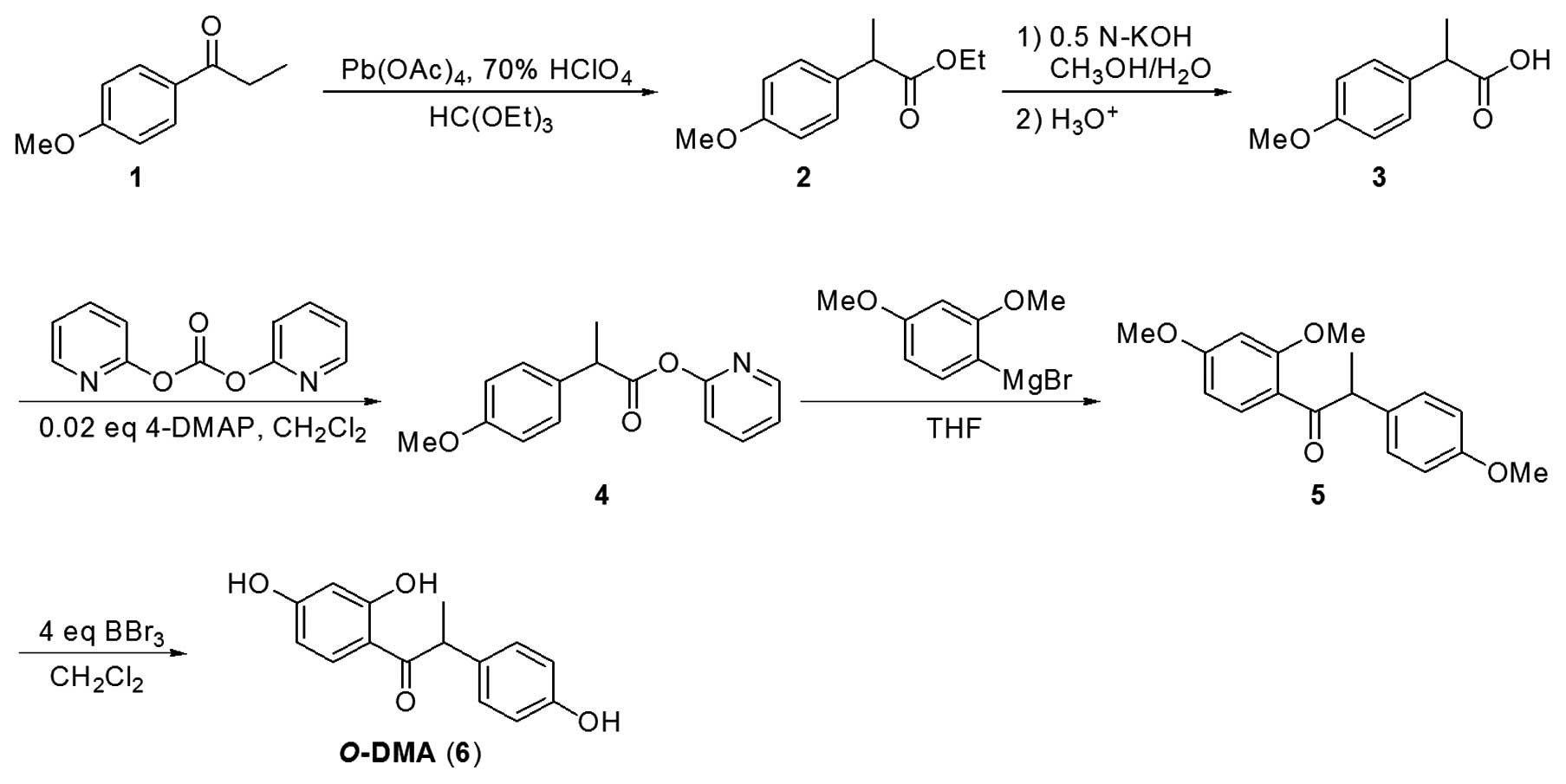
O-DMA synthesis is illustrated in Fig. 1. Ethyl
2-(p-methoxyphenyl)propionate 2 was prepared by
gentle heating of a mixture solution of
p-methoxypropiophenone 1, 70% perchloric acid and
lead(IV) acetate in triethyl orthoformate for 2 h at 50°C in an 87%
yield (Scheme 1). Compound 2 was hydrolyzed under basic
condition by the treatment of 0.5 N-KOH in
CH3OH/H2O for 24 h at room temperature to
afford 2-(p-methoxyphenyl)propionic acid 3 in a 93%
yield after acidic work-up. The reaction of 3 with
di-2-pyridyl carbonate in the presence of 0.02 equiv of
4-dimethylaminopyridine (4-DMAP) in dichloromethane proceeded well
for 3 h at room temperature to afford 2-pyridyl
2-(p-methoxyphenyl)propionate 4 in an 81% yield. The
acyl substitution of 4 by 2,4-dimethoxyphenylmagnesium
bromide in THF proceeded rapidly at 0°C via 6-membered chelate to
give 2,4,4′-trimethoxy-α-methyldesoxybenzoin 5 in a 95%
yield after acidic hydrolysis. Compound 5 was demethylated
by the treatment of 4 equiv of boron tribromide in dichloromethane
for 48 h at room temperature to afford O-DMA 6 in a
90% yields after aqueous work-up. O-DMA was dissolved in
dimethyl sulfoxide (DMSO) (final concentration 0.1% in medium) and
kept in a refrigerator.
MTT assay
Hep3B cells were plated at a density of
1×10⁵ cells/well in a 96-well tissue culture plate
(Corning, NY, USA), and incubated at 37°C for 24 h. Plated cells
were treated with the indicated concentrations of O-DMA for
24, 48 and 72 h. After treatment, plated cell were incubated with
3-(4, 5-dimethylthiazol-2-yl)-2,5-diphenyltetrazolium bromide (MTT;
0.5 mg/ml final concentration; Sigma-Aldrich) for 4 h at 37°C.
After discarding all medium from the plates, 100 μl of DMSO
was added to each well. The plates were placed for 5 min at room
temperature with shaking, so that complete dissolution of formazan
was achieved. The absorbance of the MTT formazan was determined at
540 nm by a UV spectrophotometric plate reader (EMax; Molecular
Devices). The value of IC50 (i.e., the concentration of
the extract required to inhibit cancer cell growth by 50% of the
control level) was estimated from the plot. Control cells were
treated with only the compound solvent.
Cell cycle distribution
Cell cycle distribution and apoptosis were
determined by FACS analysis using propidium iodide (PI) staining to
measure DNA content. Hep3B cells were plated at a density of
5×10⁵ cells/well in a 6-well tissue culture plate
(Corning), and incubated at 37°C for 24 h. Plated cells were
treated with an IC50 concentration of O-DMA for
72 h. Cells were then harvested, washed with cold PBS and processed
for cell cycle analysis. Briefly, the cells were fixed in absolute
ethanol and stored at −20°C for later analysis. The fixed cells
were centrifuged at 1,000 rpm and washed with cold PBS twice. RNase
A (20 μg/ml final concentration) and PI staining solution
(50 μg/ml final concentration) were added to the cells and
incubated for 30 min at 37°C in the dark. The cells were analyzed
using a FACS Calibur instrument (BD Biosciences, San Jose, CA, USA)
equipped with CellQuest 3.3 software.
Detection of apoptosis
Cells were treated and harvested similarly as
mentioned in the preceding section. Phosphatidylserine is a
biomarker of apoptosis, which is located on the cytoplasmic surface
of the cell membrane. Phosphatidylserine exposure on the outer
leaflet was detected using the Annexin V-FITC Apoptosis Detection
kit (Calbiochem; EMD Chemicals Inc., Darmstadt, Germany). Annexin V
and PI solution were added to the cell preparations, and incubation
was carried out for 25 min in the dark. Binding buffer (400
μl) was then added to each tube, and the samples were
analyzed by flow cytometry.
Cytochrome c release assay
Release of cytochrome c from the mitochondria
to the cytosol was measured by immunoblotting assay. To detect the
cytochrome c release, the cell lysates were centrifuged at
100,000 rpm for 30 min at 4°C in order to obtain the supernatant
(cytosolic fraction) and the pellet (fraction that contains the
mitochondria). Proteins (25 μg/well) denatured with sample
buffer were separated by 12% SDS-polyacrylamide gel and analyzed by
immunoblot assay using anti-cytochrome c antibody (Santa
Cruz Biotechnology, Inc., Santa Cruz, CA, USA).
Immunoblot assay
Cells were lysed in RIPA buffer (1% NP-40, 150 mM
NaCl, 0.05% DOC, 1% SDS, 50 mM Tris; pH 7.5) containing protease
inhibitor at 4°C for 1 h. The supernatant was separated by
centrifugation, and protein concentration was determined using the
Bradford protein assay kit II (Bio-Rad). Proteins (25
μg/well) denatured with sample buffer were separated by 10%
SDS-polyacrylamide gel. Proteins were transferred onto
0.45-μm nitrocellulose membranes. The membranes were blocked
with a 1% BSA solution for 3 h and washed twice with PBS containing
0.2% Tween-20, and incubated with the primary antibody at 4°C
overnight. Antibodies against CDK1, cyclin A, cyclin B, Bcl-2, Bax,
precursor caspase-3, cleaved caspase-3, cytochrome c and
β-actin (all from Santa Cruz Biotechnology, Inc.) were used to
probe the separate membranes. On the next day, the immunoreaction
was continued using the secondary goat anti-rabbit horseradish
peroxidase-conjugated antibody after washing for 2 h at room
temperature. The specific protein bands were detected using the
Opti-4CN substrate kit (Bio-Rad).
Statistical analyses
All values are expressed as the means ± SD. Data
were analyzed by unpaired Student’s t-test or one-way analysis of
variance followed by Dunnett’s multiple comparison test (SigmaStat;
Jandel, San Rafael, CA, USA). For all comparisons, differences were
considered statistically significant at P<0.05.
Results
Effect of O-DMA on cell proliferation of
Hep3B cells
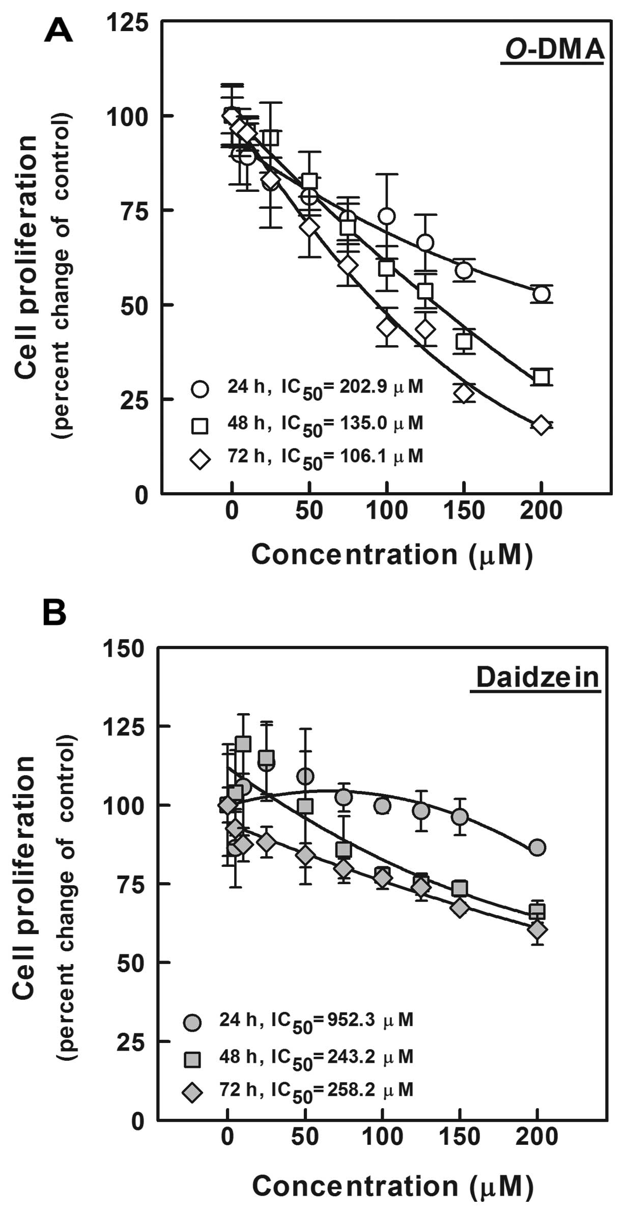
The antiproliferative effect of O-DMA was
determined using an MTT assay in human HCC Hep3B cells exposed to
O-DMA at various concentrations (5, 10, 25, 50, 75, 100,
125, 150 and 200 μM) for 24, 48 and 72 h (Fig. 2). O-DMA significantly
reduced cell proliferation in a dose- and time-dependent manner
(P<0.05), but inhibition of the cell proliferation increased
significantly after 72 h. The IC50 values at 48 and 72 h
were 135.02 and 106.14 μM, respectively. To compare
O-DMA with the precursor daidzein, we investigated the
anti-proliferative activity of daidzein under the same conditions.
We found that daidzein had a higher IC50 (4.7-fold
higher compared to O-DMA) at 24 h. The IC50
values at 48 and 72 h were 243.2 and 258.2 μM,
respectively.
Effect of O-DMA on the cell cycle
distribution of Hep3B cells
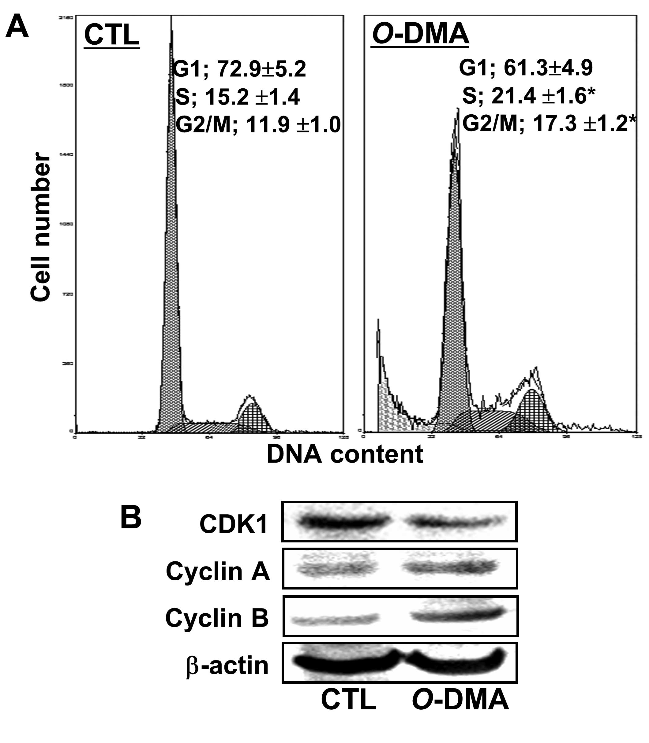
Based on these results, the IC50
concentration after 72 h exposure to O-DMA was used to
verify cell cycle arrest. After exposure of Hep3B cells to
O-DMA, the proportion of G1 phase cells was decreased by
15.9%, and the proportion of S and G2/M-phase cells was
significantly increased by 40.7 and 45.4%, respectively, compared
with control cells (Fig. 3A,
P<0.05). With respect to G2/M-related proteins, reduced CDK1
expression and slightly increased cyclin A and B was noted in the
Hep3B cells following O-DMA treatment (Fig. 3B).
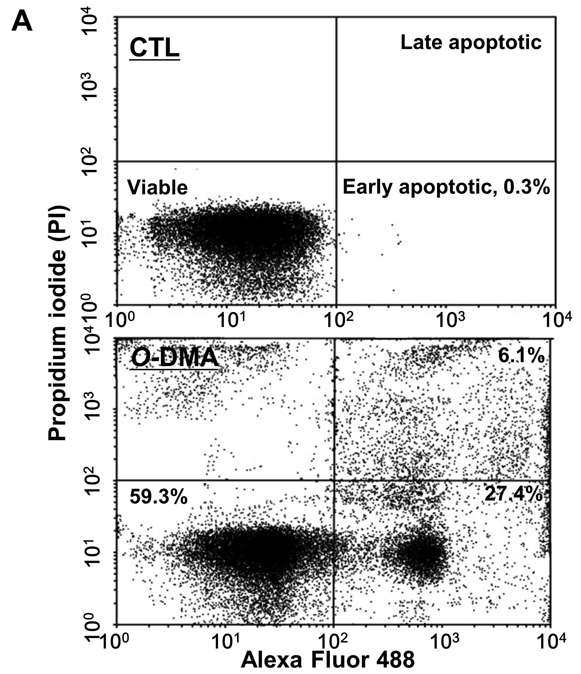
Under the same conditions, exposure to O-DMA
significantly shifted the cell populations (Fig. 4A). The percentages of early and
late apoptotic cells were 27.4 and 6.1%, respectively, in Hep3B
cells exposed to O-DMA. Moreover, small DNA fragments in the
sub-G1 phase increased by 18.5% after O-DMA treatment
(Fig. 3A).
Effect of O-DMA on the expression of
apoptosis-related genes in Hep3B cells
O-DMA had an effect on Bcl-2, Bax and
caspase-3 expression, as well as cytochrome c release
(Fig. 4B and C). Apoptosis
induced by O-DAM decreased Bcl-2 and increased Bax
expression levels. Moreover, the increase in cytochrome c
release induced by O-DMA suggests that O-DAM triggers
caspase-activated apoptosis. The caspase-3 cleavage product was
also observed after O-DMA treatment.
Discussion
HCC is a global health problem. According to
GLOBOCAN 2008 estimates (16),
HCC is the fifth and seventh most common cancer, and the second and
sixth most fatal cancer in men and women, respectively (17).
In the present study, we first examined the
antiproliferative effect of O-DMA on human HCC Hep3B cells
to investigate its anticancer activity. O-DMA demonstrated
antiproliferative activity in a dose- and time-dependent manner,
and its IC50 value was significantly lower than that of
its precursor, daidzein. Moreover, daidzein stimulated cell growth
at concentrations lower than 50 μM after 24 h, consistent
with previous reports stating that daidzein may have biphasic
effects on the growth of several types of cancer cells (18,19). This observation is explained by
the hypothesis that phytoestrogens, which are plant-derived, are
structurally similar to estradiol (17β-estradiol) and bind to
estrogen receptors (ERs) such as daidzein and its metabolite equol.
This can stimulate or inhibit cell proliferation by functioning as
an estrogen agonist or antagonist (20–22). Alternatively, O-DMA has a
very weak affinity to the ERs (23–25), and O-DMA was shown to
reduce the proliferation of breast cancer MCF-7 cells in an
estrogen-independent manner (18).
Although O-DMA and daidzein have similar
effects at higher concentrations after 72 h, the fact that daidzein
stimulates cell proliferation under certain conditions (as an
estrogenic) suggests that O-DMA may be a good anticancer
agent. However, O-DMA exposure at an IC50
concentration for 72 h caused cell cycle arrest at the G2/M phase
and subsequent apoptosis, which may be an issue for its use in
cancer chemotherapy.
O-DMA reduces CDK1 (Cdc2) and increases
cyclin A and B, which may explain the G/M phase arrest. G2/M
transition controls the onset of mitosis and is regulated by cyclin
A (in the earlier stages) and the CDK1-cyclin B complex (at later
stages) (26). CDK is often
mutated, deleted or silenced in cancer, leading to the search for
small-molecule CDK inhibitors in cancer therapy (27–29).
O-DMA treatment induced apoptosis in Hep3
cells, confirming the sub-G1 peak and the shift in cell populations
shown by the Annexin V assay. Note that the main mechanism of
apoptosis induction by O-DMA may not involve the p53 pathway
since Hep3B is a p53-deficient cell line. In our preliminary
experiments, O-DMA at concentrations over 100 μM
significantly inhibited approximately 30% of cell viability of
HepG2 cells. However, at concentrations lower than 100 μM,
cell growth was not affected by the addition of O-DMA (data
not shown). Generally, HepG2 cells are highly susceptible to
chemical agents and drugs, while Hep3B cells are more resistance
(30,31). Therefore, the significant
anticancer effect of O-DAM in Hep3B cells supports its use
as a cancer drug candidate.
In addition, expression of Bcl-2 decreased while Bax
increased in the Hep3B cells after exposure to O-DMA.
Members of the Bcl-2 family of proteins are critical regulators of
the apoptotic pathway, controlling mitochondrial permeability and
cytochrome c expression (32,33). These proteins include major
anti-apoptotic family members, Bcl-2 and Bcl-xL, and the major
pro-apoptotic proteins, Bax and Bak. This is consistent with the
anticancer mechanism of daidzein involving the modulation of Bcl
and Bax in several cancer cell lines such as breast, prostate and
head and neck (7,34,35).
Additionally, O-DMA induced cytochrome
c release and decreased the expression of a caspase-3
precursor. Release of cytochrome c from the mitochondria to
the cytoplasm is a key step in the initiation of
mitochondrial-dependent apoptosis (36). As a downstream product of
cytochrome c, caspases are critical mediators of the
principal factors found in apoptotic cells. Among them, caspase-3
is a frequently activated death protease, catalyzing the specific
cleavage of many cellular proteins (37,38).
In conclusion, our results demonstrated that
O-DMA inhibited cancer cell growth via cell cycle arrest and
apoptosis in HCC Hep3B cells. O-DMA-treated cells were
arrested at the G2/M phase, which was accompanied by a reduction in
CDK1. Downregulation of Bcl-2 and upregulation of Bax led to
cytochrome c release from the mitochondria and activation of
caspase-3. These findings indicate that O-DMA is a promising
anticancer drug candidate.
Acknowledgements
This research was supported by the
Basic Research Program through the National Research Foundation of
Korea (NRF) funded by the Ministry of Education, Science and
Technology (2012-0006811 and 2012-041653).
References
|
1
|
L’homme R, Brouwers E, Al-Maharik N,
Lapcík O, Hampl R, Mikola H, Wähälä K and Adlercreutz H:
Time-resolved fluoroimmunoassay of plasma and urine
O-desmethylangolensin. J Steroid Biochem Mol Biol.
81:353–361. 2002.
|
|
2
|
Heinonen S, Wähälä K and Adlercreutz H:
Identification of isoflavone metabolites dihydrodaidzein,
dihydrogenistein, 6’-OH-O-dma, and cis-4-OH-equol in
human urine by gas chromatography-mass spectroscopy using authentic
reference compounds. Anal Biochem. 274:211–219. 1999.PubMed/NCBI
|
|
3
|
Hwang J, Wang J, Morazzoni P, Hodis HN and
Sevanian A: The phytoestrogen equol increases nitric oxide
availability by inhibiting superoxide production: an antioxidant
mechanism for cell-mediated LDL modification. Free Radic Biol Med.
34:1271–1282. 2003. View Article : Google Scholar : PubMed/NCBI
|
|
4
|
Turner R, Baron T, Wolffram S, Minihane
AM, Cassidy A, Rimbach G and Weinberg PD: Effect of circulating
forms of soy isoflavones on the oxidation of low density
lipoprotein. Free Radic Res. 38:209–216. 2004. View Article : Google Scholar : PubMed/NCBI
|
|
5
|
Rüfer CE and Kulling SE: Antioxidant
activity of isoflavones and their major metabolites using different
in vitro assays. J Agric Food Chem. 54:2926–2931. 2006.PubMed/NCBI
|
|
6
|
Choi EJ and Kim GH: Daidzein causes cell
cycle arrest at the G1 and G2/M phases in human breast cancer MCF-7
and MDA-MB-453 cells. Phytomedicine. 15:683–690. 2008. View Article : Google Scholar : PubMed/NCBI
|
|
7
|
Jin S, Zhang QY, Kang XM, Wang JX and Zhao
WH: Daidzein induces MCF-7 breast cancer cell apoptosis via the
mitochondrial pathway. Ann Oncol. 21:263–268. 2010. View Article : Google Scholar : PubMed/NCBI
|
|
8
|
Green JM, Alvero AB, Kohen F and Mor G:
7-(O)-Carboxymethyl daidzein conjugated to N-t-Boc-hexylenediamine:
a novel compound capable of inducing cell death in epithelial
ovarian cancer stem cells. Cancer Biol Ther. 8:1747–1753. 2009.
View Article : Google Scholar : PubMed/NCBI
|
|
9
|
Gercel-Taylor C, Feitelson AK and Taylor
DD: Inhibitory effect of genistein and daidzein on ovarian cancer
cell growth. Anticancer Res. 24:795–800. 2004.PubMed/NCBI
|
|
10
|
Rabiau N, Kossaï M, Braud M, Chalabi N,
Satih S, Bignon YJ and Bernard-Gallon DJ: Genistein and daidzein
act on a panel of genes implicated in cell cycle and angiogenesis
by polymerase chain reaction arrays in human prostate cancer cell
lines. Cancer Epidemiol. 34:200–206. 2010. View Article : Google Scholar : PubMed/NCBI
|
|
11
|
Su SJ, Chow NH, Kung ML, Hung TC and Chang
KL: Effects of soy isoflavones on apoptosis induction and G2-M
arrest in human hepatoma cells involvement of caspase-3 activation,
Bcl-2 and Bcl-XL downregulation, and Cdc2 kinase activity. Nutr
Cancer. 45:113–123. 2003. View Article : Google Scholar : PubMed/NCBI
|
|
12
|
Borradaile NM, de Dreu LE, Wilcox LJ,
Edwards JY and Huff MW: Soya phytoestrogens, genistein and
daidzein, decrease apolipoprotein B secretion from HepG2 cells
through multiple mechanisms. Biochem J. 366:531–539. 2002.
View Article : Google Scholar : PubMed/NCBI
|
|
13
|
Jiang Q, Payton-Stewart F, Elliott S,
Driver J, Rhodes LV, Zhang Q, Zheng S, Bhatnagar D, Boue SM,
Collins-Burow BM, Sridhar J, Stevens C, McLachlan JA, Wiese TE,
Burow ME and Wang G: Effects of 7-O substitutions on estrogenic and
anti-estrogenic activities of daidzein analogues in MCF-7 breast
cancer cells. J Med Chem. 53:6153–6163. 2010. View Article : Google Scholar : PubMed/NCBI
|
|
14
|
Singh-Gupta V, Zhang H, Yunker CK, Ahmad
Z, Zwier D, Sarkar FH and Hillman GG: Daidzein effect on hormone
refractory prostate cancer in vitro and in vivo compared to
genistein and soy extract: potentiation of radiotherapy. Pharm Res.
27:1115–1127. 2010. View Article : Google Scholar : PubMed/NCBI
|
|
15
|
Guo JM, Xiao BX, Dai DJ, Liu Q and Ma HH:
Effects of daidzein on estrogen-receptor-positive and -negative
pancreatic cancer cells in vitro. World J Gastroenterol.
10:860–863. 2004.PubMed/NCBI
|
|
16
|
Ferlay J, Shin HR, Bray F, Forman D,
Mathers C and Parkin DM: Estimates of worldwide burden of cancer in
2008: GLOBOCAN 2008. Int J Cancer. 127:2893–2917. 2010. View Article : Google Scholar : PubMed/NCBI
|
|
17
|
Jemal A, Bray F, Center MM, Ferlay J, Ward
E and Forman D: Global cancer statistics. CA Cancer J Clin.
61:69–90. 2011. View Article : Google Scholar
|
|
18
|
Schmitt E, Dekant W and Stopper H:
Assaying the estrogenicity of phytoestrogens in cells of different
estrogen sensitive tissues. Toxicol In Vitro. 15:433–439. 2001.
View Article : Google Scholar : PubMed/NCBI
|
|
19
|
Guo JM, Xiao BX, Liu DH, Grant M, Zhang S,
Lai YF, Guo YB and Liu Q: Biphasic effect of daidzein on cell
growth of human colon cancer cells. Food Chem Toxicol.
42:1641–1646. 2004. View Article : Google Scholar : PubMed/NCBI
|
|
20
|
Magee PJ and Rowland IR: Phyto-oestrogens,
their mechanism of action: current evidence for a role in breast
and prostate cancer. Br J Nutr. 91:513–531. 2004. View Article : Google Scholar : PubMed/NCBI
|
|
21
|
Gogel WC and Tietz JD: Absence of
compensation and reasoning-like processes in the perception of
orientation in depth. Percept Psychophys. 51:309–318. 1992.
View Article : Google Scholar : PubMed/NCBI
|
|
22
|
Bingham SA, Atkinson C, Liggins J, Bluck L
and Coward A: Phyto-oestrogens: where are we now? Br J Nutr.
79:393–406. 1998. View Article : Google Scholar : PubMed/NCBI
|
|
23
|
Tang BY and Adams NR: Effect of equol on
oestrogen receptors and on synthesis of DNA and protein in the
immature rat uterus. J Endocrinol. 85:291–297. 1980. View Article : Google Scholar : PubMed/NCBI
|
|
24
|
Shutt DA and Cox RI: Steroid and
phyto-oestrogen binding to sheep uterine receptors in vitro. J
Endocrinol. 52:299–310. 1972. View Article : Google Scholar : PubMed/NCBI
|
|
25
|
Pfitscher A, Reiter E and Jungbauer A:
Receptor binding and transactivation activities of red clover
isoflavones and their metabolites. J Steroid Biochem Mol Biol.
112:87–94. 2008. View Article : Google Scholar : PubMed/NCBI
|
|
26
|
Shackelford RE, Kaufmann WK and Paules RS:
Oxidative stress and cell cycle checkpoint function. Free Radic
Biol Med. 28:1387–1404. 2000.PubMed/NCBI
|
|
27
|
Hunter T and Cooper JA: Protein-tyrosine
kinases. Annu Rev Biochem. 54:897–930. 1985. View Article : Google Scholar
|
|
28
|
Collins I and Garrett MD: Targeting the
cell division cycle in cancer: CDK and cell cycle checkpoint kinase
inhibitors. Curr Opin Pharmacol. 5:366–373. 2005. View Article : Google Scholar : PubMed/NCBI
|
|
29
|
Sandhu C and Slingerland J: Deregulation
of the cell cycle in cancer. Cancer Detect Prev. 24:107–118.
2000.PubMed/NCBI
|
|
30
|
Chi TY, Chen GG and Lai PB:
Eicosapentaenoic acid induces Fas-mediated apoptosis through a
p53-dependent pathway in hepatoma cells. Cancer J. 10:190–200.
2004. View Article : Google Scholar : PubMed/NCBI
|
|
31
|
Murakami Y, Hayashi K, Hirohashi S and
Sekiya T: Aberrations of the tumor suppressor p53 and
retinoblastoma genes in human hepatocellular carcinomas. Cancer
Res. 51:5520–5525. 1991.PubMed/NCBI
|
|
32
|
Oakes SA, Lin SS and Bassik MC: The
control of endoplasmic reticulum-initiated apoptosis by the BCL-2
family of proteins. Curr Mol Med. 6:99–109. 2006. View Article : Google Scholar : PubMed/NCBI
|
|
33
|
van Delft MF and Huang DC: How the Bcl-2
family of proteins interact to regulate apoptosis. Cell Res.
16:203–213. 2006.PubMed/NCBI
|
|
34
|
Alonso V, Pérez-Martínez FC, Calahorra FJ
and Esbrit P: Phytoestrogen modulation of bone-related cytokines
and its impact on cell viability in human prostate cancer cells.
Life Sci. 85:421–430. 2009. View Article : Google Scholar : PubMed/NCBI
|
|
35
|
Alhasan SA, Ensley JF and Sarkar FH:
Genistein induced molecular changes in a squamous cell carcinoma of
the head and neck cell line. Int J Oncol. 16:333–338.
2000.PubMed/NCBI
|
|
36
|
Jiang X and Wang X: Cytochrome C-mediated
apoptosis. Annu Rev Biochem. 73:87–106. 2004. View Article : Google Scholar
|
|
37
|
Porter AG and Jänicke RU: Emerging roles
of caspase-3 in apoptosis. Cell Death Differ. 6:99–104. 1999.
View Article : Google Scholar : PubMed/NCBI
|
|
38
|
Abu-Qare AW and Abou-Donia MB: Biomarkers
of apoptosis: release of cytochrome c, activation of caspase-3,
induction of 8-hydroxy-2’-deoxyguanosine, increased
3-nitrotyrosine, and alteration of p53 gene. J Toxicol Environ
Health B Crit Rev. 4:313–332. 2004.PubMed/NCBI
|