Introduction
Apoptosis is essential in many aspects of normal
development and is required for maintaining homeostasis (1). In liver, apoptosis is a
physiological process involved in the clearance of injured cells
and in homeostatic control (2).
Physiologically, apoptosis is virtually undetectable in the liver,
with only 1–5 apoptotic cells/10,000 cells detected (3). In the context of liver disease,
overactivation of the apoptotic process may lead to hepatocellular
damage, while the inhibition of apoptosis may promote cell
proliferation and transformation. Fulminant hepatic failure,
induced by drugs, toxins or viral hepatitis, is characterized by
severe hepatocellular dysfunction with a massive death of
hepatocytes, in which apoptosis may play a role in addition to
necrosis (4). d-galactosamine (d-GalN) is a specific hepatotoxic
agent metabolized exclusively in hepatocytes, which reduces the
intracellular pool of uracil nucleotides, thus inhibiting the
synthesis of RNA and proteins (5). When administered in combination with
a low dose of lipopolysaccharide (LPS), d-GalN highly sensitizes animals
to develop lethal liver injury, showing biochemical and metabolic
changes akin to fulminant hepatic failure (6). d-GalN and the LPS-induced liver
failure model takes advantage of the ability of a transcriptional
inhibitor d-GalN to
potentiate the toxic effects of LPS, producing typical hepatic
necrosis and apoptosis followed by fulminant hepatitis (7). Although typical hepatic apoptosis
has been shown to emerge in the d-GalN/LPS-induced liver damage
model, the hepatocyte apoptotic changes induced by d-GalN/LPS are not evaluated
systematically and its mechanisms are poorly understood.
The role of apoptosis in various liver diseases and
the mechanisms by which apoptosis occurs in the liver may provide
insight into these diseases and suggest possible treatments
(8). The aim of the present study
was to clarify d-GalN/LPS-induced hepatocyte
apoptotic changes and the related gene expression using serum
analysis, histopathological and immunohistochemical analyses, DNA
ladder detection, terminal deoxynucleotidyl transferase-mediated
dUTP nick end-labeling (TUNEL) detection, western blot analysis,
flow cytometry and electron microscopy analysis.
Materials and methods
Reagents
LPS Escherichia coli 005:B5 and d-GalN were purchased from Sigma
Chemical Co. (St. Louis, MO, USA). The alanine aminotransferase
(ALT) diagnostic kit was provided by Nanjing Jiancheng
Bioengineering Institute (Nanjing, China). The apoptosis DNA ladder
detection kit and apoptotic cell Hoechst 33342/propidium iodide
(PI) detection kit were obtained from Nanjing KeyGen Biotech Co.,
Ltd. (Nanjing, China). Caspase-3 primary antibody, Fas/Fas ligand
(FasL) primary antibodies, mouse tumor necrosis factor-α (TNF-α)
enzyme immunoassay kit and mouse transforming growth factor-β1
(TGF-β) enzyme immunoassay kit were purchased from Boster
Biological Technology, Ltd. (Wuhan, China). The mouse SP-9002
immunohistochemical detection kit was provided by Beijing
Zhongshan-Golden Bridge Biological Technology Co., Ltd. (Beijing,
China). The goat anti-rabbit IgG-horseradish peroxidase (HRP) and
glyceraldehyde-3-phosphate dehydrogenase (GAPDH) were obtained from
Santa Cruz Biotechnology, Inc. (Santa Cruz, CA, USA). ECL substrate
kit for western blot analysis was purchased from Pierce
Biotechnology Inc. (Rockford, IL, USA). In situ cell death
detection kit, POD was provided by Roche Diagnostics GmbH
(Mannheim, Germany). Tris-HCl, Coomassie brilliant blue R250 was
provided by Amresco Inc. (Solon, OH, USA). All other reagents were
of the highest commercial grade available.
Animals
Male ICR mice weighing 20–25 g were provided by the
Experimental Animal Center of Zhejiang Province. The mice were kept
in a room maintained at 22±2°C and at relative humidity of 40–70%.
The animal experimental protocol was approved by the Animal Ethics
Committee of Zhejiang University, in accordance with the Guiding
Principles in the Use of Animals in Toxicology, adopted by the
Society of Toxicology (USA) in July 1989 and revised in March
1999.
Induction of hepatocyte apoptosis by
d-GalN/LPS in
mice
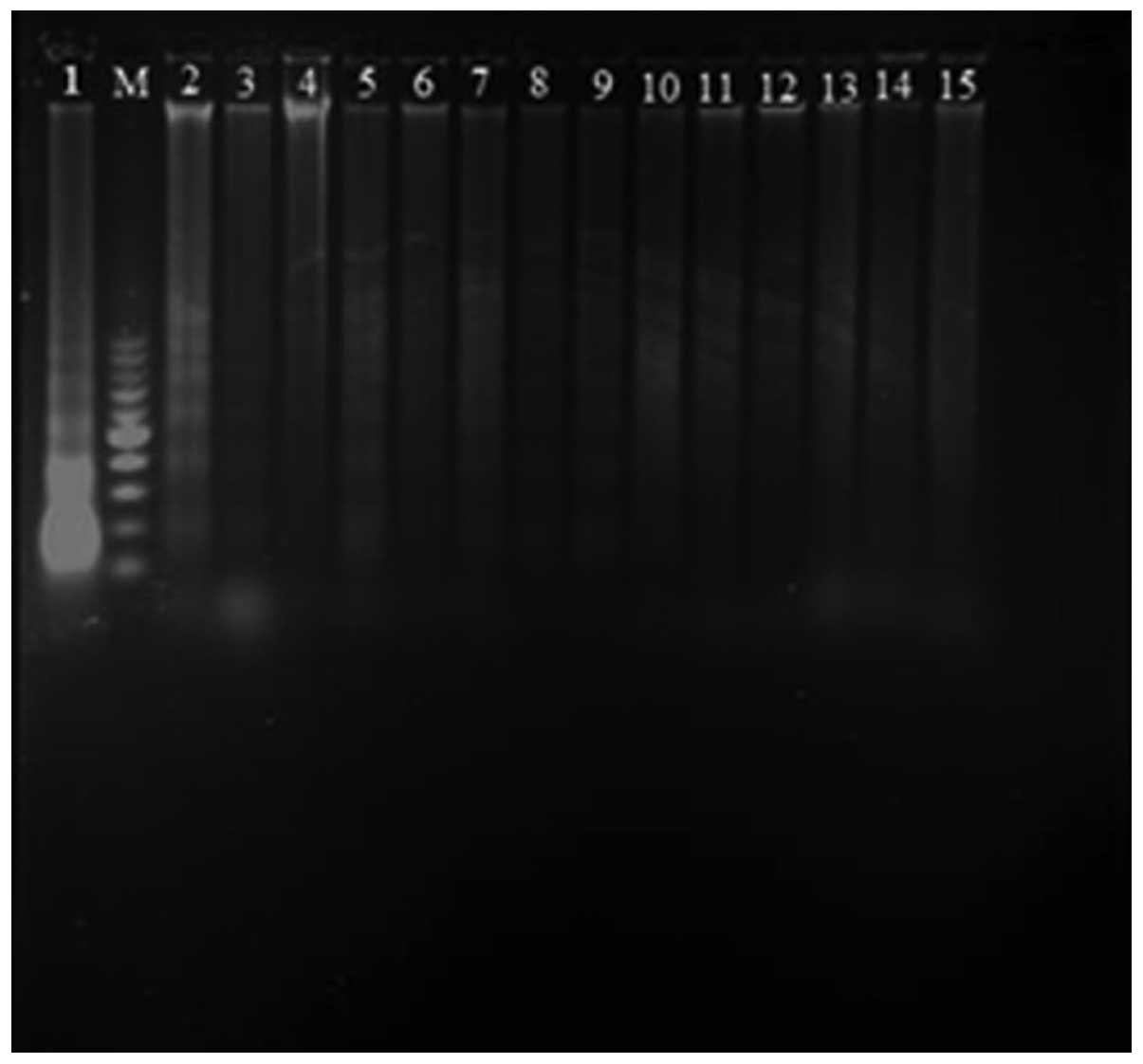
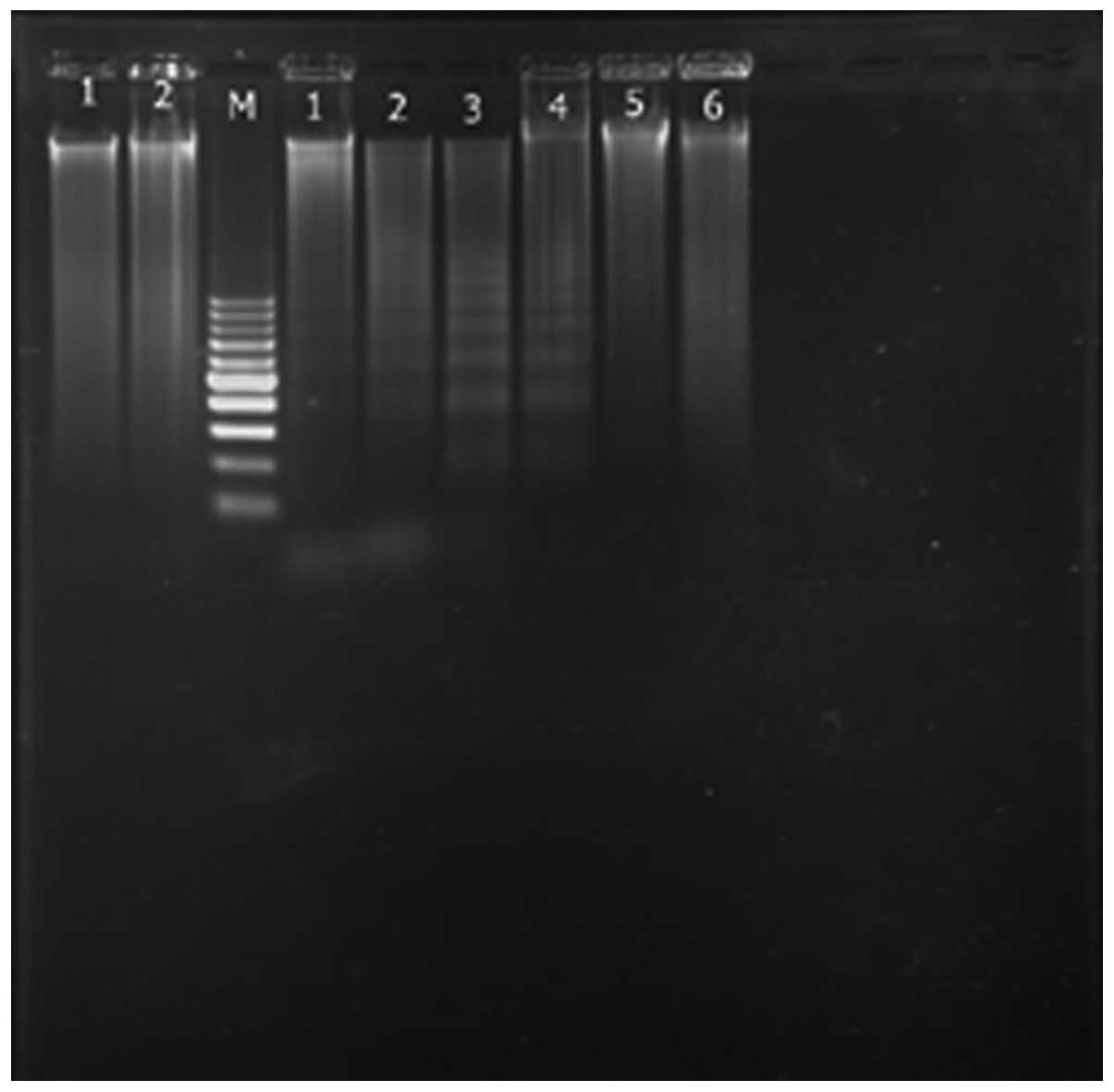
To induce hepatocyte apoptosis by d-GalN/LPS, DNA ladder detection
was employed as the evaluation method. Analysis of DNA
fragmentation indicated that the typical hepatocyte apoptosis was
observed at 6–10 h after the intraperitoneal injection of
d-GalN (700 mg/kg)
and LPS (10 μg/kg) (Fig. 1).
Therefore, the liver apoptosis model was established as follows:
the animals were divided into the vehicle control group (n=7) and
the model group (n=7). The mice in model group were injected
intraperitoneally with d-GalN (700 mg/kg) and LPS (10
μg/kg) dissolved in normal saline. The vehicle control groups
received the same volume of normal saline. Eight hours after the
last administration, the mice were slightly anaesthetized with
ether and blood samples were obtained from the eyepit. The animals
were sacrificed by cervical dislocation and liver samples were
collected for further examination.
Determination of serum ALT, TNF-α and
TGF-β1 levels
The serum was separated for the measurement of ALT,
TNF-α and TGF-β1. According to the manufacturer’s instructions, the
serum ALT activity was determined using the ALT detection kits; the
serum TNF-α and TGF-β1 levels were quantified by the murine TNF-α
and TGF-β1 enzyme immunoassay kits, respectively.
Liver histology and
immunohistochemistry
For histopathological analysis, the liver specimens
were fixed in 10% neutral-buffered formalin, embedded in paraffin,
cut into 5-μm sections, and stained with hematoxylin and eosin
(H&E). For the immunohistochemical analysis of caspase-3, Fas
and FasL, the staining steps of the paraffin sections following
dewaxing and hydration were as follows: the sections were incubated
in 0.3% H2O2-methanol solution for 10 min and
then washed with phosphate-buffered saline (PBS) three times. To
block endogenous peroxidase activity, 50 μl of peroxidase blocking
solution was added to each section and incubated for 10 min at room
temperature. Each section was then incubated with 50 μl of
non-immune goat serum for 10 min at room temperature, rinsed with
PBS and incubated with 50 μl of primary antibody (rat monoclonal
IgG for caspase-3 or rabbit polyclonal IgG for FAS and FasL) at 4°C
overnight. The sections were then incubated with 50 μl of
biotin-labeled secondary antibody (Bio-IgG) at room temperature for
10 min. After washing three times with Tris-buffered saline (TBS),
each section was overlaid with 50 μl of SP-9002
(peroxidase-conjugated goat anti-mouse IgG) solution at room
temperature for 10 min. The reaction was developed with freshly
prepared 3,3′-diaminobenzidine (DAB), and observed under a Leica
DM4000 microscope (Leica Microsystems, Wetzlar, Germany) for 3–10
min, with brown or red indicating positive. The slides were then
counterstained with hematoxylin. The pathological and
immunohistochemical changes were evaluated and photographed under a
Leica DM4000 microscope.
Analysis of DNA fragmentation of
apoptotic cells
The collected liver specimens were washed and then
homogenized in cold PBS. The homogenate was transfered to a 1.5 ml
microcentrifuge (Eppendorf) tube. The DNA of liver tissue was
extracted using the apoptosis DNA ladder detection kit as per the
manufacturer’s instructions. The DNA fragmentation was assayed by
electrophoresis on a 1.5% agarose gel containing 0.5 μg/ml ethidium
bromide and its pattern was examined on the images obtained under
ultraviolet illumination.
TUNEL detection of apoptotic cells
The TUNEL assay was used to quantify liver apoptosis
and was performed using the in situ cell death detection POD
kit according to the manufacturer’s instructions. Briefly,
paraffin-embedded tissue sections were dewaxed and rehydrated
according to the standard protocols (e.g., by heating at 60°C
followed by washing in xylene and rehydration through a graded
series of ethanol and double distilled water). The sections were
then incubated with proteinase K (20 μm/ml in 10 mM Tris-HCl, pH
7.4) for 30 min at 37°C. Slides were rinsed with PBS and incubated
with 3% H2O2 in methanol for 30 min at room
temperature to block endogenous POD activity, followed by PBS
washing and incubation in 0.1% Triton X-100 in 0.1% sodium citrate
for 2 min at 4°C. After washing in PBS, the sections were incubated
with the TUNEL reaction mixture containing terminal
deoxynucleotidyl transferase in a moist chamber at 37°C for 60 min
in the dark. This was followed by washing with PBS and incubation
with converter-POD (anti-fluorescein-HRP) in a humidified chamber
at 37°C for 30 min. After washing with PBS, the sections were
incubated with DAB substrate for 10 min at room temperature, rinsed
again with PBS, mounted under glass coverslip and analyzed under a
light microscope. The negative control was obtained by replacing
the TdT solution with distilled water.
Western blot analysis for Fas, FasL and
caspase-3 expression
Total proteins from the collected liver samples were
prepared according to the method described in the protein extract
kit. Protein concentrations were determined by Coomassie blue
dye-binding assay. Protein extracts were fractionated on 12%
polyacrylamide-sodium dodecyl sulfate gel and then transferred to a
polyvinylidene fluoride membrane. The membrane was blocked with 5%
(w/v) fat-free milk in TBS containing 0.05% Tween-20, followed by
incubation with rat primary anti-FAS (1:150), anti-FasL (1:150) and
anti-caspase-3 (1:150) polyclonal antibody at room temperature for
2 h, respectively. After washing the membrane with TBS, the
membrane was treated with HRP-conjugated goat anti-rat secondary
antibody IgG-HRP (1:3,000) for 1 h at room temperature via
agitation. The enhanced HRP-DAB substrate solution was added to the
membrane and incubated for 10 min. Bands were visualized by
chemiluminescence and exposed to X-ray. The GAPDH antibody was used
as an internal control. The relative optical density (ROD, ratio to
GAPDH) of each blot band was quantified by BandScan 4.5 image
software.
Flow cytometric analysis of apoptotic
cells
Apoptotic cells in the liver tissue were quantified
by flow cytometry. Briefly, the collected liver specimens were
homogenized in cold PBS. The homogenate was filtered through nylon
net, centrifuged and washed with PBS twice. The collected cells
were resuspended in RPMI-1640 medium and adjusted to
5×10⁵ cells/ml. According to the manufacturer’s
instructions, the cell suspension was double-stained with Heochst
33342 at 37°C for 10 min and PI for 10 min at 37°C in the dark,
respectively. Apoptotic analysis was immediately performed on a BD
FACSAria flow cytometer (Becton-Dickinson and Co., Franklin Lakes,
NJ, USA). The population was separated into three groups: live
cells showing only a low level of fluorescence; apoptotic cells
showing a higher level of blue fluorescence, and dead cells showing
low-blue and high-red fluorescence.
Electron microscopy analysis of apoptotic
cells
For electron microscopy examination, the collected
liver tissues were cut into ~1 mm3 and fixed in 2.5%
glutaraldehyde for 2 h. After rinsing with PBS, the tissues were
fixed in 1.5% osmium tetroxide, dehydrated through a graded
alcohol, embedded in epon 812, ultrathin-sectioned, and stained
with uranyl acetate and lead citrate. Labeled ultrathin sections
were observed and images were captured under a JEM-1200EX
transmission electron microscope (Jeol Ltd., Tokyo, Japan).
Statistical analysis
Experimental data were expressed as mean ± standard
deviations (SD) and subjected to a one-way analysis of variance
(ANOVA) and the Student’s t-test. P<0.05 was considered to
indicate statistical significance.
Results
Serum ALT, TNF-α and TGF-β1 levels
ALT is a vital parameter for liver necrosis, whereas
TNF-α and TGF-β1 are the critical mediators of liver apoptosis. We
hypothesized that the levels of three mediators would increase in
mice after the d-GalN/LPS challenge. The results
showed that the serum ALT, TNF-α and TGF-β1 levels of the
d-GalN/LPS-treated
mice were significantly elevated with respect to the vehicle
control (Table I).
 | Table IThe serum ALT activity, TNF-α and
TGF-β1 levels of hepatocyte apoptotic model induced by d-GalN/LPS in mice. |
Table I
The serum ALT activity, TNF-α and
TGF-β1 levels of hepatocyte apoptotic model induced by d-GalN/LPS in mice.
| Groups | ALT (IU/l) | TNF-α (pg/ml) | TGF-β1 (pg/ml) |
|---|
| Vehicle
control | 15.91±1.13 | 18.65±4.92 | 20.82±4.49 |
| d-GalN/LPS-treated |
123.60±22.22b |
134.83±80.35a |
277.23±92.88b |
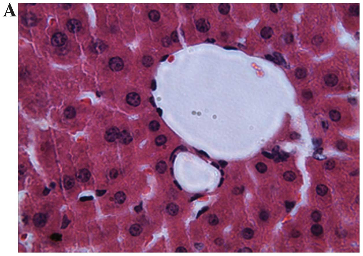
Liver histopathology
Histopathological analysis showed that hepatic
lobular architecture was clear and intact without any abnomalities
in the liver section of the control group. At 8 h after
d-GalN/LPS injection,
large apoptotic liver cells and sections of hepatic necrosis with
leukocyte infiltration were found (Fig. 2). Strong apoptotic positive
signals including chromatin condensation and margination,
disruption of nuclear membrane, and fragmentation of chromatin and
the formation of apoptotic bodies was observed.
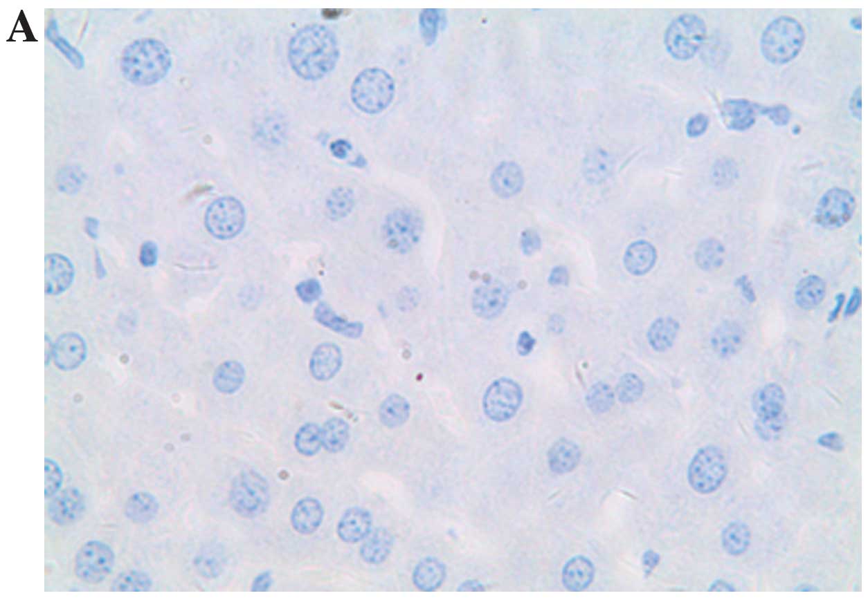
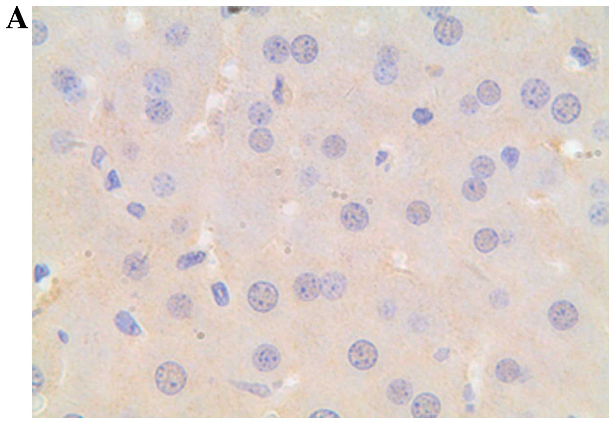
Liver immunohistochemistry
Caspase-3 plays a central role in the execution
phase of cell apoptosis. The Fas/FasL system provides a major
apoptotic mechanism for many cell types, including liver cells. We
examined the expression of caspase-3, Fas and FasL of the isolated
liver at 8 h after d-GalN/LPS treatment.
Immunohistochemical analysis revealed that d-GalN/LPS treatment markedly
increased hepatic expression of caspase-3, as well as the
pro-apoptotic receptors Fas and FasL in mice (Figs. 3–5).
Analysis of DNA fragmentation
Genomic DNA fragmentation was assayed to confirm the
occurrence of hepatocyte apoptosis. DNA fragmentation was observed
in murine liver after d-GalN/LPS treatment, while no
DNA fragmentation was found in the untreated control untreated
(Fig. 6).
TUNEL detection of apoptotic cells
Apoptotic cells were detected by TUNEL staining. A
large number of TUNEL-positive hepatocytes were observed in liver
tissues from mice following d-GalN/LPS treatment. However, no
TUNEL-positive hepatocytes were observed in the livers from the
untreated control mice (Fig.
7).
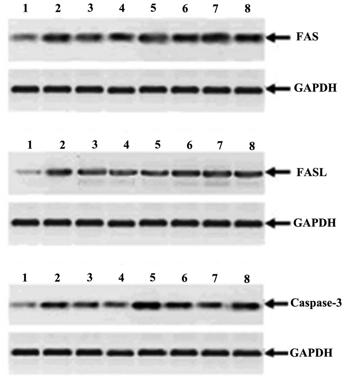
Western blot analysis for Fas, FasL and
caspase-3 expression
The expression levels of Fas, FasL and caspase-3 in
liver tissues from the mouse after d-GalN/LPS treatment were
quantified by western blotting. The GAPDH antibody was used as an
equal loading internal control. As shown in Fig. 8 and Table II, compared with the untreated
control mice, the expression levels of caspase-3, Fas and FasL were
markedly enhanced after the d-GalN/LPS treatment.
| Table IIThe relative expression of Fas, FasL
and caspase-3 proteins in liver tissues from the mice at 8 h after
the d-GalN/LPS
treatment. |
Table II
The relative expression of Fas, FasL
and caspase-3 proteins in liver tissues from the mice at 8 h after
the d-GalN/LPS
treatment.
| Samples | Relative expression
of Fas protein | Relative expression
of FasL protein | Relative expression
of caspase-3 protein |
|---|
| 1 | 0.52±0.08 | 0.33±0.04 | 0.42±0.05 |
| 2 | 1.96±0.17 | 1.18±0.18 | 1.32±0.23 |
| 3 | 1.58±0.15 | 1.02±0.24 | 1.05±0.15 |
| 4 | 1.52±0.11 | 1.01±0.31 | 0.98±0.17 |
| 5 | 1.87±0.21 | 1.05±0.24 | 2.36±0.15 |
| 6 | 1.73±0.12 | 1.15±0.25 | 1.88±0.22 |
| 7 | 2.24±0.23 | 1.20±0.18 | 1.35±0.30 |
| 8 | 1.91±0.10 | 1.07±0.19 | 1.95±0.12 |
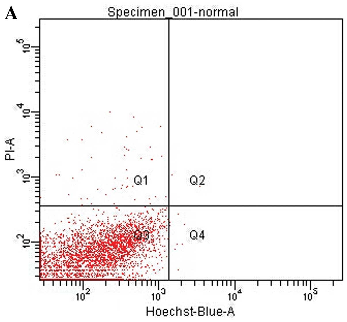
Flow cytometric analysis of apoptotic
cells
The hepatocytes were isolated from the murine liver
following administration of d-GalN/LPS and staining with
Hoechst 33342. Hoechst 33342, a type of blue-fluorescence dye,
stains the condensed chromatin in apoptotic cells more brightly
than normal chromatin. PI a red-fluorescence dye, is only permeant
to dead cells. The staining pattern resulting from the simultaneous
use of these dyes makes it possible to distinguish normal,
apoptotic, and dead cell populations by flow cytometry. The result
showed that the injury, necrotic and apoptotic cells were observed
following d-GalN/LPS
treatment and that apoptotic cells were mainly at the late stage of
cell apoptosis (Fig. 9).
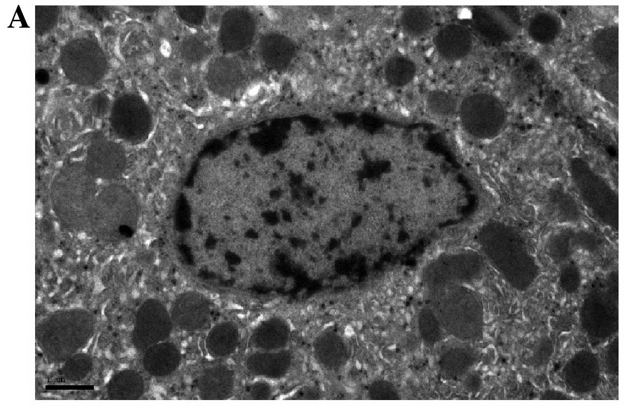
Electron microscopy analysis of apoptotic
cells
To analyze the typical morphological signs of
apoptosis, the ultrathin sections from the mouse liver tissues
after administering D-GalN/LPS were observed under electron
microscope. The chromatin of apoptitic liver cell appeared
condensation, approaching to nuclear membrane, gathering to the
edge and forming typical apoptosis body (Fig. 10).
Discussion
Liver disease is often associated with enhanced
hepatocyte apoptosis, which is the case in viral and autoimmune
hepatitis, cholestatic diseases, and metabolic disorders. Use and
abuse of certain drugs, especially alcohol, chemotherapeutic
agents, and acetaminophen, have been associated with increased
apoptosis and liver damage (8).
The availability of animal models relevant to apoptosis may
facilitate the identification of potential therapies. d-GalN is a typical hepatotoxin
and often used in pharmacodynamics research to induce hepatic
injury. This model of liver damage most closely resembles the
changes observed during human hepatitis, and thus provides a useful
system for screening and investigating drugs that can be used in
the treatment of disease (5).
LPS, the major structural component of the outer membrance of
gram-negative bacteria, causes liver injury at high doses but a
modest, non-injurious inflammation at low doses in several animal
models (9–11). In addition, LPS can induce lethal
liver failure when simultaneously administered with d-GalN (7). In this investigation, we reported
the nature of d-GalN/LPS-induced hepatocyte
apoptotic changes and the related gene expression in mice.
DNA ladder detection indicated that hepatocyte
apoptosis occurred at 6–10 h after the intraperitoneal injection of
d-GalN (700 mg/kg)
and LPS (10 μg/kg). Based on the initial test, we demonstrated the
occurrence of the hepatocyte apoptosis at 8 h after administering
d-GalN/LPS by
histopathological analysis, TUNEL detection, flow cytometry and
electron microscopy analysis. Histopathological analysis showed
that strong apoptotic positive signals were observed including
chromatin condensation and margination, disruption of nuclear
membrane, and fragmentation of chromatin and the formation of
apoptotic bodies. TUNEL detection indicated that a large number of
TUNEL-positive hepatocytes were observed in liver tissues. Flow
cytometric analysis showed that the apoptotic liver cells were
mainly at the late stage of cell apoptosis, suggesting that DNA
fragmentation may occur at late stage of cell apoptosis. Electron
microscopy analysis indicated the typical morphological signs of
apoptosis especially the formation of apoptotic bodies. These
results suggest that typical hepatocyte apoptosis was definitely
induced by d-GalN/LPS
in mice.
Overactivation involving mediators such as Fas,
TNF-α and TGF-β, can lead to significant acute injuries, such as
fulminant hepatic failure or even chronic sustained hepatocellular
damage, as occurs with toxic liver injury, viral hepatitis,
alcoholic and non-alcoholic liver disease (12). To investigate apoptosis-related
gene expression changes, we examined the expression of TNF-α,
TGF-β1, Fas, FasL and caspase-3 by serological measurement,
immunohistochemical analysis and western blot analysis. The
serological detection showed that the ALT, TNF-α and TGF-β1 levels
of the d-GalN/LPS-treated mice were
significantly enhanced, suggesting that apoptosis and necrosis
occurred together following d-GalN/LPS challenge.
Immunohistochemical analysis indicated that d-GalN/LPS treatment markedly
increased the expression of caspase-3, as well as the pro-apoptotic
receptors Fas and FasL in mice. Western blot analysis showed the
relative expression of Fas, FasL, and caspase-3 proteins, which
were markedly elevated after d-GalN/LPS treatment. The above
results suggest that the apoptosis-related gene expressions (TNF-α,
TGF-β1, Fas/FasL and caspase-3) were significantly increased
following d-GalN/LPS
treatment.
Apoptosis plays an important role in liver
pathogenesis, and disarrangement of death receptor pathways has
been identified as a major contributor to the initiation and
aggravation of acute and chronic liver injury (12). Apoptotic signalling within the
cells is transduced mainly via two molecular pathways: the
extrinsic or death receptor pathway and the intrinsic or
mitochondrial pathway. Apoptotic events in hepatocytes can be
regulated by different stimuli that bind to death receptors in the
cell membranes, such as FasL, TNF or TNF-related apoptosis-induced
ligand, which activate the extrinsic pathway. Binding of FasL or
TNF-β to their corresponding death receptors induce the recruitment
of procaspase 8–10 to form the death-inducing signaling complex,
leading to cell death (13).
Other factors, particularly TGF-β, do not bind the death receptors,
but its intracellular signals couple to the apoptotic machinery
through activation of the intrinsic pathway. Apoptosis and the
elimination of apoptotic cells are crucial factors in the
maintenance of liver health. Apoptosis allows hepatocytes to die
without provoking a potentially harmful inflammatory response. In
contrast to necrosis, apoptosis is closely controlled and regulated
via several mechanisms, including Fas/FasL interactions, the
effects of cytokines such as TNF-α and TGF-β, and the influence of
pro- and anti-apoptotic mitochondria-associated proteins of the
B-cell lymphoma-2 (Bcl-2) family (8). The d-GalN/LPS challenge can
significantly enhance the expression of Fas/FasL and TNF-α in the
liver, suggesting that the hepatocyte apoptosis induced by
d-GalN/LPS may be
regulated by the death receptor pathway such as Fas/FasL
interactions.
Caspases are broadly categorized into upstream
regulatory caspases and downstream effector caspases. Caspase-3 is
usually regarded as the downstream effector protease most important
for the classic nuclear changes associated with apoptosis (14). The cleavage of the anti-apoptotic
protein Bcl-2 into a proapoptotic form is a necessary step in
Fas-mediated apoptosis, with caspase-3 mediating this processing
(15). Caspase-3 is considered to
be a key apoptotic ‘executioner’ enzyme in mammalian cells because
its activation triggers the cascade of enzymatic events that
culminates in the death of the cell (16). Fas-mediated cell death of
hepatocytes is involved in many human liver pathologies, including
hepatitis B virus-related cirrhosis, autoimmune hepatitis, acute
liver failure, rejection of transplanted livers, and alcoholic and
toxin-induced liver diseases. Moreover, caspase-3 is required for
the initial events that occur in hepatocyte cell death following
Fas ligation (17). In this
study, the relative expression of Fas, FasL and caspase-3 proteins
in murine liver were markedly elevated following d-GalN/LPS challenge. This
finding suggests that the Fas-mediated death receptor pathway is
one of the main mechanisms of hepatocyte apoptosis induced by
d-GalN/LPS.
TGF-β is a multifunctional cytokine, whose numerous
cell and tissue activities include cell-cycle control,
differentiation, extracellular matrix formation, and the induction
of apoptosis. The important role of TGF-β in orchestrating
apoptosis in the liver is indicated by the hepatic fibrosis and
apoptotic cell death of hepatocytes in transgenic mice that
ectopically express TGF-β1 in the liver (18). TGF-β1-regulated apoptosis is
cell-type and context-dependent, with TGF-β1 providing signals for
cell survival or apoptosis. The molecular mechanisms underlying the
role of TGF-β1 in apoptosis remain unclear. The proteins that
primarily mediate the intracellular signaling of TGF-β1 are the
members of the Smad family (19).
Multiple apoptotic mediators and signaling pathways are involved in
TGF-β-induced apoptosis. The activator protein (AP)-1 complex is
also involved in TGF-β1 signaling for apoptosis (20). Bim is a crucial mediator of the
apoptotic effects elicited by TGF-β (21). TGF-β1 signaling also cooperates
with the death receptor apoptotic pathway (Fas and TNF). Moreover,
the involvement of TGF-β1 in the production of oxidative stress and
in preventing the inflammatory processes required for the clearance
of apoptotic bodies is further evidence of its integration into
apoptotic pathways (22). Since
the expression of TGF-β1 in the d-GalN/LPS-induced apoptotic
hepatocyte was significantly increased, it is suggested that the
TGF-β signaling pathway plays a vital role in this process of the
hepatocyte apoptosis induced by d-GalN/LPS.
There are several reasons that make a cell undergo
apoptosis, including oxidative stress, DNA damage, accumulation of
unfolded or misfolded proteins, occupation of death receptors by
extracellular signals and lack of stimulation from other cells
(12). Oxidative stress has been
noted to contribute to the pathogenesis of acute hepatitis. Free
radicals are toxic to hepatocytes and initiate a reactive oxygen
species-mediated cascade causing hepatocyte cell death and leading
to acute hepatitis (23,24). Oxygen-derived free radicals
released from activated hepatic macrophages are the primary cause
of d-GalN-induced
liver damage (25,26). Increased production of reactive
oxygen species has been reported in the primary culture of rat
hepatocyte damage induced by d-GalN (27). d-GalN-induced hepatocyte injury
is closely associated with the formation of oxidative stress.
Oxidative stress is a factor that is capable of triggering
apoptosis. Thus, the d-GalN/LPS-induced hepatocyte
apoptosis is probably associated with oxidative stress.
This study has evaluated the d-GalN/LPS-induced hepatocyte
apoptotic changes and clarified apoptosis-related gene expression
(TNF-α, TGF-β1, Fas/FasL and caspase-3) in mice. The hepatocyte
apoptosis induced by d-GalN/LPS is mainly regulated by
the death receptor pathway. The TGF-β signaling pathway also plays
a vital role in this process of hepatocyte apoptosis. These data
provide a deeper understanding of the occurrence of the
d-GalN/LPS-induced
liver apoptosis.
Acknowledgements
This study was supported by a grant from the Key
Project of the Chinese Ministry of Education (no. 212073), the
Experimental Animal Science and Technology Project of Zhejiang
Province (no. 2008F800169) and the Public Welfare Technology
Applied Research Project of Zhejiang Province - Experimental Animal
Science and Technology Project (no. 2013C37020). The authors
acknowledge Dr Jian Chen and Ms. Xiaoyuan Jia for technical
assistance for the flow cytometric analysis.
References
|
1
|
White E: Death-defying acts: a meeting
review on apoptosis. Genes Dev. 7:2277–2284. 1993. View Article : Google Scholar : PubMed/NCBI
|
|
2
|
Patel T and Gores GJ: Apoptosis and
hepatobiliary disease. Hepatology. 21:1725–1741. 1995.PubMed/NCBI
|
|
3
|
Eichhorst ST: Modulation of apoptosis as a
target for liver disease. Expert Opin Ther Targets. 9:83–89. 2005.
View Article : Google Scholar : PubMed/NCBI
|
|
4
|
Prétet JL, Pelletier L, Bernard B,
Coumes-Marquet S, Kantelip B and Mougin C: Apoptosis participates
to liver damage in HSV induced fulminant hepatitis. Apoptosis.
8:655–663. 2003.PubMed/NCBI
|
|
5
|
Keppler D, Lesch R, Reutter W and Decker
K: Experimental hepatitis induced by d-galactosamine. Exp Mol
Pathol. 9:279–290. 1968. View Article : Google Scholar : PubMed/NCBI
|
|
6
|
Galanos C, Freudenberg MA and Reutter W:
Galactosamine-induced sensitization to the lethal effects of
endotoxin. Proc Natl Acad Sci USA. 76:5939–5943. 1979. View Article : Google Scholar : PubMed/NCBI
|
|
7
|
Nguyen NT, Banskota AH, Tezuka Y, Le Tran
Q, Nobukawa T, Kurashige Y, Sasahara M and Kadota S:
Hepatoprotective effect of taxiresinol and
(7′R)-7′-hydroxylariciresinol on d-galactosamine and
lipopolysaccharide-induced liver injury in mice. Planta Med.
70:29–33. 2004.PubMed/NCBI
|
|
8
|
Neuman MG: Apoptosis in diseases of the
liver. Crit Rev Clin Lab Sci. 38:109–166. 2001. View Article : Google Scholar
|
|
9
|
Rietschel ET, Kirikae T, Schade FU, Mamat
U, Schmidt G, Loppnow H, Ulmer AJ, Zähringer U, Seydel U, Di Padova
F, et al: Bacterial endotoxin: molecular relationships of structure
to activity and function. FASEB J. 8:217–225. 1994.PubMed/NCBI
|
|
10
|
Kosai K, Matsumoto K, Funakoshi H and
Nakamura T: Hepatocyte growth factor prevents endotoxin-induced
lethal hepatic failure in mice. Hepatology. 30:151–159. 1999.
View Article : Google Scholar : PubMed/NCBI
|
|
11
|
Ganey PE and Roth RA: Concurrent
inflammation as a determinant of susceptibility to toxicity from
xenobiotic agents. Toxicology. 169:195–208. 2001. View Article : Google Scholar : PubMed/NCBI
|
|
12
|
Salido GM and Rosado JA: Apoptosis:
Involvement of oxidative stress and intracellular Ca2+
homeostasis. Apoptosis in the Liver. González-Gallego J and Tũnón
MJ: Springer; Spain: pp. 73–92. 2009
|
|
13
|
Yoon JH and Gores GJ: Death
receptor-mediated apoptosis and the liver. J Hepatol. 37:400–410.
2002. View Article : Google Scholar : PubMed/NCBI
|
|
14
|
Enari M, Sakahira H, Yokoyama H, Okawa K,
Iwamatsu A and Nagata S: A caspase-activated DNase that degrades
DNA during apoptosis, and its inhibitor ICAD. Nature. 391:43–50.
1998. View Article : Google Scholar : PubMed/NCBI
|
|
15
|
Cheng EH, Kirsch DG, Clem RJ, Ravi R,
Kastan MB, Bedi A, Ueno K and Hardwick JM: Conversion of Bcl-2 to a
Bax-like death effector by caspases. Science. 278:1966–1968. 1997.
View Article : Google Scholar : PubMed/NCBI
|
|
16
|
Thornberry NA and Lazebnik Y: Caspases:
enemies within. Science. 281:1312–1316. 1998. View Article : Google Scholar : PubMed/NCBI
|
|
17
|
Woo M, Hakem A, Elia AJ, Hakem R, Duncan
GS, Patterson BJ and Mak TW: In vivo evidence that caspase-3 is
required for Fas-mediated apoptosis of hepatocytes. J Immunol.
163:4909–4916. 1999.PubMed/NCBI
|
|
18
|
Lee KY and Bae SC: TGF-beta-dependent cell
growth arrest and apoptosis. J Biochem Mol Biol. 35:47–53. 2002.
View Article : Google Scholar : PubMed/NCBI
|
|
19
|
Perlman P, Schiemann WP, Brooks MW, Lodish
HF and Weinberg RA: TGF-β induced apoptosis is mediated by the
adapter protein Daxx that facilitates JNK activation. Nature Cell
Biol. 3:708–714. 2001.
|
|
20
|
Schuster N and Krieglstein K: Mechanisms
of TGF-beta-mediated apoptosis. Cell Tissue Res. 307:1–14. 2002.
View Article : Google Scholar : PubMed/NCBI
|
|
21
|
Ramesh S, Wildey GM and Howe PH:
Transforming growth factor beta (TGFbeta)-induced apoptosis: the
rise and fall of Bim. Cell Cycle. 8:11–7. 2009. View Article : Google Scholar : PubMed/NCBI
|
|
22
|
Sánchez-Capelo A: Dual role for TGF-β1 in
apoptosis. Cytokine Growth Factor Rev. 16:15–34. 2005.
|
|
23
|
Bedda S, Laurent A, Conti F, Chéreau C,
Tran A, Tran-Van Nhieu J, Jaffray P, Soubrane O, Goulvestre C,
Calmus Y, Weill B and Batteux F: Mangafodipir prevents liver injury
induced by acetaminophen in the mouse. J Hepatol. 39:765–772. 2003.
View Article : Google Scholar : PubMed/NCBI
|
|
24
|
Okuyama H, Nakamura H, Shimahara Y, Araya
S, Kawada N, Yamaoka Y and Yodoi J: Overexpression of thioredoxin
prevents acute hepatitis caused by thioacetamide or
lipopolysaccharide in mice. Hepatology. 37:1015–1025. 2003.
View Article : Google Scholar : PubMed/NCBI
|
|
25
|
Shiratori Y, Kawase T, Shiina S, Okano K,
Sugimoto T, Teraoka H, Matano S, Matsumoto K and Kamii K:
Modulation of hepatotoxicity by macrophages in the liver.
Hepatology. 8:815–821. 1988. View Article : Google Scholar : PubMed/NCBI
|
|
26
|
Hu HL and Chen RD: Changes in free
radicals, trace elements, and neurophysiological function in rats
with liver damage induced by d-galactosamine. Biol Trace Elem Res.
34:19–25. 1992. View Article : Google Scholar : PubMed/NCBI
|
|
27
|
Quintero A, Pedraza CA, Siendones E, Kamal
ElSaid AM, Colell A, García-Ruiz C, Montero JL, De la Mata M,
Fernández-Checa JC, Miño G and Muntané J: PGE1 protection against
apoptosis induced by d-galactosamine is not related to the
modulation of intracellular free radical production in primary
culture of rat hepatocytes. Free Radic Res. 36:345–355. 2002.
View Article : Google Scholar
|