Introduction
Acute lung injury (ALI) is a distinct form of acute
respiratory failure characterized by diffuse pulmonary infiltrates,
progressive hypoxemia, reduced lung compliance and abnormal
hydrostatic pressure (1). Over
the past 30 or so years, this syndrome has come to be one of the
central concerns of intensive care: lung injury arising from a
variety of different etiologies, each characterized by bilateral
diffuse infiltrates on X-ray, hypoxemia and non-cardiogenic
pulmonary edema (2–4). Actually, ALI is caused by any
stimulus of local or systemic inflammation, principally sepsis
(5). It is most often observed as
part of a systemic inflammatory process, particularly systemic
sepsis, where the lung manifestations parallel those of other
tissues, including widespread destruction of the capillary
endothelium, extravasations of protein rich fluid and interstitial
edema (6–8). In addition, the alveolar basement
membrane is damaged, and fluid seeps into the airspaces, stiffening
the lungs and causing ventilation-perfusion mismatch (9,10).
According to the clinical diagnosis, there are two major stages: i)
the acute phase characterized by the disruption of the
alveolar-capillary interface, leakage of protein rich fluid into
the interstitium and alveolar space and the excessive release of
cytokines and the migration of neutrophils (11–14); and ii) the later reparative phase
which is characterized by fibroproliferation, and the organization
of lung tissue. ALI is a disease with a high mortality rate
(30–50%) (15). However, there
are still no effective therapeutic strategies or specific drugs to
treat it. Therefore, the development of novel therapies for ALI is
urgently needed.
Baicalin, a type of flavonoid, can be found and
isolated from several species in the genus Scutellaria,
including Scutellaria baicalensis and Scutellaria
lateriflora, and has been reported to exert anti-hypertensive,
antitumor and anti-inflammatory effects (16–18). As previously demonstrated, ALI
involves a typical inflammatory response, and the nuclear factor
(NF)-κB signaling pathway is directly involved in the developmental
process of lung injury (19,20). Thus, the inhibition of the
activation of the NF-κB pathway may prove to be key to the
effective treatment of ALI. In addition, the CX3CL1-CX3CR1 axis has
also been shown to play an important role in inflammation-related
disorders, including hepatitis and fibrosis, nerve damage and tumor
development (21–25). The CX3CL1-CX3CR1 axis has the
ability to enhance the levels of phosphorylated NF-κB; in turn, the
activation of NF-κB continually promotes the CX3CL1-CX3CR1
interaction (26). The crosstalk
between the CX3CL1-CX3CR1 axis and NF-κB pathway plays a critical
role in inflammation (27–29).
Hence, in this study, we wished to determine whether baicalin
inhibits lipopolysaccharide (LPS)-induced ALI by interfering with
the activation of the CX3CL1-CX3CR1 axis and NF-κB pathway.
Furthermore, we used CX3CL1-knockout (CX3CL1-KO or
CX3CL1−/−) mice as a model with which to evaluate the
effects of CX3CL1 on inflammatory responses.
Materials and methods
Reagents and animals
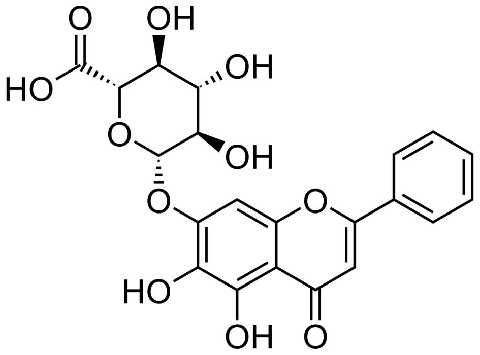
Baicalin (CAS: 21967-41-9, HPLC analysis ≥98%,
C21H18O11, MW: 446.36102) was
purchased from the National Institute for the Control of
Pharmaceutical and Biological Products (Beijing, China) (chemical
strucure shown in Fig. 1). LPS
was obtained from Sigma-Aldrich China, Inc. (Shanghai, China) and
prepared in phosphate-buffered saline (PBS) at the concentration of
100 µg/ml. Anti-CX3CL1 (rabbit; ab25088) and anti-CX3CR1
(rabbit; ab8021) antibodies were obtained from Abcam (Shanghai,
China). Anti-AKT (rabbit; 4961), anti-phosphorylated (p-) AKT
(rabbit; 9614), anti-IκB (rabbit; 4812), anti-p-IκB (rabbit; 5209),
anti-NF-κB (rabbit; 8242), anti-p-NF-κB (rabbit; 3033),
anti-cyclooxygenase-2 (COX-2; rabbit; 12282) and anti-p-IKKβ
(rabbit; 14938) antibodies were all obtained from Cell Signaling
Technology, Inc. (Beverly, MA, USA). The tumor necrosis factor
(TNF)-α, interleukin (IL)-1β, IL-6 and transforming growth factor
(TGF)-β ELISA kits were obtained from R&D Systems, Inc.,
(Minneapolis, MN, USA). The IL-18 kit was from BioLegend (San
Diego, CA, USA). Male BALB/c mice (n=120; 6–8 weeks old, weighing
20–25 g) were obtained from Charles River Laboratories (Wilmington,
MA, USA) and housed in a temperature and humidity-controlled
environment (25±2°C and 50±10% humidity) with a standard 12-h
light/dark cycle with food and water provided in the cages. This
study was approved by the Ethics Committee on Animal Research at
the Capital Medical University, Beijing, China. All mice were
randomly divided into 5 groups as follows: i) the control group;
ii) the group treated with 3 mg/kg LPS; iii) the group treated with
3 mg/kg LPS + 50 mg/kg baicalin; iv) the group treated with 3 mg/kg
LPS + 100 mg/kg baicalin; and v) the group treated with 3 mg/kg LPS
+ 200 mg/kg baicalin. The mice were orally administered with the
various concentrations of baicalin for 7 days prior to the
administration of LPS. LPS was administered to the mice via
intraperitoneal injection for 48 h. All animal experiments were
performed in accordance with the guide for the Care and Use of
Laboratory Animals established by the US National Institutes of
Health. In addition, 70 male C57BL/6 mice (6–8 weeks old, weighing
20–25 g) lacking the CX3CL1 gene (CX3CL1-KO or
CX3CL1−/−) and wild-type (WT) mice (6–8 weeks old,
weighing 20–25 g) were purchased from Beijing Biocytogen Co., Ltd.
(Beijing, China). The C57BL/6 mice and WT mice were administered
LPS in the same manner as the BALB/c mice.
Isolation and culture of lung epithelial
cells
The immune adhesion method was used to isolate lung
epithelial cells. In brief, after euthanizing the mice by ether
anesthesia, the lungs were excised and the lung tissues of the
CX3CL1-KO mice and WT mice were carefully isolated and mantained in
Hanks' buffer. Subsequently, 0.25% trypsin (0.5 ml/5 g;
Sigma-Aldrich China, Inc.) was added to homogenize the lung tissues
at 4°C overnight. The tissues were then treated with an equal
volume of trypsin and passed through a stainless steel mesh (size
60;Sigma-Aldrich China, Inc.), and suspended in DMEM/F12 medium
(Gibco/Thermo Fisher Scientific, Inc., Waltham, MA, USA). The cell
suspensions were centrifuged at 500 rpm for 5 min to remove the
debris and impurities. The suspensions were collected and washed
with Hanks' buffer 3 times. Subsequently, the cells at
3×10⁶ ml were cultured in IgG-coated 6-wells plate at
37°C in 5% CO2 for 2.5 h. The unattached cells were
removed to a new tube and collected by centrifugation at 800 rpm
for 4 min. The isolated cells were then re-cultured in 6-wells
plates. Cells at passage 3 were used in our experiments. The cells
were serum-starved fro 36 h and then stimulated with TNF-α (50
ng/ml) for various periods of time (0, 2, 5, 10 and 15 min). The
cells were then subjected to western blot analysis, RT-qPCR and the
analysis of NF-κB activity.
Lung wet-to-dry weight ratio
After euthanizing the mice by ether anesthesia, the
lungs were excised, blotted dry, weighed to obtain the 'wet'
weight, and then placed in an oven at 80°C for 48 h to obtain the
'dry' weight. The ratio of the wet lung to dry lung was calculated
to assess tissue edema.
Inflammatory cell counts in
bronchoalveolar lavage fluid (BALF)
After the mice were euthanized, the total lung
tissues (containing the trachea) of 8–10 mice were collected and
subjected to perfusion with Hanks' buffer (0.3 ml, 3 times). The
irrigation solution was collected and centrifuged (4°C, 3,000 rpm
for 10 min) to pellet the cells The BALF samples were centrifuged
(4°C, 3,000 rpm for 10 min) to pellet the cells. The cell pellets
were resuspended in PBS for the total cell counts using a
hemocytometer, and cytospins were prepared for differential cell
counts by staining using the Wright-Giemsa staining method.
Histopathological examination of lung
tissues
Histopathological evaluation was performed on the
lungs of 8–10 mice that were not subjected to BALF collection. The
lungs were fixed with 10% buffered formalin, imbedded in paraffin
and sliced. Following hematoxylin and eosin (H&E) staining, the
pathological changes of the lung tissues were observed under a
light microscope. Some samples were also was subjected to
immunohistochemical (IHC) staining (CX3CL1 and IκBα antibody)
according to the instructions of the manufacturer (Cell Signaling
Technology, Inc.) and were performed by Shanghai Zhenda
Biotechnology, Co., Ltd. (Sanghai, China).
Western blot analysis and reverse
transcription-quantitative PCR (RT-qPCR)
Six hours after the injection of LPS, the lung
tissues were harvested and frozen in liquid nitrogen immediately
until homogenization. Proteins were extracted from the lung tissue
using T-PER Tissue Protein Extraction Reagent kit (Thermo Fisher
Scientific) according to the manufacturer's instructions. Protein
concentrations were determined using the BCA protein assay kit, and
equal amounts of protein were loaded per well on a 10% sodium
dodecyl sulphatepolyacrylamide gel. Subsequently, the proteins were
transferred onto polyvinylidene difluoride membranes. The resulting
membranes were blocked with Tris-buffered saline containing 0.05%
Tween-20 (TBS-T), supplemented with 5% skim milk (Sigma-Aldrich
China, Inc.) at room temperature for 2 h on a rotary shaker,
followed by washing with TBS-T. The specific primary antibody,
diluted in TBST, was incubated with the membrane at 4°C overnight.
Subsequently, the membranes were washed with TBS-T followed by
incubation with the peroxidase-conjugated secondary antibody at
room temperature for 1 h. The immunoactive proteins were detected
by using an enhanced chemiluminescence western blotting detection
kit. The bands were observed using an ECL western blotting analysis
system (GE Healthcare, Pittsburgh, PA, USA) and exposed to Kodak
X-ray film.
RT-qPCR, was performed as previously described
(30). Total RNA was extracted
from tissues and cells using TRIzol® reagent
(Invitrogen/Thermo Fisher Scientific, Inc.) and 1 µg total
RNA was reverse transcribed using the M MLV RT system (Promega
Corp., Madison, WI, USA). This was performed at 42°C for 1 h and
terminated by the deactivation of the enzyme at 70°C for 10 min.
qPCR was conducted using SYBR®-Green (Bio-Rad
Laboratories, Inc., Hercules, CA, USA) in an ABI PRISM 7900HT
detection system (Applied Biosystems; Thermo Fisher Scientific,
Inc.). All the primers were produced by Thermo Fisher Scientific,
Inc. The amplification of the pre-denatured products was conducted
at 94°C for 60 sec; followed by 45 cycles at 95°C for 30 sec, 58°C
for 30 sec and 72°C for 30 sec; followed by 95°C for 10 sec, 65°C
for 45 sec, and 40°C for 60 sec. Fold induction values were
calculated using the to 2−ΔΔCq method, where ΔCq
represents the differences in cycle threshold number between the
target gene and GAPDH, and ΔΔCq represents the relative change in
the differences between the control and treatment groups.
Two-dimensional gel electrophoresis
Each gel was loaded with 100 µg protein and
mixed with water solution (7 mol/l urea, 2 M thiourea, 4% CHAPS and
0.2% ampholyte). The total sample volume was 290 µl. After
hydration of the IPG adhesive strip (24 cm, pH 3–10, NL), the
mineral oil was then covered. The isoelectric focusing procedure
was set as 30 V/6 h→60 V/6 h→500 V/1 h→1,000 V/1 h→8,000 V/10 h,
and the total voltage time product was 64,000 Vh. After isoelectric
focusing, the IPG strip was preserved in the SDS equilibrium
solution (6 M urea, 2% SDS, 0.02% bromophenol blue liquid reserves)
for 15 min. After completion of the equilibrium, the IPG strip was
placed above the 12.5% uniform polyacrylamide gel and 10 µl
protein marker was added on the SDS gel. The air bubbles were
removed and sealed using anhydrous ethanol. Subsequently,
two-dimensional gel electrophoresis was performed with bromophenol
blue tracking dye until the dye had move to the front of the rubber
edge with a total duration of 20 h. Cy3 and Cy5 fluorescence
staining was then applied. The protein expression of the
TLR4/MyD88/NF-κB signaling-related indicators, AKT, CX3CR1, TNF-α,
IL-6, was analyzed in this part. and the image was then subjected
to intensity correction, point detection, background subtraction,
homogenization and matching processing using DeCyder 2D 6.5
software.
Measurement of cytokine levels by
ELISA
The levels of major inflammatory cytokines, such as
TNF-α, IL-1β, IL-6, TGF-β and IL-18 levels in the cells or serum
from mice were determined using respective ELISA kits, following
the manufacturer's instructions. Blood samples were collected from
the mice by cardiac puncture following diethyl ether anesthesia.
Serum was obtained following centrifugation at 2,000 rpm for 15 min
and used for ELISA. Briefly, polyclonal rat anti-mouse cytokine
antibodies were used as capturing antibodies and biotinylated
polyclonal rat anti-mouse cytokine antibodies for detection, and
the standard curve of inflammatory cytokines was created. Color
changes were determined at 450 nm.
Statistical analysis
Data are expressed as the means ± SEM. The treated
cells, tissues and corresponding controls were compared using
GraphPad PRISM (version 6.0; GraphPad Software, Inc., La Jolla, CA,
USA) by a one-way ANOVA with Dunn's least significant difference
tests. Differences between groups were considered significant at
p<0.05.
Results
Baicalin inhibits LPS-induced
inflammatory responses in mice
We examined the protective effects of baicalin
against LPS-induced lung injury. As shown in Table I, the levels of major inflammatory
cytokines in serum, such as TNF-α, interleukin (IL)-1β, IL-6, TGF-β
and IL-18 were significantly increased following the administration
of LPS. By contrast, in the baicalin-treated mice, the levels of
these cytokines were decreased compared with the mice with ALI not
treated with baicalin. Of note, the inhibitory effects of baicalin
on LPS-induced inflammation in mice occurred in a dose-dependent
manner.
 | Table IEffects of baicalin on the expression
levels of inflammatory cytokines following LPS-induced lung
injury. |
Table I
Effects of baicalin on the expression
levels of inflammatory cytokines following LPS-induced lung
injury.
| Events (pg/ml) | Control | 3 mg/kg LPS | 3 mg/kg LPS
|
|---|
| 50 mg/kg
baicalin | 100 mg/kg
baicalin | 200 mg/kg
baicalin |
|---|
| TNF-α | 151.4±8.1 | 801.5±16.2c | 644.3±21.6e | 352.4±11.9e | 218.7±18.5f |
| IL-1β | 23.4±6.2 | 88.3±5.4c | 39.1±6.7e | 42.2±11.7e | 31.8±10.9f |
| IL-6 | 67.3±11.1 | 204.5±12.7c | 168.4 ± 5.2d | 116.4±17.2e | 121.8±14.3e |
| TGF-β | 98.5±5.7 | 235.8±6.7c | 187.6±10.7d | 143.8±7.5e | 132.9±9.9f |
| IL-18 | 195.2±8.3 | 507.2±18.4c | 327.2±14.8e | 298.2±14.7e | 265.5±11.1f |
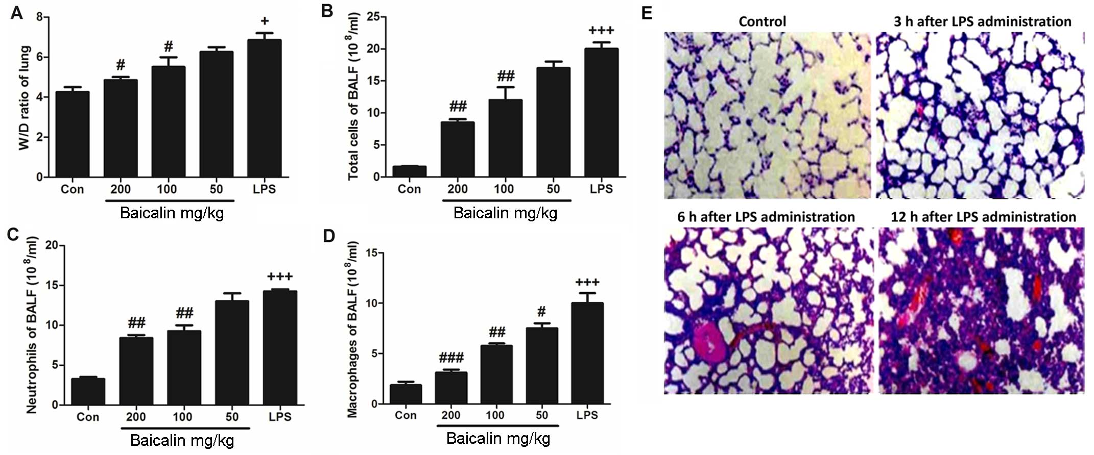
Effects of baicalin on lung wet-to-dry
weight ratio and inflammatory cell counts in BALF
We wished to determine whether the administration of
LPS affects the ratio of lung wet-to-dry weight. As shown in
Fig. 2A, the administration of
LPS induced a significant increase in the wet-to-dry ratio,
compared to the untreated control group. But in baicalin treatment,
it has ability to decrease the lung wet-to-dry ratio. However,
baicalin inhibited the LPS-induced increase in the lung wet-to-dry
weight in a dose-dependent manner. Furthermore, we also examined
the effects of baicalin on inflammatory cell counts in BALF. As
shown in Fig. 2B–D, treatment
with various concentrations of baicalin decreased the number of
inflammatory cells in BALF, including neutrophils and macrophages,
which was increased following the administration of LPS. Baicalin
decreased the number of inflammatory cells in BALF in a
dose-dependent manner.
Effects of baicalin on CX3CL1 protein
expression and NF-κB activation following LPS-induced lung
injury
In order to further examine the harmful effects of
LPS on lung tissues, LPS-induced lung injury was examined at
different time points (3, 6 and 12 h). As shown in Fig. 2E, the administration of LPS
significantly enhanced the injury levels to the lungs
(histopathological examination revealed blood capillary congestion,
necrosis and exfoliation epithelium of the pulmonary alveoli, and
the extravasation of fluid in lungs) compared to the control group
in a time-dependent manner. In addition, at the 6-h time point (6 h
post-LPS administration), we further examined whether treatment
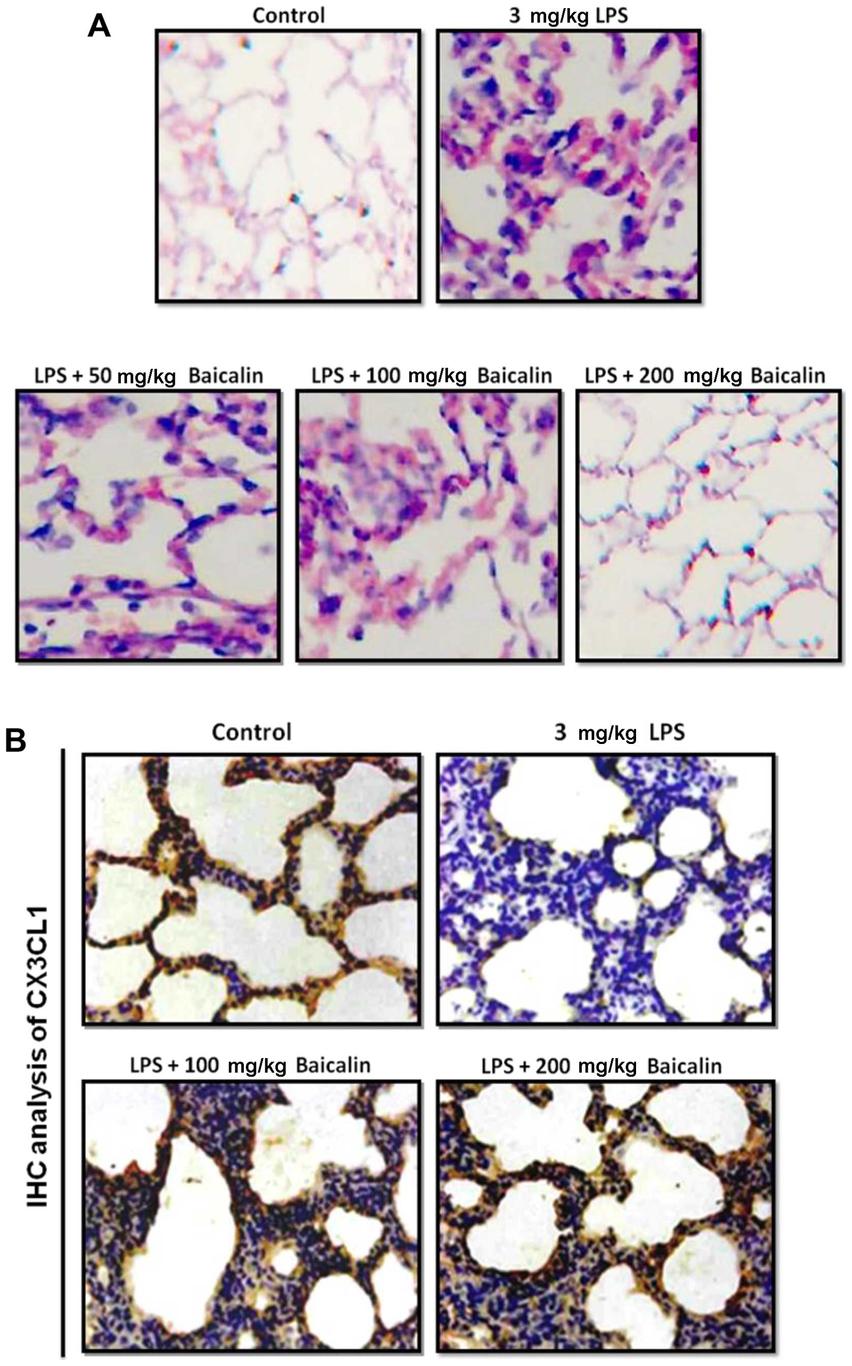
with baicalin would reduce the injury levels. As shown in Fig. 3A, treatment with baicalin
inhibited the LPS-induced lung injury in a dose-dependent manner.
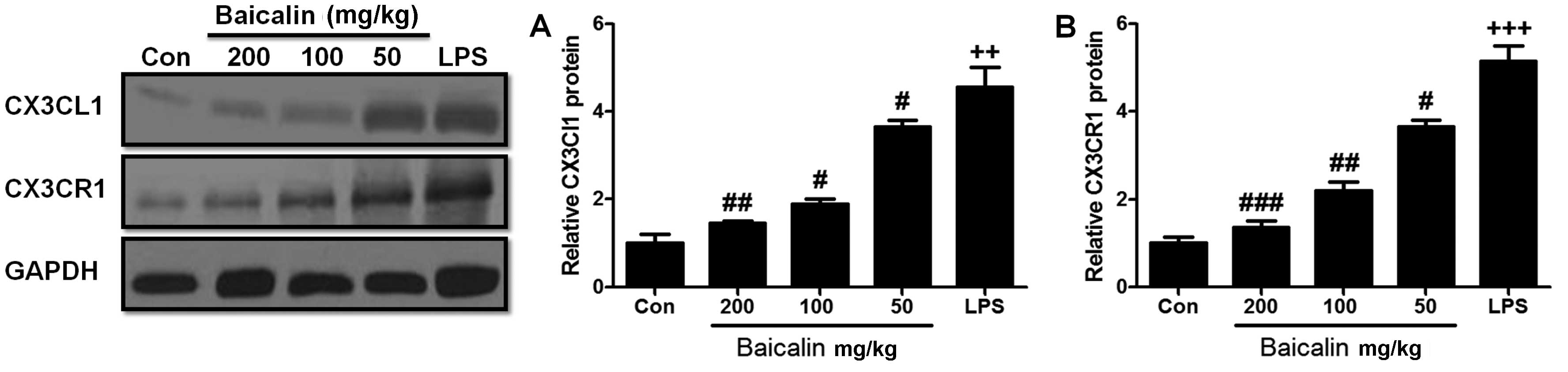
Furthermore, we examined CX3CL1 expression in the lung tissues of
mice with LPS-induced ALI (Figs.
3B and 4). The activation of
the CX3CL1-CX3CR1 axis was observed in the LPS-treated group, but
was inhibited in the baicalin-treated groups. As shown in Fig. 5, the levels of phosphorylated AKT,
NF-κB and COX-2 were significantly upregulated following the
administration of LPS, but were suppressed following treatment with
various concentrations of baicalin.
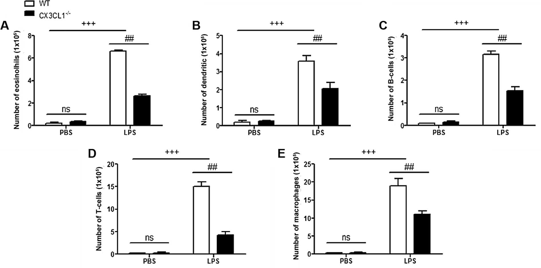
Inflammatory responses in
CX3CL1−/− mice with LPS-induced lung injury
In order to determine the role of CX3CL1 in the
pathogenesis of inflammation, WT and CX3CL1−/− mice were
sensitized and challenged with LPS. BALF was collected 48 h after
the final challenge and analyzed for the infiltration of
inflammatory cells. The administration of LPS to the WT mice
induced a significant increase in the number of inflammatory cells,
including eosinophils, dendritic cells, and lymphocytes and
macrophages, compared with non-LPS-challenged mice (Fig. 6). Among the inflammatory cell
populations, eosinophils were the most dominant, followed by
macrophages, T-cells, dendritic cells and B-cells. The number of
inflammatory cells, including the number of eosinophils and
lymphocytes in the LPS-challenged CX3CL1−/− mice was
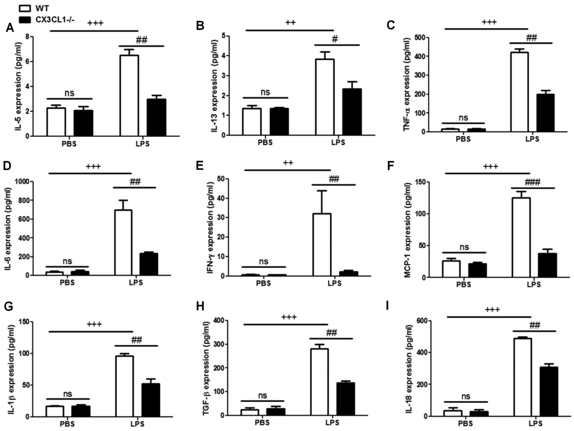
significantly lower than in the LPS-challenged WT mice. In
addition, the increase in the number of inflammatory cytokines in
the LPS-challenged CX3CL1−/− mice was significantly
inhibited, compared with the WT mice (Fig. 7). These results demonstrate that a
deficiency of CX3CL1 results in reduced inflammatory cell
infiltration in BALF following challenge with LPS. We also
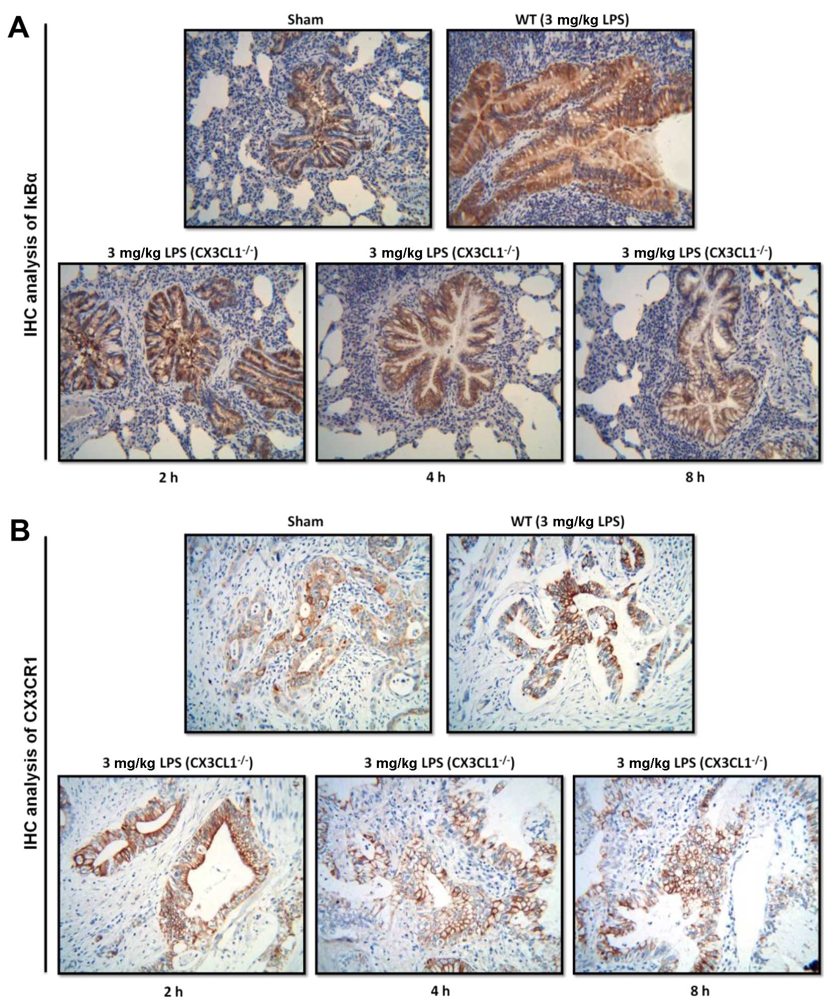
performed IHC analysis (Fig. 8)
to examine the changes in the expression levels of IκBα and CX3CR1
receptor following challenge with LPS. The CX3CL1−/−
mice exhibited lower expression levels of IκBα and CX3CR1 than the
WT mice. Of note, the decrease in the expression of IκBα and CX3CR1
was observed in a time-dependent manner. This illustrated that a
deficiency of CX3CL1 indirectly or directly affects the activation
of IκBα and CX3CR1 expression.
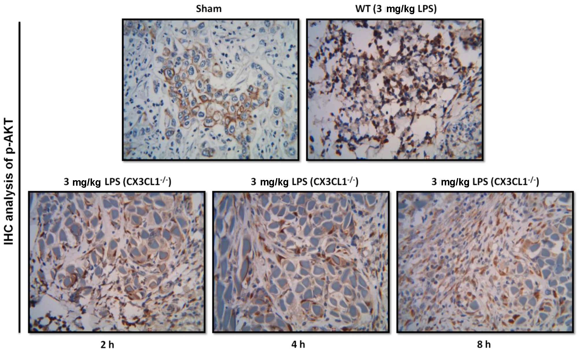
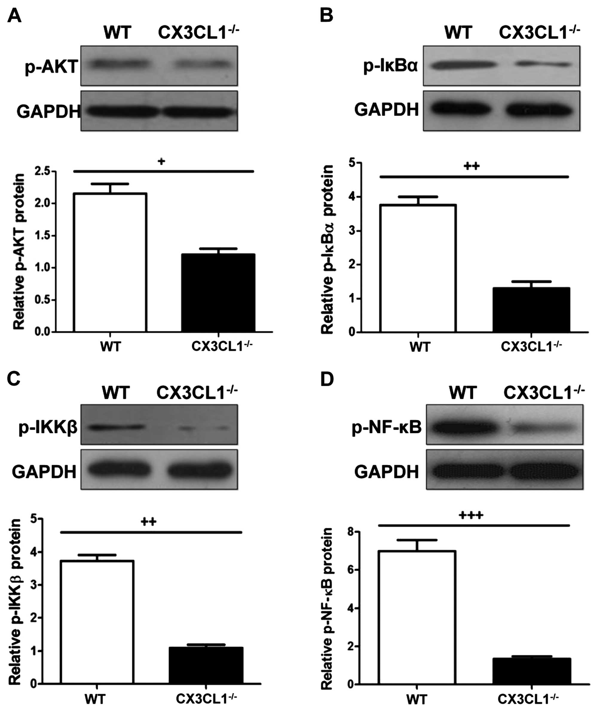
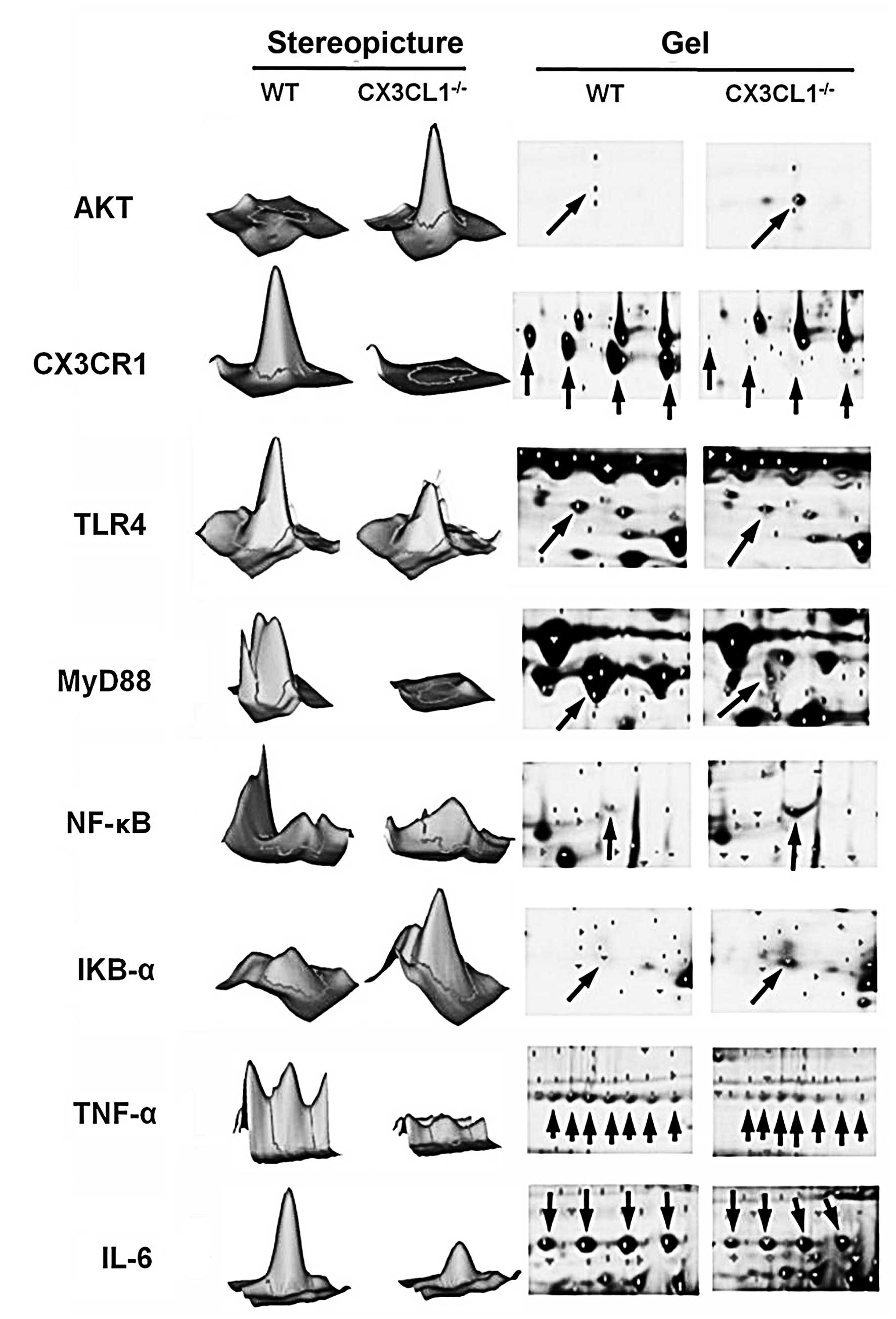
AKT-induced activation of the NF-κB
signaling pathway in CX3CL1−/− mice challenged with
LPS
As shown in Figs.
9, 10 and 11, IHC analysis demonstrated that the
AKT pathway was significantly activated following the
administration of LPS. In the CX3CL1−/− mice, the
phosphorylation levels of AKT were lower than those of the WT mice,
and the inhibition of the AKT pathway in the CX3CL1−/−
mice was observed in a time-dependent manner. In addition, the
results of western blot analysis indicated that the AKT-induced
activation of the TLR4/MyD88/NF-κB signaling pathway was
significantly inhibited in the CX3CL1−/− mice compared
with the WT mice. These findings suggest that the AKT pathway plays
a role in the development of LPS-induced lung inflammation, which
also directly involves the activation of the CX3CL1-CX3CR1 axis.
The AKT-induced activation of the NF-κB signaling pathway was
significantly inhibited in the CX3CL1−/− mice challenged
with LPS.
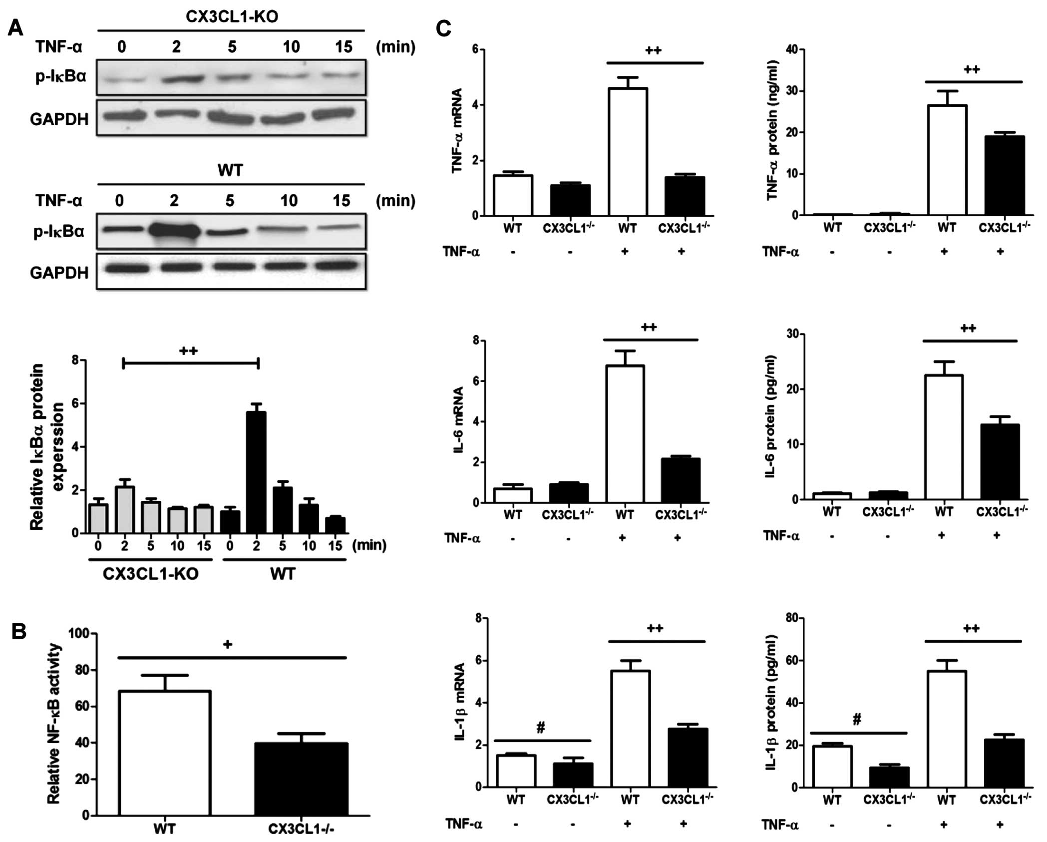
Deficiency of CX3CL1 inhibits the
inflammatory response through the NF-κB pathway in lung epithelial
cells
It has been demonstrated that the activation of the
NF-κB pathway is involved in lung-related diseases both in
experimental models and in humans (31). Moreover, the CX3CL1-CX3CR1 axis
has been shown to be activated in lung inflammation and to regulate
the NF-κB pathway in multiple cell systems (32). Thus, in this study, we wished to
examine the effects of the CX3CL1-CX3CR1 axis on the NF-κB pathway
and on the induction of inflammatory responses following the
administration of LPS. As shown in Fig. 12A, the IκBα phosphorylation
levels were significantly decreased in the CX3CL1-KO mice compared
with the WT mice. The increase in the IκBα phosphorylation level
resulted in the activation of the NF-κB pathway in the WT mice
compared to the CX3CL1-KO mice (Fig.
12B). Of note, in the epithelial cells in which CX3CL1 was
silenced, the levels of inflammatory cytokines were a lower than
the WT group (Fig. 12C). Thus,
these findings demonstrate that CX3CL1 plays a key role in
LPS-induced inflammatory responses.
Discussion
ALI as a critical illness consisting of hypoxemic
respiratory failure with bilateral pulmonary infiltrates, and poses
a serious threat to the health and lives populations worldwide
(1,11). Moreover, as with acute respiratory
distress syndrome, the formation and development of ALI are
associated with high morbidity and mortality. As previously
demonstated, some natural products have been used in animal
experiments and in clinical studies in an aim to prevent ALI, with
some promising results (2,4).
However, the detailed underlying molecular mechanisms controlling
lung injury are not yet fully understood. Effective treatments for
lung-related diseases have yet to be developed. Baicalin, as a type
of flavonoid, can be found and extracted from natural plants. It
has been shown to exert anti-hypertensive, antitumor and
anti-inflammatory effects (17,18). Thus, in this regard, we wished to
determine whether baicalin has the ability to suppress LPS-induced
ALI and to elucidate the molecular mechanisms through which it
inhibits lung damage.
Firstly, we used BALB/c mice to construct a model of
LPS-induced ALI in order to examine the protective effects of
baicalin. Our findings indicated that treatment with baicalin
inhibited the release of inflammatory cytokines, including TNF-α,
IL-1β, IL-6, TGF-β and IL-18 in serum following the administration
of LPS. Moreover, the inflammatory cell counts in BALF obtained
from mice with LPS-induced lung injury were significantly reduced
following treatment with various concentrations of baicalin.
Treatment with baicalin also restored the lung wet-to-dry weight
ratio. Furthermore, we examined the crucial role of the
CX3CL1/CX3CR1 axis in LPS-induced lung injury. The results revealed
that the CX3CL1/CX3CR1 axis is involved in the process of lung
injury and is activated by LPS. The activation of the CX3CL1/CX3CR1
axis further increased the levels of NF-κB phosphorylation. As
previously reported, the activation of the NF-κB pathway leads to
inflammatory responses and cytokine production (33). In this study, the results of
western blot analysis provide evidence of this. The protective
effects of baicalin against LPS-induced lung injury occurred in a
concentration-dependent manner. Some researchers have also
indicated that the activation of the CX3CL1/CX3CR1 axis is often
associated with the activation of the AKT pathway (34). Thus, we examined the association
of the of AKT pathway with the CX3CL1/CX3CR1 axis. Our findings
demonstrated that the administration of LPS to mice activated the
AKT pathway and increased the phosphorylation levels of AKT.
Phosphorylated AKT significantly activated the CX3CL1/CX3CR1 axis
and the NF-κB pathway. Treatment with various concentrations of
baicalin inhibited the activation of the NF-κB pathway, the
CX3CL1/CX3CR1 crosstalk with other proteins, as well as the
phosphorylation of AKT.
In addition, in order to examine the possible role
of the CX3CL1/CX3CR1 axis in LPS-induced lung injury in mice, we
used CX3CL1-KO mice as a model to examine the effects of the
crosstalk between the CX3CL1-CX3CR1 axis and NF-κB pathway. IHC
analysis of IκB at different time points (2, 4 and 8 h) of LPS
administration indicated that IκB was involved in the development
of LPS-induced lung injury in a time-dependent manner. Compared
with the WT mice, the CX3CL1−/− mice exhibited a
decreased in the levels of phosphorylated IκBα in a time-dependent
manner. As the receptor of CX3CL1, we examined CX3CR1 expression
between the WT and CX3CL1−/− mice by IHC analysis. The
results indicated that the knockdown of CX3CL1 directly affects the
expression of CX3CR1. The high expression of CX3CL1 induces the
expression of CX3CR1. These data are consistent with those of
another published study (35). As
we mentioned above, the AKT signaling pathway may directly or
indirectly be associated with LPS-induced lung injury. In the
knockout mice, the expression of phosphorylated AKT was determined
to be involved in the process of injury. These data indicated that
AKT (upstream of the CX3CL1-CX3CR1 axis) may affect CX3CL1-mediated
inflammatory responses. The inhibition of the activation of AKT may
also suppress CX3CL1-CX3CR1 activation.
In conclusion, ALI as a serious diseases with a high
mortality and poses a threat to human health and life. Nowadays,
chemoprophylaxis using natural products isolated from plants and
flora is a safe way of preventing a number of diseases, including
hepatitis, cancer, diabetes, cardiac-cerebral vascular disease,
kidney injury and lung injury (36,37). As regards lung injury, a number of
drugs have been shown to exert anti-inflammatory effects (37); however, the molecular mechanisms
responsible for the development of lung injury are not yet fully
understood. Thus, in this regard, we used a mouse model of
LPS-induced lung injury to determine the role of the CX3CL1-CX3CR1
axis and its related indicators in the development of lung injury,
as well as the anti-inflammatory effects of baicalin. We found that
baicalin, as a natural product isolated from the genus
Scutellaria, exerts potent anti-inflammatory effects by
inhibiting the activation of the CX3CL1-CX3CR1 axis and NF-κB, thus
preventing the crosstalk between the CX3CL1-CX3CR1 axis and NF-κB
pathway. In addition, the phosphorylation of AKT was significantly
increased following LPS-induced lung injury through the
CX3CL1-CX3CR1 axis, and was inhibited by treatment with baicalin.
We also examined the role of the CX3CL1-CX3CR1 axis in the
development of lung injury. We examined the differences in the
expression of CX3CR1 between WT and CX3CL1−/− mice with
LPS-induced lung injury. Our results indicated that CX3CL1 may be
the central and major indicator in the process of lung injury,
which mediates the CX3CR1 receptor and activates the AKT pathway
and further promotes the activation of NF-κB.
References
|
1
|
Phua J, Badia JR, Adhikari NK, Friedrich
JO, Fowler RA, Singh JM, Scales DC, Stather DR, Li A, Jones A, et
al: Has mortality from acute respiratory distress syndrome
decreased over time?: a systematic review. Am J Respir Crit Care
Med. 179:220–227. 2009. View Article : Google Scholar
|
|
2
|
Rubenfeld GD, Caldwell E, Peabody E,
Weaver J, Martin DP, Neff M, Stern EJ and Hudson LD: Incidence and
outcomes of acute lung injury. N Engl J Med. 353:1685–1693. 2005.
View Article : Google Scholar : PubMed/NCBI
|
|
3
|
Muñoz NM, Meliton AY, Meliton LN, Dudek SM
and Leff AR: Secretory group V phospholipase A2 regulates acute
lung injury and neutrophilic inflammation caused by LPS in mice. Am
J Physiol Lung Cell Mol Physiol. 296:L879–L887. 2009. View Article : Google Scholar : PubMed/NCBI
|
|
4
|
Xu XL, Xie QM, Shen YH, Jiang JJ, Chen YY,
Yao HY and Zhou JY: Mannose prevents lipopolysaccharide-induced
acute lung injury in rats. Inflamm Res. 57:104–110. 2008.
View Article : Google Scholar : PubMed/NCBI
|
|
5
|
Matthay MA and Zemans RL: The acute
respiratory distress syndrome: Pathogenesis and treatment. Annu Rev
Pathol. 6:147–163. 2011. View Article : Google Scholar :
|
|
6
|
Ware LB and Matthay MA: The acute
respiratory distress syndrome. N Engl J Med. 342:1334–1349. 2000.
View Article : Google Scholar : PubMed/NCBI
|
|
7
|
Li B, Yang J, Huang Q, Zhang Y, Peng C,
Zhang Y, He Y, Shi J, Li W, Hu J, et al: Biodistribution and
pulmonary toxicity of intratracheally instilled graphene oxide in
mice. NPG Asia Mater. 5:e442013. View Article : Google Scholar
|
|
8
|
Spragg RG, Bernard GR, Checkley W, Curtis
JR, Gajic O, Guyatt G, Hall J, Israel E, Jain M, Needham DM, et al:
Beyond mortality: future clinical research in acute lung injury. Am
J Respir Crit Care Med. 181:1121–1127. 2010. View Article : Google Scholar : PubMed/NCBI
|
|
9
|
Lam CW, James JT, McCluskey R and Hunter
RL: Pulmonary toxicity of single-wall carbon nanotubes in mice 7
and 90 days after intratracheal instillation. Toxicol Sci.
77:126–134. 2004. View Article : Google Scholar
|
|
10
|
Song Y, Fukuda N, Bai C, Ma T, Matthay MA
and Verkman AS: Role of aquaporins in alveolar fluid clearance in
neonatal and adult lung, and in oedema formation following acute
lung injury: studies in transgenic aquaporin null mice. J Physiol.
525:771–779. 2000. View Article : Google Scholar : PubMed/NCBI
|
|
11
|
Martin TR and Matute-Bello G: Experimental
models and emerging hypotheses for acute lung injury. Crit Care
Clin. 27:735–752. 2011. View Article : Google Scholar : PubMed/NCBI
|
|
12
|
Reiss LK, Uhlig U and Uhlig S: Models and
mechanisms of acute lung injury caused by direct insults. Eur J
Cell Biol. 91:590–601. 2012. View Article : Google Scholar : PubMed/NCBI
|
|
13
|
Liu L, Gao Z, Xia C, Xu Y, Ma Z, Dong C
and Li B: Comparative study of trans-oral and trans-tracheal
intratracheal instillations in a murine model of acute lung injury.
Anat Rec (Hoboken). 295:1513–1519. 2012. View Article : Google Scholar
|
|
14
|
Su X, Bai C, Hong Q, Zhu D, He L, Wu J,
Ding F, Fang X and Matthay MA: Effect of continuous hemofiltration
on hemodynamics, lung inflammation and pulmonary edema in a canine
model of acute lung injury. Intensive Care Med. 29:2034–2042. 2003.
View Article : Google Scholar : PubMed/NCBI
|
|
15
|
Matthay MA, Ware LB and Zimmerman GA: The
acute respiratory distress syndrome. J Clin Invest. 122:2731–2740.
2012. View
Article : Google Scholar : PubMed/NCBI
|
|
16
|
Evers DL, Chao CF, Wang X, Zhang Z, Huong
SM and Huang ES: Human cytomegalovirus-inhibitory flavonoids:
studies on antiviral activity and mechanism of action. Antiviral
Res. 68:124–134. 2005. View Article : Google Scholar : PubMed/NCBI
|
|
17
|
Wan QF, Gu LG, Yin SJ, Qi GJ, Zhang S, Li
GM and Ge DY: Protection effect of baicalin on lung injury of mice
infected with influenza FM1. Chin J Tradit Chin Med Pharm.
2848–2851. 2011.In Chinese.
|
|
18
|
Wan QF, Gu LG, Yin SJ, Ge DY and Li GM:
Effect of baicalin on cell apoptosis FAS/FAS-L system of pneumonia
mice lung tissue infected with FM1. Chin Pharmacol Bull.
27:1555–1559. 2011.In Chinese.
|
|
19
|
Vergadi E, Vaporidi K, Theodorakis EE,
Doxaki C, Lagoudaki E, Ieronymaki E, Alexaki VI, Helms M, Kondili
E, Soennichsen B, et al: Akt2 deficiency protects from acute lung
injury via alternative macrophage activation and miR-146a induction
in mice. J Immunol. 192:394–406. 2014. View Article : Google Scholar
|
|
20
|
Balakrishna S, Song W, Achanta S, Doran
SF, Liu B, Kaelberer MM, Yu Z, Sui A, Cheung M, Leishman E, et al:
TRPV4 inhibition counteracts edema and inflammation and improves
pulmonary function and oxygen saturation in chemically induced
acute lung injury. Am J Physiol Lung Cell Mol Physiol.
307:L158–L172. 2014. View Article : Google Scholar : PubMed/NCBI
|
|
21
|
Wasmuth HE, Zaldivar MM, Berres ML, Werth
A, Scholten D, Hillebrandt S, Tacke F, Schmitz P, Dahl E,
Wiederholt T, et al: The fractalkine receptor CX3CR1 is involved in
liver fibrosis due to chronic hepatitis C infection. J Hepatol.
48:208–215. 2008. View Article : Google Scholar
|
|
22
|
Aoyama T, Inokuchi S, Brenner DA and Seki
E: CX3CL1-CX3CR1 interaction prevents carbon tetrachloride-induced
liver inflammation and fibrosis in mice. Hepatology. 52:1390–1400.
2010. View Article : Google Scholar : PubMed/NCBI
|
|
23
|
Isse K, Harada K, Zen Y, Kamihira T,
Shimoda S, Harada M and Nakanuma Y: Fractalkine and CX3CR1 are
involved in the recruitment of intraepithelial lymphocytes of
intrahepatic bile ducts. Hepatology. 41:506–516. 2005. View Article : Google Scholar : PubMed/NCBI
|
|
24
|
Zhuang ZY, Kawasaki Y, Tan PH, Wen YR,
Huang J and Ji RR: Role of the CX3CR1/p38 MAPK pathway in spinal
microglia for the development of neuropathic pain following nerve
injury-induced cleavage of fractalkine. Brain Behav Immun.
21:642–651. 2007. View Article : Google Scholar :
|
|
25
|
Lee KM, Jeon SM and Cho HJ: Interleukin-6
induces microglial CX3CR1 expression in the spinal cord after
peripheral nerve injury through the activation of p38 MAPK. Eur J
Pain. 14:682.e1–12. 2010. View Article : Google Scholar
|
|
26
|
Nishiyori A, Minami M, Ohtani Y, Takami S,
Yamamoto J, Kawaguchi N, Kume T, Akaike A and Satoh M: Localization
of fractalkine and CX3CR1 mRNAs in rat brain: does fractalkine play
a role in signaling from neuron to microglia? FEBS Lett.
429:167–172. 1998. View Article : Google Scholar : PubMed/NCBI
|
|
27
|
Andréasson U, Ek S, Merz H, Rosenquist R,
Andersen N, Jerkeman M, Dictor M and Borrebaeck CA: B cell
lymphomas express CX3CR1 a non-B cell lineage adhesion molecule.
Cancer Lett. 259:138–145. 2008. View Article : Google Scholar
|
|
28
|
Erreni M, Solinas G, Brescia P, Osti D,
Zunino F, Colombo P, Destro A, Roncalli M, Mantovani A, Draghi R,
et al: Human glioblastoma tumours and neural cancer stem cells
express the chemokine CX3CL1 and its receptor CX3CR1. Eur J Cancer.
46:3383–3392. 2010. View Article : Google Scholar : PubMed/NCBI
|
|
29
|
Rodero M, Marie Y, Coudert M, Blondet E,
Mokhtari K, Rousseau A, Raoul W, Carpentier C, Sennlaub F, Deterre
P, et al: Polymorphism in the microglial cell-mobilizing CX3CR1
gene is associated with survival in patients with glioblastoma. J
Clin Oncol. 26:5957–5964. 2008. View Article : Google Scholar : PubMed/NCBI
|
|
30
|
Babar IA, Czochor J, Steinmetz A, Weidhaas
JB, Glazer PM and Slack FJ: Inhibition of hypoxia-induced miR-155
radiosensitizes hypoxic lung cancer cells. Cancer Biol Ther.
12:908–914. 2011. View Article : Google Scholar : PubMed/NCBI
|
|
31
|
Dolcet X, Llobet D, Pallares J and
Matias-Guiu X: NF-kB in development and progression of human
cancer. Virchows Arch. 446:475–482. 2005. View Article : Google Scholar : PubMed/NCBI
|
|
32
|
McComb JG, Ranganathan M, Liu XH, Pilewski
JM, Ray P, Watkins SC, Choi AM and Lee JS: CX3CL1 up-regulation is
associated with recruitment of CX3CR1+ mononuclear
phagocytes and T lymphocytes in the lungs during cigarette
smoke-induced emphysema. Am J Pathol. 173:949–961. 2008. View Article : Google Scholar : PubMed/NCBI
|
|
33
|
Valdivia-Silva JE, Franco-Barraza J, Silva
AL, Pont GD, Soldevila G, Meza I and García-Zepeda EA: Effect of
pro-inflammatory cytokine stimulation on human breast cancer:
implications of chemokine receptor expression in cancer metastasis.
Cancer Lett. 283:176–185. 2009. View Article : Google Scholar : PubMed/NCBI
|
|
34
|
Shulby SA, Dolloff NG, Stearns ME, Meucci
O and Fatatis A: CX3CR1-fractalkine expression regulates cellular
mechanisms involved in adhesion, migration, and survival of human
prostate cancer cells. Cancer Res. 64:4693–4698. 2004. View Article : Google Scholar : PubMed/NCBI
|
|
35
|
Jamieson WL, Shimizu S, D'Ambrosio JA,
Meucci O and Fatatis A: CX3CR1 is expressed by prostate epithelial
cells and androgens regulate the levels of CX3CL1/fractalkine in
the bone marrow: potential role in prostate cancer bone tropism.
Cancer Res. 68:1715–1722. 2008. View Article : Google Scholar : PubMed/NCBI
|
|
36
|
Newman DJ and Cragg GM: Natural products
as sources of new drugs over the 30 years from 1981 to 2010. J Nat
Prod. 75:311–335. 2012. View Article : Google Scholar : PubMed/NCBI
|
|
37
|
Cragg GM and Newman DJ: Natural products:
a continuing source of novel drug leads. Biochim Biophys Acta.
1830:3670–3695. 2013. View Article : Google Scholar : PubMed/NCBI
|