Introduction
Lychee (Litchi in Chinese) is an edible fruit of the
Sapindaceae family in China and Southeast Asia and it is
botanically designated as Litchi chinensis Sonn (1,2).
Lychee seed is the dry mature seed of a lychee and used as a
traditional Chinese medicine named 'Li-zhi-he' in Chinese and was
recorded by the Bencao Yanyi (Development of Herbal Medicine) and
Bencao Gangmu (Compendium of Materia Medica) for regulating Qi,
dispelling cold, alleviating pain and relieving polydipsia
(3). Modern pharmacological
studies have identified that some components of lychee seed have
the effects of modulation of blood sugar by improving insulin
resistance (IR) (4,5), lowering blood lipids (6), antioxidation (7), antivirus (8), anti-tumor (9) and preventing liver injury (10).
Type II diabetes mellitus (T2DM) is one of the most
common chronic diseases in the world (11). T2DM is a metabolic disorder
characterized by hyperglycemia and its development is due to
pancreatic β cells failing to secrete sufficient amounts of insulin
to meet metabolic demand (12,13). T2DM causes various serious
complications of heart, eyes, nerves, liver and kidneys and is
associated with decrements in cognitive function and changes in
brain structure (14). IR in
pancreatic β cells contributes to the pathogenesis of T2DM
(13).
Alzheimer's disease (AD) is the major causative
disease of dementia and it is characterized pathologically by the
accumulation of senile plaques (SPs) and neurofibrillary tangles
(NFTs) in the brain (15).
Emerging evidence suggests that T2DM can contribute significantly
to the onset and/or progression of AD either directly or as a
cofactor (16). Numerous studies
have evidenced that T2DM is a major risk factor in the pathology of
AD, and that the two diseases share common biological mechanisms at
the molecular level including IR, impaired glucose metabolism,
β-amyloid (Aβ) formation, oxidative stress and presence of advanced
glycation end products (AGEs) (16–19). IR is likely to be the basis for a
variety of pathological changes in AD, and improvement of IR can
delay the onset and progression of AD (20,21).
Currently, the drugs for AD treatment can only
improve patients' cognition, however they do not slow the
progression or cure the disease (22). Development of drugs for the
treatment of AD have a very high failure rate due to lack of
therapeutic efficacy or emergence of serious adverse effects; no
new drug has been approved for AD treatment since 2003 (23). Therefore, discovery and
development of novel anti-AD drugs is urgently needed. Previously,
active ingredients from natural products and medicinal herbs for
treatment of AD and/or diabetes mellitus have attracted substantial
attention. Studies have shown that some natural products have
neuroprotective effects (24,25). The aqueous extract from lychee
seed improved the ability of learning and memory in mice (26). The saponins isolated and extracted
from lychee seeds exhibited an anti-diabetic effect (27). Our preliminary unreported data
also showed that saponins from lychee seeds significantly relieved
cognitive dysfunction by improving IR in rats. In the present
study, we analyzed the chemical profile of lychee seed extract
(LSE) by ultrahigh performance liquid chromatography (UHPLC)-SPD.
In order to study the effects of LSE on neuroprotection and
cognitive function improvement, we also established a rat model of
T2DM with cognitive impairment and investigated the ability of
spatial learning and memory in rats by Morris water maze. In
addition, the effects of LSE on neuropathology of the neurons were
studied by pathohistological analysis. Furthermore, to explore the
possible mechanisms associated with the effects of LSE on
neuroprotection and cognitive function improvement, the levels of
glucose, insulin, Aβ, acetylcholinesterase (AChE), AGEs, and Tau
protein in blood and/or hippocampus were determined by
blood-glucose meter, radioimmunoassay, chemical chromatometry,
enzyme-linked immunosorbent assay (ELISA) and immunohistochemical
analysis, respectively.
Materials and methods
Reagents
Donepezil hydrochloride tablets (Aricept, cat. no.
H20070181) were purchased from Eisai China Inc. (Chengdu, China),
and prepared to a mixed suspension at the concentration of 0.1
mg/ml as a positive control. Streptozotocin (STZ; cat. no. S0130)
was purchased from Sigma-Aldrich; Merck KGaA (Darmstadt, Germany).
Aβ release analysis kit (cat. no. 200805) and serum insulin
radioimmunoassay kit (cat. no. 200808) were purchased from the
Chinese PLA General Hospital (Beijing, China). Tau protein assay
kit (cat. no. BM3928) was purchased from Wuhan Boster Biological
Technology, Ltd. (Hubei, China). AChE assay kit (cat. no. A024) was
purchased from Nanjing Jiancheng Bioengineering Institute (Jiangsu,
China). AGEs assay kit (cat. no. CEB353Ge) was purchased from Wuhan
USCN Business Co., Ltd. (Hubei, China). Morris water maze system
(MT-2000) was purchased from Chengdu Techman Software Co., Ltd.
(Sichuan, China).
Experimental animals
A total of 150 8–10-week old (body weight, 180–220
g) specific pathogen free grade male Sprague Dawley rats were
purchased from the Experimental Animal Centre, Sichuan Provincial
Academy of Medical Sciences (Chengdu, China; SCXK201302). All rats
were housed in sterile plastic cages (up to 4 rats per cage) with
free access to water and food ad libitum in a controlled room
temperature (22±1°C), humidity (50–70%) and under a 12-h light/dark
cycle. All animal experiments were performed strictly in accordance
with institutional guidelines and followed an approved protocol
(permit no. 250114) by the Committee on Use and Care of Animals of
Southwest Medical University (Sichuan, China). There were 10–12
rats for each experimental group.
Collection, extraction and isolation of
lychee seeds
Lychee seeds were purchased from a local market in
Luzhou, China, and were authenticated by Professor Can Tang of
School of Pharmacy, Southwest Medical University (Luzhou, China). A
total mass of 1,000 g of air-dried lychee seeds were ground and
soaked with 1,000 ml of 70% ethanol overnight, extracted by
percolation with 8,000 ml of 70% ethanol at the speed of 5
ml/min/kg. Following this, the solvents were evaporated under
vacuum, then the LSE was concentrated to dryness using a rotary
vacuum evaporator and collected to yield a total mass of 185.2 g
dried powder.
Analysis of chemical profile of LSE
The chemical profile of LSE was performed at 254 nm
by an LCMS-8040 UHPLC system (Shimadzu Corp., Kyoto, Japan).
Chromatographic analyses were performed at 45°C with an Inert
Sustain C18 column (2.0 μm particle size, 50×2.1 mm; GL
Sciences, Tokyo, Japan), using water: Formic acid (100:0.1, v/v)
and methanol as the mobile phase A and B, respectively. The mobile
phase was delivered at a rate of 0.25 ml/min with injection volume
of 1 μl. The gradient separation process was as follows:
15–15% B at 0–2.0 min, 15–80% B at 2.0–5.0 min, 80–80% B at 5.0–7.0
min, 80–15% B at 7.0–7.5 min. The data were analyzed by
LabSolutions software version 5.75 (Shimadzu Corp.).
Morris water maze test
The spatial learning and memory ability was
investigated using the Morris water maze test as described
previously (28,29). Briefly, the test was conducted in
a round white pool (94 cm in diameter and 31 cm deep) filled with
water (30 cm depth) with the temperature ~25°C. The escape platform
was a 25 cm2 Plexiglas square, placed in the center of
one quadrant of the pool, 15 cm from the pool's edge and submerged
1 cm beneath the water surface.
Hidden platform test: Each rat was trained in a
circular pool, which randomly divided into four equal quadrants by
a hidden platform with four trials for 120 sec per trial daily with
30 min intervals between the trials for 5 consecutive days. The
time for the rat to seek the platform was record by an online image
video tracking system and within 120 sec as the escape latency.
Spatial probe test: The platform was removed from
the pool after the hidden platform test, each rat was left to one
quadrant of the pool which was farthest from the primary platform.
The number of rats crossing the platform, the time spent in the
target quadrant and the percentage of time spent in the target
quadrant were recorded by a tracking system.
Establishment of the rat model of T2DM
with cognitive impairment
Rats were selected by Morris water maze test as the
standard of 40–120 sec to find the platform, the rat was eliminated
if it found the platform in <40 sec or more than 120 sec, as
described previously (29,30).
The selected rats were randomly divided into control group (n=12)
and study group (n=100). The rats in the control group were fed
with normal standard diet, while the rats in the study group were
fed with high fat, high sugar and high protein diet (HFSPD, food
ratio: carbohydrate 25%, protein 15.2%, fat (lard) 58.8%, and
sodium cholate 1% (31,32). The body weights of rats were
weighed every two weeks. Then, the rats in the study group were
intraperitoneally (IP) injected with STZ (27 mg/kg) once and the
rats in the control group were injected the same volume of citric
acid buffer for 8 weeks. The blood glucose and serum insulin of the
rats were measured 72 h following STZ injection and was calculated
by Homeostasis Model Assessment (HOMA = fasting blood glucose ×
fasting insulin/22.5) for IR index to judge development of T2DM.
The rats were then continuously fed the HFSP diet and weighed
weekly for 4 more weeks. Finally, the rats were further examined by
a Morris water maze test for cognitive impairment and other
experiments.
Drug preparation and administration
The T2DM rats with cognitive impairment were
randomly divided into five groups with 10 rats for each group and
treated with normal saline (NS) 1 ml/kg (negative control),
donepezil 0.42 mg/kg (positive control), LSE 0.7, 1.4 and 2.8 g/kg
by intragastric (IG) administration once a day (daily) at 8 a.m.
for 28 consecutive days. In addition, 10 normal rats on a normal
diet were given equal volume of NS as additional control. The dose
of donepezil was selected as previous described from literature
(33).
Measurement of blood glucose
The blood samples of rats were collected from the
tail veins, and blood glucose was measured by blood-glucose meter,
as described previously (34).
HOMA was calculated for IR index and the method of calculation is
described above.
Determination of the levels of glucose,
insulin, AChE, Aβ and AGEs in the blood and/or hippocampus of
rats
A total of 10 rats for each group were randomly
selected after Morris water maze test, then weighed and
anesthetized with 1% pentobarbital sodium (0.4 ml/kg) by IP
injection. Bloods were taken from the abdominal aorta for detection
of the levels of insulin, AChE and Aβ. The tissues of brain were
collected. The freshly dissected hippocampal tissues were isolated
to prepare hippocampal tissue homogenates for detection of the
contents of AChE, Aβ and AGEs. Insulin, AChE, Aβ and AGEs were
measured by serum insulin radioimmunoassay kit, AChE assay kit, Aβ
release analysis kit and AGEs assay kit, respectively.
Determination of the expression of Tau
protein in hippocampal neurons of rats by immunohistochemical
analysis
The expression of Tau protein in the hippocampus of
rats was carried out with immunohistochemical staining and examined
under microscope, as previous described (33). The positive rate of the expression
of Tau protein was calculated by GD-10.0 image analysis system. A
total of 10 rats were used for each group.
Histopathological examination of
hippocampus neuron CA1 pyramidal cells of rats with hematoxylin and
eosin (H&E) staining
The rats were anesthetized by IP injection of 1.0%
pentobarbital and perfusion with 4% paraformaldehyde to fix the
brains for 24 h after treatment. Then, the brains of rats were
removed and the brain tissues were weighed, fixed in 10% neutral
buffered formalin, dehydrated and embedded in paraffin for coronal
microtome sections (~4–5 μm) with H&E staining for the
study, as described previously (35). The sections were observed for
pathohistological changes under light microscope.
Statistical analysis
All the data were presented as mean ± standard
deviation (SD). Statistical differences of the data among the means
of two or more groups were analyzed by Student's t-test and/or
one-way univariate analysis of variance (ANOVA). A value of
P<0.05 was considered to indicate a statistically significant
difference, and a value of P<0.01 was considered to indicate a
highly statistically significant difference.
Results
The chemical profile of LSE
To determine the chemical profile of LSE isolated
and extracted from lychee seed, we studied its chemical fingerprint
at 254 nm by a UHPLC-SPD chromatogram and the data are presented in
Fig. 1. The data display the main
chemical compositions of LSE and indicate that it mainly consists
of eight major (Fig. 1) and
around 20 minor ingredients.
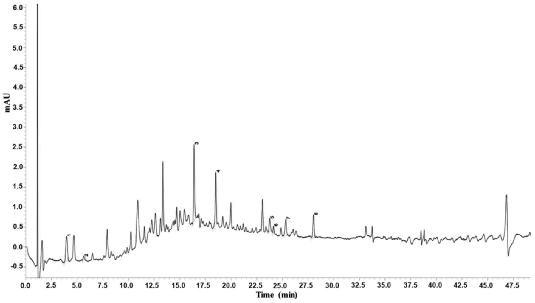 | Figure 1Analysis of the chemical profile of
LSE by a UHPLC-SPD-MS-MS chromatogram. The chemical fingerprint of
LSE was analyzed at 254 nm by a Shimadzu LCMS-8040 UHPLC system,
which comprised two LC-30AD pumps, a SIL-30AC autosampler with a
CTO-30AC column oven, a DGU-20A5 degasser and a Shimadzu CBM-20A
system controller. Chromatographic analyses were achieved at 45°C
with an InertSustain C18 column using water-formic acid (100:0.1,
v/v) and methanol as the mobile phase A and B, respectively. The
delivered rate of mobile phase was 0.25 ml/min with the injection
volume of 1 μl. For the gradient separation, the process was
as follows: 15–15% B at 0–2.0 min, 15–80% B at 2.0–5.0 min, 80–80%
B at 5.0–7.0 min, and 80–15% B at 7.0–7.5 min. The data were
analyzed by LabSolutions software (version 5.75). The major
ingredients of LSE are indicated as 1–8. The numbers indicate the
following substances: 1, adenosine; 2, 5-hydroxymethyluridine; 3,
4-p-coumaroylquinic acid; 4, procyanidin B; 5, procyanidin A; 6,
5′-β-D-glucopyranosyloxy jasmonic acid; 7, 4-O-(trans-p-coumaroyl)
quinic acid; and 8, procyanidin tetramer. LSE, lychee seed extract;
UHPLC, ultrahigh performance liquid chromatography. |
Establishment of a rat model of T2DM with
cognitive impairment
To establish the rat model of T2DM with cognitive
impairment, the rats were initially fed with HFSP diet for 8 weeks
and observed the general condition including food and water
intakes, hair color, activities and body weight changes and
compared to those of the rats with normal standard diet (control).
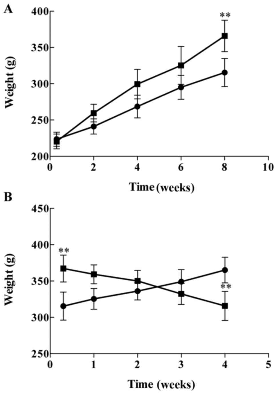
These was no difference in general condition between the two
groups, however, the body weights of the rats with HFSP diet were
significantly increased (P<0.01) compared to those of control
rats (Fig. 2A). Then, the rats
were injected with a single dose of STZ (27 mg/kg, IP ×1) after 8
weeks on the HFSP diet. Interestingly, the food and water intakes,
as well as urine volumes were gradually increased, while the body
weights of the rats were decreased (P<0.01) following injection
with STZ compared to the normal control rats (Fig. 2B), indicating the rats displayed
the symptoms of T2DM.
Next, the blood glucose and insulin levels were
measured and HMOA was calculated in the T2DM rats and compared to
those of the normal rats (control); the results are illustrated in
Fig. 3. The data show that there
were no significant differences of blood glucose (Fig. 3A), insulin (Fig. 3B) and HMOA (Fig. 3C) between the rats on the HFSP
diet and the rats on regular standard diet (P>0.05) in the 8
weeks before STZ injection as 6.33±3.01 vs. 6.57±3.21 mmol/l,
8.57±3.18 vs. 9.14±2.66 μIU/l, and 2.1±0.43 vs. 2.67±0.38,
respectively. However, the blood glucose, insulin and HMOA were
increased significantly (P<0.01) following STZ injection in the
rats on the HFSP diet compared to those of the rats on regular
standard diet without STZ treatment, as 3.62-fold (22.93±3.74 vs.
6.34±2.81 mmol/l), 4.95-fold (44.23±5.21 vs. 8.94±2.95
μIU/l), and 17.89-fold (45.08±0.87 vs. 2.52±0.37) increases,
respectively. These data indicated that the rat model of T2DM was
successfully established (Fig.
3A–C).
 | Figure 3Effects of HFSP diet alone and in
combination with STZ on (A) serum glucose (white bars, control
rats; black bars, T2DM rats), (B) insulin (white bars, control
rats; black bars, T2DM rats), (C) HOMA (white bars, control rats;
black bars, T2DM rats) and (D) escape latency of the normal
(control, ●; and T2DM, ■) rats. The rats were fed with HFSP diet
for 8 weeks, then injected a single dose of STZ (27 mg/kg,
intraperitoneal injection) and fed the HFSP diet for another 4
weeks. There were 10 rats used for each experimental group. The
results are presented as mean ± standard deviation.
**P<0.01 vs. STZ group; #P<0.01 vs. before HFSP
diet. HFSP, high fat, high sugar and high protein; STZ,
streptozocin; HOMA, homeostasis model assessment. |
Finally, we evaluated the cognitive function of T2DM
rats by a navigation test starting 1 h after STZ treatment during
the first 5 days to see whether the cognition was impaired or not
compared to the normal rats (control) and the results are presented
in Fig. 3D. The escape latency of
T2DM rats was slightly increased but no statistically significant
differences were observed in the first and the second days after
SZT treatment. However, the escape latency of T2DM rats was
significantly higher than that of the normal rats on the days 3–5
(P<0.01). In the last navigation test (on day 5), the escape
latency was 22.35±5.12 sec (Fig.
3A) for normal rats and 59.42±5.32 sec (Fig. 3B) for T2DM rats, respectively.
Therefore, the number was 62.39% by the calculation formula of
(B-A)/B%, suggesting that T2DM rats have obvious cognitive
impairment according to the judgment standard of dementia criteria
of (B-A)/B% >20% (36).
Effects of LSE on the cognitive function
of the T2DM rats with cognitive impairment
Following successfully establishing the rat model of
T2DM with cognitive impairment, the effects of LSE on cognitive
functions of the T2DM rats were investigated using Morris water
maze test, a commonly used method for assessing cognitive
functions, and compared to that of NS (as negative control) and
donepezil (as positive control) treatments, as well as the normal
rats treated with NS. The results are illustrated in Fig. 4. First, the Hidden platform test
was performed in the normal rats and T2DM rats on the day 1–5 after
STZ treatment to investigate the ability of learning and memory. As
presented in Fig. 4A, the escape
latency time of the T2DM rats was significantly increased compared
to that of the normal rats (P<0.01). However, LSE at the doses
of 0.7, 1.4 and 2.8 g/kg/day and donepezil at 0.42 mg/kg/day by
intragastric administration for 28 consecutive days significantly
shortened the escape latency compared to NS treatment (P<0.01)
in the T2DM rats (Fig. 4A).
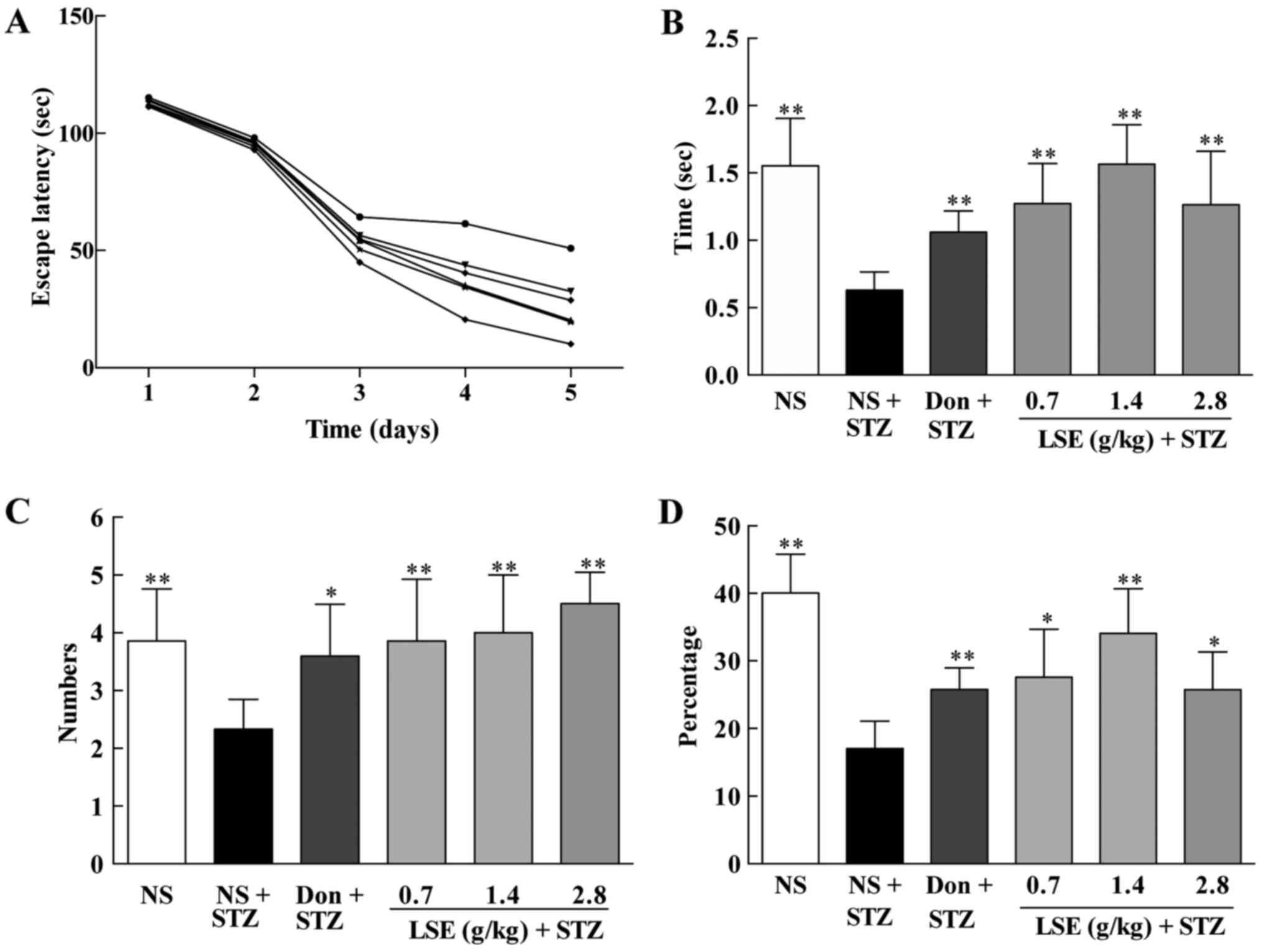 | Figure 4Effects of LSE on the ability of
spatial learning and memory in T2DM rats with cognitive impairment
induced by high fat, high sugar and high protein diet and STZ (27
mg/kg, intraperitoneal ×1). The normal control rats were treated
with NS (1 ml/kg/day), and T2DM rats were treated with NS (1
ml/kg/day), Don (0.42 mg/kg/day) or LSE (0.7, 1.4 and 2.8 g/kg/day
×28, IG). The cognitive function was assessed by Morris water maze
test. (A) The escape latency of rats in days 1–5 after STZ
injection. Normal control rats (■) treated with NS (1 ml/kg/day, IG
×28); T2DM rats (●) treated with NS (1 ml/kg/day, IG ×28); T2DM
rats (▲) treated with donepezil (0.42 mg/kg/day, IG ×28); T2DM rats
(▼) treated with LSE (0.7 g/kg/day, IG ×28); T2DM rats (◆) treated
with LSE (1.4 g/kg/day, IG ×28); and T2DM rats (*) treated with LSE
(2.8 g/kg/day, IG ×28). (B) The time that rat spent in target
quadrant; (C) the number of rats crossing the platform; and (D) the
percentage of time spent in the target quadrant. (B–D) The rats
were assessed on day 5 after STZ treatment. There were 10 rats used
for each experimental group and results are expressed as mean ±
standard deviation. *P<0.05 vs. T2DM rats treated
with NS; **P<0.01 vs. T2DM rats treated with NS. NS,
normal saline; IG, intragastric; LSE, lychee seed extract; T2DM,
type 2 diabetes mellitus; STZ, streptozotocin; Don, donezapil. |
Next, the spatial probe test was performed for the
rats. Similarly, the residence time (Fig. 4B), the numbers of crossing
platform (Fig. 4C), and the
percentage of time spent in the target quadrant (Fig. 4D) were significantly shortened in
the T2DM rats (P<0.01) compared to those of the normal rats. LSE
(0.7, 1.4 and 2.8 g/kg/day ×28 days) significantly increased the
residence time (Fig. 4B), the
numbers of those crossing the platform (Fig. 4C), and the the percentage of time
spent in the target quadrant (Fig.
4D) compared to NS treatment (P<0.01) in the T2DM rats. In
addition, donepezil (0.42 mg/kg/day ×28 days) has similarly effect
on the spatial learning and memory of the T2DM rats as LSE at the
dose of 2.8 g/kg/day.
The detailed classical path of the latency to get to
the platform for the normal rats treated with NS and the T2DM rats
treated with NS, donepezil or LSE during the acquisition trials are
shown in Fig. 5. The data
indicate that LSE and donepezil can improve the ability of spatial
learning and memory of the T2DM rats (Figs. 4 and 5).
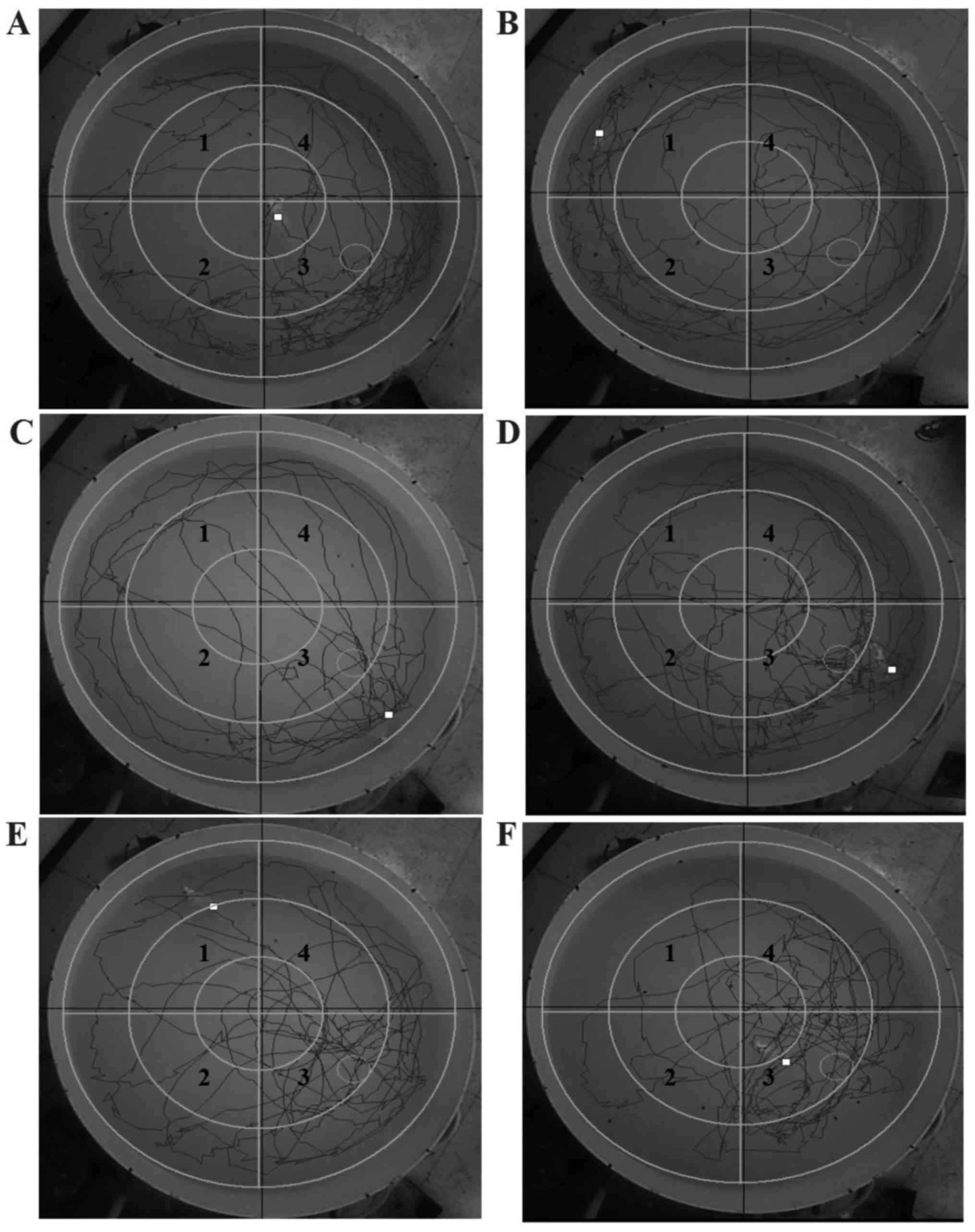 | Figure 5Effects of LSE on the ability of
spatial learning and memory in type 2 diabetes mellitus rats with
cognitive impairment induced by HFSP diet and (STZ, 27 mg/kg,
intraperitoneal ×1). Detail running tracts: (A) Normal rats on
regular diet (control) treated with NS (1 ml/kg/day, IG ×28); (B)
rats on HFSP diet and treated with STZ and NS (1 ml/kg/day, IG
×28); (C) rats on HFSP diet and treated with STZ and donepezil
(0.42 mg/kg/day, IG ×28); (D) rats on HFSP diet and treated with
STZ and LSE (0.7 g/kg/day, IG ×28); (E) rats on HFSP diet and
treated with STZ and LSE (1.4 g/kg/day, IG ×28); and (F) rats on
HFSP diet and treated with STZ and LSE (2.8 g/kg/day, IG ×28).
There were 10 rats used for each experimental group. 1, First
quadrant; 2, beta quadrant; 3, third quadrant; 4, delta quadrant.
LSE, lychee seed extract; HFSP, high fat, high sugar and high
protein; STZ, streptozotocin; NS, normal saline; IG,
intragastric. |
Effects of LSE on the blood glucose,
insulin and HMOA in T2DM rats
The development of T2DM is due to pancreatic β cells
failed to secrete sufficient insulin to meet metabolic demand to
cause hyperglycemia (12). T2DM
and AD share common biological mechanisms, including IR and
impaired glucose metabolism (16). Therefore, we examined the effects
of LSE on blood glucose, insulin and HMOA in the T2DM rats, and the
results are shown in Fig. 6. The
blood glucose (Fig. 6A), insulin
(Fig. 6B) and HMOA (Fig. 6C) levels of the T2DM rats were
significantly higher than those of the normal rats (P<0.01) as
24.35±3.30 vs. 6.34±2.81 mmol/l, 42.42±4.88 vs. 8.94±2.95
μIU/l and 45.90±0.72 vs. 2.52±0.37, respectively. There are
3.84-, 4.74-fold and 18-, 21-fold increases in blood glucose,
insulin and HMOA in the T2DM rats compared to normal rats,
respectively.
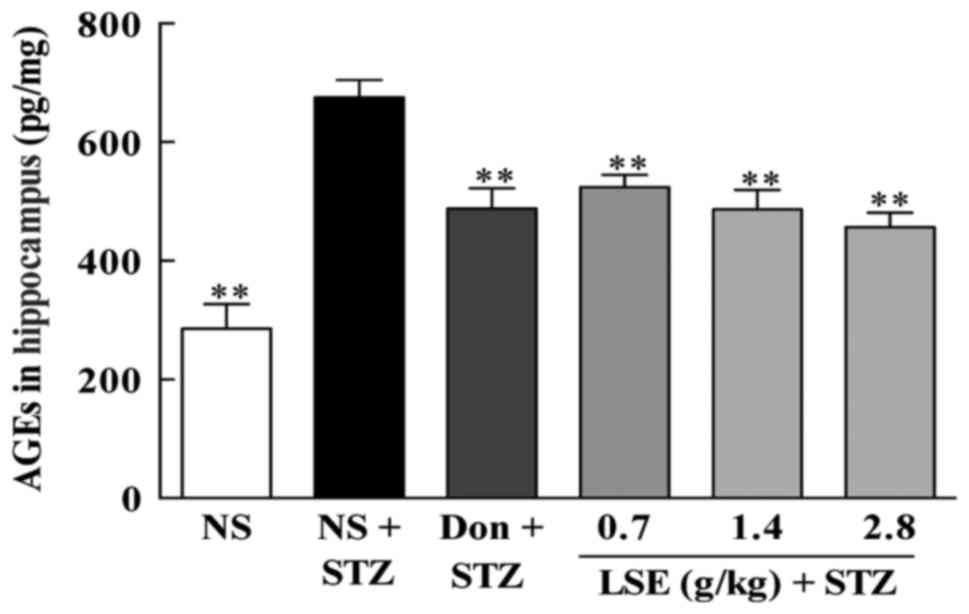
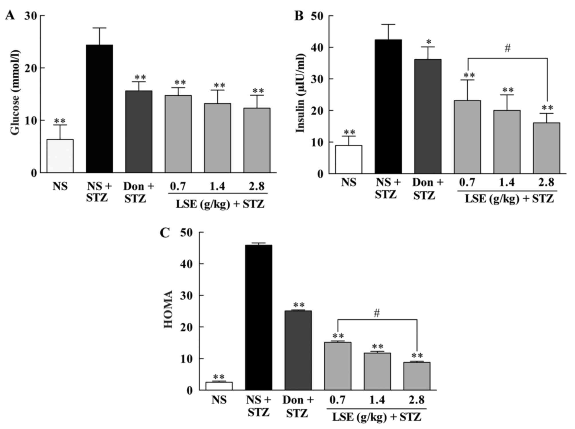 | Figure 6Effects of LSE on the (A) blood
glucose, (B) insulin and (C) HOMA in T2DM rats. The normal rats
were treated with NS (1 ml/kg/day, IG ×28) and the T2DM rats
induced by high fat, high sugar and high protein diet and STZ (27
mg/kg, intraperitoneal ×1) were treated with NS (1 ml/kg/day, IG
×28), Don (0.42 mg/kg/day, IG ×28) or LSE (0.7, 1.4 and 2.8
g/kg/day, IG ×28). There were 10 rats used for each experimental
group and are expressed as the mean ± standard deviation.
*P<0.05 vs. T2DM rats treated with NS;
**P<0.01 vs. T2DM rats treated with NS;
#P<0.01 vs. LSE 0.7 g/kg group. LSE, lychee seed
extract; T2DM, type 2 diabetes mellitus; HOMA, homeostasis model
assessment; NS, normal saline; STZ, streptozotocin; Don, donepezil;
IG, intragastric. |
However, LSE at the doses of 0.7, 1.4 and 2.8
g/kg/day significantly decreased the blood glucose (14.74±1.50,
13.19±2.57 and 12.33±2.46 mmol/l), insulin (23.09±6.54, 20.04±4.93
and 16.09±3.00 μIU/l), and HMOA (15.13±0.44, 11.75±0.56 and
8.82±0.33) in a dose-dependent manner (Fig. 6) compared to NS treatment in the
T2DM rats (P<0.01), although the reductions did not reach the
same level in normal rats. Interestingly, donepezil (4.2 mg/kg/day)
also significantly decreased the blood glucose (15.60±1.77
μIU/l), insulin (36.19±3.95 μIU/l) and HMOA
(25.09±0.31) in the T2DM rats compared to NS treatment (P<0.01),
but it was less effective compared to those of LSE treatment even
at the lowest dose of 0.7 g/kg/day (Fig. 6). These data indicate that LSE and
donepezil are efficacy to decrease elevated blood glucose and
insulin to overcome IR in T2DM rats, and that LSE is more effective
than that of donepezil.
Effects of LSE on AChE and Aβ in the
blood and hippocampus of T2DM rats
It has been found that AD is associated with
reduction of cholinergic neurons activity (37). AChE inhibitors such as donepezil
reduce the rate of acetylcholine degradation to increase its
concentration in the brain to execute their anti-AD effect.
Therefore, we evaluated the concentrations of AChE in the blood and
in the hippocampus of T2DM rats and compared to that of the normal
rats (Fig. 7A and B).
Interestingly, the concentrations of AChE were significantly
increased (P<0.01) in the blood (14.08±1.55 vs. 5.34±0.99
μmol/ml) but markedly decreased (P<0.01) in the
hippocampus (0.51±0.14 vs. 2.66±0.37 U/mg) in the T2DM rats
compared to that of normal control rats. However, LSE at the doses
of 0.7, 1.4 and 2.8 g/kg/day significantly decreased (P<0.01)
the concentrations of AChE in the blood (9.19±0.90, 8.57±1.00 and
7.51±0.75 μmol/ml, respectively), while increased
(P<0.01) the concentrations of AChE in the hippocampus
(1.10±0.24, 1.13±0.43 and 1.22±0.40 U/mg, respectively) compared to
NS treatment in the T2DM rats (Fig.
7A and B). The AChE inhibitor donepezil also significantly
decreased (P<0.01) the concentrations of AChE in the blood
(5.94±1.04 μmol/ml) and increased (P <0.01) the
concentrations of AChE in the hippocampus (1.77±0.47 U/mg).
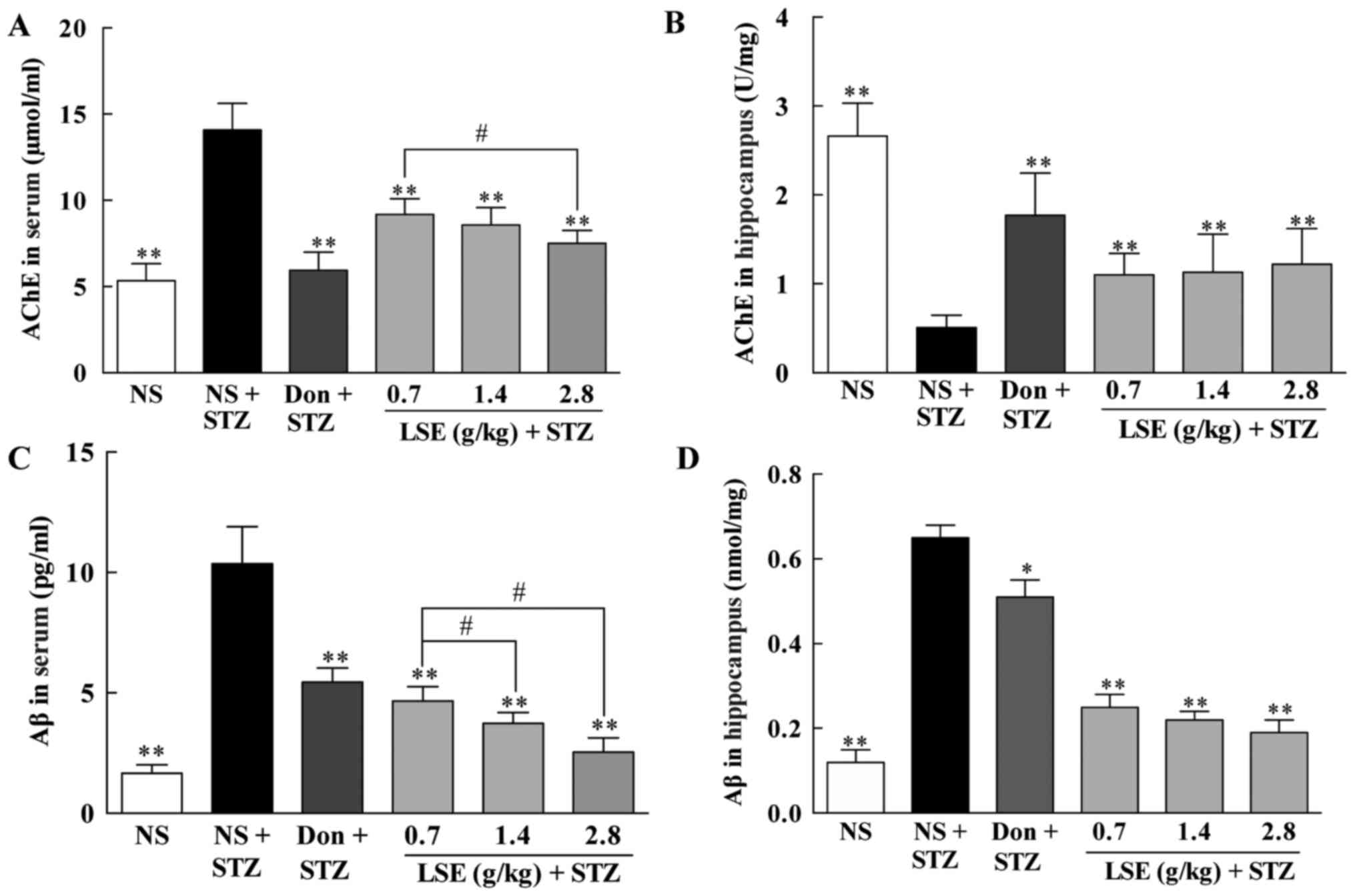 | Figure 7Effects of LSE on AChE (A and B) and
Aβ (C and D) in the blood and the hippocampus of T2DM rats. The
normal rats were treated with NS (1 ml/kg/day), and T2DM rats
induced by high fat, high sugar and high protein diet and (STZ, 27
mg/kg, intraperitoneal ×1) were treated with NS (1 ml/kg/day), Don
(0.42 mg/kg/day), or LSE (0.7, 1.4 and 2.8 g/kg/day, IG ×28 days).
There were 10 rats used for each experimental group and are
expressed as the mean ± standard deviation. *P<0.05
vs. T2DM rats treated with NS; **P<0.01 vs. T2DM rats
treated with NS; #P<0.01 vs. LSE 0.7 g/kg group. LSE,
lychee seed extract; AChE, acetylcholinesterase; NS, normal saline;
STZ, streptozotocin; Don, donepezil; NS, normal saline. |
One of the histopathological hallmarks of AD is
deposition of Aβ plaques. Therefore, the effect of LSE on Aβ in the
blood and hippocampus of T2DM rats was studied and compared to that
of donepezil; the results are illustrated in Fig. 7C and D. The concentrations of Aβ
are significantly increased (P<0.01) in the blood and in the
hippocampus in T2DM rats compared to that of normal control rats
(10.36±1.54 vs. 1.66±0.35 pg/ml in the blood and 0.65±0.03 vs.
0.12±0.03 mmol/mg in the hippocampus, respectively). Similarly, LSE
at the doses of 0.7, 1.4 and 2.8 g/kg/day significantly decreased
(P<0.01) the concentrations of Aβ in the blood (4.65±0.60,
3.73±0.45 and 2.54±0.59 pg/ml, respectively) and in the hippocampus
(0.25±0.03, 0.22±0.02 and 0.19±0.03 mmol/mg, respectively) compared
to NS treatment in T2DM rats (Fig. 7C
and D). Donepezil also significantly decreased the
concentrations of Aβ in the blood (5.45±0.58 pg/ml, P<0.01) and
in the hippocampus (0.51±0.04 mmol/mg, P<0.05) but is less
effective than that of LSE.
The data indicate that LSE and donepezil are
efficacy to decrease AChE in the blood and increase AChE in the
hippocampus as well as to reduce elevated Aβ in the blood and the
hippocampus in T2DM rats. LSE is more effective in reduction of Aβ
than that of donepezil while donepezil has a profound effect on
AchE than that of LSE (Fig.
7).
Effects of LSE on reductions of AGEs and
Tau protein in the hippocampus of T2DM rats
AGEs have been implicated in diabetes related
complications and serve an important role in the pathogenesis of
AD. Therefore, we studied the effect of LSE on AGEs in the
hippocampus of T2DM rats and the results are presented in Fig. 8. AGEs in the hippocampus of T2DM
rats are much higher (P<0.01) compared to that of the normal
rats (675.48±29.41 vs. 285.90±41.22 pg/ml). However, LSE (0.7, 1.4
and 2.8 g/kg/day) and donepezil (4.2 mg/kg/day) partially but
significantly decreased (P<0.01) AGEs in the hippocampus of T2DM
rats compared to NS treatment, the concentrations of AGEs were
reduced to 524.19±20.92, 486.77±32.90, 456.94±20.06 and
488.29±33.87 pg/ml, respectively.
The intracellular accumulation of aggregated Tau
protein is another histopathological hallmark of AD. Therefore, we
investigated the effect of LSE on the expression of Tau protein in
the hippocampus of T2DM rats with immunohistochemical staining
under the microscope, and the representative photographs are
illustrated in Fig. 9. The image
of normal control rat treated with NS shows little brown particle
deposition in the cytoplasm of neuronal cells (Fig. 9A); however, the image of the T2DM
rat treated with NS indicates strong positive expression with the
emergence of a large number of brown yellow granules (as denoted by
arrows) in the cytoplasm of neuronal cells (Fig. 9B). Interestingly, the brown
particle depositions in the cytoplasm of neuronal cells of the rats
treated with donepezil (Fig. 9C);
and LSE (0.7, 1.4 or 2.8 g/kg) (Fig.
9D–F) are significantly decreased compared to that of NS
treatment, while the most profound reduction of brown particles in
the cytoplasm of cells is observed with the rat treated with high
dose of LSE (2.8 g/kg/day).
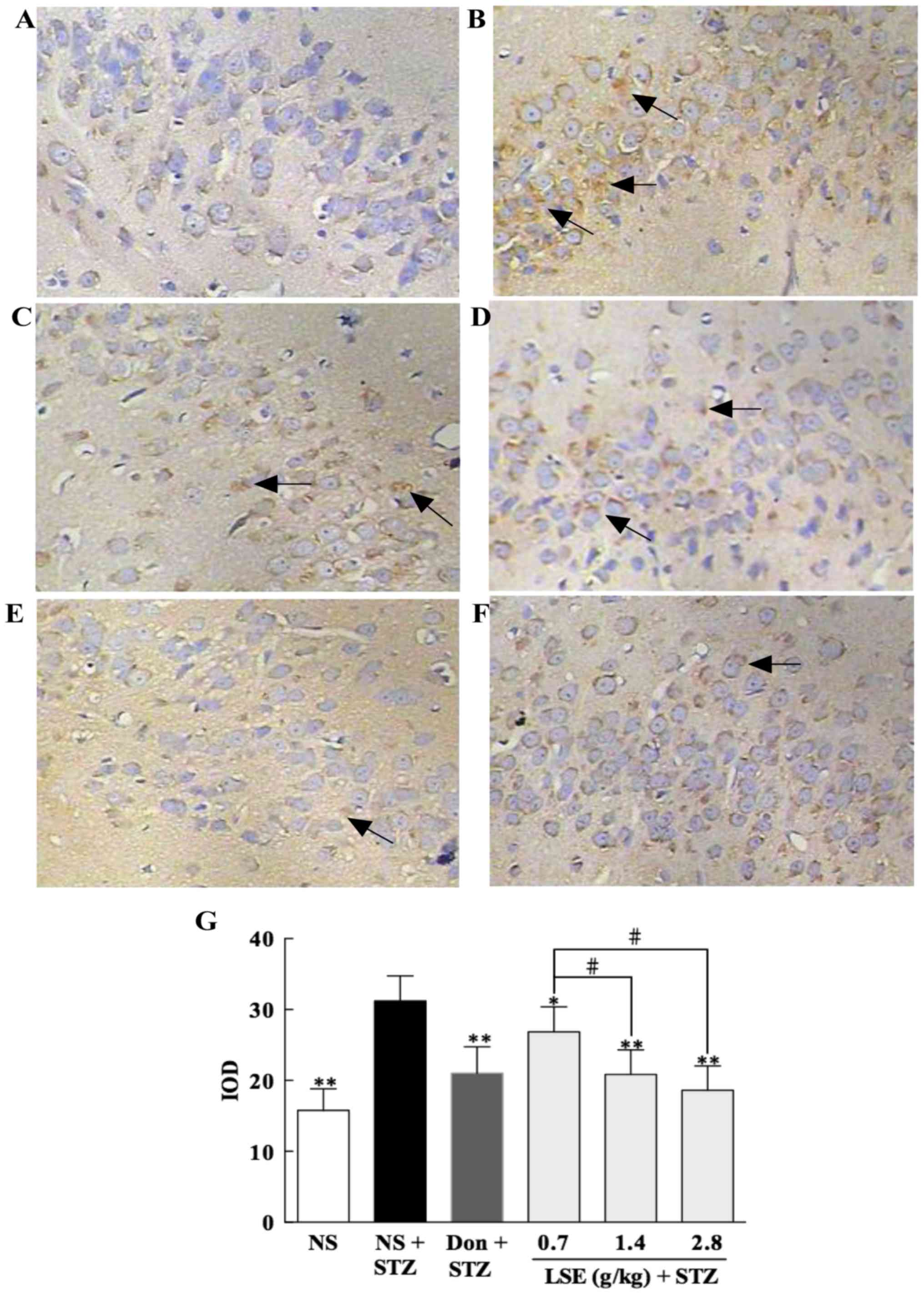 | Figure 9Effect of LSE on the expression of
Tau protein in the hippocampus of T2DM rats. The control rats were
treated with NS (1 ml/kg/day, IG ×28), and the T2DM rats were
treated with NS (0.2 ml/day, IG ×28), donepezil (0.42 mg/kg/day, IG
×28) or LSE (0.7, 1.4 and 2.8 g/kg/day, IG ×28). The brain tissues
of rats were removed at 24 h after drug treatment (magnification,
×400). The representative histologic photographs are shown as: (A)
Histological image of hippocampal neuronal cells of a control rat
treated with NS, only little brown particle deposition in the
cytoplasm of neuronal cells. (B) Hippocampal neuronal cells of a
T2DM rat treated with NS, strong positive expression of Tau protein
with the emergence of a large number of brown yellow granules
(arrows) in the cytoplasm of neuronal cells. (C) Hippocampal
neuronal cells of a T2DM rat treated with Don, brown particle
depositions (arrows) in the cytoplasm of neuronal cells are
significantly decreased compared to NS treatment. (D) Hippocampal
neuronal cells of a T2DM rat treated with LSE (0.7 g/kg), brown
particle depositions (arrows) in the cytoplasm of neuronal cells
are significantly decreased compared to NS treatment. (E)
Histologic picture of hippocampal neuronal cells of a T2DM rat
treated with LSE (1.4 g/kg), brown particle depositions (arrow) in
the cytoplasm of neuronal cells are significantly decreased
compared with NS treatment. (F) Histologic picture of hippocampal
neuronal cells of a T2DM rat treated with LSE (2.8 g/kg), brown
particle depositions (arrow) in the cytoplasm of neuronal cells are
also significantly decreased compared to NS treatment and the
decrease is more obvious than those of the rats treated with
donepezil and lower doses of LSE. (G) Quantitative results of bar
graph for each group. Arrows indicate Tau protein as brown particle
depositions. There were three rats for each group and results are
presented as mean ± standard deviation. *P<0.05 vs.
T2DM rats treated with NS; **P<0.01 vs. T2DM rats
treated with NS; #P<0.01 vs. LSE 0.7 g/kg group. LSE,
lychee seed extract; T2DM, type 2 diabetes mellitus; Don,
donezapil; IOD, integrated option density; STZ, streptozotocin; NS,
normal saline. |
The summary results of the expression of Tau protein
in the neuronal cells are presented in Fig. 9G. The data indicate that the
positive expression of Tau protein in the neuronal cells in T2DM
rats is significantly increased (P<0.01) compared to normal rats
(control). Interestingly, the Tau protein in the neuronal cells is
significantly decreased (P<0.05 or P<0.01) when the rats were
treated with LSE or donepezil compared to that of NS treatment, and
the most obvious decrease is observed with the group administered
with LSE at a concentration of 2.8 g/kg. The results indicate that
LSE and donepezil can effectively prevent the depositions of AGEs
and Tau protein in the neuronal cells of the hippocampus of T2DM
rats and high dose of LSE is more effective than that of
donepezil.
Effect of LSE on neuronal injury
protection in the CA1 area of hippocampus in T2DM rats by
histopathological exanimation
We further investigated the effect of LSE on
neuronal injury protection and compared to that of donepezil in
T2DM rats by histopathological examination and the results are
illustrated in Fig. 10.
 | Figure 10Protective effect of LSE on the
hippocampal neurons injury in T2DM rats. The representative
photographs of the hippocampal neurons in the normal (control) and
T2DM rats: The control rats were treated with NS (1 ml/kg/day) and
T2DM rats were treated with NS (1 ml/kg/day), donepezil (0.42
mg/kg/day) or LSE (0.7, 1.4 and 2.8 g/kg/day) ×28 days with
intragastric administration. The brain tissues of rats were removed
at 24 h after NS or drug treatment. Conventional formalin fixed
paraffin-embedded sections were stained with hematoxylin and eosin
and observed under light microscope (magnification, ×200). The
representative histologic photographs are shown as: (A)
Histological image of hippocampal neurons of a control rat treated
with NS, shows preserved normal histological features of neurons.
(B) Hippocampal neurons of a T2DM rat treated with NS, shows severe
damaged neurons. (C) Hippocampal neurons of a T2DM rat treated with
donepezil, shows still damaged neurons but less severe than that of
the T2DM rat treated with NS. (D) Hippocampal neurons of a T2DM rat
treated with LSE 0.7 g/kg, shows near normal histological features
of neurons. (E) Hippocampal neurons of a T2DM rat treated with LSE
1.4 g/kg, shows near normal histological features of neurons. (F)
Hippocampal neurons of a T2DM rat treated with LSE 2.8 g/kg, shows
normal histological features of neurons. Arrows indicate damaged
hippocampal neurons with loose arrangement, cytoplasmic and nuclear
condensation. There were three rats for each group. LSE, lychee
seed extract; T2DM, type 2 diabetes mellitus; NS, normal
saline. |
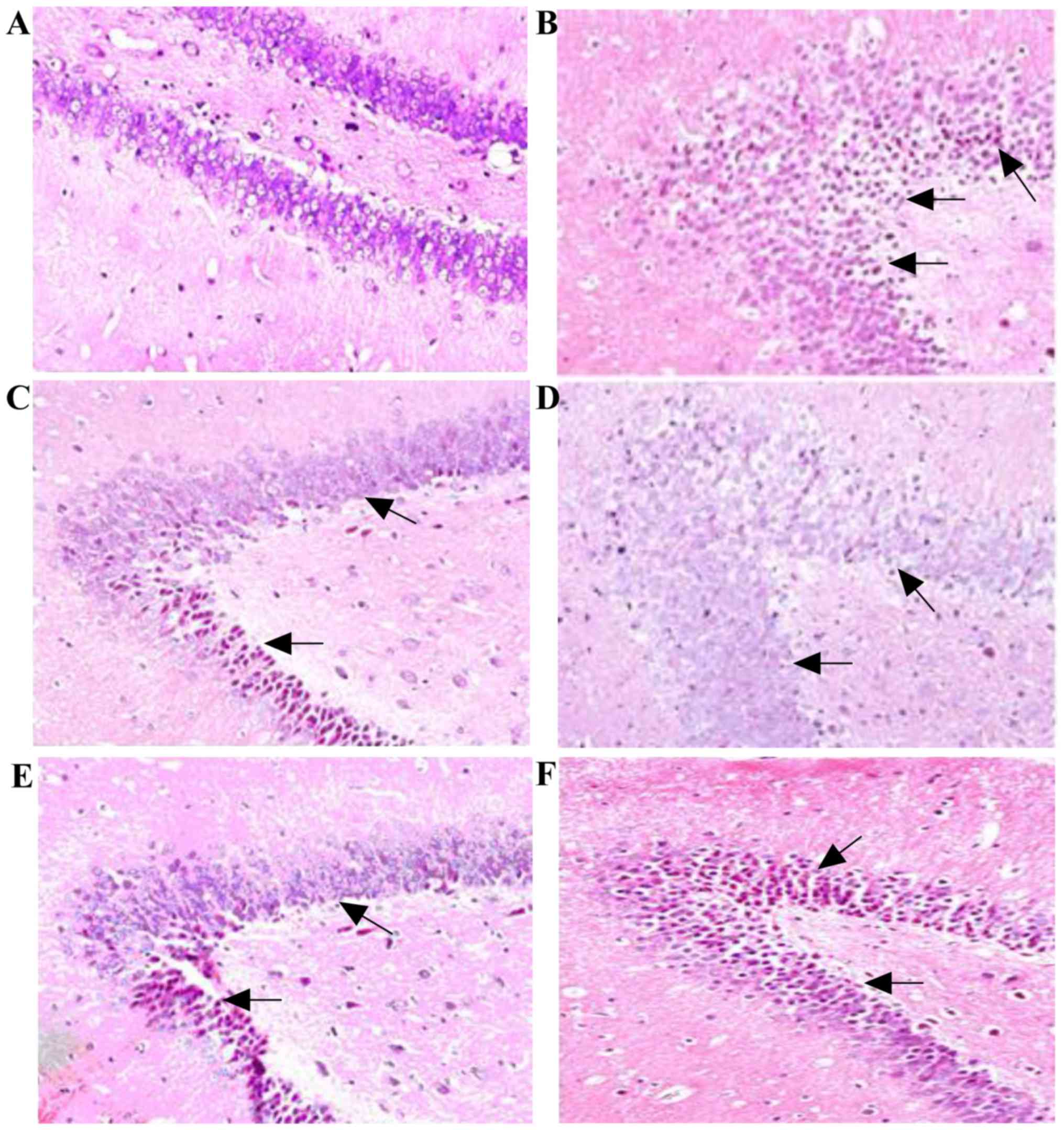
The histological changes were observed in the
sections of the neurons of the rats with H&E stain under a
light microscope (magnification, ×200). The picture in Fig. 10A presents the characteristic of
preserved normal histological features of the pyramidal cells in
the CA1 area of hippocampus of the normal rat (control) treated
with NS. The cells were closely arranged in order, the nucleus was
clearly visible in large and round, and there was no obvious
degeneration of neurons, such as nuclear condensation. However, as
shows in Fig. 10B, the neuronal
cells arranged in disorder and are sparse, cell morphology is
irregular or spindle shaped and a large number of cells appeared
shrieked volume and different layers of nuclear condensation in the
T2DM rat treated with NS. However, the hippocampal neurons of the
T2DM rats treated with donepezil are still damaged but much less
severe than that of the T2DM rat treated with NS (Fig. 10C). Interestingly, the morphology
of the neuronal cells is nearly (LSE, 0.7 and 1.4 g/kg) or
completely normal (LSE, 2.8 g/kg) after LSE treatments, the cells
arrange more closely and orderly with increased cell numbers and
normal shape, a large and round nucleus, and a clear nucleolus
(Fig. 10D–F).
The results indicated that LSE effectively protects
the hippocampal neuronal cells injury in a dose-dependent manner in
T2DM rats, and is more effective than donepezil.
Discussion
T2DM and AD are very common diseases. The rapid
increases of T2DM and AD incidences in the elderly population have
been observed. Evidence has proven that there is a close link
between T2DM and AD, and that the two diseases share common
biological mechanisms at a molecular level. T2DM can significantly
contribute to the onset and/or progression of AD, and T2DM patients
are at higher risk of AD (38).
The common pathology for both T2DM and AD is IR, impaired glucose
metabolism, Aβ formation, oxidative stress and presence of AGEs. IR
may be the pathological basis of the link between T2DM and AD
(39,40). IR promotes AD pathological changes
of Aβ deposition and Tau protein phosphorylation and induces
sustained high blood glucose to accelerate the formation of AGEs
(41–44). Studies have indicated that
abnormal structural components such as amyloid precursor (APP),
apolipoprotein E (ApoE), very low density lipoprotein (VLDL), AD,
Aβ and Tau protein are all related to the pathological changes of
AD in the brain tissues (43–45). Aβ and Tau protein are the
molecular basis for the formation of SPs and NFTs. IR indirectly
enhances the activity of glycogen synthase kinase-3β (GSK-3β) and
promotes the AD-like Tau hyperphosphorylation in the hippocampus of
the mice and rats in animal models of insulin dysfunction (46). In addition, IR can regulate the
activity of γ-secreting enzyme by GSK-3β (47), promote the synthesis of Aβ40 and
Aβ42, inhibit the activity of insulin degrading enzyme and reduce
the degradation of Aβ (48).
Furthermore, insulin plays a pivotal role in regulation of energy
metabolism, growth, survival and differentiation of neuronal cells
via insulin signaling in the brain (49,50). Therefore, improving IR may be one
of the important strategies for effective prevention and treatment
of AD (51,52). Recent studies have demonstrated
that improvement of IR may indeed delay the onset and/or
progression of AD in the animals and patients (53,54). In the present studies, the blood
glucose, insulin and HMOA are significantly increased in T2DM rats
and LSE significantly decreases the elevated glucose, insulin and
HMOA in a dose-dependent manner compared to NS treatment
(P<0.01) in T2DM rats (Fig.
6). The data indicate that LSE can indeed improve IR in T2DM
rats.
Currently, the drugs used for AD management can only
improve cognition and behavior of AD patients, but cannot slow
progression or cure the disease (22). In order to discover and develop
more effective and less toxic novel anti-AD drugs, we established a
rat model of T2DM with AD-like neurodegeneration and cognitive
impairment by feeding the rats with HSFP diet for 8 weeks and then
intraperitoneal injection of STZ (27 mg/kg) and continued HSFP diet
for another 4 weeks. The rat model of T2DM was validated by high
levels of blood glucose, insulin and HOMA as well as impaired
spatial learning and memory of the rats (Fig. 3). The model has the
characteristics and pathogenesis of both T2DM and AD. It is
reliable, reproducible and easy to be produced and shows high
resemblance as the T2DM and/or AD in the aging patients. Therefore,
it is an excellent model for study of cognitive impairment and drug
screening of T2DM and/or AD.
Natural products and medicinal herbs are important
sources for drug discovery against various diseases including AD
and T2DM (55). The use of drug
substances derived from natural sources has a long tradition in
medicine (56). In the present
study, we evaluated the effect of LSE, which was isolated and
extracted from lychee seeds on the anti-diabetic and anti-AD
efficacy, and compared to donepezil, the standard drug for AD
treatment. The main components of LSE were determined and it was
found that is primarily consists of eight major ingredients and
around 20 minor ingredients by a UHPLC-SPD chromatogram (Fig. 1). It has been reported that the
chemical components of lychee seeds comprise volatile categories,
saponins, flavonoids, organic, fatty, amino acids and sugar
(7). The preliminary studies
revealed that the saponins isolated and extracted from lychee seeds
are the main effective ingredients for the anti-diabetic and
anti-AD efficacy. In addition, it was demonstrated that LSE
significantly improves cognitive function in T2DM rats with
cognitive impairment for shortening the escape latency, increasing
the number across the platform, platform quadrant dwell time and
the percentage of time spent in the target quadrant compared to NS
treatment (Figs. 4 and 5). Furthermore, LSE markedly alleviates
neuronal injury in T2DM rats by morphological study (Fig. 10).
It has been reported that AD is associated with
reduction of cholinergic activity of neurons (37). The dysfunction of cholinergic
system is closely related to AD (21). Four of five FDA approved
medications (donepezil, tacrine, rivastagmine and galantamine) for
AD management are AChE inhibitors by reducing the rate of
acetylcholine degradation, and thereby increase its concentration
in the brain. AChE in the blood and the hippocampus of T2DM rats
were investigated and the concentrations of AChE were significantly
increased in the blood but significantly decreased in the
hippocampus compared to that of normal control rats (Fig. 7A and B). The authors are unsure as
to how to explain why the concentration of AChE is different in the
blood and hippocampus in T2DM rats. Both LSE and donepezil
significantly decreased the concentrations of AChE in the blood and
increased the concentrations of AChE in the hippocampus compared to
NS treatment (P<0.01) in T2DM rats (Fig. 7A and B).
Histopathological hallmarks of AD are deposition of
Aβ plaques and formation of NFTs, which are primarily made up of
aggregated Tau protein (57,58). The neurotoxic activities of Aβ
include generation of reactive oxygen species (ROS), lipid
peroxidation, calcium overload and eventually leading to neuronal
death (59). The progressive
accumulation of Tau protein leads to instability of the
microtubular structure and results in loss of effective
intracellular transport and neuronal death (60). AGEs also play an important role in
the pathogenesis of both T2DM and AD. The present results showed
that Aβ, Tau protein and AGEs in the hippocampus of T2DM rats were
significantly increased compared to that of normal control rats
(P<0.01). LSE can effectively prevent the depositions of Aβ, Tau
protein and AGEs in the neuronal cells of hippocampus in T2DM rats
and LSE is more effective than that of donepezil for reducing the
formation of Aβ, Tau protein, and AGEs (Figs. 7D, 8 and 9). Therefore, LSE may be developed as
the agent for the treatment of T2DM and/or AD. However, further
studies are needed to find out and purify the active ingredient(s)
from LSE and investigate the associated mechanistic action and
molecular pathways of neuronal protection.
In conclusion, LSE mainly consists of eight major
and around 20 minor ingredients, and significantly improves
cognitive function such as the ability of spatial learning and
memory and obviously protects from neuronal injury in T2DM rats
with cognitive impairment through reducing blood glucose, improving
IR and inhibiting the formation of Aβ, Tau protein and AGEs in the
brain of rats. LSE is similar or even more effective for
neuroprotection than that of donepezil, a classic drug for the
treatment of AD. Therefore, LSE may be developed as the agent for
the treatment of T2DM and/or AD clinically. However, further
studies are required to find out the active contents of LSE and
related long-term toxicity.
Acknowledgments
The present study was supported by grants from the
Science and Technology Planning Project of Sichuan Province, China
(grant nos. 2008SZ0050, 14JC0798 and LZ-LY-38), Science and
Technology Program of Luzhou, Sichuan, China (grant nos. 14JC0056,
2015-S-43 and 2016LZXNYD-T03), Educational Commission of Sichuan
Province, China (grant nos. 10ZA035, 15ZA0155 and 16ZA0187), Key
Development Program of Southwest Medical University (grant nos.
2010ZD-010 and 2014QN-043) and the Distinguished Professor Research
Startup Funding (S.C.) from Southwest Medical University (grant no.
2015-RCYJ0002). The authors would like to thank Professor Can Tang
of the Department of Chinese Materia Medica, School of Pharmacy,
Southwest Medical University (Luzhou, China) for authenticating the
lychee seeds used in the studies.
Glossary
Abbreviations
Abbreviations:
|
Aβ
|
β-amyloid
|
|
AChE
|
acetylcholinesterase
|
|
AD
|
Alzheimer's disease
|
|
AGEs
|
advanced glycation end products
|
|
APOE
|
apolipoprotein E
|
|
APP
|
amyloid precursor
|
|
ELISA
|
enzyme-linked immunosorbent assay
|
|
GSK-3β
|
glycogen synthase kinase-3β
|
|
H&E
|
hematoxylin and eosin
|
|
HFSPD
|
high fat, high sugar, and high protein
diet
|
|
IG
|
intragastric
|
|
IP
|
intraperitoneal
|
|
IR
|
insulin resistance
|
|
LSE
|
lychee seed extract
|
|
NFTs
|
neurofibrillary tangles
|
|
NS
|
normal saline
|
|
ROS
|
reactive oxygen species
|
|
SPs
|
senile plaques
|
|
STZ
|
streptozotocin
|
|
T2DM
|
type II diabetes mellitus
|
|
UHPLC
|
ultrahigh performance liquid
chromatography
|
|
VLDL
|
very low density lipoprotein
|
References
|
1
|
Jiang YM, Yao L, Amon L and Li JR:
Postharvest biology and technology of litchi fruit. J Food Agric
Environ. 1:76–81. 2003.
|
|
2
|
Yang B, Wang J, Zhao M, Liu Y, Wang W and
Jiang Y: Identification of polysaccharides from pericarp tissues of
litchi (Litchi chinensis Sonn.) fruit in relation to their
antioxidant activities. Carbohydr Res. 341:634–638. 2006.
View Article : Google Scholar : PubMed/NCBI
|
|
3
|
Kilari EK and Putta S: Biological and
phytopharmacological descriptions of Litchi chinensis. Pharmacogn
Rev. 10:60–65. 2016. View Article : Google Scholar : PubMed/NCBI
|
|
4
|
Xiao ZJ, Guo JW and Xu F: Effect of litchi
saponin and litchi flavones on insulin resistance in HepG2 cells. J
Pharm Pract. 4:316–318. 2015.
|
|
5
|
Ye HM, Zhong CY and Lv JH: Improving
effect of litchi seed saponins on learning and memory impairment in
mice. Acad J Guangzhou Med Univ. 2:10–14. 2015.
|
|
6
|
Zhang YM, Yuan H and Tian JX: Effects of
saponin of litchi seed on gluconeogenesis and metabolism of blood
lipid in mce. J Hangzhou Teach Coll. 6:435–436. 2005.
|
|
7
|
Zhang J and Zhang C: Research progress on
the antineoplastic pharmacological effects and mechanisms of Litchi
seeds. Chin Med. 6:20–26. 2015. View Article : Google Scholar
|
|
8
|
Li W, Zhu YT, Huang ZY, He JJ, Pei J and
Song JP: Experimental studies on anti-fluvirus effect of litchi
seed in vivo. Chin J Ethnomed Ethnopharm. 18:34–36. 2011.
|
|
9
|
Zhao M, Yang B, Wang J, Liu Y, Yu L and
Jiang Y: Immunomodulatory and anticancer activities of flavonoids
extracted from litchi (Litchi chinensis Sonn.) pericarp. Int
Immunopharmacol. 7:162–166. 2007. View Article : Google Scholar
|
|
10
|
Xiao LY, Pan JQ and Rao WN: The research
of protective effect of Litchi seed of experimental liver injury in
mice. Chin J Tradit Chin Med Pharm. 20:42–43. 2005.
|
|
11
|
Whiting DR, Guariguata L, Weil C and Shaw
J: IDF diabetes atlas: Global estimates of the prevalence of
diabetes for 2011 and 2030. Diabetes Res Clin Pract. 94:311–321.
2011. View Article : Google Scholar : PubMed/NCBI
|
|
12
|
Kasuga M: Insulin resistance and
pancreatic beta cell failure. J Clin Invest. 116:1756–1760. 2006.
View Article : Google Scholar : PubMed/NCBI
|
|
13
|
Spellman CW: Pathophysiology of type 2
diabetes: Targeting islet cell dysfunction. J Am Osteopath Assoc.
110(Suppl 2): S2–S7. 2010.
|
|
14
|
Moheet A, Mangia S and Seaquist ER: Impact
of diabetes on cognitive function and brain structure. Ann NY Acad
Sci. 1353:60–71. 2015. View Article : Google Scholar : PubMed/NCBI
|
|
15
|
Kimura N: Diabetes mellitus induces
Alzheimer's disease pathology: Histopathological evidence from
animal models. Int J Mol Sci. 17:5032016. View Article : Google Scholar : PubMed/NCBI
|
|
16
|
Mushtaq G, Khan JA and Kamal MA:
Biological mechanisms linking Alzheimer's disease and type-2
diabetes mellitus. CNS Neurol Disord Drug Targets. 13:1192–1201.
2014. View Article : Google Scholar : PubMed/NCBI
|
|
17
|
Aliev G, Shahida K, Gan SH, Firoz C, Khan
A, Abuzenadah AM, Kamal W, Kamal MA, Tan Y, Qu X, et al: Alzheimer
disease and type 2 diabetes mellitus: The link to tyrosine
hydroxylase and probable nutritional strategies. CNS Neurol Disord
Drug Targets. 13:467–477. 2014. View Article : Google Scholar
|
|
18
|
Riederer P, Bartl J, Laux G and Grünblatt
E: Diabetes type II: A risk factor for
depression-Parkinson-Alzheimer. Neurotox Res. 19:253–265. 2011.
View Article : Google Scholar
|
|
19
|
Yarchoan M and Arnold SE: Repurposing
diabetes drugs for brain insulin resistance in Alzheimer disease.
Diabetes. 63:2253–2261. 2014. View Article : Google Scholar : PubMed/NCBI
|
|
20
|
Gasparini L, Netzer WJ, Greengard P and Xu
H: Does insulin dysfunction play a role in Alzheimer's disease.
Trends Pharmacol Sci. 23:288–293. 2002. View Article : Google Scholar : PubMed/NCBI
|
|
21
|
Mushtaq G, Greig NH, Khan JA and Kamal MA:
Status of acetylcholinesterase and butyrylcholinesterase in
Alzheimer's disease and type 2 diabetes mellitus. CNS Neurol Disord
Drug Targets. 13:1432–1439. 2014. View Article : Google Scholar : PubMed/NCBI
|
|
22
|
Lee JH, Jeong SK, Kim BC, Park KW and Dash
A: Donepezil across the spectrum of Alzheimer's disease: Dose
optimization and clinical relevance. Acta Neurol Scand.
131:259–267. 2015. View Article : Google Scholar : PubMed/NCBI
|
|
23
|
Godyń J, Jończyk J, Panek D and Malawska
B: Therapeutic strategies for Alzheimer's disease in clinical
trials. Pharmacol Rep. 68:127–138. 2016. View Article : Google Scholar
|
|
24
|
Kim J, Lee HJ and Lee KW: Naturally
occurring phytochemicals for the prevention of Alzheimer's disease.
J Neurochem. 112:1415–1430. 2010. View Article : Google Scholar : PubMed/NCBI
|
|
25
|
Yang YJ and Liang BM: Determination of
anti-diabete saponins from Litchi chinensis sonn. J Guangdong
Pharm. 14:13–15. 2004.
|
|
26
|
Spencer JP: Beyond antioxidants: The
cellular and molecular interactions of flavonoids and how these
underpin their actions on the brain. Proc Nutr Soc. 69:244–260.
2010. View Article : Google Scholar : PubMed/NCBI
|
|
27
|
Ye HM, Zhong CY, Huang MX, Wang CY, Feng
X, Chen XY and Lv JH: Effect of litchi seed aqueous extracts on
learning and memory obstacles induced by D-galactose in mice and
its mechanism. Zhong Yao Cai. 36:438–440. 2013.In Chinese.
PubMed/NCBI
|
|
28
|
Barnhart CD, Yang D and Lein PJ: Using the
Morris water maze to assess spatial learning and memory in weanling
mice. PLoS One. 10:e01245212015. View Article : Google Scholar : PubMed/NCBI
|
|
29
|
Morris R: Developments of a water-maze
procedure for studying spatial learning in the rat. J Neurosci
Methods. 11:47–60. 1984. View Article : Google Scholar : PubMed/NCBI
|
|
30
|
Saxena G, Singh SP, Agrawal R and Nath C:
Effect of donepezil and tacrine on oxidative stress in
intracerebral streptozotocin-induced model of dementia in mice. Eur
J Pharmacol. 581:283–289. 2008. View Article : Google Scholar : PubMed/NCBI
|
|
31
|
Ghiasi R, Ghadiri Soufi F, Somi MH,
Mohaddes G, Mirzaie Bavil F, Naderi R and Alipour MR: Swim training
improves HOMA-IR in type 2 diabetes induced by high fat diet and
low dose of streptozotocin in male rats. Adv Pharm Bull. 5:379–384.
2015. View Article : Google Scholar : PubMed/NCBI
|
|
32
|
Srinivasan K, Viswanad B, Asrat L, Kaul CL
and Ramarao P: Combination of high-fat diet-fed and low-dose
streptozotocin- treated rat: A model for type 2 diabetes and
pharmacological screening. Pharmacol Res. 52:313–320. 2005.
View Article : Google Scholar : PubMed/NCBI
|
|
33
|
Liu G, Liu C and Zhang XN: Comparison of
the neuropsychological mechanisms of 2,6-diisopropylphenol and
N-methyl-D-aspartate receptor antagonist against electroconvulsive
therapy-induced learning and memory impairment in depressed rats.
Mol Med Rep. 12:3297–3308. 2015. View Article : Google Scholar : PubMed/NCBI
|
|
34
|
Wang S, Luo Y, Feng A, Li T, Yang X,
Nofech-Mozes R, Yu M, Wang C, Li Z, Yi F, et al: Ethanol induced
impairment of glucose metabolism involves alterations of GABAergic
signaling in pancreatic β-cells. Toxicology. 326:44–52. 2014.
View Article : Google Scholar : PubMed/NCBI
|
|
35
|
Mehdizadeh R, Parizadeh MR, Khooei AR,
Mehri S and Hosseinzadeh H: Cardioprotective effect of saffron
extract and safranal in isoproterenol-induced myocardial infarction
in wistar rats. Iran J Basic Med Sci. 16:56–63. 2013.PubMed/NCBI
|
|
36
|
He X, Ji O and Li JQ: The pharmacological
effect of Puerarin extract on learning and memory ability of rats
with Alzheimer disease. Pharm Clin Chin Mater Med. 5:39–42.
2014.
|
|
37
|
Rees TM and Brimijoin S: The role of
acetylcholinesterase in the pathogenesis of Alzheimer's disease.
Drugs Today (Barc). 39:75–83. 2003. View Article : Google Scholar
|
|
38
|
Plum L, Belgardt BF and Brüning JC:
Central insulin action in energy and glucose homeostasis. J Clin
Invest. 116:1761–1766. 2006. View Article : Google Scholar : PubMed/NCBI
|
|
39
|
Steen E, Terry BM, Rivera EJ, Cannon JL,
Neely TR, Tavares R, Xu XJ, Wands JR and de la Monte SM: Impaired
insulin and insulin-like growth factor expression and signaling
mechanisms in Alzheimer's disease - is this type 3 diabetes. J
Alzheimers Dis. 7:63–80. 2005. View Article : Google Scholar : PubMed/NCBI
|
|
40
|
Narasimhan K, Govindasamy M, Gauthaman K,
Kamal MA, Abuzenadeh AM, Al-Qahtani M and Kanagasabai R: Diabetes
of the brain: Computational approaches and interventional
strategies. CNS Neurol Disord Drug Targets. 13:408–417. 2014.
View Article : Google Scholar
|
|
41
|
Craft S, Peskind E, Schwartz MW,
Schellenberg GD, Raskind M and Porte D Jr: Cerebrospinal fluid and
plasma insulin levels in Alzheimer's disease: Relationship to
severity of dementia and apolipoprotein E genotype. Neurology.
50:164–168. 1998. View Article : Google Scholar : PubMed/NCBI
|
|
42
|
Craft S, Asthana S, Newcomer JW, Wilkinson
CW, Matos IT, Baker LD, Cherrier M, Lofgreen C, Latendresse S,
Petrova A, et al: Enhancement of memory in Alzheimer disease with
insulin and somatostatin, but not glucose. Arch Gen Psychiatry.
56:1135–1140. 1999. View Article : Google Scholar : PubMed/NCBI
|
|
43
|
Giacobini E and Gold G: Alzheimer disease
therapy - moving from amyloid-β to tau. Nat Rev Neurol. 9:677–686.
2013. View Article : Google Scholar : PubMed/NCBI
|
|
44
|
Ashraf GM, Greig NH, Khan TA, Hassan I,
Tabrez S, Shakil S, Sheikh IA, Zaidi SK, Akram M, Jabir NR, et al:
Protein misfolding and aggregation in Alzheimer's disease and type
2 diabetes mellitus. CNS Neurol Disord Drug Targets. 13:1280–1293.
2014. View Article : Google Scholar : PubMed/NCBI
|
|
45
|
Deutsch SI, Rosse RB and Lakshman RM:
Dysregulation of tau phosphorylation is a hypothesized point of
convergence in the pathogenesis of alzheimer's disease,
frontotemporal dementia and schizophrenia with therapeutic
implications. Prog Neuropsychopharmacol Biol Psychiatry.
30:1369–1380. 2006. View Article : Google Scholar : PubMed/NCBI
|
|
46
|
Soro-Paavonen A, Watson AM, Li J, Paavonen
K, Koitka A, Calkin AC, Barit D, Coughlan MT, Drew BG, Lancaster
GI, et al: Receptor for advanced glycation end products (RAGE)
deficiency attenuates the development of atherosclerosis in
diabetes. Diabetes. 57:2461–2469. 2008. View Article : Google Scholar : PubMed/NCBI
|
|
47
|
Marszałek M: Diabetes type 2 and Alzheimer
disease - one or two diseases? Mechanisms of association. Postepy
Hig Med Dosw. 67:653–671. 2013.In Polish. View Article : Google Scholar
|
|
48
|
El Khoury NB, Gratuze M, Papon MA,
Bretteville A and Planel E: Insulin dysfunction and Tau pathology.
Front Cell Neurosci. 8:222014. View Article : Google Scholar : PubMed/NCBI
|
|
49
|
Wang H, Wang R, Zhao Z, Ji Z, Xu S,
Hölscher C and Sheng S: Coexistences of insulin signaling-related
proteins and choline acetyltransferase in neurons. Brain Res.
1249:237–243. 2009. View Article : Google Scholar
|
|
50
|
Kang JH, Korecka M, Toledo JB, Trojanowski
JQ and Shaw LM: Clinical utility and analytical challenges in
measurement of cerebrospinal fluid amyloid-β(1-42) and τ proteins
as Alzheimer disease biomarkers. Clin Chem. 59:903–916. 2013.
View Article : Google Scholar : PubMed/NCBI
|
|
51
|
Bingham EM, Hopkins D, Smith D, Pernet A,
Hallett W, Reed L, Marsden PK and Amiel SA: The role of insulin in
human brain glucose metabolism: An 18fluoro-deoxyglucose positron
emission tomography study. Diabetes. 51:3384–3390. 2002. View Article : Google Scholar : PubMed/NCBI
|
|
52
|
Li Y, Tsui W, Rusinek H, Butler T, Mosconi
L, Pirraglia E, Mozley D, Vallabhajosula S, Harada R, Furumoto S,
et al: Cortical laminar binding of PET amyloid and tau tracers in
Alzheimer disease. J Nucl Med. 56:270–273. 2015. View Article : Google Scholar : PubMed/NCBI
|
|
53
|
Takalo M, Haapasalo A, Martiskainen H,
Kurkinen KM, Koivisto H, Miettinen P, Khandelwal VK, Kemppainen S,
Kaminska D, Mäkinen P, et al: High-fat diet increases tau
expression in the brain of T2DM and AD mice independently of
peripheral metabolic status. J Nutr Biochem. 25:634–641. 2014.
View Article : Google Scholar : PubMed/NCBI
|
|
54
|
Hung AS, Liang Y, Chow TC, Tang HC, Wu SL,
Wai MS and Yew DT: Mutated tau, amyloid and neuroinflammation in
Alzheimer disease-A brief review. Prog Histochem Cytochem. 51:1–8.
2016. View Article : Google Scholar : PubMed/NCBI
|
|
55
|
Anekonda TS and Reddy PH: Can herbs
provide a new generation of drugs for treating Alzheimer's disease.
Brain Res Brain Res Rev. 50:361–376. 2005. View Article : Google Scholar : PubMed/NCBI
|
|
56
|
Farver D: The use of 'natural products' in
clinical medicine. S D J Med. 49:129–130. 1996.PubMed/NCBI
|
|
57
|
Blass JP, Ko L and Wisniewski HM:
Pathology of Alzheimer's disease. Psychiatr Clin North Am.
14:397–420. 1991.PubMed/NCBI
|
|
58
|
Iqbal K, Liu F, Gong CX and Grundke-Iqbal
I: Tau in Alzheimer disease and related tauopathies. Curr Alzheimer
Res. 7:656–664. 2010. View Article : Google Scholar : PubMed/NCBI
|
|
59
|
Wei W, Wang X and Kusiak JW: Signaling
events in amyloid beta-peptide-induced neuronal death and
insulin-like growth factor I protection. J Biol Chem.
277:17649–17656. 2002. View Article : Google Scholar : PubMed/NCBI
|
|
60
|
Alonso AD, Grundke-Iqbal I, Barra HS and
Iqbal K: Abnormal phosphorylation of tau and the mechanism of
Alzheimer neurofibrillary degeneration: Sequestration of
microtubule-associated proteins 1 and 2 and the disassembly of
microtubules by the abnormal tau. Proc Natl Acad Sci USA.
94:298–303. 1997. View Article : Google Scholar : PubMed/NCBI
|