|
1
|
Cohen S, Nathan JA and Goldberg AL: Muscle
wasting in disease: Molecular mechanisms and promising therapies.
Nat Rev Drug Discov. 14:58–74. 2015. View
Article : Google Scholar : PubMed/NCBI
|
|
2
|
Jackman RW and Kandarian SC: The molecular
basis of skeletal muscle atrophy. Am J Physiol Cell Physiol.
287:C834–C843. 2004. View Article : Google Scholar : PubMed/NCBI
|
|
3
|
Sillau AH and Banchero N: Effects of
hypoxia on capillary density and fiber composition in rat skeletal
muscle. Pflugers Arch. 370:227–232. 1977. View Article : Google Scholar : PubMed/NCBI
|
|
4
|
Zattara-Hartmann MC, Badier M, Guillot C,
Tomei C and Jammes Y: Maximal force and endurance to fatigue of
respiratory and skeletal muscles in chronic hypoxemic patients: The
effects of oxygen breathing. Muscle Nerve. 18:495–502. 1995.
View Article : Google Scholar : PubMed/NCBI
|
|
5
|
Sanders KJ, Kneppers AE, van de Bool C,
Langen RC and Schols AM: Cachexia in chronic obstructive pulmonary
disease: New insights and therapeutic perspective. J Cachexia
Sarcopenia Muscle. 7:5–22. 2016. View Article : Google Scholar : PubMed/NCBI
|
|
6
|
Clark AL, Poole-Wilson PA and Coats AJ:
Exercise limitation in chronic heart failure: Central role of the
periphery. J Am Coll Cardiol. 28:1092–1102. 1996. View Article : Google Scholar : PubMed/NCBI
|
|
7
|
Coats A: The 'Muscle Hypothesis' of
chronic heart failure. J Mol Cell Cardiol. 28:2255–2262. 1996.
View Article : Google Scholar : PubMed/NCBI
|
|
8
|
Coats AJ, Clark AL, Piepoli M, Volterrani
M and Poole-Wilson PA: Symptoms and quality of life in heart
failure: The muscle hypothesis. Br Heart J. 72(suppl 2): S36–S39.
1994. View Article : Google Scholar : PubMed/NCBI
|
|
9
|
Piepoli MF, Kaczmarek A, Francis DP,
Davies LC, Rauchhaus M, Jankowska EA, Anker SD, Capucci A, Banasiak
W and Ponikowski P: Reduced peripheral skeletal muscle mass and
abnormal reflex physiology in chronic heart failure. Circulation.
114:126–134. 2006. View Article : Google Scholar : PubMed/NCBI
|
|
10
|
Mancini DM, Walter G, Reichek N, Lenkinski
R, McCully KK, Mullen JL and Wilson JR: Contribution of skeletal
muscle atrophy to exercise intolerance and altered muscle
metabolism in heart failure. Circulation. 85:1364–1373. 1992.
View Article : Google Scholar : PubMed/NCBI
|
|
11
|
Farkas J, von Haehling S, Kalantar-Zadeh
K, Morley JE, Anker SD and Lainscak M: Cachexia as a major public
health problem: Frequent, costly, and deadly. J Cachexia Sarcopenia
Muscle. 4:173–178. 2013. View Article : Google Scholar : PubMed/NCBI
|
|
12
|
Hajahmadi M, Shemshadi S, Khalilipur E,
Amin A, Taghavi S, Maleki M, Malek H and Naderi N: Muscle wasting
in young patients with dilated cardiomyopathy. J Cachexia
Sarcopenia Muscle. 8:542–548. 2017. View Article : Google Scholar : PubMed/NCBI
|
|
13
|
Buller NP, Jones D and Poole-Wilson PA:
Direct measurement of skeletal muscle fatigue in patients with
chronic heart failure. Br Heart J. 65:20–24. 1991. View Article : Google Scholar : PubMed/NCBI
|
|
14
|
Stugiewicz M, Tkaczyszyn M, Kasztura M,
Banasiak W, Ponikowski P and Jankowska EA: The influence of iron
deficiency on the functioning of skeletal muscles: Experimental
evidence and clinical implications. Eur J Heart Fail. 18:762–773.
2016. View Article : Google Scholar : PubMed/NCBI
|
|
15
|
Loncar G, Springer J, Anker M, Doehner W
and Lainscak M: Cardiac cachexia: Hic et nunc. J Cachexia
Sarcopenia Muscle. 7:246–260. 2016. View Article : Google Scholar : PubMed/NCBI
|
|
16
|
Zizola C and Schulze PC: Metabolic and
structural impairment of skeletal muscle in heart failure. Heart
Fail Rev. 18:623–630. 2013. View Article : Google Scholar :
|
|
17
|
Gosker HR, Wouters EF, van der Vusse GJ
and Schols AM: Skeletal muscle dysfunction in chronic obstructive
pulmonary disease and chronic heart failure: Underlying mechanisms
and therapy perspectives. Am J Clin Nutr. 71:1033–1047. 2000.
View Article : Google Scholar : PubMed/NCBI
|
|
18
|
von Haehling S, Lainscak M, Springer J and
Anker SD: Cardiac cachexia: A systematic overview. Pharmacol Ther.
121:227–252. 2009. View Article : Google Scholar
|
|
19
|
Minotti JR, Christoph I, Oka R, Weiner MW,
Wsells L and Massie BM: Impaired skeletal muscle function in
patients with congestive heart failure. Relationship to systemic
exercise performance. J Clin Invest. 88:2077–2082. 1991. View Article : Google Scholar : PubMed/NCBI
|
|
20
|
Jankowska EA, Rozentryt P, Witkowska A,
Nowak J, Hartmann O, Ponikowska B, Borodulin-Nadzieja L, Banasiak
W, Polonski L, Filippatos G, et al: Iron deficiency: An ominous
sign in patients with systolic chronic heart failure. Eur Heart J.
31:1872–1880. 2010. View Article : Google Scholar : PubMed/NCBI
|
|
21
|
Jankowska EA, Malyszko J, Ardehali H,
Koc-Zorawska E, Banasiak W, von Haehling S, Macdougall IC, Weiss G,
McMurray JJ, Anker SD, et al: Iron status in patients with chronic
heart failure. Eur Heart J. 34:827–834. 2013. View Article : Google Scholar :
|
|
22
|
Anker SD, Comin Colet J, Filippatos G,
Willenheimer R, Dickstein K, Drexler H, Lüscher TF, Bart B,
Banasiak W, Niegowska J, et al: Ferric carboxymaltose in patients
with heart failure and iron deficiency. N Engl J Med.
361:2436–2448. 2009. View Article : Google Scholar : PubMed/NCBI
|
|
23
|
Okonko DO, Grzeslo A, Witkowski T, Mandal
AKJ, Slater RM, Roughton M, Foldes G, Thum T, Majda J, Banasiak W,
et al: Effect of intravenous iron sucrose on exercise tolerance in
anemic and nonanemic patients with symptomatic chronic heart
failure and iron deficiency. J Am Coll Cardiol. 51:103–112
|
|
24
|
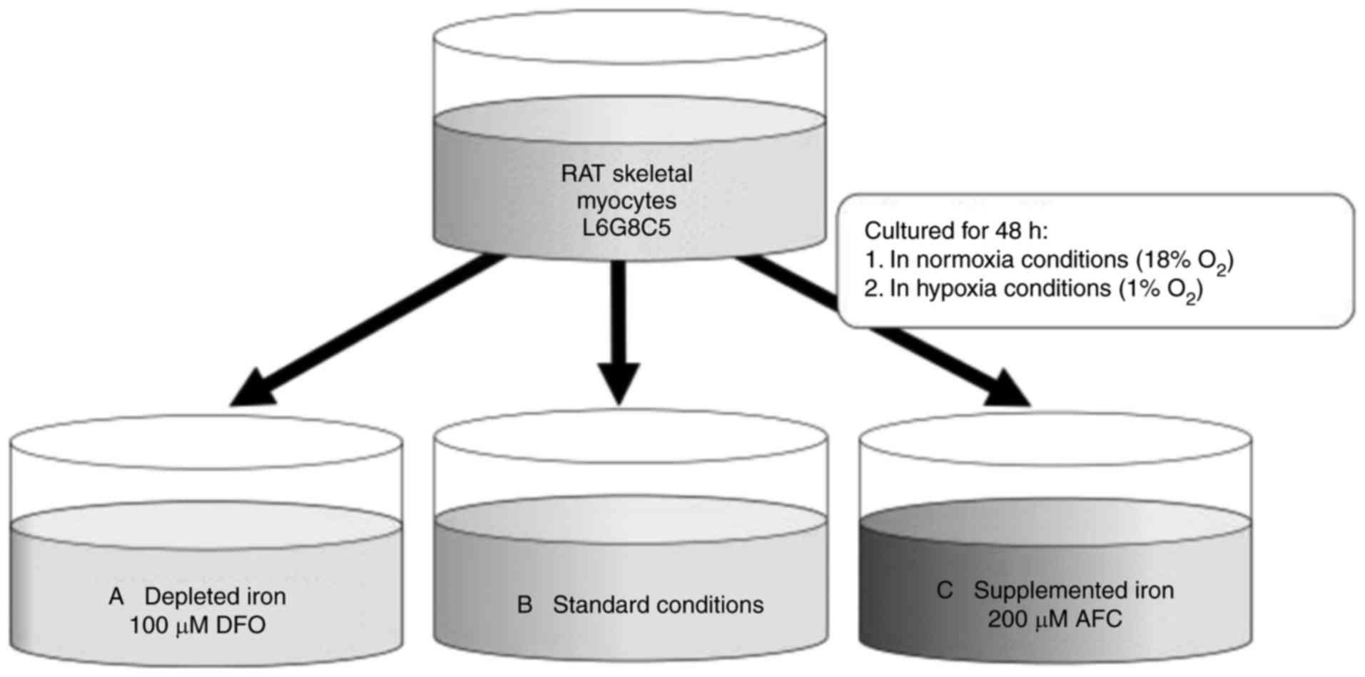
Dziegala M, Kasztura M, Kobak K, Bania J,
Banasiak W, Ponikowski P and Jankowska EA: Influence of the
availability of iron during hypoxia on the genes associated with
apoptotic activity and local iron metabolism in rat H9C2
cardiomyocytes and L6G8C5 skeletal myocytes. Mol Med Rep.
14:3969–3977. 2016. View Article : Google Scholar : PubMed/NCBI
|
|
25
|
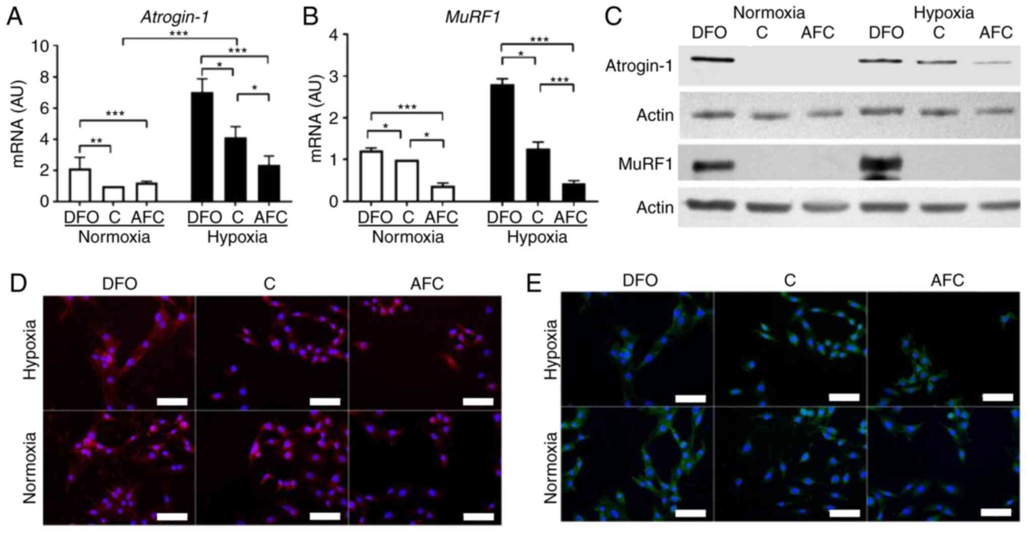
Gomes MD, Lecker SH, Jagoe RT, Navon A and
Goldberg AL: Atrogin-1, a muscle-specific F-box protein highly
expressed during muscle atrophy. Proc Natl Acad Sci.
98:14440–14445. 2001. View Article : Google Scholar : PubMed/NCBI
|
|
26
|
Bodine SC, Latres E, Baumhueter S, Lai VK,
Nunez L, Clarke BA, Poueymirou WT, Panaro FJ, Na E, Dharmarajan K,
et al: Identification of ubiquitin ligases required for skeletal
muscle atrophy. Science. 294:1704–1708. 2001. View Article : Google Scholar : PubMed/NCBI
|
|
27
|
Palus S, von Haehling S and Springer J:
Muscle wasting: An overview of recent developments in basic
research. J Cachexia Sarcopenia Muscle. 5:193–198. 2014. View Article : Google Scholar : PubMed/NCBI
|
|
28
|
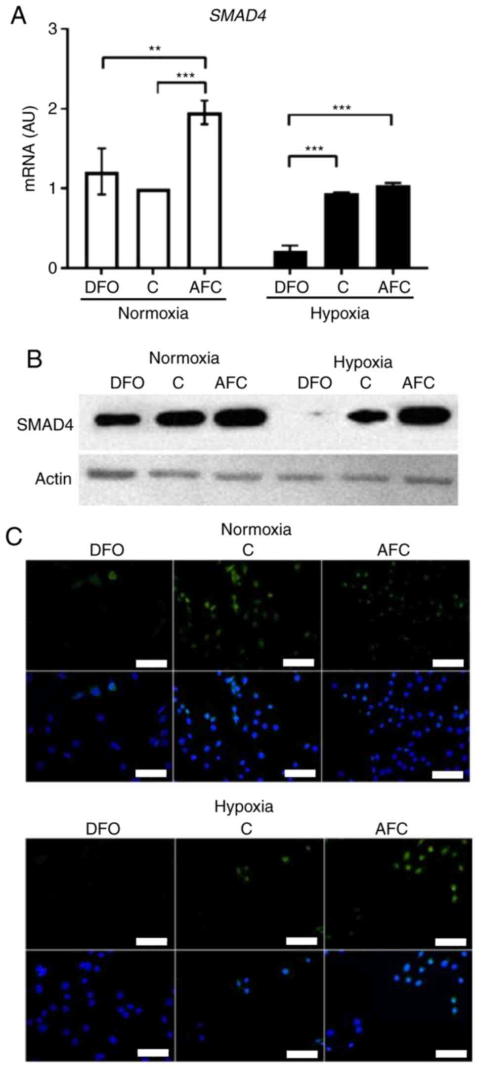
Liu F, Pouponnot C and Massagué J: Dual
role of the Smad4/DPC4 tumor suppressor in TGFbeta-inducible
transcriptional complexes. Genes Dev. 11:3157–3167. 1997.
View Article : Google Scholar
|
|
29
|
Seong HA, Jung H, Kim KT and Ha H:
3-Phosphoinositide-dependent PDK1 negatively regulates transforming
growth factor-β-induced signaling in a kinase-dependent manner
through physical interaction with Smad Proteins. J Biol Chem.
282:12272–12289. 2007. View Article : Google Scholar : PubMed/NCBI
|
|
30
|
Sartori R, Schirwis E, Blaauw B,
Bortolanza S, Zhao J, Enzo E, Stantzou A, Mouisel E, Toniolo L,
Ferry A, et al: BMP signaling controls muscle mass. Nat Genet.
45:1309–1318. 2013. View Article : Google Scholar : PubMed/NCBI
|
|
31
|
Aliparasti MR, Alipour MR, Almasi S and
Feizi H: Ghrelin administration increases the Bax/Bcl-2 gene
expression ratio in the heart of chronic hypoxic rats. Adv Pharm
Bull. 5:195–199. 2015. View Article : Google Scholar : PubMed/NCBI
|
|
32
|
Fletcher J: Iron transport in the blood.
Proc R Soc Med. 63:1216–1218. 1970.PubMed/NCBI
|
|
33
|
Woo KJ, Lee TJ, Park JW and Kwon TK:
Desferrioxamine, an iron chelator, enhances HIF-1alpha accumulation
via cyclooxy-genase-2 signaling pathway. Biochem Biophys Res
Commun. 343:8–14. 2006. View Article : Google Scholar : PubMed/NCBI
|
|
34
|
Parkes JG, Hussain RA, Olivieri NF and
Templeton DM: Effects of iron loading on uptake, speciation, and
chelation of iron in cultured myocardial cells. J Lab Clin Med.
122:36–47. 1993.PubMed/NCBI
|
|
35
|
Hoepken HH, Korten T, Robinson SR and
Dringen R: Iron accumulation, iron-mediated toxicity and altered
levels of ferritin and transferrin receptor in cultured astrocytes
during incubation with ferric ammonium citrate. J Neurochem.
88:1194–1202. 2004. View Article : Google Scholar : PubMed/NCBI
|
|
36
|
Chandio ZA, Talpur FN, Khan H, Afridi HI
and Khaskheli GQ: Determination of cadmium and zinc in vegetables
with online FAAS after simultaneous pre-concentration with
1,5-diphenylthiocarbazone immobilised on naphthalene. Food Addit
Contam Part A Chem Anal Control Expo Risk Assess. 30:110–115. 2013.
View Article : Google Scholar
|
|
37
|
Lowry OH, Rosebrough NJ, Farr AL and
Randall RJ: Protein measurement with the folin phenol reagent. J
Biol Chem. 193:265–275. 1951.PubMed/NCBI
|
|
38
|
Pfaffl MW: A new mathematical model for
relative quantification in real-time RT-PCR. Nucleic Acids Res.
29:e452001. View Article : Google Scholar : PubMed/NCBI
|
|
39
|
Walker JM: The bicinchoninic acid (BCA)
assay for protein quantitation. Methods Mol Biol. 32:5–8.
1994.PubMed/NCBI
|
|
40
|
Ikeda Y, Imao M, Satoh A, Watanabe H,
Hamano H, Horinouchi Y, Izawa-Ishizawa Y, Kihira Y, Miyamoto L,
Ishizawa K, et al: Iron-induced skeletal muscle atrophy involves an
Akt-forkhead box O3-E3 ubiquitin ligase-dependent pathway. J Trace
Elem Med Biol. 35:66–76. 2016. View Article : Google Scholar : PubMed/NCBI
|
|
41
|
Reardon TF and Allen DG: Iron injections
in mice increase skeletal muscle iron content, induce oxidative
stress and reduce exercise performance. Exp Physiol. 94:720–730.
2009. View Article : Google Scholar : PubMed/NCBI
|
|
42
|
Kasztura M, Dzięgała M, Kobak K, Bania J,
Mazur G, Banasiak W, Ponikowski P and Jankowska EA: Both iron
excess and iron depletion impair viability of rat H9C2
cardiomyocytes and L6G8C5 myocytes. Kardiol Pol. 75:267–275. 2017.
View Article : Google Scholar
|
|
43
|
Sequeira V, Nijenkamp LL, Regan JA and van
der Velden J: The physiological role of cardiac cytoskeleton and
its alterations in heart failure. Biochim Biophys Acta.
1838:700–722. 2014. View Article : Google Scholar
|
|
44
|
Lazarides E and Hubbard BD: Immunological
characterization of the subunit of the 100 A filaments from muscle
cells. Proc Natl Acad Sci USA. 73:4344–4348. 1976. View Article : Google Scholar : PubMed/NCBI
|
|
45
|
Koutakis P, Miserlis D, Myers SA, Kim JK,
Zhu Z, Papoutsi E, Swanson SA, Haynatzki G, Ha DM, Carpenter LA, et
al: Abnormal accumulation of desmin in gastrocnemius myofibers of
patients with peripheral artery disease. J Histochem Cytochem.
63:256–269. 2015. View Article : Google Scholar : PubMed/NCBI
|
|
46
|
Russ DW and Grandy JS: Increased desmin
expression in hindlimb muscles of aging rats. J Cachexia Sarcopenia
Muscle. 2:175–180. 2011. View Article : Google Scholar : PubMed/NCBI
|
|
47
|
Walter PB, Knutson MD, Paler-Martinez A,
Lee S, Xu Y, Viteri FE and Ames BN: Iron deficiency and iron excess
damage mitochondria and mitochondrial DNA in rats. Proc Natl Acad
Sci USA. 99:2264–2269. 2002. View Article : Google Scholar : PubMed/NCBI
|