Introduction
Osteoarthritis (OA) is a common type of degenerative
arthritis that is characterized by subchondral bone damage,
synovitis and articular cartilage degradation (1). Due to joint pain, inflammation,
movement difficulty and crepitus, knee OA poses a major burden on
patient daily life, and even causes disability (2). The World Health Organization has
reported that >10% of the population aged <60 years suffer
from OA worldwide (3). Nuclear
factor (NF)-κB was reported to enhance the synthesis of nitric
oxide and other catabolic factors that are drivers of OA
chondrocyte apoptosis and cartilage inflammation (4). A previous study demonstrated that
in vitro injection or oral administration of hyaluronic acid
(HA) may be a viable treatment option for patients with knee OA
(5). Furthermore, it has also
been demonstrated that curcuminoid inhibited the progression of OA
and effectively alleviated pain symptoms (6). Therefore, the aim of the present
study was to examine whether HA and curcuminoid can act
synergistically to provide the best therapeutic efficacy in knee
OA.
HA is a mucopolysaccharide biopolymer forming from
cyclical units of disaccharides (7). Intra-articular HA is often applied
in knee OA treatment to relieve pain and reduce knee inflammation
(8). Moreover, evidence from a
recent study suggests that curcuminoid inhibited chondrocyte
apoptosis and the inflammatory signaling pathway in OA (9). However, the use of curcuminoid is
limited in therapeutics due to its low aqueous solubility, unstable
chemical characteristics, and poor bioavailability following oral
administration. However, the low bioavailability of curcuminoid may
be improved by nanoparticles (10). In addition, chitosan with high
molecular weight may improve colloidal stability during the
preparation of curcuminoid nanoplexes and does not substantially
affect the dissolution capacity for other materials (11). Furthermore, HA interacts with
intercellular adhesion molecule-1 to decrease Akt expression in a
lipopolysaccharide-stimulated macrophage cell line, and may also
inhibit NF-κB pathway activation (12). Curcumin significantly inhibited
matrix metalloproteinase (MMP)-13 mRNA and suppressed NF-κB pathway
activation by repressing I-κBα phosphorylation and degradation, as
well as P65 nuclear translocation (13). In addition, inhibition of the
NF-κB pathway was reported to attenuate inflammation (14). Thus, it was hypothesized that HA
and curcuminoid may be better dissolved with the help of chitosan
and nanoparticles to form HA/chitosan nanoparticles (CNP) for the
delivery of curcuminoid, and that HA/CNP for the delivery of
curcuminoid can treat knee OA by targeting the NF-κB pathway. The
aim of the present study was to determine whether the use of
intra-articular HA/CNP for the delivery of curcuminoid has a better
efficacy compared with HA or curcuminoid treatment individually, in
order to investigate new approaches to the treatment of knee
OA.
Materials and methods
Ethics statements
The breeding and sacrifice of the experimental
animals, as well as tissue retrieval, were all for medical research
and performed in strict accordance with the recommendations in the
Guide for the Care and Use of Laboratory Animals of the National
Institutes of Health. The procedures were approved by the Animal
Committee of Shanghai Ninth People's Hospital, Shanghai Jiao Tong
University School of Medicine.
Study subjects
A total of 70 specific-pathogen-free (SPF) healthy
male Sprague Dawley (SD) rats (age, 8 weeks; weight range, 180–220
g) were provided by SLAC Laboratory Animal Co. Ltd. (Shanghai,
China). The rats were housed in an environment of 68% humidity at
25°C with 12 h of light every day, with free access to food and
running water supplied by the feeders.
Establishment of animal model
The Hulth method was applied to establish a knee OA
model in SD rats (15). After a
12-h fasting period, the SD rats were fully anesthetized by
intraperitoneal injection of 0.3% pentobarbital sodium
(Sigma-Aldrich; Merck KGaA, Darmstadt, Germany) at 10 ml/kg and
were then fixed in the supine position. The patellae were pushed
outward until dislocated. When the knees were at maximum bending
angle, the articular cavity was exposed, and the right knee
anterior cruciate ligaments (ACLs), medial collateral ligaments
(MCLs) and medial menisci were cut off without any damage to the
joint cartilage surface. After the knee was determined to be
positive by the drawer test, the wounds were fully stanched and
sutured layer by layer, while treated with drops of penicillin
solution (Sigma-Aldrich; Merck KGaA). After the operation, the rats
were treated by intramuscular injection of 40,000 units of
penicillin daily for 4 days. On the fifth day, the rats were forced
to run for 30 min per day. The sham group consisted of SD rats
which had undergone articular cavity surgery without any damage to
the ACL, MCL or medial menisci. The subsequent treatments were the
same as those in the rats in the OA group. After being forced to
run every day for 4 weeks, the walking status of the rats was
observed, and their joints were examined by safranin O-fast green
staining. There were 5 SD rats in the sham group and 5 SD rats in
the OA group; a total of 60 rats (20 rats per group) were used for
the subsequent experiments.
Chondrocyte isolation and culture
Ten SD rats were euthanized using 100%
CO2, were immersed in 75% ethanol for 10 min, and were
then transferred to a superclean bench. The tibial cartilages of
the knee joints were removed using a surgical knife and washed with
phosphate-buffered saline (PBS) 3 times. The cartilage was cut into
pieces, collected in a 50-ml centrifuge tube, and treated with 2 ml
0.25% trypsin (Sigma-Aldrich; Merck KGaA) for 60 min. The trypsin
was then removed, and the cartilages were rinsed twice with PBS and
then treated with 0.2% type II collagenase (Sigma-Aldrich; Merck
KGaA) for 4 h. After removal of the collagenase, turbid liquid was
poured into a 120-pore mesh screen (Solarbio Science and Technology
Ltd., Co., Beijing, China) and the filtered liquid was collected
and centrifuged at 100 × g for 10 min. Subsequently, the filtered
liquid was washed with PBS twice and incubated in Dulbecco's
Modified Eagle's Medium (DMEM) (Gibco; Thermo Fisher Scientific,
Inc., Waltham, MA, USA) containing 10% fetal bovine serum (FBS)
(Gibco; Thermo Fisher Scientific Inc.) to resuspend cells. The cell
resuspension was evenly inoculated in a 35-mm culture dish and
cultured in a 5% CO2 incubator at 37°C. Subsequently,
the growth of chondrocytes was observed under an inverted
microscope (Promega Corporation, Madison, WI, USA). When confluence
reached 90%, separated primary chondrocytes were passaged.
Immunofluorescence method
Chondrocytes were digested, resuspended, replated in
a 6-well plate with an adhesive cover-glass inside at density of
1×10⁶ cells/well, and then cultured in a 5%
CO2 incubator at 37°C. The coverglass was removed when
the cells reached 90% confluence. The cells were washed with PBS,
fixed with 1 ml 4% paraformaldehyde for 15 min, rinsed twice with
PBS, and permeabilized with 1 ml 0.2% Triton-PBS for 30 min. The
cells were then rinsed twice with PBS, sealed with 1 ml 5% bovine
serum albumin (BSA) for 20 min, and incubated in 1 ml rabbit
anti-rat collagen II (1:100, ab34712, Abcam, Cambridge, MA, USA) at
4°C overnight. The cells were rinsed with PBS and then incubated
with secondary goat anti-rabbit antibody immunoglobulin G (IgG;
1:1,000, ab150077, Abcam) at room temperature for 4 h in the dark.
Cells were rinsed twice with PBS, stained with 100 µl
Hoechst 33258 staining solution at room temperature for 5 min, and
washed twice with PBS. Then, the cells were fast-rinsed with 95%
ethanol and dried. An appropriate amount of fluorescence quenching
solution was dropped onto the glass slide, and then the coverslip
was applied. Subsequent fluorescence was observed using an inverted
fluorescence phase-contrast microscope (EclipseTi-S, Nikon, Tokyo,
Japan).
Ionic cross-linking method
Chitosan (9 mg, 10–40 kD, Jinke Company, Zhejiang,
China) was precisely weighed, collected, dissolved in 3.6 ml
distilled water, and added to 5 ml curcuminoid solution (1 mg/ml in
absolute ethanol, Geleixiya Company, Chengdu, China) for 10 min.
This reaction mixture was added to 1 ml triphenyl phosphate (2
mg/ml in water) (Sangon Biotech Company, Shanghai, China) and
incubated at room temperature for 30 min. The suspension was then
centrifuged at 23,649 × g at 4°C for 40 min. Sediments were
collected, washed and treated with lyophilization to obtain a
powder. A total of 30 ml distilled water was added into a 50-ml
centrifuge tube. Then, 90 mg HA (Sangon Biotech Company), 180 mg
endocrine-disrupting chemical (Sangon Biotech Company) and 195 mg
N-hydroxysuccinimide (Sangon Biotech Company) were added in that
order, and the solution was adjusted to pH 7.5. The tube was
incubated in a water bath at 37°C for 3 h. Then, 15 mg of the
aforementioned lyophilized powder was added, and the mixture was
incubated for 24 h at 300 × g to obtain a nanosuspension. The
nanosus-pension was centrifuged at 23,469 × g at 4°C for 60 min,
and sediments were obtained. Finally, HA/CNP for the delivery of
curcuminoid was obtained after the sediments were washed and
treated via lyophilization. The molecular weight of the resulting
compound was 500–1,000 kD.
Morphological observation of HA/CNP for
the delivery of curcuminoid
The appropriate amount of HA/CNP for the delivery of
curcuminoid powder was diluted in distilled water, dropped on a
copper grid-covered carbon film, and incubated with 2%
phosphotungstic acid for 10 sec. The reaction products were dried
and observed under a transmission electron microscope (TEM)
(Hitachi Ltd., Tokyo, Japan).
Ultraviolet (UV) assay
Curcuminoid solutions were prepared in absolute
ethanol at concentrations of 1, 2, 3, 4, 5 and 6 g/ml. The optical
densities (ODs) of each solution were measured at 430 nm wavelength
with a blank control of absolute ethanol. From these data, a
standard curve was calculated. Nanoparticles of freeze-dried
HA/CNPs were destroyed by 10% hydrochloric acid, and HA/CNPs were
then diluted with absolute ethanol to 10 ml. ODs were measured at
430 nm to calculate the drug loading capacity.
Bag filter method
HA/CNPs were collected and then suspended in 3 ml
PBS containing 1% sodium dodecyl sulfate (SDS) (0.2 mol/l, pH 6.6)
to adjust the drug concentration to 0.4 mg/ml. The final solution
was put in a dialysis bag (Sinopharm Chemical Reagent Co., Ltd.,
Shanghai, China) (Mr2000). The dialysis bag was then placed in a
beaker with 30 ml PBS and incubated in a water bath at 37°C, while
shaking at 60 × g. A total of 1 ml of the solution was collected
after 1, 2, 4, 8, 12, 18, 24, 36, 48, 60 and 72 h (isothermal
medium was appended), and the OD values at 430 nm were measured.
These values were drawn into a standard curve and used to calculate
related concentrations and total release rates of the samples in
two groups at each time point.
3-(4,5-dimethylthiazol-2-yl)-2,5-diphenyltetrazolium bromide (MTT)
assay
SD rat chondrocytes (3.0×10⁴ cells/ml
diluted in 10% FBS/DMEM) were inoculated in a 96-well culture
plate. Each group had three duplicates with 100 µl in each
well. The cells were cultured in 5% CO2 at 37°C for 24
h. A total of 100 µl of media containing different
concentrations of curcuminoid (4, 8, 16, 32 and 64 µg/ml)
and HA/CNPs (8, 16, 32, 64 and 128 µg/ml) was added to each
well, and the cells were cultured for 24 and 48 h. Then, 20
µl MTT (5 mg/ml) was added to 80 µl serum-free
medium, and the mixture was added to the cells at different time
points. The cells were incubated in a 5% CO2 incubator
at 37°C for 4 h, treated with 150 µl dimethyl-sulphoxide and
rocked on a shaking table for 10 min until the crystals were fully
dissolved. The OD values of each well were measured at 570 nm by a
microplate reader for the calculation of chondrocyte
IC50.
Cell model construction
After the third passage, chondrocytes were plated
into a 6-well plate at a density of 1×10⁶ cells/well,
and then the medium was replaced with DMEM complete medium
containing 10 ng/ml interleukin (IL)-1β (Sigma-Aldrich; Merck KGaA)
and 50 ng/ml tumor necrosis factor (TNF)-α (Sigma-Aldrich; Merck
KGaA). After 12 h, drugs were added (curcuminoid as the benchmark)
as follows: The negative control (NC) group was treated with fresh
culture medium, the HA group was treated with complete medium
containing 10 µg/ml HA, the curcuminoid group was treated
with complete medium containing 10 µg/ml curcuminoid, and
the HA/CNP group was treated with complete medium containing 30
µg/ml HA/CNP. The chondrocytes were further cultured for 48
h for subsequent experiments.
5-Ethynyl-2′-deoxyuridine (EdU)
assay
SD rat chondrocytes in the logarithmic growth phase
were collected and plated in a 96-well plate at a density of 3,000
cells/well. The NC, HA, curcuminoid and HA/CNP groups were analyzed
in three experiments of technical duplicates. Chondrocytes were
cultured in a thermostatic incubator for 68 h and treated with 20
µmol/l EdU solution for 4 h. The chondrocytes were fixed
with 4% paraformaldehyde for 15 min, and 0.5% Triton X-100 was
added to permeabilize the membrane. The cells were then incubated
with Click-iT™ in the dark for 30 min. Finally, the nuclei were
stained with Hoechst 33342, photographed and counted under a
fluorescence microscope. Five fields from each well were randomly
selected under the microscope. Cells that stained positive for EdU
were proliferating. The cell proliferation rate was calculated as
the number of EdU-positive cells/the number of total cells.
Flow cytometry
After 48 h of drug treatment, the cells were
collected in flow tubes and centrifuged at 1,000 × g for 5 min.
Then, the cells were washed three times with cold PBS and
centrifuged. According to the instructions of Annexin V-FITC Cell
Apoptosis assay kit (Sigma-Aldrich; Merck KGaA), cells in each tube
were added to 150 µl binding buffer and 5 µl Annexin
V-FITC, shaken to completely mix, and incubated at room temperature
in the dark for 15 min, after which cells in each tube were
combined with 100 µl binding buffer and 5 µl
propidium iodide (PI) dye (Sigma-Aldrich; Merck KGaA), and shaken
to completely mix. Flow cytometers (FACSCanto II, BD Biosciences,
Franklin Lakes, NJ, USA) were used to detect cell apoptosis.
Western blot analysis
Cartilage tissues and chondrocytes were dissociated
in RIPA lysis buffer (Beyotime Institute of Biotechnology,
Shanghai, China), and centrifuged at 14,000 × g at 4°C. The
supernatant was collected to obtain total protein, and preserved at
−80°C. Nuclear and cytoplasmic proteins from chondrocytes were
obtained using the Cell nuclear protein and Cytoplasmic Protein
Extraction Kits according to the manufacturer's instructions
(Beyotime Institute of Biotechnology). Then, the proteins were
stored at −80°C. A bicinchoninic acid assay (Pierce; Thermo Fisher
Scientific, Inc.) was used to assess the protein concentration of
the protein extraction solution. Proteins were separated by
electrophoresis on a 4% spacer gel and a 10% a spacer gel, and
transferred onto a membrane. The membrane was blocked with 0.5% BSA
and incubated with the following primary antibodies overnight:
rabbit anti-rat collagen II (1:1,000; ab34712; Abcam), rabbit
anti-rat MMP-13 (1:1,000; ab39012; Abcam), rabbit anti-rat I-κBα
(1:1,000; ab7217; Abcam), rabbit anti-rat p-I-κBα (1:1,000;
ab133462; Abcam) and rabbit anti-rat P65 (1:1,000; ab16502; Abcam).
The membrane was washed and incubated with horseradish
peroxidase-labeled goat anti-rabbit immunoglobulin G (1:1,000;
ab205718; Abcam) at room temperature for 2 h, and then developed
with enhanced chemiluminescence (Invitrogen; Thermo Fisher
Scientific, Inc.). The images were obtained using the Bio-Rad
microscopic imaging system (Bio-Rad Laboratories, Inc., Hercules,
CA, USA) and analyzed by ImageJ Software. The experiment was
performed in triplicate.
Reverse transcription-quantitative
polymerase chain reaction (RT-qPCR)
Cartilage tissues and chondrocytes were ground using
a mortar in liquid nitrogen, and RNAs were extracted with TRIzol
(Invitrogen; Thermo Fisher Scientific, Inc.). RNA concentration was
measured using a spectrophotometer. RNA was reverse-transcribed
into cDNA through oligodT assay. RT-qPCR was performed with a SYBR
Green assay. The reaction system was as follows: 1.0 µl
cDNA, 10 µl 2X SYBR Green Mix, 0.5 µl forward primer
(10 µM), 0.5 µl reverse primer (10 µM) and
RNase-free water to a total of 20 µl. The reaction
conditions were as follows: 95°C for 3 min, 40 cycles of 95°C for
15 sec, 60°C for 30 sec and 72°C for 20 sec, and then 72°C for 5
min. Primers were synthesized by the Beijing Genomics Institute
(Beijing, China) (Table I). The
solubility curve was used to evaluate the reliability of the PCR
results, while a quantitative method was applied for calculation.
The 2−ΔCq value represented the relative expression of
each target gene (16). Each
experiment was performed in triplicate to obtain the mean
value.
 | Table IPrimers for RT-qPCR. |
Table I
Primers for RT-qPCR.
| Gene | Sequence |
|---|
| MMP-1 | F:
CGGCAGAATGTGGAA |
| R:
AACGAGGATTGTTGTGAGTA |
| MMP-13 | F:
GCCATTACTCACAACAATCCTCG |
| R:
AACACAATATCACCTTCCTCCTCAA |
| Collagen II | F:
TCCGCTGGTCTGATGGACAC |
| R:
CCAGATCATCACTACGCAGTCCTC |
| GAPDH | F:
CCCCAATGTATCCGTTGTG |
| R:
CTCAGTGTAGCCCAGGATGC |
Collection of knee articular tissues
HA and HA/CNP for the delivery of curcuminoid were
prepared at a 10 mg/ml solution with normal saline. At 4 weeks
after knee OA model establishment, the knee articular cavity was
injected with either 0.1 ml drugs or normal saline every week for 4
weeks. A total of 5 SD rats were euthanized by cervical dislocation
each week, and their knee-joint tissues were collected,
photographed and quickly stored in liquid nitrogen.
Hematoxylin and eosin (H&E)
staining
Rat knee joints were fixed with 10% formaldehyde
solution for 24 h, embedded in paraffin, and cut into sections. The
sections were dewaxed with xylene, dehydrated using an ethanol
gradient (100% I, 100% II, 95, 85 and 75%; 3 min for each
concentration), and soaked in hematoxylin dye liquor for 5 min. The
sections were then washed with running water for 1 min to remove
floating color, dipped in 1% NS ethanol for 30 sec, and washed with
running water for 1 min. Next, the sections were immersed in 0.5%
eosin dye liquor for 3 min, rinsed with running water for 1 min,
dehydrated using an ethanol gradient (75, 85, 95, 100% II and 100%
I; 3 min for each concentration), cleared by xylene, and sealed by
neutral gum.
Toluidine blue staining
Rat knee joints were fixed with 10% formaldehyde
solution for 24 h, embedded in paraffin, and cut into sections. The
sections were dewaxed by xylene, dehydrated using an ethanol
gradient (100% I, 100% II, 95, 85 and 75%; 3 min for each
concentration), and soaked in 2% toluidine blue dye liquor for 10
min. After that, the sections were rinsed with running water for 1
min to remove floating color, dehydrated using an ethanol gradient
(75, 85, 95, 100% II and 100% I; 3 min for each concentration),
cleared by xylene, and sealed with neutral gum.
Safranin O-fast green staining
The experiment was performed according to the
instructions of the Safranin O-fast green reagent manufacturer
(Newcomer Supply, Middleton, WI, USA). Rat knee joints were fixed
with 10% formaldehyde solution for 24 h, embedded in paraffin, and
cut into sections. The sections were dewaxed by xylene, dehydrated
using an ethanol gradient (100% I, 100% II, 95, 85 and 75%; 3 min
for each concentration), and soaked in safranin O-fast green dye
liquor for 10 min. After that, the sections were rinsed with
running water for 1 min to remove floating color, dehydrated using
an ethanol gradient (75, 85, 95, 100% II and 100% I; 3 min for each
concentration), cleared by xylene, and sealed with neutral gum.
Mankin pathological score of Outerbridge
classification
Three orthopedists were invited by Shanghai Ninth
People's Hospital, Shanghai Jiao Tong University School of Medicine
to individually assess SD rat knee joints according to Tables II and III. From this assessment, the Mankin
pathological score of Outerbridge classification was obtained.
| Table IIOuterbridge classification of rat
knee joints. |
Table II
Outerbridge classification of rat
knee joints.
| Outerbridge
class | Pathological
characteristics |
|---|
| 0 | Joint cartilage is
light white, translucent, glossy, without cracks or erosions. |
| I | Joint cartilage is
yellowish, with flat surface, and mostly without cracks or
erosions. |
| II | Joint cartilage has
visible cracks and erosions, and is thinner. |
| III | Joint cartilage is
of a darker color and bears an obvious fracture with the lower
layer. |
| Table IIIMankin pathological score of rat knee
joints. |
Table III
Mankin pathological score of rat knee
joints.
| Cartilage
structure | Score | Cellularity | Score | Tideline
completeness | Score |
|---|
| Normal | 0 | Normal | 0 | Fully complete | 0 |
| Cracks only on the
surface | 1 | Diffuse
hypercellularity | 1 | Multiple
tideline | 1 |
| Cracks in
transition layer | 2 | Local cell
clustering | 2 | Tideline deep into
cartilage blood vessel | 2 |
| Cracks in emission
layer | 3 |
Hypocellularity | 3 | | |
| Cracks in calcified
cartilage zone | 4 | | | | |
| Cartilaginous
sclera disappeared | 5 | | | | |
Statistical analysis
SPSS21.0 (IBM Corp. Armonk, NY, USA) was used to
analyze data and calculate the mean value and standard deviation.
All experiments were repeated at least 3 times. Differences between
two groups were compared by the t-test. Multiple groups were
compared by one-way analysis of variance (ANOVA). P<0.05 was
considered to indicate statistically significant differences.
Results
HA/CNP for the delivery of curcuminoid
promotes chondrocyte proliferation and has sustained release
action
The optimum drug loading capacity of HA/CNP for the
delivery of curcuminoid was 38.44%, as measured by the UV assay.
The MTT assay (Table IV)
demonstrated that both curcuminoid and HA/CNP treatments enhanced
rat chondrocyte proliferation. Additionally, HA/CNP treatment
exerted a stronger enhancing effect on chondrocyte proliferation
compared with that of curcuminoid treatment. The IC50 of
HA/CNP for the delivery of curcuminoid at 48 h was 8.93±1.14
µg/ml. The in vitro bag filter method (Table V) showed that the cumulative
release rate of curcuminoid after 1 h in PBS reached 78% and
maintained that level over time. Under the same conditions, the
release rate of HA/CNP for the delivery of curcuminoid increased
from 40 to 74% over time. This result suggested that HA/CNP for the
delivery of curcuminoid has a better sustained release function.
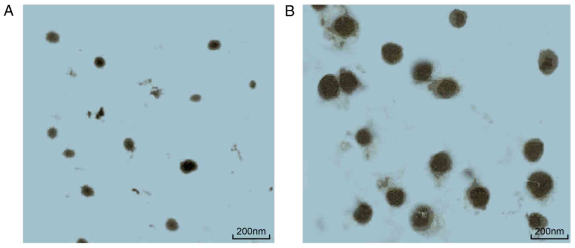
TEM observation (Fig. 1) revealed
that curcuminoid and HA/CNP deliveries were both ball-shaped and
regular. The diameter of the curcuminoid particle was 42.46±3.23
nm, while the diameter of HA/CNPs was 164.68±14.21 nm. These three
experiments demonstrated that HA/CNP for the delivery of
curcuminoid was able to accelerate chondrocyte proliferation and
has a sustained release.
| Table IVIC50 of curcuminoid and
HA/CNP for delivery of curcuminoid to rat chondrocytes
(µg/ml). |
Table IV
IC50 of curcuminoid and
HA/CNP for delivery of curcuminoid to rat chondrocytes
(µg/ml).
| Drug | 24 h | 48 h |
|---|
| Curcuminoid | 82.31±5.46 | 46.75±5.73 |
| HA/CNP for delivery
of curcuminoid | 24.23±3.85 | 8.93±1.14 |
| Table VCumulative release rates of
curcuminoid and HA/CNP for delivery of curcuminoid (%). |
Table V
Cumulative release rates of
curcuminoid and HA/CNP for delivery of curcuminoid (%).
| Drug | 1 h | 2 h | 4 h | 8 h | 12 h | 24 h | 36 h | 48 h | 72 h |
|---|
| Curcuminoid | 78.42±3.12 | 78.81±3.76 | 80.21±2.13 | 80.95±4.33 | 81.58±3.35 | 82.27±2.79 | 82.91±3.84 | 83.53±2.96 | 83.84±4.27 |
| HA/CNP for delivery
of curcuminoid | 40.24±2.66 | 48.45±3.52 | 54.37±3.55 | 58.67±4.12 | 62.25±3.87 | 66.75±3.48 | 69.52±2.25 | 72.47±3.27 | 74.13±3.45 |
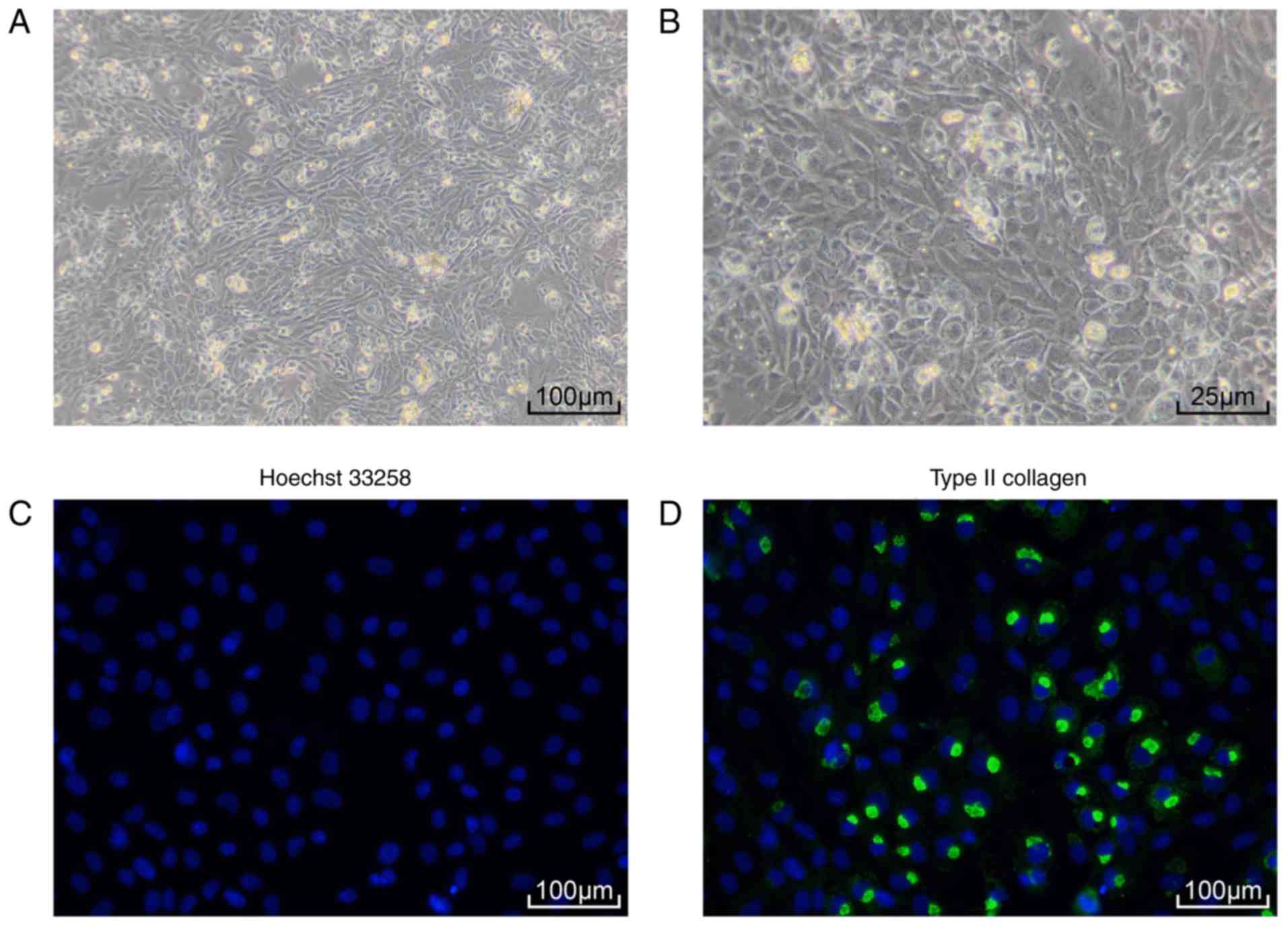
Identification of rat chondrocytes
Primary culture of SD rat chondrocytes (Fig. 2A and B) demonstrated that rat
chondrocytes were tightly packed and had an elongated spindle
shape. Immunofluorescence assay revealed strong collagen II
expression in rat chondrocytes (Fig.
2C and D). These results demonstrated that the cells that were
isolated and cultured were rat chondrocytes.
HA/CNP for the delivery of curcuminoid
enhances proliferation but suppresses apoptosis and inflammatory
response in chondrocytes
The biological functions of HA/CNP for the delivery
of curcuminoid were detected by EdU assay, RT-qPCR assay, western
blot analysis, P65 protein nuclear translocation detection and flow
cytometry. The EdU assay (Fig.
3A) determined that the cell proliferation rate of the NC group
was 13.62±1.22%, the rate of the HA group was 20.33±1.83%, the rate
of the curcuminoid group was 19.14±2.12%, while the rate of the
HA/CNP for the delivery of curcuminoid group was 36.42±3.26%.
RT-qPCR (Fig. 3B) revealed that,
compared with the NC group, the HA and curcuminoid groups exhibited
increased collagen II mRNA levels but decreased MMP-1 and MMP-13
mRNA levels (all P<0.05). Additionally, the HA/CNP group also
exhibited significantly increased collagen II mRNA levels, but
significantly decreased MMP-1 and MMP-13 mRNA levels (all
P<0.01). Western blot analysis (Fig. 3C and D) demonstrated that, in
comparison with the NC group, the protein levels of collagen II and
I-κBα were increased, but the protein levels of MMP-1 and MMP-13
and the extent of I-κBα phosphorylation were decreased in the
curcuminoid and HA groups. In addition, compared with the
curcuminoid group, the protein levels of collagen II and I-κBα were
elevated, while the protein levels of MMP-1 and MMP-13 and the
extent of I-κBα phosphorylation were decreased in the HA/CNP group
(all P<0.01). P65 protein nuclear translocation detection
(Fig. 3E and F) revealed that the
curcuminoid and the HA groups had more P65 in the cytoplasm but
less P65 in the cell nucleus compared with the NC group.
Furthermore, the HA/CNP had more P65 in the cytoplasm but less P65
in the cell nucleus compared with the curcuminoid group (all
P<0.01). Flow cytometry analysis (Fig. 3G) showed that cell apoptosis rate
was decreased in the HA and the curcuminoid groups (P<0.05) and
significantly decreased in the HA/CNP group (P<0.01) when
compared with the NC group, while the HA/CNP group had a lower cell
apoptosis rate compared with the curcumi-noid group (P<0.05).
Taken together, our results suggest that HA/CNP for the delivery of
curcuminoid enhanced proliferation but suppressed apoptosis and
inflammatory response in chondrocytes.
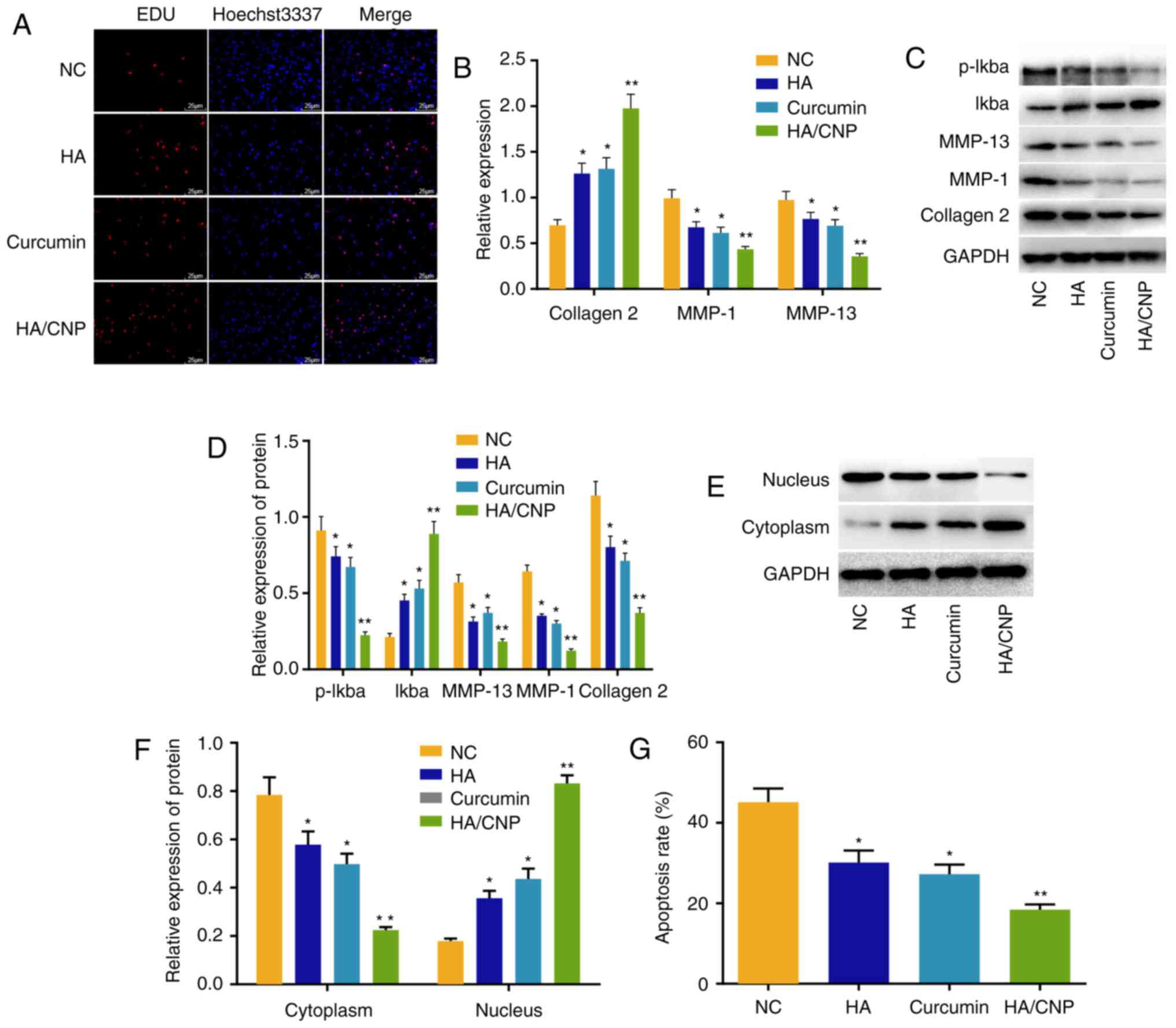 | Figure 3HA/CNP for the delivery of curcuminoid
enhances cell proliferation but suppresses apoptosis and
inflammatory responses in chondrocytes. (A) EdU assay revealed that
HA/CNP for the delivery of curcuminoid enhanced chondrocyte
proliferation; (B) RT-qPCR assay demonstrated that HA and
curcuminoid increased collagen II mRNA levels but decreased MMP-1
and MMP-13 mRNA levels, and that HA/CNP for the delivery of
curcuminoid exerted stronger effects compared with individual
treatments. (C and D) Western blot analysis showed that HA and
curcuminoid treatment elevated the protein levels of collagen II
and I-κBα, but reduced the protein levels of MMP-1 and MMP-13,
while I-κBα phosphorylation was decreased. HA/CNP treatment
significantly enhanced the response compared with the two
individual treatments. (E and F) Western blot analysis revealed
that HA/CNP for the delivery of curcuminoid promoted P65
translocation from the cell nucleus to the cytoplasm. (G) Flow
cytometry demonstrated that HA/CNP for the delivery of curcuminoid
inhibited chondrocyte apoptosis; *P<0.05 and
**P<0.01 compared with the NC group. Measurement
data, including mRNA and protein levels, cell proliferation rate
and cell apoptosis rate, were all expressed by the mean value ±
standard deviation. Statistical analysis was performed using
one-way ANOVA; n=3 replicates per experiment for 3 experiments.
RT-qPCR, reverse transcription quantitative polymerase chain
reaction; MMP, matrix metalloproteinase; NC, negative control;
ANOVA, analysis of variance; HA, hyaluronic acid; CNP, chitosan
nanoparticle. |
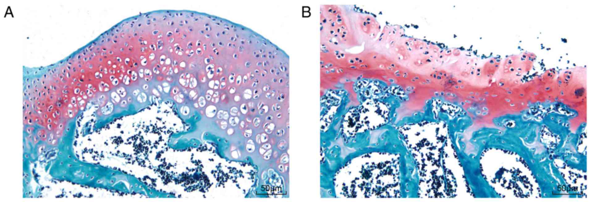
Establishment of the OA model
SD rat behavioral observation showed that SD rats in
the sham group walked normally, but SD rats in the OA group walked
slowly with the bodies tilted to the right. Safranin O-fast green
staining of knee joints (Fig. 4)
showed hyaline cartilage and calcified cartilage in the knee joints
(red) and subchondral bone (blue). The rats in the sham group had
more calcified cartilage and matrix outside the chondrocytes, more
obvious layers of chondrocytes, and a smoother surface, while rats
in the OA group had no obvious boundary lines between the
subchondral bone layer and calcified cartilage layer, with
comb-like structures, lightly stained chondrocyte lacunae in the
hyaline cartilage and scattered osteocytes in the subchondral bone
zone. Collectively, these data demonstrated that the knee OA model
in SD rats was successfully established.
HA/CNP alleviates knee joint lesions in
rats with knee OA
Finally, the effect of HA/CNP delivery on knee joint
lesions in rats with knee OA was investigated. RT-qPCR assay
(Fig. 5A) revealed that, compared
with the NC group, the mRNA levels of collagen II were elevated,
while the mRNA levels of MMP-1 and MMP-13 were decreased in the HA
group (all P<0.05). Additionally, collagen II mRNA expression
was significantly elevated and MMP-1 and MMP-13 expression was
significantly decreased in the HA/CNP for the delivery of
curcuminoid group (all P<0.01). Western blot analysis (Fig. 5B and C) demonstrated that the HA
and the HA/CNP for the delivery of curcuminoid groups had higher
collagen II and I-κBα protein levels, lower MMP-1 and MMP-13
protein levels, and decreased I-κBα phosphorylation compared with
the NC group. It was also observed that SD rat knee joints in the
NC group exhibited a rough surface and local erosion (Fig. 5D), while the knee joints in the HA
group had a complete surface without gloss, and the knee joints in
the HA/CNP group had a smooth and complete surface. H&E
staining (Fig. 5E) demonstrated
that the articular cartilage surface layer was fiberized and cell
lamination was completely disordered in the NC group, while the
knee joint surface was partially incomplete and cells were
partially disordered in the HA group, whereas the knee joint
surface was smooth and the cells were regularly arranged in the
HA/CNP group. Following toluidine blue staining (Fig. 5F), some of the layers were not
dyed, and the chondrocytes were significantly decreased with
disorderly layers in the NC group. In comparison, articular
cartilage was lightly stained and the cells were somewhat
disorderly but arranged in clearer layers in the HA group, while
the knee joints were deeply stained blue, cartilages had a flat
surface, the cells were orderly arranged and there were distinct
layers in the HA/CNP group. Following Safranin O-fast green
staining (Fig. 5G), some of the
layers were not dyed and the chondrocytes were significantly
decreased with disordered layers in the NC group, while articular
cartilage was lightly stained and the cells were somewhat
disordered, but in clearer layers in the HA group. However, the
knee joints were deeply stained with red, the cartilages had a flat
surface, the cells were orderly arranged and there were distinct
layers in the HA/CNP group. The Outerbridge classification and
Mankin pathological score of knee chondrocytes are shown in
Table VI. Taken together, these
results indicate that HA/CNP alleviates knee joint lesions in rats
with knee OA.
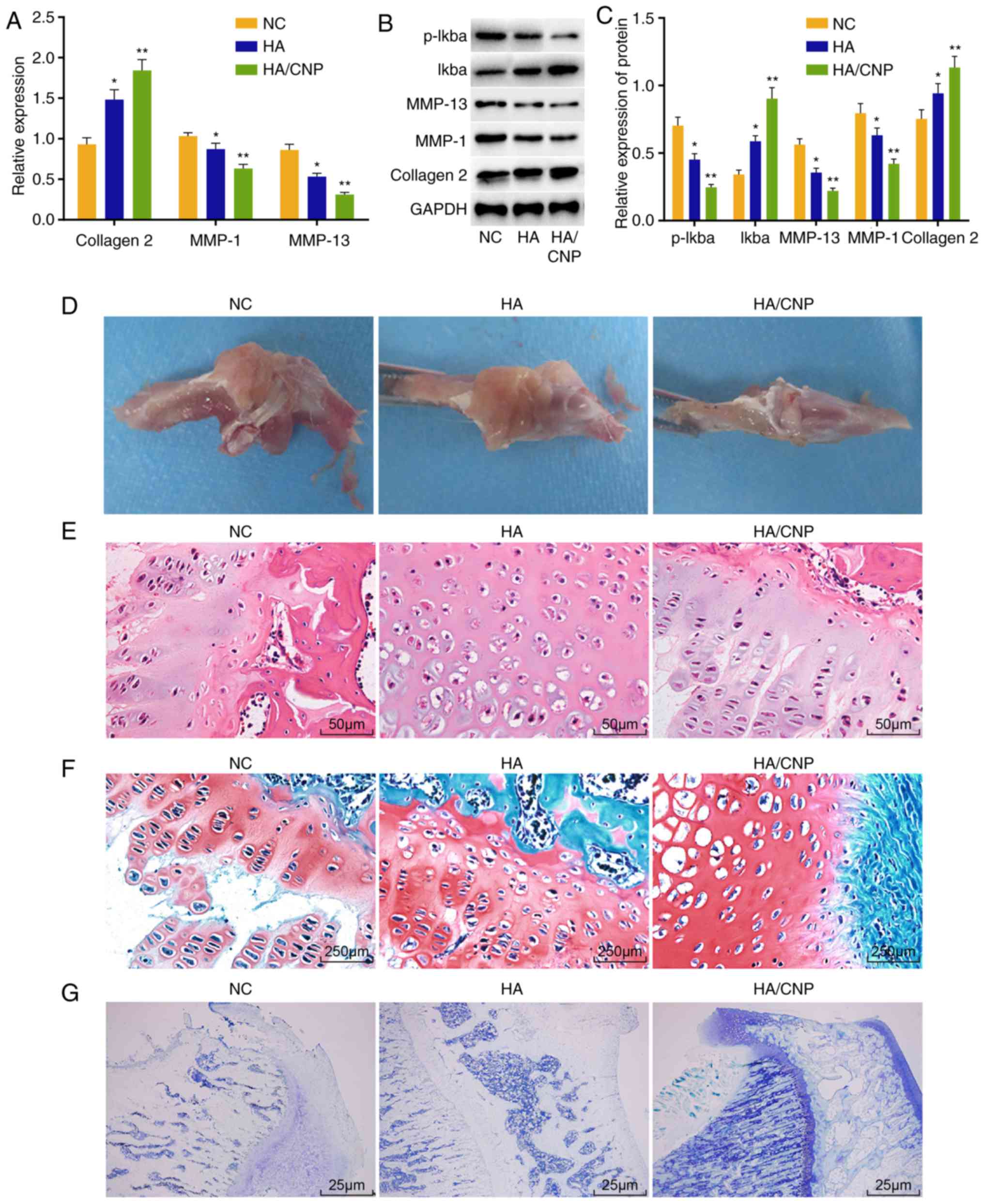 | Figure 5HA/CNP for delivery of curcuminoid
confers protection against knee joint lesions in rats with knee OA.
(A) RT-qPCR assay showed that collagen II mRNA level was
significantly increased and MMP-1 and MMP-13 mRNA levels in
chondrocytes were significantly decreased after treatment with
HA/CNP for delivery of curcuminoid. (B and C) Western blot analysis
demonstrated that collagen II and I-κBα protein levels were
significantly increased and MMP-1 and MMP-13 protein levels as well
as the extent of I-κBα phosphorylation were decreased after
treatment with HA/CNP for delivery of curcuminoid. (D) Under a
microscope, HA/CNP for delivery of curcuminoid improved articular
surface injury in OA rats. (E) H&E staining revealed that the
knee joint surface was smooth and the cells were regularly arranged
in OA rats treated with HA/CNP for delivery of curcuminoid (×400).
(F) Toluidine blue staining revealed that knee joints are deeply
stained with blue, the cartilages had a flat surface, the cells
were orderly arranged and there were distinct layers in OA rats
treated with HA/CNP for delivery of curcuminoid (×40). (G) Safranin
O-fast green staining demonstrated that the knee joints were deeply
stained with red, the cartilages had a flat surface, the cells were
orderly arranged and there were distinct layers in OA rats treated
with HA/CNP for delivery of curcuminoid (×400);
*P<0.05 and **P<0.01 compared with the
NC group. mRNA and protein value, as measurement data, were all
expressed by the mean value ± standard deviation, the result of
which were verified by one-way ANOVA; n=3; the experiments were
repeated 3 times. H&E, hematoxylin and eosin; HA, hyaluronic
acid; RT-qPCR, reverse transcription quantitative polymerase chain
reaction; MMP, matrix metalloproteinase; NC, negative control;
ANOVA, analysis of variance; OA, osteoarthritis; CNP, chitosan
nanoparticle. |
| Table VIOuterbridge classification and Mankin
pathological score of Sprague Dawley rat knee-joint
chondrocytes. |
Table VI
Outerbridge classification and Mankin
pathological score of Sprague Dawley rat knee-joint
chondrocytes.
| Drug treatment | Period (week) | Outerbridge
classification | Mankin pathological
score |
|---|
| NC | 1 | II | 7 |
| 2 | II | 7 |
| 3 | II | 7 |
| 4 | II | 7 |
| HA | 1 | II | 7 |
| 2 | II | 6 |
| 3 | I | 4 |
| 4 | I | 3 |
| HA/CNP for delivery
of curcuminoid | 1 | I | 3 |
| 2 | I | 2 |
| 3 | 0 | 1 |
| 4 | 0 | 0 |
Discussion
As a common treatment for knee OA, HA has been
recently adopted as a ligand to contribute to drug delivery
(17). Therefore, HA was selected
as the ligand for curcuminoid, an effective drug for knee OA
therapy. In addition, CNP may be used to improve curcuminoid
stability and solubility (10).
As a result, it was decided to prepare a new combination of drugs,
HA/CNP, for curcuminoid delivery and explore its therapeutic effect
in this study. The results demonstrated that HA/CNP for the
delivery of curcuminoid inhibited inflammation and chondrocyte
apoptosis by repressing the NF-κB pathway.
The major finding of the present study was that
HA/CNP decreased chondrocyte apoptosis by inhibiting the NF-κB
pathway. The NF-κB pathway, as one of the most important catabolic
pathways, participates in the pathogenesis of OA and critically
regulates inflammatory mediators that are associated with OA
(18). It was previously
demonstrated that the combination of HA and curcuminoid can act
synergistically to reduce scar formation (19). Ji et al demonstrated that
isoliquiritigenin can protect chondrocytes and prevent inflammation
by decreasing the activation of the NF-κB pathway (4). Zhuang et al demonstrated that
kaempferol may be a good therapeutic strategy for OA, since it can
inhibit NF-κB and decrease IL-1β-stimulated inflammation in
chondrocytes of rats with OA (20). Importantly, it is obvious that the
inhibition of NF-κB pathway can be a point to explore the OA
therapy. In the present study, HA/CNP for the delivery of
curcuminoid was found to promote the expression of IκB. IκB is an
inhibitor of the NF-κB pathway (21). Therefore, HA/CNP can be used to
inhibit the NF-κB pathway by increasing IκB. Moreover, Li et
al have proved that inhibiting phosphorylated P65 translocation
to the nucleus can promote the effect of baicalein against
chondrocyte inflammation and apoptosis (22). Therefore, inhibiting P65
translocation to the nucleus may be a target for OA therapy.
Interestingly, this study has demonstrated that HA/CNP for the
delivery of curcuminoid was able to relocate P65 from the cell
nucleolus to the cytoplasm, which provides evidence that HA/CNP for
the delivery of curcuminoid may be an optimal drug for OA therapy.
However, Hwang et al reported that the inhibition of
chondrocyte apoptosis, which is the main pathogenetic mechanism,
can be targeted in OA therapy by modulating cartilage degeneration
(23). Our study also
demonstrated that HA/CNP for the delivery of curcuminoid can
decrease chondrocyte apoptosis and knee joint lesions in knee OA.
NF-κB regulates inflammatory responses and mediates related
chondrocyte inflammatory responses, which may cause cartilage
degradation (24). Our findings
revealed that HA/CNP for the delivery of curcuminoid suppresses
chondrocyte apoptosis by inactivating the NF-κB pathway.
Another important finding of the present study was
that HA/CNP for the delivery of curcuminoid inhibited the
expression of inflammation-related genes, such as MMP-1 and MMP-13,
but promoted collagen II expression. Collagen II is the major
component of articular cartilage. Sufficient collagen II is crucial
for cartilage biomechanical properties (25), while previous findings determined
that MMP-13 is the primary protein involved in cartilage
degradation in the course of OA by cleaving collagen II (26). MMP-1 degrades type I, II, III,
VII, X, IX and other types of collagen and aggrecan, while MMP-13
mainly cleaves collagen type II, IV and IX, osteonectin and
perlecan in the cartilage (27).
When stimulated by catabolism and anabolism, HA suppresses MMP-1
and MMP-13 (28). Curcuminoid may
inhibit downstream MMP-1 and MMP-13 gene upregulation by oncostatin
M in chondrocytes when upstream STAT1 phosphorylation is inhibited
(29). Furthermore, our results
demonstrated that HA/CNP for the delivery of curcuminoid inhibited
the expression of MMP-1 and MMP-13, and promoted collagen II
expression.
In conclusion, the present study suggests that
intra-articular HA/CNP for the delivery of curcuminoid can decrease
chondrocyte apoptosis in rats with knee OA through repression of
the NF-κB pathway. These findings may lead to novel OA therapies in
the future. However, it should be noted that this study has
examined only SD rat models; therefore, it is not clear at present
if there are unwanted side effects in practical clinic
applications. Due to time and funding constraints, we did not study
the effect of inhibition of the NF-κB pathway on arthritis models.
Further studies may focus on the effects of inhibition of the NF-κB
pathway and other pathways on arthritis models. It was demonstrated
in rats that HA/CNP for the delivery of curcuminoid may be a
potential therapy for OA, which may represent a creative suggestion
to doctors. Furthermore, we will continue to explore materials that
can further improve the solubility and stability of HA and
curcuminoid.
Funding
The present study was supported by Science and
Technology Development Fund Project of Baoshan District (17-E-1);
Scientific Research Project of the Shanghai Municipal Health and
Planning Committee (20174Y0240).
Availability of data and materials
The data generated and analyzed in the present study
are available from the corresponding author upon reasonable
request.
Authors' contributions
JW conceived and together with XW and YC designed
the study. TH and DXS were involved in data collection. XW and HRT
performed the statistical analysis and figure preparation. YC and
TH drafted the manuscript. JW, XW, TH and DXS performed critical
revision of the article. All authors read and approved the final
version of the manuscript.
Ethics approval and consent to
participate
The breeding and sacrifice of the experimental
animals, as well as tissue retrieval, were all for medical research
and performed in strict accordance with the recommendations in the
Guide for the Care and Use of Laboratory Animals of the National
Institutes of Health. The procedures were approved by the Animal
Committee of Shanghai Ninth People's Hospital, Shanghai Jiao Tong
University School of Medicine.
Patient consent for publication
Not applicable.
Competing interests
The authors declare that they have no competing
interests to disclose.
Acknowledgments
The authors are grateful to all the staff who
contributed to this study and the construction plan of
characteristic medical specialties and community projects of
Baoshan District.
References
|
1
|
Wu Q, Sun X and Du L: Association of
fibulin-3 concentrations with the presence and severity of knee
osteoarthritis: A cross-sectional study. Knee. 24:1369–1373. 2017.
View Article : Google Scholar : PubMed/NCBI
|
|
2
|
Ogunbode AM, Adebusoye LA, Olowookere OO
and Alonge TO: Physical functionality and self-rated health status
of adult patients with knee osteoarthritis presenting in a primary
care clinic. Ethiop J Health Sci. 24:319–328. 2014. View Article : Google Scholar :
|
|
3
|
Runhaar J, van Middelkoop M, Reijman M,
Willemsen S, Oei EH, Vroegindeweij D, van Osch G, Koes B and
Bierma-Zeinstra SM: Prevention of knee osteoarthritis in overweight
females: The first preventive randomized controlled trial in
osteoarthritis. Am J Med. 128:888–895.e4. 2015. View Article : Google Scholar : PubMed/NCBI
|
|
4
|
Ji B, Guo W, Ma H, Xu B, Mu W, Zhang Z,
Amat A and Cao L: Isoliquiritigenin suppresses IL-1β induced
apoptosis and inflammation in chondrocyte-like ATDC5 cells by
inhibiting NF-κB and exerts chondroprotective effects on a mouse
model of anterior cruciate ligament transection. Int J Mol Med.
40:1709–1718. 2017.PubMed/NCBI
|
|
5
|
Ricci M, Micheloni GM, Berti M, Perusi F,
Sambugaro E, Vecchini E and Magnan B: Clinical comparison of oral
administration and viscosupplementation of hyaluronic acid (HA) in
early knee osteoarthritis. Musculoskelet Surg. 101:45–49. 2017.
View Article : Google Scholar
|
|
6
|
Zhang Z, Leong DJ, Xu L, He Z, Wang A,
Navati M, Kim SJ, Hirsh DM, Hardin JA, Cobelli NJ, et al: Curcumin
slows osteoarthritis progression and relieves
osteoarthritis-associated pain symptoms in a post-traumatic
osteoarthritis mouse model. Arthritis Res Ther. 18:1282016.
View Article : Google Scholar : PubMed/NCBI
|
|
7
|
Salwowska NM, Bebenek KA, Żądło DA and
Wcisło- Dziadecka DL: Physiochemical properties and application of
hyaluronic acid: A systematic review. J Cosmet Dermatol.
15:520–526. 2016. View Article : Google Scholar : PubMed/NCBI
|
|
8
|
Concoff A, Sancheti P, Niazi F, Shaw P and
Rosen J: The efficacy of multiple versus single hyaluronic acid
injections: A systematic review and meta-analysis. BMC
Musculoskelet Disord. 18:5422017. View Article : Google Scholar : PubMed/NCBI
|
|
9
|
Li X, Feng K, Li J, Yu D, Fan Q, Tang T,
Yao X and Wang X: Curcumin inhibits apoptosis of chondrocytes
through activation ERK1/2 signaling pathways induced autophagy.
Nutrients. 9:E4142017. View Article : Google Scholar : PubMed/NCBI
|
|
10
|
Wang F, Yang Y, Ju X, Udenigwe CC and He
R: Polyelectrolyte complex nanoparticles from chitosan and acylated
rapeseed cruciferin protein for curcumin delivery. J Agric Food
Chem. 66:2685–2693. 2018. View Article : Google Scholar : PubMed/NCBI
|
|
11
|
Yu H, Nguyen MH and Hadinoto K: Effects of
chitosan molecular weight on the physical and dissolution
characteristics of amorphous curcumin-chitosan nanoparticle
complex. Drug Dev Ind Pharm. 44:82–88. 2018. View Article : Google Scholar
|
|
12
|
Yasuda T: Type II collagen peptide
stimulates Akt leading to nuclear factor-kappaB activation: Its
inhibition by hyaluronan. Biomed Res. 35:193–199. 2014. View Article : Google Scholar
|
|
13
|
Yang Q, Wu S, Mao X, Wang W and Tai H:
Inhibition effect of curcumin on TNF-α and MMP-13 expression
induced by advanced glycation end products in chondrocytes.
Pharmacology. 91:77–85. 2013. View Article : Google Scholar
|
|
14
|
Zhou L, Hu Y, Li C, Yan Y, Ao L, Yu B,
Fang W, Liu J and Li Y: Levocorydalmine alleviates
vincristine-induced neuropathic pain in mice by inhibiting an
NF-kappa B-dependent CXCL1/CXCR2 signaling pathway.
Neuropharmacology. 135:34–47. 2018. View Article : Google Scholar : PubMed/NCBI
|
|
15
|
Xu H, Bouta EM, Wood RW, Schwarz EM, Wang
Y and Xing L: Utilization of longitudinal ultrasound to quantify
joint soft-tissue changes in a mouse model of posttraumatic
osteoarthritis. Bone Res. 5:170122017. View Article : Google Scholar : PubMed/NCBI
|
|
16
|
Xu J, Lu MX, Cui YD and Du YZ: Selection
and evaluation of reference genes for expression analysis using
qRT-PCR in Chilo suppressalis (Lepidoptera: Pyralidae). J Econ
Entomol. 110:683–691. 2017.PubMed/NCBI
|
|
17
|
Xu Y, Asghar S, Yang L, Chen Z, Li H, Shi
W, Li Y, Shi Q, Ping Q and Xiao Y: Nanoparticles based on chitosan
hydrochloride/hyaluronic acid/PEG containing curcumin: In vitro
evaluation and pharmacokinetics in rats. Int J Biol Macromol.
102:1083–1091. 2017. View Article : Google Scholar
|
|
18
|
Chen C, Zhang C, Cai L, Xie H, Hu W, Wang
T, Lu D and Chen H: Baicalin suppresses IL-1β-induced expression of
inflammatory cytokines via blocking NF-κB in human osteoarthritis
chondrocytes and shows protective effect in mice osteoarthritis
models. Int Immunopharmacol. 52:218–226. 2017. View Article : Google Scholar : PubMed/NCBI
|
|
19
|
Sharma M, Sahu K, Singh SP and Jain B:
Wound healing activity of curcumin conjugated to hyaluronic acid:
In vitro and in vivo evaluation. Artif Cells Nanomed Biotechnol.
46:1009–1017. 2018. View Article : Google Scholar
|
|
20
|
Zhuang Z, Ye G and Huang B: Kaempferol
alleviates the interleukin-1β-induced inflammation in rat
osteoarthritis chondrocytes via suppression of NF-κB. Med Sci
Monit. 23:3925–3931. 2017. View Article : Google Scholar : PubMed/NCBI
|
|
21
|
De Palma A, Cheleschi S, Pascarelli NA,
Giannotti S, Galeazzi M and Fioravanti A: Hydrostatic pressure as
epigenetic modulator in chondrocyte cultures: A study on miRNA-155,
miRNA-181a and miRNA-223 expression levels. J Biomech. 66:165–169.
2018. View Article : Google Scholar
|
|
22
|
Li Y, Wang J, Song X, Bai H, Ma T, Zhang
Z, Li X, Jiang R, Wang G, Fan X, et al: Effects of baicalein on
IL-1β-induced inflammation and apoptosis in rat articular
chondrocytes. Oncotarget. 8:90781–90795. 2017.PubMed/NCBI
|
|
23
|
Hwang HS and Kim HA: Chondrocyte apoptosis
in the pathogenesis of osteoarthritis. Int J Mol Sci.
16:26035–26054. 2015. View Article : Google Scholar : PubMed/NCBI
|
|
24
|
Lee HG and Yang JH: PCB126 induces
apoptosis of chondrocytes via ROS-dependent pathways.
Osteoarthritis Cartilage. 20:1179–1185. 2012. View Article : Google Scholar : PubMed/NCBI
|
|
25
|
Ito K and Shinomura T: Development and
application of a new Silent reporter system to quantitate the
activity of enhancer elements in the type II collagen gene. Gene.
585:13–21. 2016. View Article : Google Scholar : PubMed/NCBI
|
|
26
|
Motomura H, Seki S, Shiozawa S, Aikawa Y,
Nogami M and Kimura T: A selective c-Fos/AP-1 inhibitor prevents
cartilage destruction and subsequent osteophyte formation. Biochem
Biophys Res Commun. 497:756–761. 2018. View Article : Google Scholar : PubMed/NCBI
|
|
27
|
Stancker TG, Vieira SS, Serra AJ, do
Nascimento Lima R, Dos Santos Feliciano R, Silva JA Jr, Dos Santos
SA, Dos Santos Vieira MA, Simões MCB, Leal-Junior EC, et al: Can
photobiomodulation associated with implantation of mesenchymal
adipose-derived stem cells attenuate the expression of MMPs and
decrease degradation of type II collagen in an experimental model
of osteoarthritis. Lasers Med Sci. 33:1073–1084. 2018. View Article : Google Scholar : PubMed/NCBI
|
|
28
|
Pohlig F, Guell F, Lenze U, Lenze FW,
Mühlhofer HM, Schauwecker J, Toepfer A, Mayer-Kuckuk P, von
Eisenhart-Rothe R, Burgkart R and Salzmann GM: Hyaluronic acid
suppresses the expression of metalloproteinases in osteoarthritic
cartilage stimulated simultaneously by interleukin 1β and
mechanical load. PLoS One. 11:e01500202016. View Article : Google Scholar
|
|
29
|
Li WQ, Dehnade F and Zafarullah M:
Oncostatin M-induced matrix metalloproteinase and tissue inhibitor
of metalloproteinase-3 genes expression in chondrocytes requires
Janus kinase/STAT signaling pathway. J Immunol. 166:3491–3498.
2001. View Article : Google Scholar : PubMed/NCBI
|